Can increasing endurance help individuals who want to improve their physical abilities or extend the time they perform these activities?
Endurance
Individuals tend to think of endurance in terms of exercise and fitness, such as running, biking, swimming, and strength training. While this is true, endurance is involved in nearly every task we perform. For example, an individual has to have enough endurance to complete a full day of activities. This includes:
Carpooling the kids
Professional responsibilities
Home chores
Preparing dinner
Helping out kids with homework, etc.
Nearly every activity requires some level of endurance, which means the ability to maintain activity for an extended period of time. When endurance begins to wane, it usually results from not performing certain activities regularly. The body gets used to daily routines and activities. When it stops engaging in certain activities, like walking and exercising regularly, endurance slowly declines, and the ability to perform at the same caliber.
What Is It?
Endurance is an ability that is acquired after extensive physical and mental training. Physiological and psychological factors reinvigorate individuals to continue doing what they are doing longer. Factors include:
Fatigue
Individuals who didn’t sleep well the night before or are worn out may have difficulty following through with certain activities that require extensive output or stamina.
Fitness Levels
Current fitness levels are also a predictor of endurance.
How physically fit an individual is, coupled with their level of training, will impact endurance abilities.
Genetics is another factor, as everybody has different muscle fibers that can influence physical capabilities. While research shows that individuals can gradually alter the amount of these fibers, it also emphasizes the role of genetics in determining one’s muscle makeup. (de Souza, E. O. et al., 2014)
Individuals who constantly challenge themselves mentally and physically are continually building endurance.
Endurance and Stamina Difference
Endurance is often used interchangeably with stamina. However, the two are very different.
Stamina refers to how long an individual can perform a certain activity at maximum capacity or without getting tired.
Endurance revolves around an individual’s ability to perform a certain activity without performing at maximum capacity.
Types
Endurance can be divided into classifications defined by type. Here are the main types of endurance in physical fitness and what they mean.
Cardiovascular
Cardiovascular endurance is the stress an individual’s heart can take during physical activity.
When building cardiovascular endurance, the body becomes more efficient at pumping blood while performing a specific activity.
Individuals with more cardiovascular endurance can sustain longer and more intense overall training.
Muscular
Muscular endurance is the length of time muscles can continue to contract enough to allow the body to finish a certain activity.
An individual lacking in muscular endurance will succumb faster to excess lactic acid build-up, causing cramps.
An individual with significant muscular endurance can lift a weight for more repetitions before failure.
Anaerobic
Anaerobic means without oxygen, so anaerobic endurance refers to how long a muscle can continue working at a certain physical level without much or any oxygen.
Weightlifting is a great example of this.
Anaerobic exercise tends to be shorter in duration but more intense than aerobic exercise, like swimming or cycling.
Improvement
Through endurance training, individuals can improve their ability to carry out certain activities longer. Recommendations for how to improve include.
Interval Training
Interval training, or high-intensity interval training, involves increasing the intensity of the workout for a short period of time.
If running, intentionally push the pace harder than normal for 20-second intervals.
Followed by a slower recovery pace for about a minute.
This increases endurance and improves insulin sensitivity.
Pedaling on an air bike is another recommended activity to build strength and endurance.
Rest Less Between Sets
Resting in between certain types of physical activity is beneficial, it can also lower heart rate and endurance threshold.
Taking less rest between workout sets so that the heart rate stays elevated increases endurance with each workout.
Perform a Few More Reps On Each Set
Whatever the type of exercise being done, one way to enhance endurance is to add one more rep, one more mile, or a few more minutes to the fitness schedule.
The body will slowly adapt to that level, making it the new norm.
Increase Core Strength
No matter the workout—running, swimming, cycling, or weight lifting—it’s important to focus on strengthening the core. This will help improve endurance in any activity and prevent injuries.
Individuals having trouble taking their workouts to the next level and feeling that their endurance has flattened should consider enlisting the help of a certified personal trainer. If there is any discomfort or pain when working to increase endurance, seek advice from a healthcare professional. Injury Medical Chiropractic and Functional Medicine Clinic uses an integrated approach to treating injuries and chronic pain syndromes. It offers personalized care plans that improve ability through flexibility, mobility, and agility programs to relieve pain. Our providers use an integrated approach to create personalized care plans for each patient, including Functional Medicine, Acupuncture, Electro-Acupuncture, and Sports Medicine principles. Our goal is to relieve pain naturally by restoring health and function to the body. If other treatment is needed, Dr. Jimenez has teamed up with top surgeons, clinical specialists, medical researchers, and rehabilitation providers to provide the most effective treatments.
Unlocking Athletic Potential
References
de Souza, E. O., Tricoli, V., Aoki, M. S., Roschel, H., Brum, P. C., Bacurau, A. V., Silva-Batista, C., Wilson, J. M., Neves, M., Jr, Soares, A. G., & Ugrinowitsch, C. (2014). Effects of concurrent strength and endurance training on genes related to myostatin signaling pathway and muscle fiber responses. Journal of strength and conditioning research, 28(11), 3215–3223. https://doi.org/10.1519/JSC.0000000000000525
Can understanding the nucleus pulposus help in body positioning and prevention for individuals wanting to practice spinal hygiene and protect their discs from injury?
Nucleus Pulposus
The spinal discs are located between the spine’s vertebrae and are the body’s natural impact and shock absorbers. Within the disc is the nucleus pulposus, which plays a major role in providing the spine with shock absorption during movement. (Zhou Z. et al., 2014) The discs have a tough outer portion and a soft inner core. They are the:
It forms the tough circular exterior and comprises concentric sheets of collagen fibers or lamellae surrounding the inner core.
It has cartilaginous endplates that firmly attach to the vertebrae above and below.
Nucleus Pulposus
The nucleus pulposus is the inner core soft filling of the discs.
It contains a network of fibers suspended in a mucoprotein gel with a water base to maintain strength and pliability.
The near-liquid consistency makes it responsive to movement to handle the body’s axial load.
It helps maintain spinal suspension to prevent pressure on the bones and prevent bone-to-bone contact, reducing the potential for injuries and pain.
Shock Absorber
Each intervertebral disc is a shock-absorbing cushion, with the nucleus pulposus providing shock-absorbing properties (Zhou Z. et al., 2014). The intervertebral discs move as the body moves. For example, when arching the back, the disc moves forward slightly, and when twisting, the disc twists as well.
Spinal Action
The intervertebral disc supports spinal movements. When bending, twisting, arching, or tilting the spine, the nucleus pulposus swivels to accommodate these actions. These repeated spinal actions, which occur throughout the day and night, contribute to shifting positions while sitting, working, playing sports, carrying groceries, performing house chores, etc. An example is bending forward to pick something up. This action involves forward spinal flexion, which is bending the spine forward, flattening, or rounding. When bending using flexion, the spinal bones come closer together, pushing the nucleus pulposus toward the back.
Injuries
The disc can be pushed too far back with persistent or excessive spinal flexion. If the fibers of the annulus fibrosus become weak, they can tear, causing the nucleus pulposus to leak out and disc herniation. Generally, the nucleus pulposus will leak to the side and back; however, this corresponds to the location of the very sensitive nerve root/s with which it can come into contact, causing pain and other symptoms. The most common causes of disc herniation are degenerative wear and tear changes of the disc and trauma. Disc degeneration occurs as the body ages; it weakens the annulus fibers, allowing the nucleus pulposus to distend, bulge, or herniate.
Aging
Disc degeneration occurs with age but can also occur with injuries to the area. In young individuals, the nucleus pulposus is mostly water. For this age group, a herniation from trauma is more likely than in older individuals. (Ucar, D. et al., 2021) But as the body ages, the discs, especially the nucleus pulposus, begin to dry out. This dehydration leads to a significant loss of disc height. (UCLA Health, 2024) By age 60 or 70, the discs may be composed entirely of fiber, which can cause the shock absorption function not to work and disappear.
Chiropractic therapy is among the more conservative treatment options for a herniated disc and may be tried first before proceeding with more invasive treatments. Injury Medical Chiropractic and Functional Medicine Clinic works with primary healthcare providers and specialists to develop an optimal health and wellness solution that fully benefits the individual to get back to normal.
The Science of Functional Healing
References
Zhou, Z., Gao, M., Wei, F., Liang, J., Deng, W., Dai, X., Zhou, G., & Zou, X. (2014). Shock absorbing function study on denucleated intervertebral disc with or without hydrogel injection through static and dynamic biomechanical tests in vitro. BioMed research international, 2014, 461724. https://doi.org/10.1155/2014/461724
Nosikova, Y. S., Santerre, J. P., Grynpas, M., Gibson, G., & Kandel, R. A. (2012). Characterization of the annulus fibrosus-vertebral body interface: identification of new structural features. Journal of anatomy, 221(6), 577–589. https://doi.org/10.1111/j.1469-7580.2012.01537.x
Ucar, D., Duman, S., Bayram, Y., & Ucar, B. Y. (2021). Extruded disc herniations are experienced earlier by inactive young people in the high-tech gaming era. Journal of medicine and life, 14(3), 402–407. https://doi.org/10.25122/jml-2021-1059
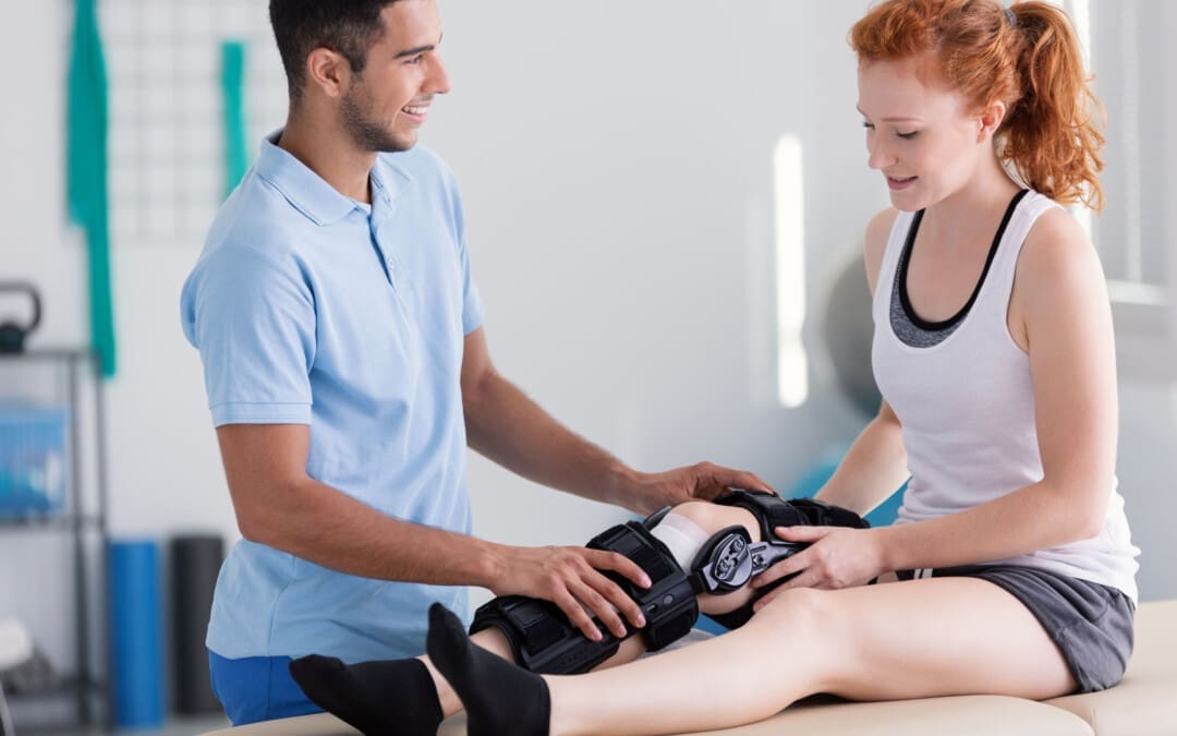
Can physical therapies help relieve muscle contractures in individuals who have endured prolonged bed rest, inactivity, or lack of use of certain muscle groups?
Muscle Contracture
A muscle contracture, or contracture deformity, is caused when a muscle loses elasticity. This causes permanent shortening and tightening of muscle fibers, which reduces flexibility and makes movement difficult. Muscles that cannot move and stretch cause the surrounding joints to lose mobility and develop pain symptoms. When trying to stretch the contracted muscle, the individual will feel the muscle become very rigid, which can increase pain. (Lieber, R. L., and Fridén, J. 2019) Delaying treatment can potentially cause irreversible and chronic symptoms.
Commonly Affected Muscles
Flexor muscles bend the joints and are those most affected by contractures. The stiffening and tightening prevent the body parts from moving out and away. The most common include:
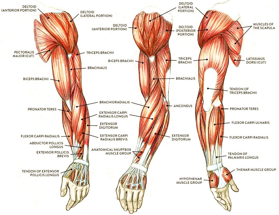
Wrist and Finger Flexors
Muscle groups that bend the wrist and fingers.
Biceps
The primary elbow flexor that bends the arm.
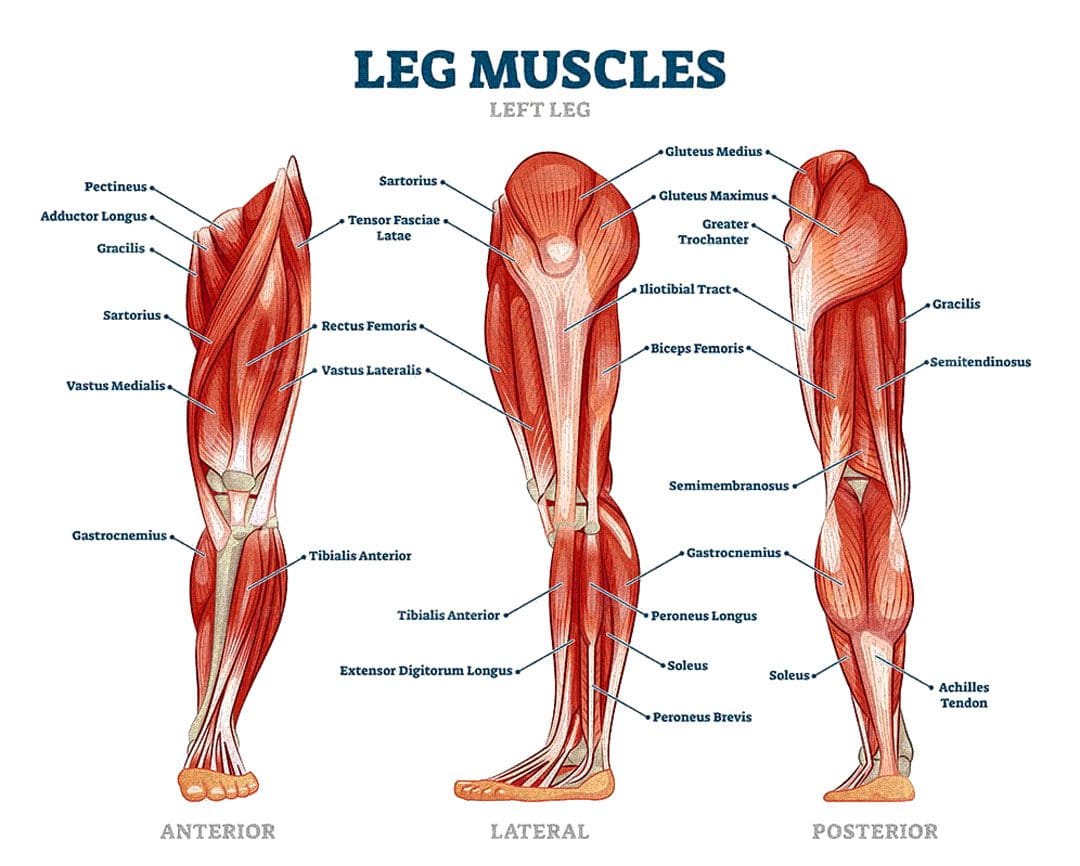
Gastrocnemius and Soleus
The calf muscles which allow the ankle to point the foot/plantarflexion.
Hamstrings
A group of three muscles behind the thigh that bend the knee.
Causes
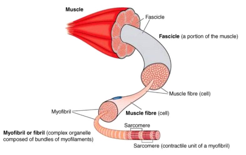
The permanent shortening of muscle fibers and changes in muscle structure cause muscle contractures or stiffer-than-normal tissue that is difficult to stretch. Sarcomeres are structural units of muscles that cause fibers to contract.
With contractures, the sarcomeres overly lengthen when the muscle fibers tighten. This increase in sarcomere length prevents the muscle from contracting normally, resulting in weakness. Muscle fibers are encased in an extracellular matrix, a mesh composed of collagen and other proteins that help transmit force and provide muscle contraction. Muscle contractures cause the amount of collagen within the extracellular matrix to increase, causing a stiffening of fibers that restricts movement. (Lieber, R. L., and Fridén, J. 2019)
Muscle contractures also form from decreased satellite cells. Satellite cells are specialized stem cells that can rebuild muscle and are necessary for muscle regeneration and repair. Without the proper amount of satellite cells, other cells like fibroblasts significantly increase in the muscle tissue, causing the fibers to become stiff and fibrotic or more fibrous. These changes to the sarcomeres, collagen within the extracellular matrix, and decreased satellite cells all result from conditions in which neurological input to the brain and spinal cord muscles becomes reduced. This is caused by lack of use, injury, or neurological and neuromuscular conditions. (Lieber, R. L., and Fridén, J. 2019)
Cerebral Palsy
Contractures often occur from upper motor neuron lesions, which prevent signals from the brain and spinal cord from reaching the motor neurons that control muscle contraction. When these signals are weakened or blocked, muscles become stiff and weak from lack of stimulation. (Lieber, R. L., and Fridén, J. 2019)
Cerebral palsy is a group of disorders affecting mobility caused by an upper motor neuron lesion that is present at birth and is the most common motor disability in children. It causes:
Cognitive impairment
Decreased muscle strength
Problems with movement, coordination, and functional motions.
Because cerebral palsy prevents the muscles of the legs from being sufficiently stimulated, contractures commonly develop in the hips, knees, and ankles. Individuals can have a 75% decrease in satellite cells to repair muscle tissue and prevent muscle fibrosis or stiffening. Specific genes linked to collagen production are also altered, causing irregular changes to the extracellular matrix of muscles. (Lieber, R. L., and Fridén, J. 2019)
Muscular Dystrophy
Muscular dystrophy is a group of inherited neuromuscular disorders characterized by muscle weakness and wasting. Deficient nerve supply to muscles causes them to become stiff and tight, inhibiting the functional range of motion needed to move joints and activate muscles to move. Clinical research suggests that individuals with muscular dystrophy have decreased levels of satellite cells to repair, increasing the risk of developing muscle contracture. (Lieber, R. L., and Fridén, J. 2019)
Disuse-induced Muscle Wasting or Disuse Atrophy
When muscles are not used for some time because of hospitalization, prolonged bed rest, or immobilization from wearing braces, splints, or casts after injuries, the blood circulation and electrical signaling from nerves to muscles decreases. This results in weakness, increased muscle tightness and stiffness, and muscle wasting/atrophy. Over time, stiff and tight muscles can progress to contractures that become extremely difficult to stretch.
Trauma or Injury
Muscle or tendon injuries can cause contractures as scar tissue develops, joining muscle fibers and joints together. This can significantly restrict movement. Large burns can also cause skin, muscles, and joint contractures. The range of motion can become significantly limited, and the changes can become irreversible if not aggressively treated.
Other Causes
Other forms of upper motor neuron lesions that can cause contractures because of weak or blocked electrical input to muscles as a result of brain or spinal cord damage include:
Neuromuscular disorders like spinal muscular atrophy – SMA.
Conditions that cause inflammation and joint stiffening, like juvenile rheumatoid arthritis.
A history of diabetes also increases the risk of developing contractures affecting finger flexors, like Dupuytren’s contractures and stenosing tenosynovitis
or trigger finger. (Lieber, R. L., and Fridén, J. 2019)
Symptoms
Symptoms include:
Extremely stiff and tight muscles resistant to stretching.
Pain from the inability to stretch.
Loss of range of motion.
Impaired joint mobility.
Severe contractures can interfere with the functional range of motion needed to move joints to complete normal tasks and movements, such as standing up from a chair and walking.
Treatment
Physical Therapy
Physical therapies can help reduce the severity through stretching and soft tissue mobilization to decrease tightness. (Lieber, R. L., and Fridén, J. 2019)
Specialized braces or splints can be custom-made to fit different body parts.
These provide a prolonged low-intensity stretch over a period of time to increase muscle length.
Once the muscle has stretched, a new brace or splint may be needed to adjust to the increased range of motion. (Lieber, R. L., and Fridén, J. 2019)
Surgery
In severe cases where muscle contractures limit the functional range of motion needed for activities of daily living or ADLs, surgical release of the contracted tissue may be recommended. This surgery can improve functional movements like walking, getting in and out of bed, and standing up from chairs. The tight muscles can be surgically cut, and the tendons can be lengthened to allow more mobility. (Lieber, R. L., and Fridén, J. 2019)
The causes of muscle contracture are not always avoidable, but various treatment options are available to help loosen up tight muscles and preserve or restore the range of motion. It’s important to move daily and stretch common areas like the fingers, arms, and legs to reduce the risk of muscle tightness and prevent contractures from developing. It is imperative to seek medical treatment for severe contractures resulting from neuromuscular disorders, including physical and occupational therapy, to prevent contractures from worsening and regaining as much functional range as possible.
Injury Medical Chiropractic and Functional Medicine Clinic uses an integrated approach personalized to the individual that focuses on what works for them and treats injuries and chronic pain syndromes through personalized care plans that improve ability through flexibility, mobility, and agility programs to relieve pain. Our providers use an integrated approach to create personalized care plans for each patient, including Functional Medicine, Acupuncture, Electro-Acupuncture, and Sports Medicine principles. Our goal is to relieve pain naturally by restoring health and function to the body. If other treatment is needed, Dr. Jimenez has teamed up with top surgeons, clinical specialists, medical researchers, and rehabilitation providers to provide the most effective treatments.
Chiropractic Treatment for Cerebral Palsy
References
Lieber, R. L., & Fridén, J. (2019). Muscle contracture and passive mechanics in cerebral palsy. Journal of applied physiology (Bethesda, Md. : 1985), 126(5), 1492–1501. https://doi.org/10.1152/japplphysiol.00278.2018
Can kimchi benefit individuals trying to incorporate more fermented foods into their diet?
Kimchi
Kimchi is a flavorful and nutritious food packed with nutritious vegetables. It is high in vitamin C, vitamin A, and iron. It is made of salted, fermented vegetables and typically served as a side dish that starts with cabbage as the base. Other varieties use different vegetables, like radish, cucumber, and onion. It has minimal calories, a low carb count, zero fat, and health benefits like an abundance of probiotics from its fermentation process.
Nutrition
Kimchi is an excellent source of vitamin C and vitamin A. A typical half-cup of kimchi is 85 grams and provides the following. (U.S. Department of Agriculture. 2017)
Calories – 20
Fat – 0g
Sodium – 290 milligrams
Carbohydrates – 4 grams
Fiber – 1 grams
Sugars – 2 grams
Protein – 1 grams
Vitamin C – 18 milligrams
Iron – 1.08 milligrams
Vitamin A – 375 micrograms
Calcium – 40 milligrams
Calories
A half-cup serving provides 20 calories, about 53% of which are carbohydrates, 21% are protein, and 26% are fat.
Carbohydrates
Kimchi comprises 4 grams of carbohydrates per serving, with 1 being fiber.
However, many kimchi recipes add sweeteners, like honey or fruit juice, to balance the sourness.
More sweeteners means more carbohydrates.
Fats
Because it is primarily vegetables, it is naturally fat-free.
Protein
Kimchi isn’t exactly a protein-power player.
A half-cup serving provides just 1 gram of plant-based protein from veggies.
However, recipes that include seafood like shrimp or squid will contain higher amounts of this macronutrient.
Vitamins and Minerals
Vitamins and minerals vary depending on the vegetables used.
A Napa cabbage-based kimchi includes abundant vitamins C and K and smaller amounts of iron, calcium, copper, and potassium.
A recipe with carrots will contain significant vitamin A.
A recipe with radishes will supply folate, potassium, and riboflavin.
All varieties are made with salt, so sodium is a mineral to watch.
A half-cup serving may provide nearly 300 milligrams or 13% Daily Value of sodium.
Benefits
Kimchi is a versatile food that can provide health benefits.
Digestion
The lactic acid that ferments the cabbage also provides healthy gut bacteria.
Consuming probiotics through kimchi promotes healthy digestion and helps alleviate constipation problems. (Higashikawa, F. et al., 2010)
Compatible with Special Diets
With simple plant-based ingredients it can be suitable for specialized diets.
It suits vegan, vegetarian, low-carb, gluten-free, and dairy-free diets.
Immune System Support
The probiotics in fermented foods improve digestion and may help improve immune function.
Research has suggested that when individuals stop eating fermented foods, their immune response decreases. (Olivares, M. et al., 2006)
Researchers isolated a compound in kimchi called HDMPPA – 3-(4′-hydroxyl-3′,5′-dimethoxyphenyl) propionic acid –
and studied its interaction with inflammatory proteins.
They discovered that HDMPPA counteracted the proteins’ inflammatory effect.
It is not enough to conclude that kimchi readily reduces inflammation, but further research could help confirm its ability. (Jeong, J. W. et al., 2015)
Improve Asthma Symptoms
A study of Korean adults with asthma found that the more kimchi they consumed, the less likely they were to experience an asthma attack.
Further research is needed, but the results are promising. (Kim, H. et al., 2014)
Allergies
Commercial and home-prepared kimchi is often free of all top eight food allergens—but check ingredient labels to be sure.
Some preparations, for example, may contain fish sauce, shrimp, or shrimp paste, which are a no-go for those with a fish or shellfish allergy.
Adverse Effects
Kimchi may have adverse effects on some individuals depending on its preparation.
It could be high in sodium, which may not be recommended for individuals on a heart-healthy or sodium-restricted diet.
With high levels of probiotics, it could cause bloating or an upset stomach.
Individuals sensitive to strong flavors may not enjoy the taste.
Varieties
Traditionally, kimchi is made from cabbage, but a wide variety of vegetables can be substituted for or combined with recipes that use alternative vegetables, spices, or other additions. Some recipes include fish or meat to turn it inta a main dish. Water kimchi is a soup version served in broth. But what makes kimchi is its base of fermented vegetables.
Storage and Safety
Fermentation can be tricky when it comes to food safety. Store-bought or homemade kimchi properly canned in a sterilized jar can be kept at room temperature for up to a week after opening. Stored in the refrigerator, it will stay fresh for three to six months. The beneficial bacteria working and fermentation process is ongoing, making the taste increasingly sour and texture mushier over time. This does not mean the jar has gone bad as long as it has no odd smell or mold.
Preparation
The process is not that complex.
Select a recipe with vegetables like cabbage, radish, and carrots.
Slice the vegetables into chunks and rub with salt.
Leave the vegetables in salt; some recipes include water for several hours to allow fermentation.
Drain the excess water, then add flavoring ingredients like sweeteners and spices.
Serve as a side dish with fried rice or noodles, or make it a main course by adding fish, meat, or tofu.
Injury Medical Chiropractic and Functional Medicine Clinic focuses on and treats injuries and chronic pain syndromes through personalized care plans that improve ability through flexibility, mobility, and agility programs to relieve pain. Our providers use an integrated approach to create personalized care plans for each patient, to restore health and function to the body through Nutrition and Wellness, Functional Medicine, Acupuncture, Electro-Acupuncture, and Sports Medicine protocols. If the individual needs other treatment, they will be referred to a clinic or physician best suited for them, as Dr. Jimenez has teamed up with the top surgeons, clinical specialists, medical researchers, nutritionists, and health coaches to provide the most effective clinical treatments.
The Healing Diet
References
U.S. Department of Agriculture. FoodData Central. (2017). Kimchi. Retrieved from https://fdc.nal.usda.gov/fdc-app.html#/food-details/516912/nutrients
Higashikawa, F., Noda, M., Awaya, T., Nomura, K., Oku, H., & Sugiyama, M. (2010). Improvement of constipation and liver function by plant-derived lactic acid bacteria: a double-blind, randomized trial. Nutrition (Burbank, Los Angeles County, Calif.), 26(4), 367–374. https://doi.org/10.1016/j.nut.2009.05.008
Olivares, M., Paz Díaz-Ropero, M., Gómez, N., Sierra, S., Lara-Villoslada, F., Martín, R., Miguel Rodríguez, J., & Xaus, J. (2006). Dietary deprivation of fermented foods causes a fall in innate immune response. Lactic acid bacteria can counteract the immunological effect of this deprivation. The Journal of dairy research, 73(4), 492–498. https://doi.org/10.1017/S0022029906002068
National Institutes of Health Office of Dietary Supplements. (2021). Vitamin C: Fact sheet for health professionals. Retrieved from https://ods.od.nih.gov/factsheets/VitaminC-HealthProfessional/
Jeong, J. W., Choi, I. W., Jo, G. H., Kim, G. Y., Kim, J., Suh, H., Ryu, C. H., Kim, W. J., Park, K. Y., & Choi, Y. H. (2015). Anti-Inflammatory Effects of 3-(4′-Hydroxyl-3′,5′-Dimethoxyphenyl)Propionic Acid, an Active Component of Korean Cabbage Kimchi, in Lipopolysaccharide-Stimulated BV2 Microglia. Journal of medicinal food, 18(6), 677–684. https://doi.org/10.1089/jmf.2014.3275
Kim, H., Oh, S. Y., Kang, M. H., Kim, K. N., Kim, Y., & Chang, N. (2014). Association between kimchi intake and asthma in Korean adults: the fourth and fifth Korea National Health and Nutrition Examination Survey (2007-2011). Journal of medicinal food, 17(1), 172–178. https://doi.org/10.1089/jmf.2013.3013
Can the thoracolumbar fascia cause or contribute to lower back pain and inflammation?
Thoracolumbar Fascia
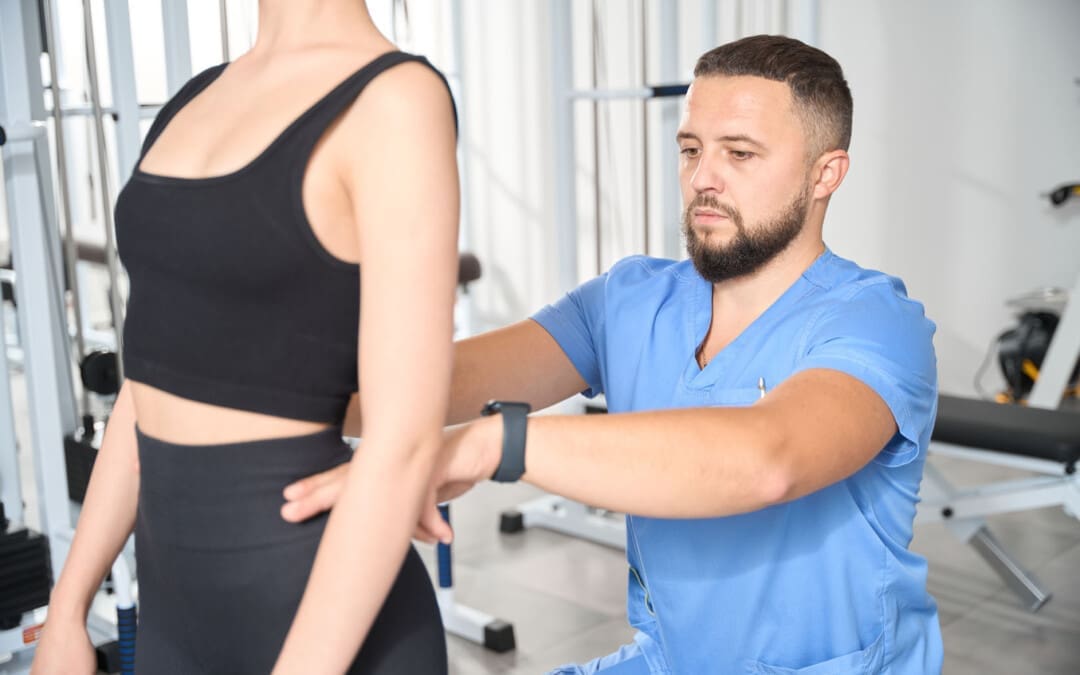
Tissue behind the spinal column, positioned at both the lower back and mid-back levels, is connected to the thoracolumbar fascia, also called the lumbodorsal fascia or LF. The fascia is a thick connective tissue that covers and supports all the body’s muscles, bones, tendons, ligaments, and organs. The fascia also contains nociceptive nerve endings, also known as free nerve endings, that arise from the central nervous system, i.e., the brain and spinal cord, which may be responsible for some forms of back pain and stiffness caused by injury or inflammation.
Anatomy
The thoracolumbar fascia is divided into three layers:
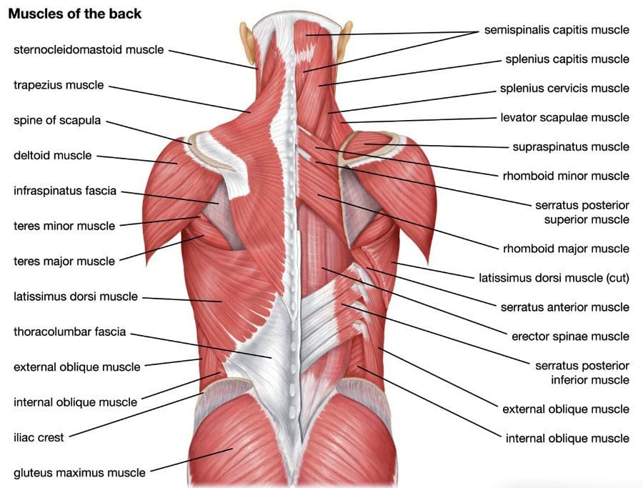
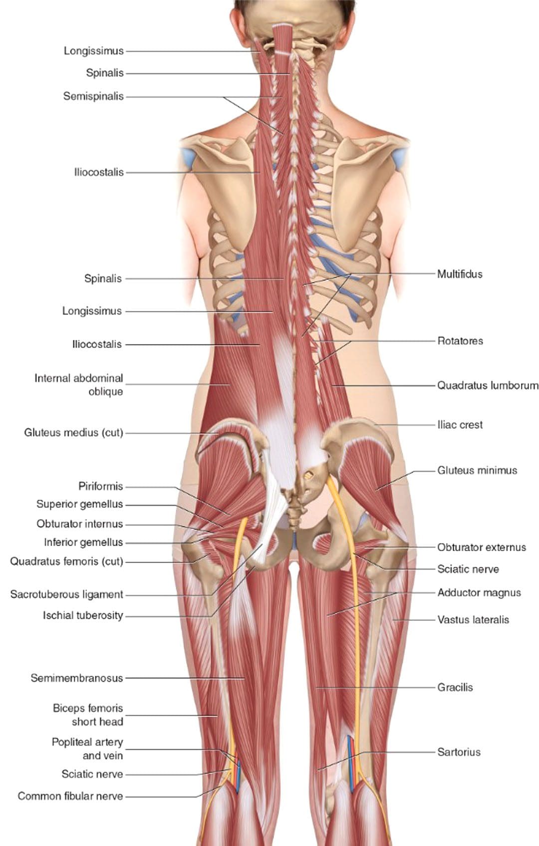
Many of the back muscles attach to the thoracolumbar fascia. The erector spinae muscle group, known as the paraspinals, runs longitudinally down the spine. They are attached to the thoracolumbar fascia and the bony spine. The lumbar part of the posterior layer of the thoracolumbar fascia extends from the lowest rib to the top of the hip bone or the iliac crest. On the same path, it connects with the transverse abdominal muscle. The thoracolumbar fascia connections help bridge the back muscles to the abdominal wall muscles. The latissimus dorsi, a large back muscle that bears and moves the body’s weight with the arms and shoulders, is also connected to the thoracolumbar fascia, with the fibers extending outward from the fascia. The front part of the thoracolumbar fascia, or anterior layer, covers a muscle called the quadratus lumborum. This muscle bends the trunk to the side, helps maintain a healthy posture, and is often focused on muscle-related lower back pain.
What the Fascia Does
The thoracolumbar fascia, examined from the back of an anatomical drawing or diagram, is diamond-shaped. Its shape, large size, and central location uniquely position it to unify and synchronize the upper body’s movements with the lower body’s. The fascia’s fibers are very strong, enabling the tissue sheath to lend support (Willard, F. H. et al., 2012) . The tissue is also flexible, enabling it to help circulate forces of movement and contralateral movements as the back muscles contract and relax. An example is walking.
Back Pain
Scientists and doctors don’t know for sure, but it’s possible that the thoracolumbar fascia may contribute to lower back pain. A study found that the fascia may generate back pain based on: (Wilke, J. et al., 2017)
Sustaining micro-injuries and/or inflammation, which are often related, may cause signal changes in the free nerve endings in the fascia. Nerve endings acquire information from the outer areas of the body, like skin and other fascia, and relay it back to the central nervous system. The theory is that when the fascia close to the skin becomes injured, damaged, and/or backed up with inflammatory chemicals and substances, it is communicated as pain and other sensations back to the brain and spinal cord.
After a back injury, tissues tighten and stiffen. Some studies of patients with back pain noted alterations in their thoracolumbar fascia.
Injuries tend to stimulate nerves, which can lead to increased sensitivity.
Injury Medical Chiropractic and Functional Medicine Clinic focuses on and treats injuries and chronic pain syndromes through personalized care plans that improve ability through flexibility, mobility, and agility programs to relieve pain. Our providers use an integrated approach to create personalized care plans for each patient, including Functional Medicine, Acupuncture, Electro-Acupuncture, and Sports Medicine principles. Our goal is to relieve pain naturally by restoring health and function to the body. If other treatment is needed, Dr. Jimenez has teamed up with top surgeons, clinical specialists, medical researchers, and rehabilitation providers to provide the most effective treatments.
Sciatica, Causes, Symptoms, and Tips
References
Willard, F. H., Vleeming, A., Schuenke, M. D., Danneels, L., & Schleip, R. (2012). The thoracolumbar fascia: anatomy, function and clinical considerations. Journal of anatomy, 221(6), 507–536. https://doi.org/10.1111/j.1469-7580.2012.01511.x
Wilke, J., Schleip, R., Klingler, W., & Stecco, C. (2017). The Lumbodorsal Fascia as a Potential Source of Low Back Pain: A Narrative Review. BioMed research international, 2017, 5349620. https://doi.org/10.1155/2017/5349620
For wrestling athletes or those thinking about getting into the sport, can knowing about common injuries help in rehabilitation and prevention?
Wrestling Injuries
Wrestling is an intense and demanding sport. Studies have found that football and wrestling are the two high school sports with the highest risk of serious injury to athletes (Center for Injury Research and Policy, 2009). The injury rate for college wrestlers is 9 injuries per 1,000 athlete exposures. (Kroshus, E. et al., 2018) While most wrestling injuries include strains and sprains, there can also be serious traumatic and unusual injuries. Using proper safety gear and learning correct techniques can significantly reduce the risk of injuries. The majority occur during competition.
Common
The most common wrestling injuries are similar to those in other sports and include:
Muscle Soreness
Muscle soreness that is experienced 12 to 48 hours after an intense workout or competition.
Resting is often all that is needed to recover.
Bruises and Contusions
Sparring, take-downs, and hard landings can result in various bruises and contusions.
Sprains and Strains
Rest, ice, compression, and elevation are recommended to treat sprains and strains immediately.
Ankle Sprains
Ankle sprains occur when surrounding ligaments stretch and tear around the joint.
Wrist Sprains
Typically, it occurs when stretching or tearing the ligaments.
Falling or landing on the hands is a common cause.
Overtraining Syndrome
Frequently occurs in athletes who train beyond the body’s ability to recover.
Dehydration
When trying to make weight, dehydration can be a serious health problem that many wrestlers experience.
Other Injuries
Other injuries common in wrestling:
Wrist tendinitis
Finger fractures
Iliotibial band syndrome
Meniscus tears
Groin pull
Hamstring pull or tear
Pulled calf muscle
Achilles tendonitis
Achilles tendon rupture
Clavicle/Collarbone fracture
Concussion
Serious
The forcing of a joint beyond its normal range of motion is the most common cause of serious injuries. The most serious wrestling injuries affect the neck, shoulder, elbow, and knee and include:
Neck
The cervical vertebrae are often forced into vulnerable positions during various techniques and movements, which can result in a neck injury. Common types include:
Neck Strain
Whiplash
Cervical Fracture
Shoulder
A combination of leverage and twisting causes most upper body and shoulder injuries during competition. Types of shoulder injuries include:
Rotator cuff injury
Shoulder separation
Shoulder dislocation
Elbow Dislocation
Elbows are under tremendous strain when maneuvering.
Dislocations of the radial head are often related to the athlete bracing for a fall on an outstretched arm during take-downs.
Knee
Most knee injuries occur to the ligaments of the knee joint.
These include anterior and posterior cruciate ligament or ACL/PCL injuries.
Safety
Wrestling requires flexibility, strength, and proper technique to prevent injury, combined with thorough instruction and coaching and following basic safety precautions. Some tips include.
Safety Gear
Wear appropriate headgear and mouthguards during practices, meets, and tournaments.
Improving Joint Flexibility
Wrestlers with a high degree of shoulder flexibility have fewer injuries.
The flexibility of the lower back, hamstrings, elbows, and cervical spine should also be worked on.
Gain or Lose Weight Safely
Avoid dramatic weight loss and weight-gaining strategies by maintaining healthy nutrition and hydration during the season.
Avoiding Dangerous Holds and Slam Moves
Safe wrestling techniques need to be followed as these can generate severe injuries.
Regardless of how common or seemingly not serious an injury or medical condition is, it’s important to rest and recover and tell a coach and health care professional, as some injuries and conditions can become serious if left untreated. Injury Medical Chiropractic and Functional Medicine Clinic focuses on and treats injuries and chronic pain syndromes through personalized care plans that improve ability through flexibility, mobility, and agility programs to relieve pain. Our providers use an integrated approach to create personalized care plans for each patient, including Functional Medicine, Acupuncture, Electro-Acupuncture, and Sports Medicine principles. Our goal is to relieve pain naturally by restoring health and function to the body. If other treatment is needed, Dr. Jimenez has teamed up with top surgeons, clinical specialists, medical researchers, and rehabilitation providers to provide the most effective treatments.
Perseverance and Power
References
Nationwide Children’s Hospital. (2024). Center for Injury Research and Policy. https://www.nationwidechildrens.org/research/areas-of-research/center-for-injury-research-and-policy
Kroshus, E., Utter, A. C., Pierpoint, L. A., Currie, D. W., Knowles, S. B., Wasserman, E. B., Dompier, T. P., Marshall, S. W., Comstock, R. D., & Kerr, Z. Y. (2018). The First Decade of Web-Based Sports Injury Surveillance: Descriptive Epidemiology of Injuries in US High School Boys’ Wrestling (2005-2006 Through 2013-2014) and National Collegiate Athletic Association Men’s Wrestling (2004-2005 Through 2013-2014). Journal of athletic training, 53(12), 1143–1155. https://doi.org/10.4085/1062-6050-154-17
Can physical therapies help treat a high steppage gait from injury or medical conditions and restore normal gait patterns for individuals who have or are developing one?
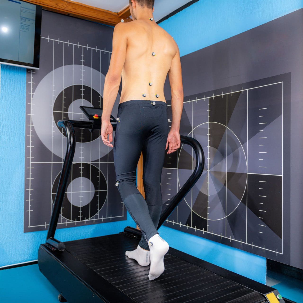
Walking or gait anthropometric analysis on a treadmill
Neuropathic Gait
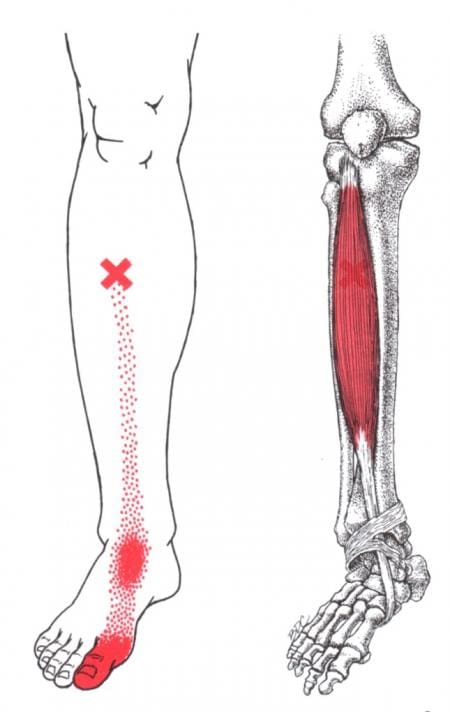
Neuropathic gait, aka equine or high steppage gait, is a type of walking abnormality that causes individuals to raise their hips to lift their legs higher than normal. It occurs when individuals have a foot drop or ankle equinus due to loss of dorsiflexion. As a result, the foot hangs with the toes pointing down, causing the toes to drag while walking. The foot may appear floppy when it drops. Foot drop is caused by weakness or paralysis of the anterior tibialis muscle in front of the shin bone. The anterior tibialis muscle contracts to help flex the foot and ankle while walking, ensuring the foot clears the floor and doesn’t drag. Individuals with anterior tibialis weakness or paralysis may have a neuropathic gait and excessively bend the hip and knee while stepping forward, lifting their leg high off the floor to clear the foot to avoid tripping. A physical therapy team can help with a high steppage gait pattern after illness or injury.
Causes
Conditions that can cause anterior tibialis weakness or paralysis and a high steppage gait pattern include:
Sciatica
Pain caused by compression or irritation of the sciatic nerve starts in the lower back and travels down the back of the leg. (McCabe, F. J., McCabe, J. P. 2016)
Peroneal Nerve Injury
Damage to the peroneal nerve branches from the sciatic nerve that help move the lower leg and foot. (Johns Hopkins Medicine. 2024)
Multiple Sclerosis
An autoimmune disease that damages nerve cells in the brain and spinal cord. (Taylor, P. N. et al., 2016)
Balance exercises will help improve overall proprioception, or the sense of the body’s position and movement.
Neuromuscular electrical stimulation, or NMES, can help improve the function of the muscle. (Hollis, S., McClure, P. 2017)
The electrical stimulation artificially contracts the muscle to restore proper function.
For anterior tibialis weakness caused by sciatica, back decompression exercises may be prescribed to relieve pressure off the sciatic nerve.
The exercises release the nerve to restore normal signal transmission up and down the nerve in the lower back.
Neuromuscular electrical stimulation may also be used to help improve muscle function.
Assistive Walking Devices
A therapist may suggest using an assistive device to help the patient walk properly. This could include a wheeled walker or a quad cane. A temporary solution to anterior tibialis weakness is to elevate the foot while walking with an elastic band. Tie a band around the leg below the knee and secure it around the ball of the foot. When swinging the leg forward, the band pulls the foot up. Using it as a temporary solution may help maintain safe mobility. Sometimes, paralysis of the anterior tibialis muscle can become permanent. In this case, individuals may benefit from a special brace called an ankle-foot orthosis. The brace helps to lift the foot and toes off the ground.
For individuals concerned about losing their balance and falling, there are ways to improve walking patterns to stay safe. A healthcare provider may recommend physical therapy to correct gait, strengthen the anterior tibialis muscle, improve balance, and educate on injury prevention. Individuals should discuss symptoms and conditions with a primary physician, healthcare provider, or specialist to guide them in the right direction and determine the best treatment.
Injury Medical Chiropractic and Functional Medicine Clinic uses an integrated approach personalized to the individual that focuses on what works for them and treats injuries and chronic pain syndromes through personalized care plans that improve ability through flexibility, mobility, and agility programs to relieve pain. If other treatment is needed, Dr. Jimenez has teamed up with top surgeons, clinical specialists, medical researchers, and rehabilitation providers to provide the most effective treatments.
Control Foot Motion and Posture
References
McCabe, F. J., & McCabe, J. P. (2016). An Unusual Presentation of Right-Sided Sciatica with Foot Drop. Case reports in orthopedics, 2016, 9024368. https://doi.org/10.1155/2016/9024368
Kaykisiz, E. K., & Unluer, E. E. (2017). An Unexpected Reason for Isolated Foot Drop: Acute Stroke. Pakistan journal of medical sciences, 33(5), 1288–1290. https://doi.org/10.12669/pjms.335.13593
Taylor, P. N., Wilkinson Hart, I. A., Khan, M. S., & Slade-Sharman, D. E. (2016). Correction of Footdrop Due to Multiple Sclerosis Using the STIMuSTEP Implanted Dropped Foot Stimulator. International journal of MS care, 18(5), 239–247. https://doi.org/10.7224/1537-2073.2015-038
Hollis, S., & McClure, P. (2017). Intramuscular Electrical Stimulation for Muscle Activation of the Tibialis Anterior After Surgical Repair: A Case Report. The Journal of orthopaedic and sports physical therapy, 47(12), 965–969. https://doi.org/10.2519/jospt.2017.7368
IFM's Find A Practitioner tool is the largest referral network in Functional Medicine, created to help patients locate Functional Medicine practitioners anywhere in the world. IFM Certified Practitioners are listed first in the search results, given their extensive education in Functional Medicine


 With contractures, the sarcomeres overly lengthen when the muscle fibers tighten. This increase in sarcomere length prevents the muscle from contracting normally, resulting in weakness. Muscle fibers are encased in an extracellular matrix, a mesh composed of collagen and other proteins that help transmit force and provide muscle contraction. Muscle contractures cause the amount of collagen within the extracellular matrix to increase, causing a stiffening of fibers that restricts movement. (

 What the Fascia Does
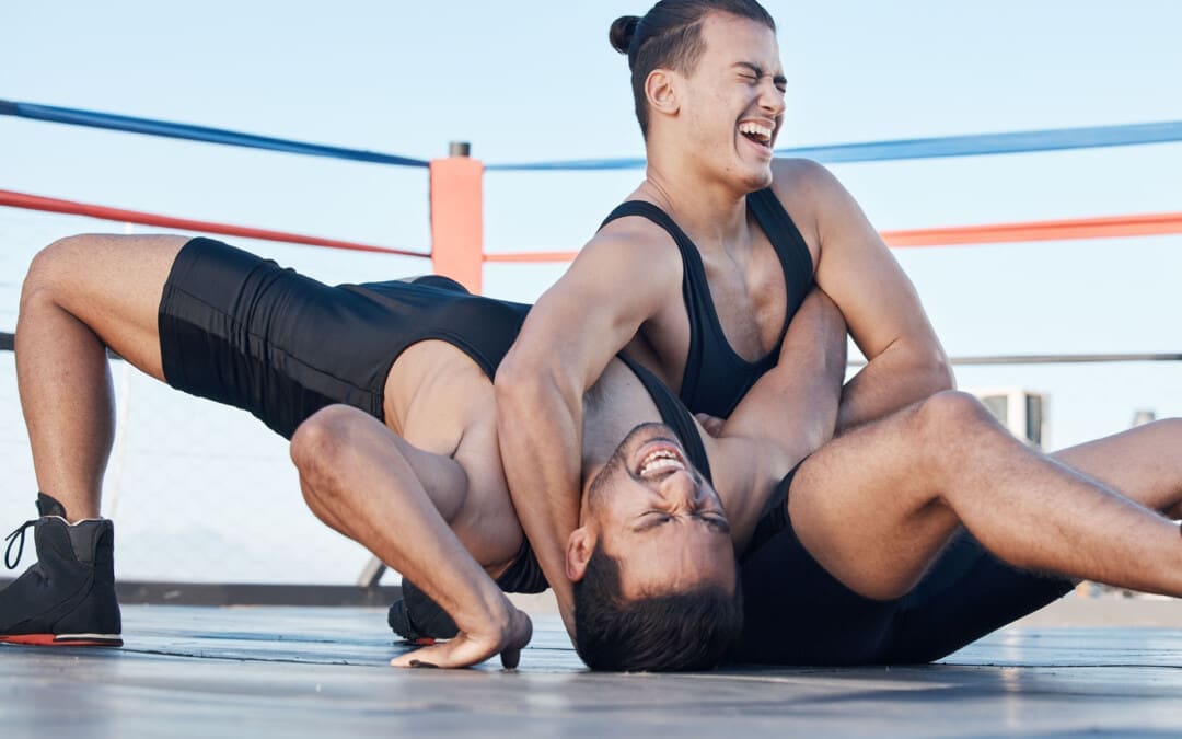


 Causes


