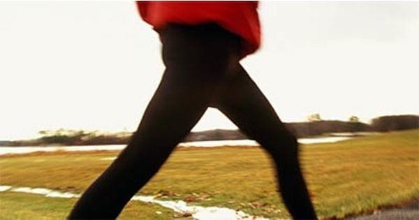
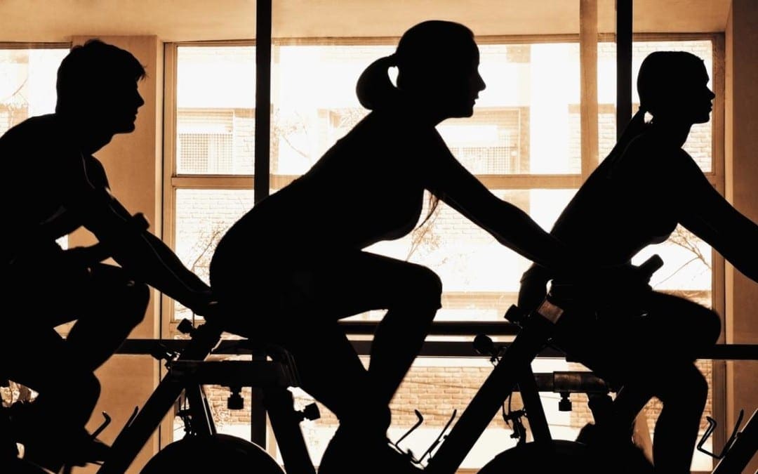
When you want to shed serious weight, walking might not even come to mind. But it should.
“Fast-paced walking, when combined with healthy eating, is hugely effective for weight loss,” says Art Weltman, PhD, director of exercise physiology at the University of Virginia. And those simple steps can have a big impact on your overall health, cutting your risk of everything from heart disease to depression. If your daily strolls haven’t made you skinny so far, your speed may be the problem. Many of us stride more like a window-shopper than a power walker. The goal�thankfully�isn’t crazy race-walker style; you just need to move at a challenging pace.
In studies, Weltman has found that women who do three short (about 30-minute) high-intensity walks plus two moderately paced recovery walks a week lose up to six times more abdominal fat than participants who simply stroll five days a week. (This despite the fact that both groups burn the exact same number of calories.)
The power walkers also drop about four times as much total body fat. “There is a strong relationship between intensity of exercise and fat-burning hormones,” says Weltman. “So if you’re exercising at a pace considered to be hard, you’re likely to release more of these hormones.” The best part: When women walk, deep abdominal fat is the first to go. That’s a scientific fact we can get excited about.
Another happy truth: Although you’re moving at a fast clip, power walking is still easier on the joints than running. “During walking one of your feet is always in contact with the ground,” says Weltman, “but during running there’s a float stage where your whole body is lifted in the air. Then you come back down and subject your body to the impact.”
That’s why walking is a smart long-term fitness plan. To get you off on the right foot, here’s a complete primer, from how to tweak your speed for maximum burn to what gear you need (hint: almost none). Follow the workouts and wisdom�along with healthy eating�and not only can you lose those extra 10 pounds in three weeks, but you will have a no-fuss plan that you can do anywhere, anytime.
Dial In Your Speed
To make sure your pace is on point, use these guidelines from exercise physiologist Tom Holland, author of Beat the Gym. For maximum fat burn, aim for 30 minutes at power-walk intensity three days a week (see the walking plan on the next page). That time can be completed all at once, or you can break it up into spurts with recovery strides (stroll or brisk walk) in between.
Stroll. Think window-shopping pace, or an intensity of 4 on a scale of 10. It burns about 238 calories an hour.
Brisk walk. This means an effort of 5 or 6 on a scale of 10. It burns up to 340 calories an hour (at a 3.5 to 4 mph pace). While you can gossip about Mad Men, you need to catch your breath every few sentences.
Power walk. You’re torching off approximately 564 calories an hour (at a 4 to 5 mph pace). Moving at this clip, using your arms to help propel you forward and taking longer strides, your effort should be a 7 or 8 on a scale of 10. Talking is possible only in spurts of three or four words, but…you’d…rather…focus…on…breathing.
The Amped-Up Plan
This program from Holland mixes a regular walking workout with interval routines to help you reach your power-walking quota of 30 minutes, three times a week. Aim to walk on three nonconsecutive days and either rest or cross-train on the other ones. If you cross-train (think power yoga or swimming), you’ll help your body recover; and with our diet, you’ll progress more quickly to dropping up to 10 pounds in three weeks.
Tempo day
Burns about 220 calories:
Warm-up: Stroll for 5 minutes.
Workout: Maintain a power-walk intensity for 30 minutes.
Cooldown: Stroll for 3 to 5 minutes.
Long-Interval Day
Burns about 355 calories:
Warm-up: Stroll for 5 minutes.
Interval Workout: Maintain a hard power-walk intensity (8 on a scale of 10) for 5 minutes. Recover at a brisk pace for 1 minute. Repeat for a total of 6 intervals.
Cooldown: Stroll for 3 to 5 minutes.
Short-Interval Day
Burns about 405 calories:
Warm-up: Stroll for 5 minutes.
Interval Workout: Maintain a hard power-walk intensity (8 on a scale of 10) for 2 minutes. Recover at a brisk pace for 1 minute. Repeat for a total of 15 intervals.
Cooldown: Stroll for 3 to 5 minutes.
Walk This Way
When it comes to walking, your body and brain know what to do. Makes sense�you’ve been doing it since you took those first wobbly baby steps. But with these three form fixes, you’ll maximize your burn, big time.
Chin up. Your gaze shouldn’t be aimed at your feet, no matter how snazzy your sneakers are. Instead, focus on a point about 10 feet ahead of you. This will keep your stride longer and your neck comfortably in line with your spine.
Activate your abs. When you brace your core�pulling your belly button toward your spine�you automatically trigger good posture.
Squeeze your glutes. Your backside literally propels you through your walk. To get the most oomph�so you can go longer and faster�keep your glutes tight. Bad visual, good strategy: Imagine squeezing a winning lottery ticket between your cheeks.
4 Ways To Burn More Fat
So you’re the impatient type? Use these tricks to up the challenge and calorie burn.
Add hills. When you hit the hills on a treadmill or in your neighborhood, you increase your calorie burn by nearly 20 percent�and that’s just on a 1 to 5 percent incline.
Go off-road. Head out for a light but brisk hike and you’ll torch about 430 calories in just an hour. Credit the uneven terrain�which forces you to work harder. Sub this in for one of your weekly power walks.
Swing your arms. With elbows bent at 90 degrees and hands in loose fists, move your arms in an arc, keeping elbows tight to your body. This helps drive you forward, says Weltman, builds upper-body strength and can increase your burn by up to 10 percent.
Make longer strides. Instead of taking more steps, “work on increasing your stride length,” Weltman says. “You’ll cover more ground,” and that means more fat fried.
Itching To Run?
Let’s face it: Some of us would rather just run. But if you go from zero to Usain Bolt on your first outing, you might end up sidelined. Use this guide from Holland to transition from walking to running safely.
For the running newbie: Do this modified version of the Short-Interval Day (see “The Amped-Up Plan,” left) three times a week: Run for one minute (work up to two minutes over the course of a couple of weeks), walk for one minute and repeat for a total of 15 intervals. Do this for a few weeks, then transition to the Long-Interval Day, running for five minutes and walking for one, repeating for a total of six intervals. The goal is to eventually tackle Tempo Day�running for 30 minutes nonstop.
For the on-and-off runner: Assuming you have some running experience under your belt, you can dive right into the Long-Interval Day plan, subbing in running for the power walks. The intervals should be challenging, and the Tempo Day run should be done at a hard but comfortable pace.
For the gym-goer: You can also use this plan to cross-train, doing the exact same routines while on the elliptical machine, rowing machine or stationary bike.
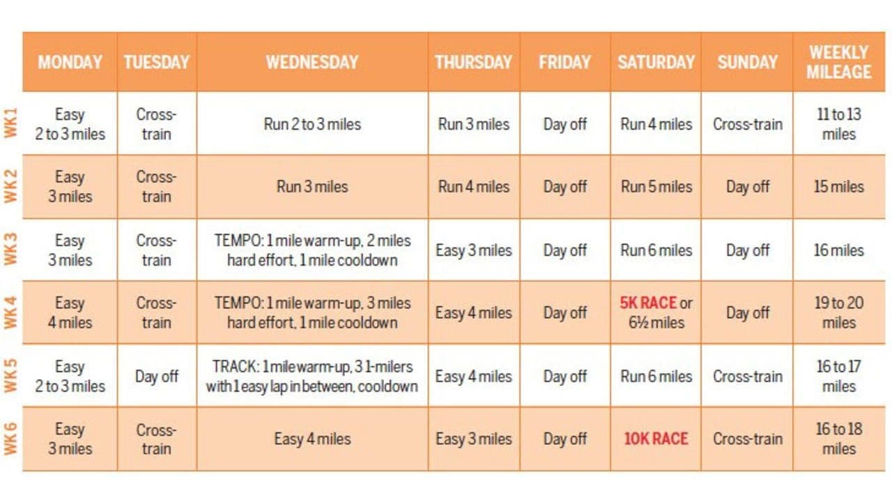
Thinking of training for a 10K race? This plan is perfect for you if you’ve already mastered a 5K race and you’re a “sometimes” runner who is able to do at least three miles without stopping a couple of days a week, most weeks. The goal for this 10K training plan, which was developed by developed by running coach Paula Harkin, co-owner of Portland Running Company in Oregon, will be to increase your endurance, run for an hour straight, and tackle a 10K by the end of 6 weeks.
The 10K training plan: This program incorporates a combo of tempo (effort of 7 or 8 on a scale of 1 to 10), and longer runs (effort of 5 or 6 ) to build endurance. “Combining these workouts will help you get faster while also making sure you can cover the distance,” says Harkin. Do a combination of running and cross-training on alternate days. As the weeks pass, alternate between building up the speed bursts and balancing out the recovery time. Focus on covering the distance, not your pace. Kick off with an easy 2 to 3 mile run. Over 6 weeks, try to work up to running 6 miles.
How to train smarter for your 10K race
1. Make three the magic number. If you’re used to running twice a week, says Jonathan Cane, an exercise physiologist and co-founder of City Coach Multisport in New York City, “three times is your sweet spot�. You’ll get a big bump in both speed and endurance, but it’s not so much more that you’ll risk getting injured.” And if weight loss is a goal, remember that adding just one extra day of running helps you burn an additional 300 to 400 calories, depending on your pace and size.
2. It’s OK to hit the treadmill. Some running purists say there’s no substitute for the outdoors, but all things being equal, “your heart and lungs don’t really know the difference between the road and the treadmill,” says Cane. So if it’s late in the day, raining or just not a good time to go outside but you really want to keep up your training, feel free to hit the “on” button. To compensate for a lack of wind resistance and natural terrain changes, keep the treadmill deck set at a 1% incline.
3. Turn down the music. Yes, pumping JT through your earbuds can power you up that hill, but don’t forget to tune in to how your body feels. “At this stage, you know you can already run for a while,” says Cane. “But it’s important to be aware of cues: how heavy you are breathing, or if you have a small twinge in your knee and need to slow down. It helps keep you from getting injured and makes you more aware of when you can bump up your pace or give a little more effort.”
For more information, please feel free to ask Dr. Jimenez or contact us at 915-850-0900 .
Chiropractic and Athletic Performance
Many athletes who are injured performing their specific sport or physical activity, frequently seek treatment from chiropractors. Chiropractic care focuses on the prevention, diagnosis and treatment of injuries and conditions affecting the musculoskeletal and nervous system. While chiropractic is a safe and effective form of conservative care for a variety of ailments, chiropractic can also be utilized to enhance athletic performance.
If you’ve been thinking of running a 5K, you should: Running just might be the most convenient workout going. You don’t need to be a skilled athlete, and there’s no fancy equipment involved; just lace up your sneaks and go. It’s also one of the most efficient ways to blast fat and burn calories��about 600 an hour.
Sure, walking has its benefits, but research shows that running kicks its butt when it comes to shedding pounds. One study of 47,000 runners and walkers, from the Lawrence Berkeley National Laboratory in Berkeley, Calif., found that the runners burned more calories and had a far greater decrease in BMI over a six-year period. The joggers who started out heaviest (those with a BMI over 28) lost up to 90 percent more weight than the walkers did.
Dropping pounds and toning up are hardly the only benefits of this killer cardio workout: You’ll also reduce your risk of heart disease and diabetes, boost your mood, temper stress, and build muscle, especially in the lower body and core. You don’t even need to dedicate a lot of time to reap these rewards; do 20 to 30 minutes, three to four days a week, and you’ll see significant improvement.
Ready to hit the road? Here’s a 5K training plan for beginning joggers. And it’s smart to add in one day of cross-training (think cycling or swimming) to rev up calorie burn and help prevent injury. Soon enough, you’ll feel as if you were born to run.
This is the 5K training plan for you if: You’re new to running and generally don’t work out consistently.
Your goal: By the end of 8 weeks, be able to run for 20 minutes straight��and build up to a 5K challenge.
The 5K training plan: This eight-week, three-days-a-week plan by Nike+ Run Club coach Julia Lucas mixes walking with running to help prevent injury and overexertion. OK running for longer? Shorten or discard the walking time.�Your ideal pace? One where you can carry on a conversation, but still feel like you’re doing a brisk walk.
How to train for your 5K smarter:�
1. Start off on the right foot.�Making a small investment in gear now will save you loads of aggravation later��you’ll feel more comfortable and avoid aches. “A good pair of�running shoes�can help ward off injuries like knee pain,” says Susan Paul, an exercise physiologist and training program director at Orlando Track Shack Fitness Club in Orlando, Florida.�Get a gait analysis at your local running store (it’s usually free) to help determine your ideal shoe type.
2. Stop side stitches.�Beginners are often plagued by this cramp, which strikes like a boxer’s body blow and happens when an overworked diaphragm begins to spasm. To ease the pain, slow down and forcefully exhale each time your opposite foot strikes (so if the stitch is on your right side, breathe out when your left foot comes down). It also helps to massage the area with two fingers. And don’t eat too much before you head out; a full stomach can be a culprit.
3. Think tortoise, not hare.�“The biggest mistake most new runners make is they start out way too fast,” says Paul. “It takes time for your body to get used to the demands of running. You have to condition your muscles, ligaments, tendons and bones, not just your heart and lungs.” No matter how tempted you are to push yourself, don’t. Slow and steady wins the calorie-burn race!
For more information, please feel free to ask Dr. Jimenez or contact us at 915-850-0900 .�
Chiropractic and Athletic Performance
Many athletes who are injured performing their specific sport or physical activity, frequently seek treatment from chiropractors. Chiropractic care focuses on the prevention, diagnosis and treatment of injuries and conditions affecting the musculoskeletal and nervous system. While chiropractic is a safe and effective form of conservative care for a variety of ailments, chiropractic can also be utilized to enhance athletic performance.
There�s no question that high intensity exercise burns mega-calories in minimum time. But when that high intensity comes in the form of running, jumping, and sprinting, you�re only as strong as your weakest link�and for many that means your hips and knees, which are more vulnerable to injury as impact levels rise. �Stress and impact are amplified with high intensity training routines and sudden force can cause damage to joint cushions, tendons, and muscles,� says Nicholas DiNubile, MD, orthopedic surgeon and best-selling author of the FrameWork series of books. �This is especially true as we age, or if you�ve had previous injuries, as your musculoskeletal frame is not as durable or limber.”
The good news is you can raise your heart rate and rev your metabolism to burn calories and fry fat without the jarring impact. Here are 10 relatively gentle workouts your joints (and your waistline) will love. Note: Just because a workout is low impact doesn�t mean it�s zero risk. You can further minimize your chances of pulling a muscle or straining a joint by starting your exercise sessions slowly so you can warm up your muscles and lubricate your joints before turning up your efforts.
For years, companies have been hawking vibration as a form of exercise�from those fat-jiggling waist belts in the �80s to the vibrating platforms found in many gyms today. Now, a new study in mice suggests there might be some truth to the idea that a vibrating machine may be able to deliver some of the same benefits as actual physical activity.
The new research, published in the journal Endocrinology, found that mice with diabetes and obesity had similar improvements in muscle mass and insulin sensitivity over 12 weeks when they were assigned to either 45 minutes of daily treadmill walking or 20 minutes of daily whole-body vibration. Both groups gained less weight and improved more in overall health than sedentary mice that received neither intervention.
How Vibration Exercise Works
Whole-body vibration consists of a person (or, in the study�s case, a mouse) sitting, standing or lying on a platform. The platform�s vibrations send tiny shockwaves through the body, causing muscles to contract and relax multiple times per second.
The obese mice in the study also had low bone density, a common side effect of excess weight in both animals and humans. While treadmill exercise did improve this measure over 12 weeks, the vibration technique did not. Both interventions did, however, increase levels of a protein involved in bone formation, suggesting that longer-term treatments could potentially help prevent future bone loss.
Vibration is not a cure-all for the problems associated with sedentary life, say the study authors, and results seen in mice don�t necessarily translate to humans. Before vibration-based treatments can be widely recommended, these results would need to be replicated in clinical trials. (A 2009 study found that vibration platforms helped obese people lose body fat, but other metabolic benefits have been less studied in people.)
The authors also point out that the study was designed to test the benefits of vibration on obese, unhealthy mice for whom regular exercise is difficult. Young, healthy mice, who were also included in the study, did not reap the same benefits from the whole-body vibration.
Lead author Meghan McGee-Lawrence, assistant professor of cellular biology and anatomy at Augusta University, says that vibration therapy might be an effective way to help people who are extremely overweight or have other limitations that keep them from regular physical activity.
�If you are able to exercise, we�d still recommend exercise as a first choice option,� says McGee-Lawrence. But for people who find it difficult to work out in a traditional way, �our study suggests it may be possible to obtain some of the same beneficial effects of exercise in a different, less strenuous way.�
For vibration to have these benefits, though, a lot of things have to be just right. �The frequency and magnitude of the stimulus, and how long it�s applied, need to be optimized to achieve the outcome you desire,� says McGee-Lawrence. It is possible to have too much of a good thing, she adds. Exposure to higher-level vibration in occupational settings, for example, can actually have a harmful effect on bone.
Pete McCall, an exercise physiologist with the American Council on Exercise, says that benefits of whole-body vibration are �100% legit.� Vibration platforms can be used for exercise warm-ups, cool-downs or for certain moves like squats, planks and Pilates poses.
�When you�re on one of these platforms, the oscillations add gravity and force, which are really important for building strength,� he says. For people who are too overweight or too out of shape to exercise safely and comfortably, he adds, vibration training can �introduce exercise to the body in a relatively low-stress environment.�
�Standing on a vibrating platform for 5, 10, 15 minutes can actually make cells stronger, maybe help them lose a little weight, and get them better prepared to eventually start exercising,� he says.
The American Council on Exercise warns that whole-body vibration machines may affect pacemakers and other electronic implants, and that pregnant women and anyone with a history of seizures, tumors or thrombosis should not use them.
For generally healthy people, McCall stresses that they should be used as a supplement to moderate-to-vigorous physical activity, not a replacement. �There�s no additional demand for oxygen, so the lungs and heart don�t have to work any harder,� he points out. �It�s not going to give you the important cardiovascular benefits that real exercise will.�
For more information, please feel free to ask Dr. Jimenez or contact us at 915-850-0900 .
Whole Body Wellness
Overall health and wellness can be achieved by following a proper nutrition and engaging in regular exercise and/or physical activities. While these are some of the most common ways to ensure whole body health and wellness, visiting a qualified and experienced healthcare professional can also grant your body additional benefits. Chiropractic care, for instance, is a safe and effective alternative treatment option utilized by people to maintain well-being.
Spinning might look about the same as outdoor cycling or riding a stationary bike, but in many ways, it�s a far more intense workout�and one of the easiest to overdo.
First, there aren�t many (if any) breaks in spin class. �When you�re biking outside, you have to be aware of road dangers like water and cars, so you have to slow down at times,� says Dr. Maureen Brogan, an assistant professor of medicine at New York Medical College who has conducted research into spinning. Especially if you�re a novice road rider, it�s going to take some time before you�re comfortable enough on two wheels to really push yourself hard for long distances. That�s not the case on a spinning bike, where newbies can hop on and ride hard from the start.
Popular spinning studios like Flywheel and SoulCycle have their riders clip their feet into the stationary bikes. As long as the wheels turn, legs keep pumping. Combine this always-working aspect with the thumping music, enthusiastic instructors and energetic group atmosphere of most spinning studios, and it�s easy to get intense exercise and burn calories by the bucketful.
�The muscles you use on a spinning bike, the gluteus maximus and the quadriceps, are some of the largest in your body, so you�re using a lot of energy,� Brogan says�600 calories an hour, and sometimes more.
Spinning: High-Intensity Workout
This puts spinning near the top of the list when it comes to high-intensity workouts. A study from Sweden found that one hour of spinning was enough to trigger the release of blood chemicals associated with heart stress or changes. While that may sound like a bad thing, these blood chemicals�or biomarkers�signal the heart is getting a good workout. �These kinds of findings have also been seen with prolonged exertion such as marathons,� says study author Dr. Smita Dutta Roy of Sahlgrenska University Hospital in Sweden. While more research is needed to tease out the risks or benefits associated with exercise of this intensity, she says that some of the biomarker shifts her team observed could lead to blood vessel repair and renewal.
It can also help improve body composition, decrease fat mass and lower blood pressure and cholesterol, says Jinger Gottschall, an associate professor of kinesiology at Penn State University. Some of her research has shown that high-intensity spinning can increase fitness levels even in trained athletes. �In every study we�ve done, we�ve seen increases in heart and lung capacity,� she says. She calls spinning �the optimal cardio workout,� and says you can get all the intensity of a treadmill or stair-climber without the impact.
The low-impact nature of spinning makes it great exercise for older adults or people recovering from orthopedic injuries, she adds. �Because you can adjust the resistance and moderate the pace and intensity of your ride, it opens the door for many people to participate,� she says.
But it�s also easy for people who are new to spinning to overexert themselves. �If you�re not used to vigorous exercise, or to exercising the large lower-body muscles involved in spinning, you can overdo it,� Brogan says. She�s a kidney expert by training, and some of her research has linked spinning to rhabdomyolysis, a condition in which muscles break down to the point that they release a protein that can poison the kidneys. �People have swollen legs or trouble walking, and sometimes they take aspirin or NSAIDs for the muscle pain, which is the last thing they should do because those can also damage the kidneys,� she says. Problems like this can set in a day or two after spin class, she says.
While overexertion is possible with any form of exercise, she says the risks during spinning may be higher�especially when you consider that some spinners lose up to a liter of water during an hour-long session.
Even for trained athletes, there�s some evidence that spinning too often may lead to trouble. A study in the Journal of Strength and Conditioning Research concluded that spinning may push some people past the threshold at which the exercise is beneficial. �If indoor cycling were used as an everyday training activity, it is possible that the overall intensity would be too high and possibly contribute to developing nonfunctional overreaching,� the authors of that study write. (�Nonfunctional overreaching� is sports science lingo for a workout that�s so strenuous it leads to fatigue and performance declines, rather than fitness improvements.)
Overall, spinning is exceptional exercise. But if you�re new to it, you need to ease in and give your muscles time to adapt to its intensity. Even if you�re an experienced athlete, pushing yourself to your limit the first or second time you get on a spinning bike may be risky, Brogan says. Even once you�ve found your spinning legs, daily sessions may still be overkill.
But if you�re looking for a high-intensity workout a few days a week�and especially if running or other forms of vigorous aerobic exercise hurt your joints�spinning may be the ideal way to keep your heart and body in shape.
For more information, please feel free to ask Dr. Jimenez or contact us at 915-850-0900 .
Whole Body Wellness
Overall health and wellness can be achieved by following a proper nutrition and engaging in regular exercise and/or physical activities. While these are some of the most common ways to ensure whole body health and wellness, visiting a qualified and experienced healthcare professional can also grant your body additional benefits. Chiropractic care, for instance, is a safe and effective alternative treatment option utilized by people to maintain well-being.
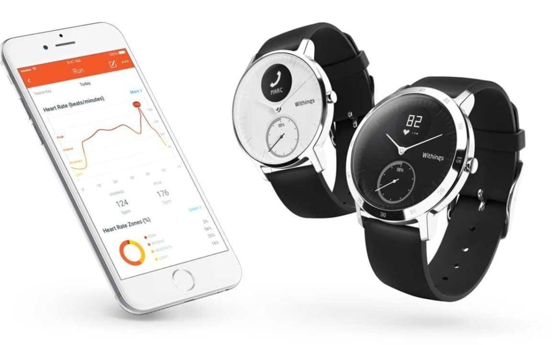
The first thing you notice when you see the Withings Steel HR is how little it resembles what most of us have come to expect from most fitness trackers in terms of shape and design. At first glance, it looks and feels like a classic analog watch: the silicone strap (or leather, if you choose that option) connects to a slim, round display with minute markers dotting the edges and hands to show you the time. The one giveaway that this is not your typical watch is the small circular OLED display at the top, and little would you know that housed behind that round face is enough hardware to power a very capable fitness tracker.
What is the Withings Steel HR?
The Withings Steel HR (from $179.95, amazon.com, bestbuy.com) has all the standard features one would expect from a fitness tracker: it lets you know how many calories you’ve burned, offers heart-rate monitoring, automatically tracks your sleep, counts your steps, pulls notifications from your phone and has an alarm to gently wake you up.
Aside from its design, one thing that really helps to set the Steel HR apart from a lot of other fitness trackers is its incredible battery life. Most fitness trackers can last you a few days before it needs to be charged again. Withings boasts that it can last up to 25 days on a single charge and that it has the longest battery life of any heart-rate tracker on the market. After spending nearly three straight weeks with the Steel HR on my wrist and over a dozen workouts tracked in that time, there was still enough juice left in the tank to leave me with little reason to doubt them on that claim.
Using the Steel HR is simple enough. There is a single button on the right side of the watch that you press to turn on the display and to cycle through the different stats. It was a minor nuisance having to press the button every time to turn the OLED display on, as the display doesn�t automatically light up if you pull the watch up toward your face. However, the goal meter below the OLED display is a nice touch on the watch face. You can quickly see how close you are to hitting your daily activity goal by looking at the meter as it climbs towards 100%.
Withings likely had to make some compromises for the sake of battery life and overall look and I found the OLED display to be one of te biggest weaknesses of the watch. While it�s relatively easy to read in regular usage if you�re just casually checking to see how many steps you walked, all of that changes if you�re engaged in a workout or on a run. If you�re running, trying to read the display or cycling through the different screens was a bit of a challenge. And you won�t get a lot of useful real-time feedback during a workout. With such a small screen, you can�t just casually glance at the display to get a quick readout. Notifications from your phone also don�t give you any real useful information.
It appears that GPS functionality was another compromise that Withings likely made. Not every fitness tracker comes equipped with GPS built into the device. Instead, they typically rely on your phone�s GPS to track you. However, the Steel HR isn�t even able to use your phone�s GPS, so it�s hard to put much faith into the accuracy of the distance measurements. It instead uses sensors and data about you to generate the distance, which is far from accurate. If you�re a runner, this is a pretty glaring omission.
Workouts are automatically detected, which seemed to work fine. Like most fitness trackers that have this feature, it will occasionally falter, but you can typically rely on it to record your workout in case you forgot to do so. You also have the option of tracking a workout manually by holding the button on the side, and workouts can be logged in the app after you sync your watch.
The heart-rate monitor on the Steel HR has two modes: workout and smart mode. Smart mode is constantly running and takes measurements about every 10 minutes, which helps to preserve battery life. However, if you switch to workout mode, it continuously runs throughout your workout. Every fitness tracker that I�ve used has had odd, random spikes that occur every now and then�this was no different for the Steel HR. I frequently wore a Fitbit Charge 2 along with the Steel HR to compare and noticed that the Steel HR always tracked higher than the Fitbit. It wasn�t enough to give me any real cause for concern, but it�s something that should be noted nonetheless. The Steel HR also seemed slower to normalize, frequently spiking at the start of a workout before eventually coming back down to a more reasonable level.
Sleep tracking worked as well as the Fitbit Charge 2, and the fact that you barely notice the watch while you�re wearing it makes it easier to wear at night compared to some of the bulkier fitness trackers out there.
If you�re the type of person that wants the features of a fitness tracker but don�t like how most of them look, the Withings Steel HR may be the tracker for you. It has a sleek and stylish design that looks good whether you�re in the gym or out on the town. Most people that see it won�t even be able to tell that you�re wearing a fitness tracker. For those that may be runners or just a little more serious about fitness, you may find the Steel HR lacks in some areas. Although, even with the compromises that Withings made with the Steel HR, this is the best hybrid option available.
For more information, Please feel free to ask Dr. Jimenez or contact us at 915-850-0900 .
Whole Body Wellness
Overall health and wellness can be achieved by following a proper nutrition and engaging in regular exercise and/or physical activities. While these are some of the most common ways to ensure whole body health and wellness, visiting a qualified and experienced healthcare professional can also grant your body additional benefits. Chiropractic care, for instance, is a safe and effective alternative treatment option utilized by people to maintain well-being.
No matter what size you are, you may have some fat between your back and arms that spills over your bra�also known as “bra bulge.” Some of that’s due to genetics, but an unbalanced workout routine can play a role as well. Many women neglect their arms, chest, and back due to a misguided fear of getting bulky. And while you may not love your bra bulge, a weak upper body can also wreck your posture and bring on back pain.
Barry’s Bootcamp instructor and celeb personal trainer Astrid Swan wants you to get over your fear of upper body workouts, so she created this exclusive routine for Health. This seven-move sequence revs your heart rate to torch calories and melt away fat from your whole body (including your back). Plus, these sculpting exercises will perk up your posture, which may minimize the appearance bra bulge. Use a pair of heavy dumbbells; Swan suggests 12 lb. or higher, depending on your strength level.
Pushups to superman
Lower all the way to the floor slowly as you do a pushup. Lay flat and extend arms forward to a superman position, lifting chest and thighs off the floor. Pull elbows down to goal-post position and lower your body down to press back up into the top of the pushup. Do 10 repetitions.
Plank renegade rows
Start in plank position, using the dumbbells as handles. Keep feet slightly wider than hip width, be sure to keep hips parallel to the floor and abs engaged. Alternate renegade rows, 10 repetitions per side, for 20 total reps.
Start with dumbbells on shoulders, feet hip-width apart with feet slightly turned out. Lower down into a squat position, keeping chest tall and abs engaged. Power from the core and glutes to press the weights above head in a press. Be sure to avoid locking out your knees as you press the weights up to the top. Do 10 reps.
Combine all three moves minus the superman. Using the dumbbells as handles, do one pushup, at the top of the pushup complete renegade rows on the right side then left side. Next, jump your feet forward sand land in the bottom of your squat. Be sure to keep your core engaged as you thrust the weights above your head. Do 10 repetitions.
Snatch passes
Using one dumbbell, bend knees slightly to hoist up the weight and snatch it to the top. Be sure to keep hips tucked and keep a small bend in the knees as you extend the arm. Return to starting position and pass the weight to the other side. Do 10 reps per side.
Triceps extensions
Depending on your strength, you can continue using both weights or drop down to one. Do a triceps extension slowly; think three counts to lower and one count to press up. Keep your elbows tight, framing your face. Do 10 reps, slowly and with intention.
Hold one dumbbell on each end. Start with the dumbbell right under your chin, and pass it around your head clockwise for 10 reps, then counterclockwise for another 10 reps. Be sure to keep your elbows tight, framing your face, and bring the weight around your head (like a “halo”) with elbows bent.
For more information, please feel free to ask Dr. Jimenez or contact us at 915-850-0900 .
Chiropractic and Athletic Performance
Many athletes who are injured performing their specific sport or physical activity, frequently seek treatment from chiropractors. Chiropractic care focuses on the prevention, diagnosis and treatment of injuries and conditions affecting the musculoskeletal and nervous system. While chiropractic is a safe and effective form of conservative care for a variety of ailments, chiropractic can also be utilized to enhance athletic performance.
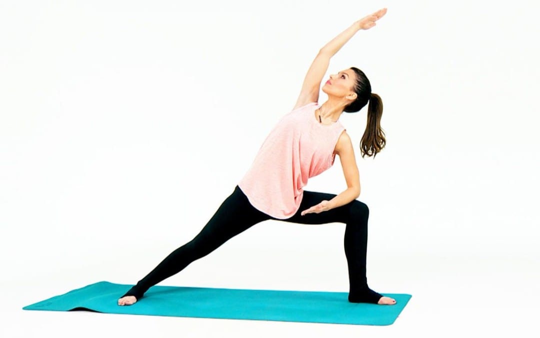
In celebrity yoga instructor Hilaria Baldwin�s new book The Living Clearly Method: 5 Principles for a Fit Body, Healthy Mind & Joyful Life, she outlines her method for combining movement and mindfulness to lead a more balanced life. Her strategy includes five simple principles: perspective, breathing, grounding, balance, and letting go. But how exactly does the celeb and mother of three stay so centered with such a crazy schedule? One of her go-to ways to bring all her principles into practice is through yoga. Watch this video to learn one of Baldwin�s go-to yoga sequences that incorporates all elements of her method into a movement format.
Here, she guides you through tree pose, high lunge, warrior II, side angle A, plank, chaturanga, upward facing dog, and finally downward facing dog. After completing the sequence on one side, you roll up slowly and repeat it on the other. Within this sequence, you get a touch of balance and grounding, thanks to tree pose. And as you conquer the challenging transitions from high lunge to side angle A, consistent breathing plays a role, helping you get calm and centered.
Baldwin describes this practice as an �all-purpose flow,� meaning you can do it at any time of day�whether you want to wake your body up in the morning, get your heart rate up in the afternoon, or close out your day the right way with a moving meditation before you go to sleep. Whenever you do this sequence, it�s an efficient workout that can easily become a part of your daily routine. The goal is to simply set aside some time for yourself to help unwind and connect with your body and mind. Watch the video to learn more about how to master this flow.
For more information, please feel free to ask Dr. Jimenez or contact us at 915-850-0900 .�
If just thinking about a HIIT workout seems tiring, let the music play. A Journal of Sports Sciences study found that when people performed four 30-second all-out sprint intervals on a bike while listening to music, they had a more positive workout experience than when they pedaled without tunes�possibly because music helps distract you from the, uh, discomfort of a tough sprint. Try biking (or running or rowing) it out to one of these songs recommended by Steph Dietz, lead instructor at Cyc Fitness, an indoor-cycling studio chain.
“They�re perfect for intervals because they slowly build to the chorus, where the beat drops, picking up speed and intensity,” says Dietz. “Each song has about two or three HIIT interval builds.”
IFM's Find A Practitioner tool is the largest referral network in Functional Medicine, created to help patients locate Functional Medicine practitioners anywhere in the world. IFM Certified Practitioners are listed first in the search results, given their extensive education in Functional Medicine