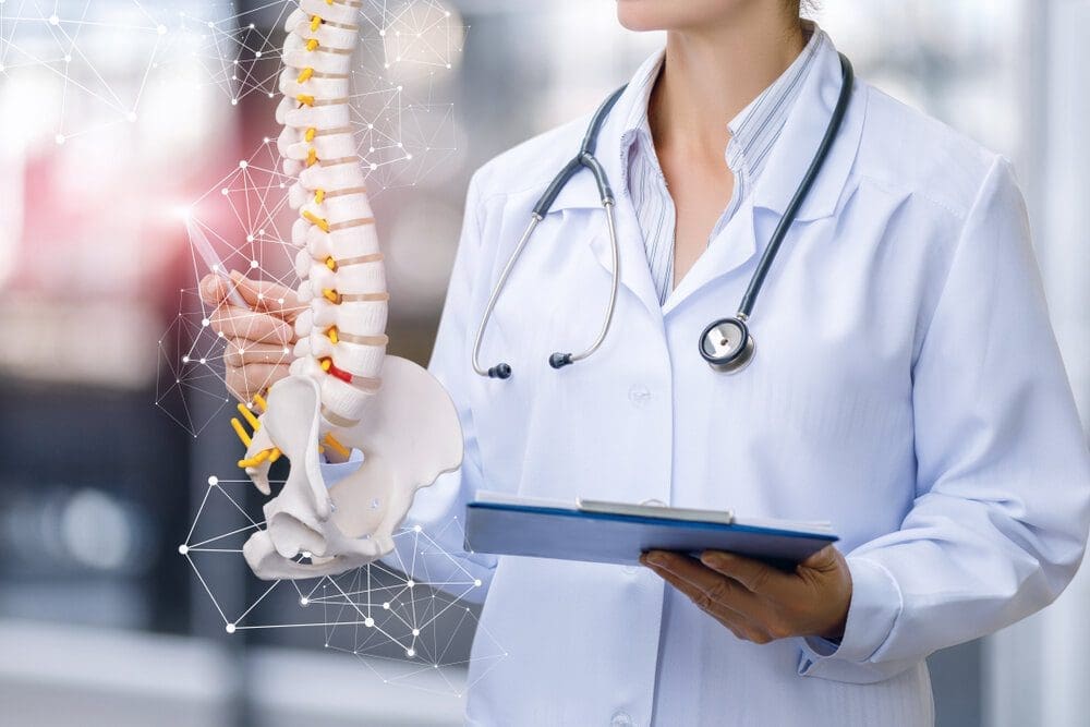
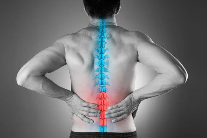
The body utilizes the spine to make sure that everything is moving, bending, twisting, and turning without feeling any pain from the back. The spine is an S-shaped curve protected by ligaments, soft tissue from the musculoskeletal system, the spinal cord, and spinal discs. When the back suffers from an injury or has pulled a muscle, it can cause chronic issues to the back and make a person’s life miserable. Luckily there are therapeutic ways to relieve chronic back issues and can alleviate the symptoms it has caused to the individual. In this article, we will be looking at musculoskeletal disorders and their symptoms and how decompression therapy has an effect on alleviating musculoskeletal disorders from the back. By referring patients to qualified and skilled providers specializing in spinal decompression therapy. To that end, and when appropriate, we advise our patients to refer to our associated medical providers based on their examination. We find that education is the key to asking valuable questions to our providers. Dr. Alex Jimenez DC provides this information as an educational service only. Disclaimer
Can my insurance cover it? Yes, it may. If you are uncertain, here is the link to all the insurance providers we cover. If you have any questions, please call Dr. Jimenez at 915-850-0900.
What Are Musculoskeletal Disorders?
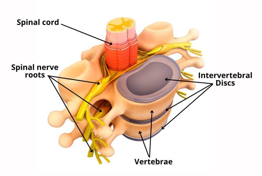
As research studies have stated, the musculoskeletal system combines bone, muscles, tendons, ligaments, and soft tissues that work together to support the body’s weight and help individuals move. The spine is located at the back of the body, where it connects to the musculoskeletal tissues and keeps it upright. Many individuals must keep their musculoskeletal system healthy and functional; however, a wide range of disorders and conditions can affect the musculoskeletal system making the body succumb to diseases and injuries that can limit its movement. This is known as musculoskeletal disorders.
Research studies have found that musculoskeletal pain and disorders affect the bones, joints, ligaments, muscles, and tendons throughout the entire body. Sometimes the pain can become acute, and it can become sudden and severe or chronic, which can hinder a person’s ability to do any daily activities. Some of the most common types of musculoskeletal disorders that can affect the body include:
Tendon and ligament pain: Sprains, strains, and overused tissues
The Symptoms
Research studies have shown that musculoskeletal disorders are the leading source of pain and disability worldwide. With a variety of back and neck disorders, arthritic conditions, and soft tissue syndromes that involve the tendons, ligaments, muscles, and cartilages that make up the main bulk of musculoskeletal disorders, it can cause many people to go to their primary physician and get time off work. Other research studies have shown that the symptoms that are caused by musculoskeletal disorders include:
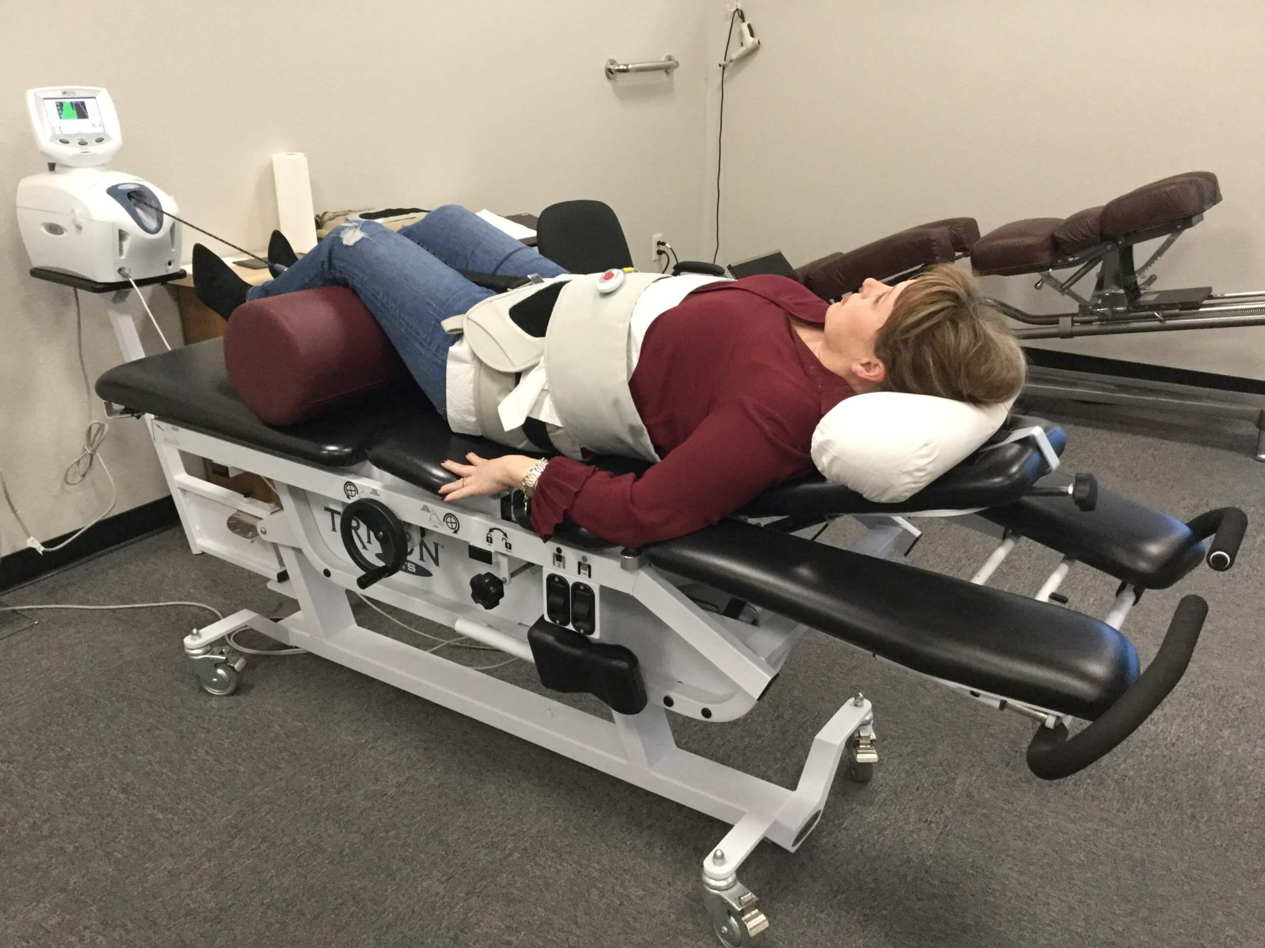
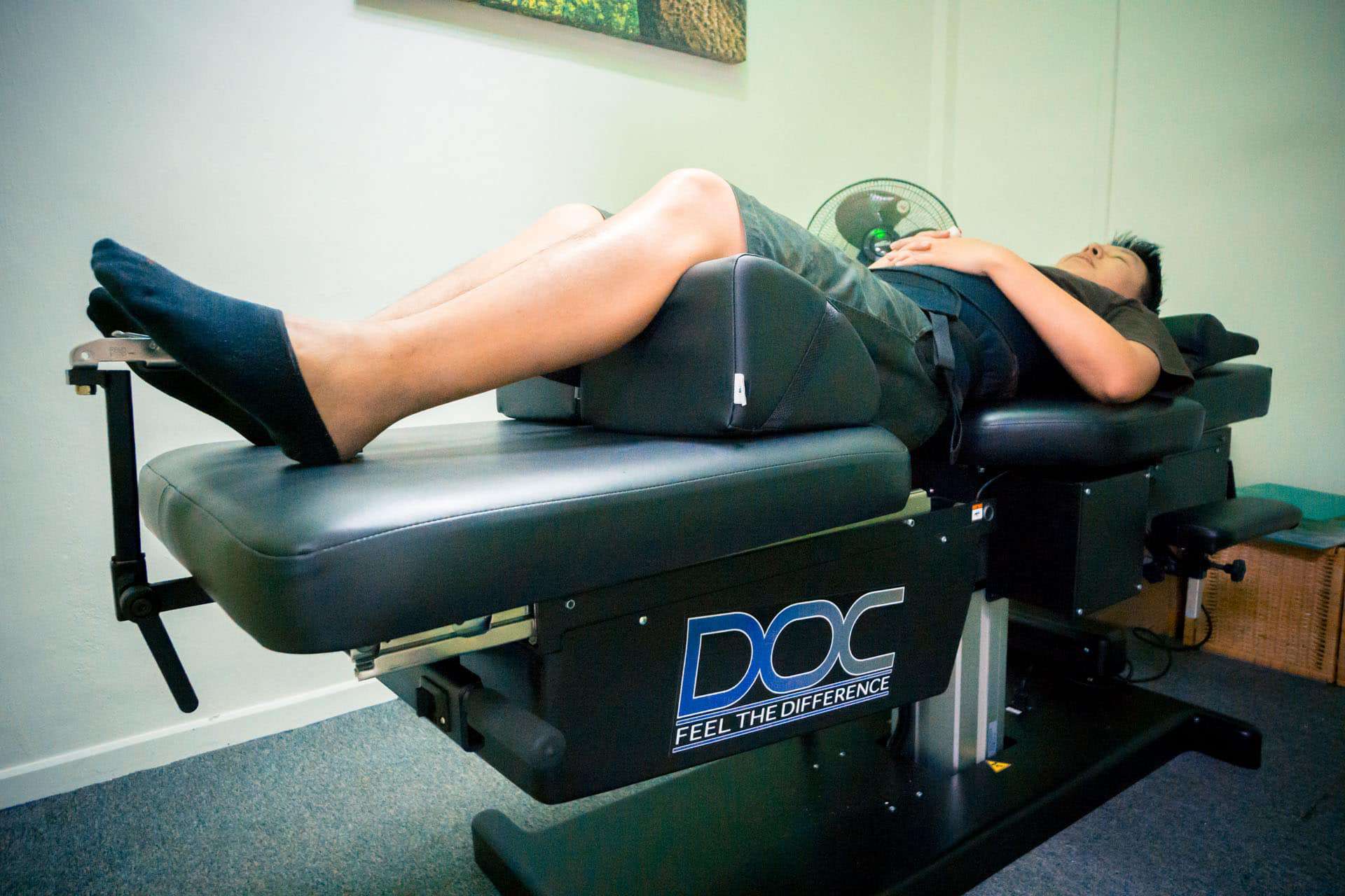
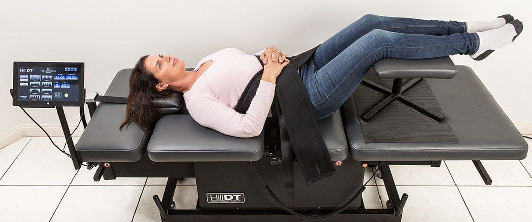
The video above shows how the Chattanooga Triton is being used to alleviate back issues that have been affected by musculoskeletal disorders. Traction therapy is a form of spinal decompression therapy that utilizes traction on a person’s spine, gently stretching it. This will cause the beneficial nutrients and oxygen to go back into the spine and alleviate pain in the back. Since back pain is one of the most common types of musculoskeletal pain that can make a person miserable, decompression therapy can help with low back pain and make a person get back their quality of life. If you want to learn more about spinal decompression therapy, this link will explain the benefits of spinal decompression and how it can alleviate low back pain symptoms.
How Does Decompression Therapy Help Musculoskeletal Disorders?
Decompression therapy is a non-surgical treatment that allows for individuals who have musculoskeletal disorders like low back pain. Decompression therapy allows the individual to lie down on the traction table, be strapped in, and the traction machine gently pulls on the spine to cause instant relief. Research studies have found that utilizing decompression therapy and even physical therapy can help improve the lumbar range of motion, back muscle endurance, and functional disability that musculoskeletal disorders have caused. Other research studies have also shown that non-surgical spinal decompression therapy can reduce pain in the back and promote an increase in the spinal disc height and restore it. When individuals start to feel relief from their back, they can continue with their wellness journey.
Conclusion
All in all, musculoskeletal disorders are a wide variety of disorders that affects the bones, ligaments, tissues, and muscles all over the body. Low back pain is one of the most common musculoskeletal disorders that causes the individual to have pain and stiffness in the back muscles. Luckily treatments like spinal decompression therapy allow individuals who are suffering from feel relief from chronic back issues and gently stretch the spine to allow the beneficial nutrients to re-hydrate the spinal disc. With the combination of physical therapy, many individuals won’t have to suffer any longer, knowing that decompression may be their relief.
References
Amjad, Fareeha, et al. “Effects of Non-Surgical Decompression Therapy in Addition to Routine Physical Therapy on Pain, Range of Motion, Endurance, Functional Disability and Quality of Life versus Routine Physical Therapy Alone in Patients with Lumbar Radiculopathy; a Randomized Controlled Trial.” BMC Musculoskeletal Disorders, BioMed Central, 16 Mar. 2022, https://www.ncbi.nlm.nih.gov/pmc/articles/PMC8924735/.
Apfel, Christian C, et al. “Restoration of Disk Height through Non-Surgical Spinal Decompression Is Associated with Decreased Discogenic Low Back Pain: A Retrospective Cohort Study.” BMC Musculoskeletal Disorders, BioMed Central, 8 July 2010, https://www.ncbi.nlm.nih.gov/pmc/articles/PMC2912793/.
Malik, Khalid M, et al. “Musculoskeletal Disorders a Universal Source of Pain and Disability Misunderstood and Mismanaged: A Critical Analysis Based on the U.S. Model of Care.” Anesthesiology and Pain Medicine, Kowsar, 15 Dec. 2018, https://www.ncbi.nlm.nih.gov/pmc/articles/PMC6348332/.
Medical Professionals, Cleveland Clinic. “Musculoskeletal Pain: Types, Causes, Symptoms & Treatment.” Cleveland Clinic, 10 Mar. 2021, https://my.clevelandclinic.org/health/diseases/14526-musculoskeletal-pain.
Medical Professionals, Cleveland Clinic. “Musculoskeletal System: Arthritis, Lower Back Pain, Bones, Muscles.” Cleveland Clinic, 11 Dec. 2020, https://my.clevelandclinic.org/health/articles/12254-musculoskeletal-system-normal-structure–function.
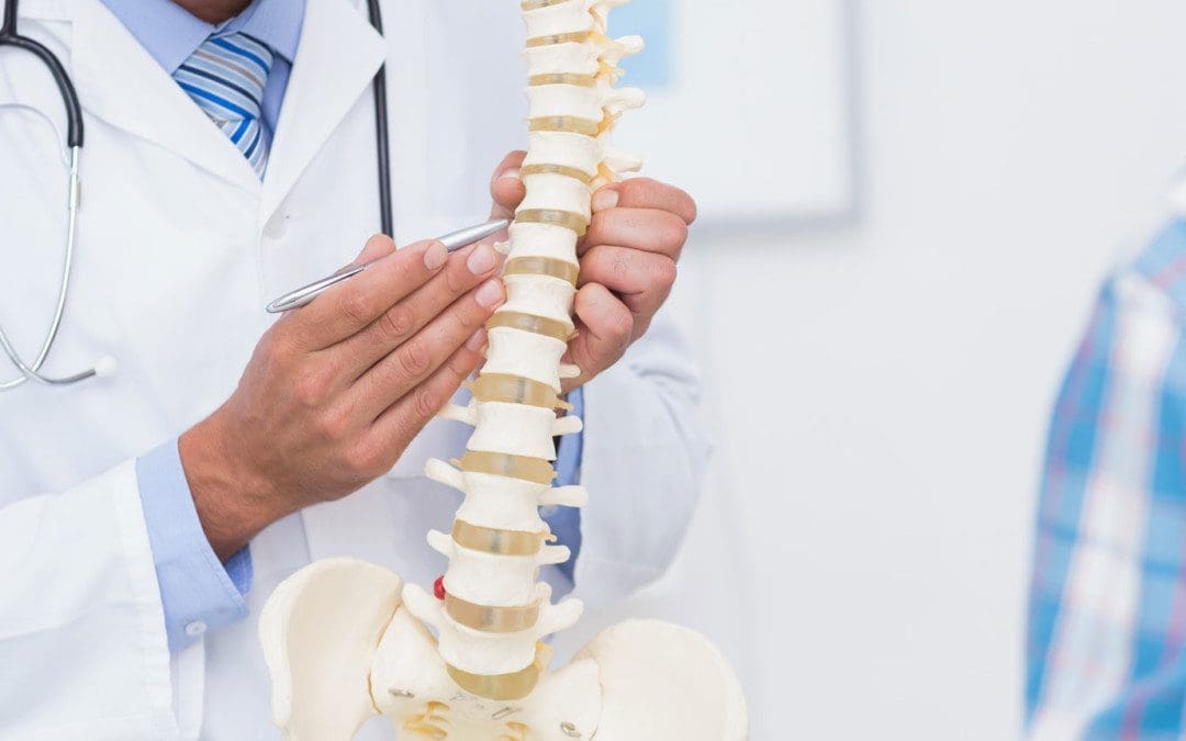
Inside the body, the spine allows it to move around and do all sorts of things without pain. The spine is protected by ligaments, soft tissue from the musculoskeletal system, spinal discs, and the spinal cord in an S-shaped curve that holds the body together. When the back gets injured or pulls a muscle, it can cause unwanted back issues that can cause a person to be in pain. When this happens, the individual suffering from back pain will be hindered from their daily activities and be miserable if it is not treated right away. Luckily, treatments like spinal decompression therapy can help alleviate back pains and other issues that affect the body’s back and spine. In this article, we will be looking at what DDD is, its symptoms, and how spinal decompression can help relieve DDD. By referring patients to qualified and skilled providers specializing in spinal decompression therapy. To that end, and when appropriate, we advise our patients to refer to our associated medical providers based on their examination. We find that education is the key to asking valuable questions to our providers. Dr. Alex Jimenez DC provides this information as an educational service only. Disclaimer
Can my insurance cover it? Yes, it may. If you are uncertain, here is the link to all the insurance providers we cover. If you have any questions, please call Dr. Jimenez at 915-850-0900.
What Is DDD?
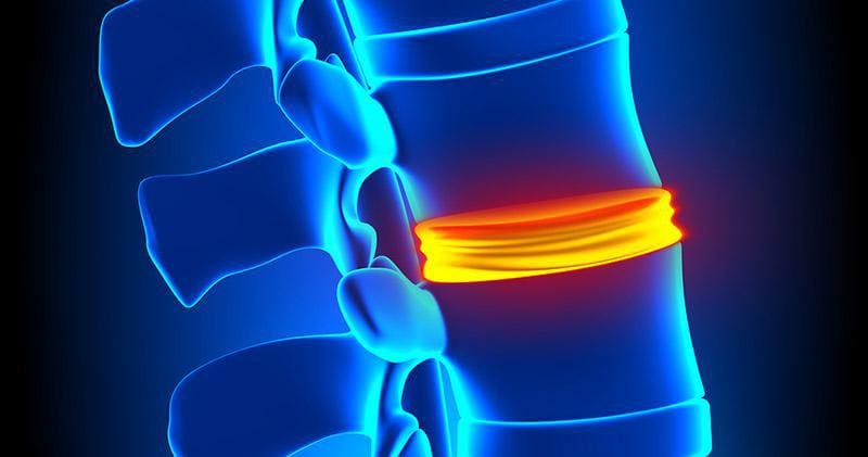
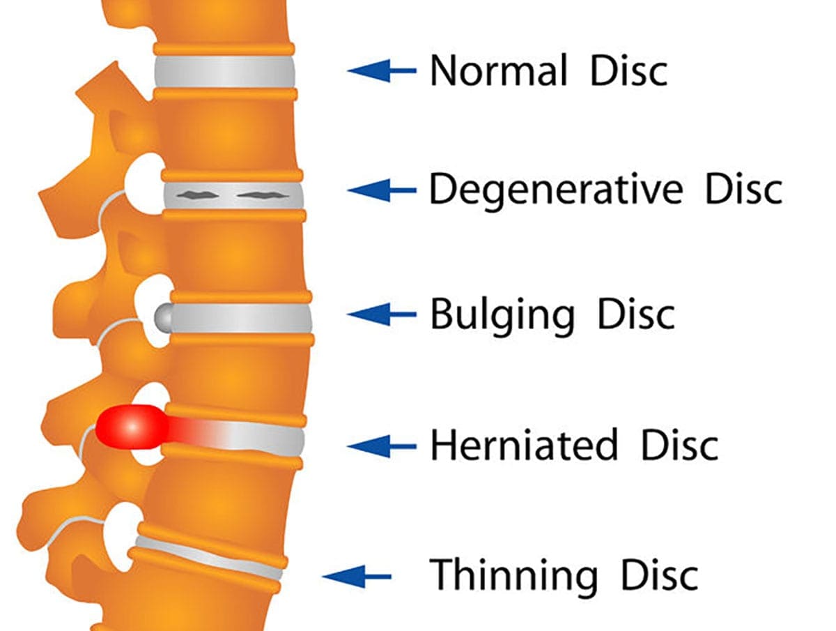
Since low back pain is common for many individuals worldwide, some of the causes of low back pain are DDD or degenerative disc disease. Research studies show that DDD (degenerative disc disease) usually happens when the spinal discs start to wear down naturally due to age. The spinal disc is a rubbery cushion between the spine’s vertebrae, and they help people move comfortably. When the spinal disc starts to wear and tear naturally through age, it can cause the vertebrae to rub against each other and cause pain.
Other research studies have found that DDD is often misunderstood since the symptoms affect either the neck or the back, causing sudden shooting pain in the arms or legs. DDD can also progress over time if it is not treated right away, causing the individual to be in pain and can make them unstable. Research studies have found that the two main factors of DDD are inflammation and abnormal micro-motion instability. How inflammation plays in DDD is that the inflammatory proteins from the spinal disc interiors are leaked when degeneration affects the spinal disc and causes swelling around the spinal structure. Abnormal micro-motion instability starts to affect the spinal discs’ outer layer by causing small, unnatural motions in-between the vertebrae, thus causing irritation and tension to the surrounding muscles, joints, and nerve roots, making the person become unstable and be in more pain.
The Symptoms Of DDD
Research studies have shown that when DDD affects the spine, it also affects the nervous system surrounding the spinal disc. DDD also causes structural failure, a radial tear in the annulus fibrosis, herniated disc, and calcification to the endplate of the spine. Since flare-up pains and abnormal stress on the spine can be due to recent activities or suddenly come up for no apparent reason, research studies have shown that pain episodes from DDD can last between a few days to several weeks before going back to be low-level back pain. Some of the common symptoms of DDD can include:
Increased pain from lifting heavy objects, bending or twisting the spine
Sudden sharp, radiating pain from the cervical or lumbar parts of the spine
Increased pain from holding a position for too long
Spinal Decompression Therapy & DDD-Video
The video above shows how spinal decompression can help alleviate DDD (degenerative disc disease). Spinal decompression therapy is utilized for many individuals suffering from chronic back issues like DDD, herniated disc, and low back pain. What spinal decompression therapy does is that it allows the individual to lay on a traction table and start to gently stretch their spine to relieve any issues that were causing back pain. The beneficial nutrients are reabsorbed into the spinal disc when the spine is gently pulled. The individual will begin to feel instant relief after a couple of sessions. If you want to learn more about spinal decompression therapy, this link will explain the benefits of spinal decompression and how it can alleviate low back pain symptoms.
How Spinal Decompression Therapy Relieves DDD
Many treatments help alleviate DDD symptoms and chronic low back pain as they provide relief to many individuals. One of the treatments that have been getting attention is spinal decompression therapy. Research studies have shown that many individuals suffering from DDD utilize non-surgical spinal decompression therapy to reduce pain and cause an increase in spinal disc height. This will allow the compressed spinal disc to be decompressed and improve disc health. Other research studies have also shown that since the degenerative process and mechanical effects of DDD can affect the spine, spinal decompression therapy allows traction to reduce the pressure off the spinal disc by gravity and soft tissue, enabling sufficient tension to extend spinal separation and the intervertebral disc. Spinal decompression also allows negative pressure within the intervertebral disc by increasing its hydration and reducing pressure off the nerve root.
Conclusion
The spine is an S-shaped curve protected by ligaments, soft tissue from the musculoskeletal system, the spinal discs, and the spinal cord allowing it to hold the body together. The body is home to the spine, where it can move around without feeling any sort of pain. When a person injures their back or pulls a muscle, it can cause unwanted back issues to hinder them from doing various daily activities. Sometimes the spinal disc wear and tear naturally causes symptoms like a herniated disc or DDD (degenerative disc disease) to affect the spine and the back by causing sharp, shooting radiate pain to affect the body. Luckily, treatments like spinal decompression therapy alleviate these symptoms by gently stretching the spine and causing instant relief to the individual.
References
Apfel, Christian C, et al. “DRX9000 BMC Study.” DRX9000® & DRX9000c® Global Trusted Suppliers Excite Medical, 18 Apr. 2022, https://excitemedical.com/drx9000-research/drx9000-bmc-study/#section-tab|0.
Choi, Jioun, et al. “Influences of Spinal Decompression Therapy and General Traction Therapy on the Pain, Disability, and Straight Leg Raising of Patients with Intervertebral Disc Herniation.” Journal of Physical Therapy Science, The Society of Physical Therapy Science, Feb. 2015, https://www.ncbi.nlm.nih.gov/pmc/articles/PMC4339166/.
Choi, Yong-Soo. “Pathophysiology of Degenerative Disc Disease.” Asian Spine Journal, Korean Society of Spine Surgery, June 2009, https://www.ncbi.nlm.nih.gov/pmc/articles/PMC2852042/.
The back is part of the musculoskeletal system, held by the spine to keep the body upright. The spine allows the body and the back to twist, turn, bend, and move side to side without feeling pain. However, when the body suffers from a pulled muscle or an injury, it can strain the back and cause back issues over time if not treated right away. Luckily, many treatments for low back pain can help a person get back to their daily activities. In this article, we will be looking at what causes low back pain and its symptoms and how lumbar traction decompression can help alleviate low back pain for individuals. By referring patients to qualified and skilled providers specializing in spinal decompression therapy. To that end, and when appropriate, we advise our patients to refer to our associated medical providers based on their examination. We find that education is the key to asking valuable questions to our providers. Dr. Alex Jimenez DC provides this information as an educational service only. Disclaimer
Can my insurance cover it? Yes, it may. If you are uncertain, here is the link to all the insurance providers we cover. If you have any questions, please call Dr. Jimenez at 915-850-0900.
What Are The Causes Of Low Back Pain?
The spine is encompassed by ligaments, soft tissue, the spinal cord, and nerve roots that allow the body to twist and bend. The lower back allows the motion of twisting and turning to happen, as research studies have shown the lumbar spine provides the support, strength, and flexibility to all the muscles, joints, and nerves in the body. Sadly, the lumbar spine is susceptible to injury and pain, as it supports the upper body’s weight and anything from a pulled muscle from lifting heavy objects to being injured in an accident. Since low back pain is common for many individuals, the causes of low back pain occur at any moment, as research studies have shown. Some of the reasons that occur for low back pain include:
Fractures
Spinal disc problems: herniated disc or DDD (degenerative disc disease)
Other research studies have shown that low back pain causes can also be due to mechanical and soft tissue issues that can damage the intervertebral disc, compress the nerve roots, and even cause improper movement to the spinal joints, causing the individual to be in immense pain.
Low Back Pain Symptoms
When a person is suffering from low back pain, the pain can range from a mild, dull ache in the lower back to a sharp shooting pain that can travel from the lower back all the way down to the foot. Research studies have shown that low back pain symptoms can begin as an acute symptom that can turn into chronic if it is not treated right away. Some of the most common low back pain symptoms that can occur include:
Dull aching pain due to muscle spasms, limited mobility, and aches on the hips and pelvis
Traveling pain down to the buttocks, legs, and feet causing sciatica to form
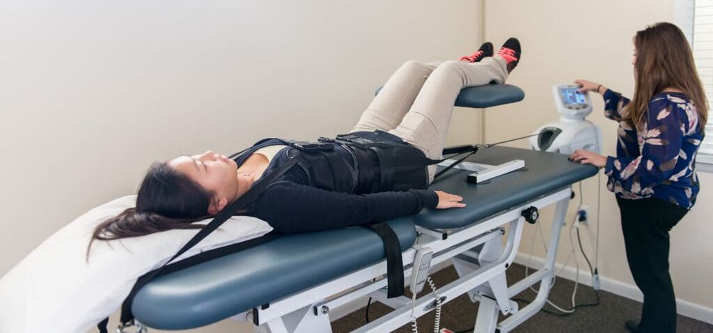
The video above shows how traction decompression therapy is used for individuals suffering from low back pain. Research studies have found that low back pain is common and one of the reasons many individuals see their primary physicians and even miss work. Some of the treatments like traction decompression utilize a traction table to gently pull the spine to cause instant relief to the individuals suffering from low back pain. What traction decompression does is that it allows the beneficial nutrients to be put back into the spine as well as decompressing the compressed discs back to their original form and alleviating the pain. With the combination of physical therapy, many individuals suffering from low back pain will begin to feel much better and continue on their wellness journey. If you want to learn more about spinal decompression therapy, this link will explain the benefits of spinal decompression and how it can alleviate low back pain symptoms.
Lumbar Traction Decompression For Low Back Pain
As many people don’t know, lumbar traction is one of the oldest known treatments for low back pain. Lumbar traction decompression has been used to reduce muscle contraction and reduce the symptoms of low back pain in prone. Research studies have shown that the efficacy of lumbar traction therapy for treating low back pain will allow a significant reduction in the pain intensity that the individual is feeling. Utilizing physical therapy that incorporates local heat and exercise and traction decompression therapy will provide excellent results in dampening the effects of low back pain. Other research studies have shown that mechanical traction on individuals suffering from low back pain due to having herniated discs will be significantly improved and restore the spinal discs to their original state. This will allow many individuals to be pain-free and continue their daily activities.
Conclusion
All in all, low back pain is common for many individuals and is one of the reasons why many individuals visit their primary physicians and get out of work. Low back pain can range from a dull, mild ache to a sudden, sharp pain that can cause muscle weakness and other back issues that hinder a person from performing their daily activities. Luckily, treatments like lumbar traction decompression therapy allow the individual suffering from low back pain to be lying on a traction table and have their spine be gently pulled. This gentle stretching allows the beneficial nutrients back into the spine and restores the compressed disc by increasing their height and reducing the pain. Afterward, many individuals will continue with their daily activities without suffering anymore.
References
Borman, Pinar, et al. “The Efficacy of Lumbar Traction in the Management of Patients with Low Back Pain.” Rheumatology International, U.S. National Library of Medicine, Mar. 2003, https://pubmed.ncbi.nlm.nih.gov/12634941/.
Cheng, Yu-Hsuan, et al. “The Effect of Mechanical Traction on Low Back Pain in Patients with Herniated Intervertebral Disks: A Systemic Review and Meta-Analysis.” Clinical Rehabilitation, U.S. National Library of Medicine, Jan. 2020, https://pubmed.ncbi.nlm.nih.gov/31456418/.
Medical Professionals, Cleveland Clinic. “Low Back Pain: Causes, Diagnosis & Treatments.” Cleveland Clinic, 18 Jan. 2021, https://my.clevelandclinic.org/health/diseases/7936-lower-back-pain.
Peloza, John. “Causes of Lower Back Pain.” Spine, Spine-Health, 20 Apr. 2017, https://www.spine-health.com/conditions/lower-back-pain/causes-lower-back-pain.
Peloza, John. “Lower Back Pain Symptoms, Diagnosis, and Treatment.” Spine, Spine-Health, 20 Apr. 2017, https://www.spine-health.com/conditions/lower-back-pain/lower-back-pain-symptoms-diagnosis-and-treatment.
Staff, Mayo Clinic. “Back Pain.” Mayo Clinic, Mayo Foundation for Medical Education and Research, 21 Aug. 2020, https://www.mayoclinic.org/diseases-conditions/back-pain/diagnosis-treatment/drc-20369911.
Spinal stress can affect nerve health. Neuropathy happens when disease or damage is sustained in the nerves that transmit messages from the brain through the spinal cord to the whole body. The source of the damage can be inside the spine, where a herniated disc could be squeezing the nerves, impeding or completely blocking blood circulation until deterioration begins to disease or damage nerve receptors. Removing the pressure from the spine and reversing the stress on the nerves can be done through manual or motorized spinal decompression.
Spinal Stress and the Nerves
The peripheral nervous system is comprised of three types of nerves that are directly influenced by the central nervous system, each with a distinct function which is why there is a wide range of symptoms associated with neuropathy. The types of nerves include:
Sensory nerves receive sensations from the skin like heat, cold, pleasure, and pain.
Spinal nerves contain sensory and motor fibers giving them sensory and motor functions. The spinal nerves receive sensory messages from the skin, internal organs, and bones. Any disruption from a bent, crushed, or entangled nerve group will not allow proper blood circulation and message transmission, causing delayed responses, tingling, numbness, and pain. If left untreated, it could cause permanent damage that can lead to chronic pain. Decompression therapy accelerates healing as it floods the spine with blood, oxygen, and nutrients.
Peripheral nerves originate from the spinal cord and extend a network of lines throughout the body called dermatomes. Injury to one dermatome can radiate/spread out to other dermatomes and the peripheral areas like the hands and feet. Once communication with the brain is compromised, results can lead to sensations like numbness and severe pain. Several factors can result in peripheral neuropathy, including:
Gordon, Tessa. “Peripheral Nerve Regeneration and Muscle Reinnervation.” International journal of molecular sciences vol. 21,22 8652. 17 Nov. 2020, doi:10.3390/ijms21228652
Menorca, Ron M G et al. “Nerve physiology: mechanisms of injury and recovery.” Hand clinics vol. 29,3 (2013): 317-30. doi:10.1016/j.hcl.2013.04.002
Wang, Mark L et al. “Peripheral nerve injury, scarring, and recovery.” Connective tissue research vol. 60,1 (2019): 3-9. doi:10.1080/03008207.2018.1489381
The spine makes sure that the body is staying upright while making sure that it stands, twists, bends, and turns without feeling any sort of pain. However, as the body begins to naturally age, so does the spine as the spinal discs begin to start wear and tear causing unwanted back issues that will affect a person’s quality of life. Luckily there are treatments that help alleviate back pain issues and can help restore the spine back to its original function. In this article, we will be taking a look at what is sciatica, the symptoms it causes to a person, and how decompression therapy can help alleviate sciatica symptoms. By referring patients to qualified and skilled providers specializing in spinal decompression therapy. To that end, and when appropriate, we advise our patients to refer to our associated medical providers based on their examination. We find that education is the key to asking valuable questions to our providers. Dr. Alex Jimenez DC provides this information as an educational service only. Disclaimer
Can my insurance cover it? Yes, it may. If you are uncertain, here is the link to all the insurance providers we cover. If you have any questions, please call Dr. Jimenez at 915-850-0900.
What Is Sciatica?
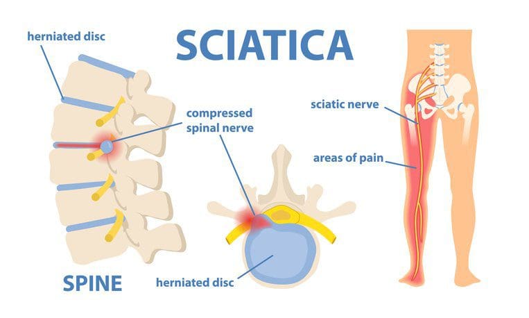
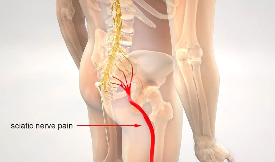
Since the spine is encompassed by ligaments, the spinal cord, soft tissues, and trillions of nerves spread out throughout the entire body, these nerves ensure that the sensations are being felt when a person is feeling something they are touching or feeling impacted on. When the body begins to feel pain and starts to affect the nerves, it can send a sharp shooting pain that can cause a person to feel weakness in the leg muscles, known as sciatica. Research studies have shown that sciatica radiates pain along the sciatic nerve that travels down from the lower back to the leg. Sciatica usually occurs when the spine is suffering from a herniated disc, and that affected disc is touching the sciatic nerve causing sharp shooting pain down the leg.
Other research studies have found that when individuals describe how sciatica pain feels, there are many different ways to express it depending on the cause and how severe the pain is. Sometimes the pain would often be described as sharp, shooting pain that goes down on one leg or as excruciating burning pain that either comes or goes or even may be constant. Sciatica can also come suddenly or gradually on the leg when the sciatic nerve has been pinched.
What Are The Symptoms?
Research studies have shown that sciatica symptoms can range from being infrequent and irritating to severe and debilitating. Since the sciatic nerve root is compressed or pinched, the symptoms can affect the specific spinal nerve root originating from the sciatic nerve. Some of the common symptoms that are caused by sciatica usually involve one leg at a time and are seen as:
Pain that is shooting down the leg
Numbness or tingling sensation that is felt in the back of the leg
Muscle weakness that is in the leg and foot
Posture change can alleviate or aggravate the pain
Treating Sciatica With Decompression-Video
The video above shows where the sciatic nerve is and how sciatica affects the leg. One of the many treatments that can alleviate sciatica nerve pain is spinal decompression therapy. Spinal decompression allows the spine to be gently pulled by traction, causing instant relief to the individual. Spinal decompression therapy also allows the beneficial nutrients to enter the spinal cord and increases the disc height on the spine. Since a herniated disc causes sciatica, spinal decompression allows the herniated disc, which affects the sciatic nerve, to retreat to the spine before it was herniated. This will cause instant relief to the individual that was affected by sciatica, and they can start on their wellness journey pain-free. If you want to learn more about spinal decompression therapy, this link will explain the benefits of spinal decompression and how it can alleviate low back pain symptoms.
Utilizing Decompression Therapy For Sciatica
With sciatica causing many individuals pain, many treatments are utilized to alleviate sciatica nerve pain and dampen the inflammatory effects it has caused. Research studies have found that non-surgical spinal decompression therapy is used to reduce low back pain and pain associated with sciatica and increase disc height in the spine. When individuals are lying down on the decompression table, they are strapped in. The machine allows the spine to be gently stretched out through traction, causing instant relief to the individual. Other research studies have shown that decompression therapy allows the decompression machine to effectively stretch the spine gently that has been suffering from back issues like sciatica, herniated discs, and low back pain. This gentle stretch allows the herniated disc to stop pressing on the sciatic nerve and causes relief to the individual.
Conclusion
The spine is encompassed by ligaments, the spinal cord, soft tissues, and nerves that help protect the spine from injury. However, when the spine does get injured, it can cause the spinal disc to bulge out or herniate and touch the sciatic nerve to cause immense shooting pain down the leg. This is known as sciatica, and it can cause a person to have immense shooting sharp pain that can affect a person’s quality of life. Treatments like decompression therapy allow the individuals suffering from sciatica to feel instant relief as their compressed spine is being gently pulled and causing the herniated disc to stop touching the sciatic nerve. Combined with physical therapy, spinal decompression allows the individual to be pain-free from sciatica and will enable them to continue their wellness journey.
References
Apfel, Christian C, et al. “Restoration of Disk Height through Non-Surgical Spinal Decompression Is Associated with Decreased Discogenic Low Back Pain: A Retrospective Cohort Study.” BMC Musculoskeletal Disorders, U.S. National Library of Medicine, 8 July 2010, https://pubmed.ncbi.nlm.nih.gov/20615252/.
Hochschuler, Stephen. “Sciatica Symptoms.” Spine, Spine-Health, 5 June 2019, https://www.spine-health.com/conditions/sciatica/sciatica-symptoms.
Kang, Jeong-Il, et al. “Effect of Spinal Decompression on the Lumbar Muscle Activity and Disk Height in Patients with Herniated Intervertebral Disk.” Journal of Physical Therapy Science, The Society of Physical Therapy Science, Nov. 2016, https://www.ncbi.nlm.nih.gov/pmc/articles/PMC5140813/.
Medical Professionals, Cleveland Clinic. “Sciatica: Causes, Symptoms, Treatment, Prevention & Pain Relief.” Cleveland Clinic, 25 Mar. 2020, https://my.clevelandclinic.org/health/diseases/12792-sciatica.
Staff, Mayo Clinic. “Sciatica.” Mayo Clinic, Mayo Foundation for Medical Education and Research, 1 Aug. 2020, https://www.mayoclinic.org/diseases-conditions/sciatica/symptoms-causes/syc-20377435.
The body is home to the spine, where it is allowed to move, twist, bend, and run around without being in pain. When a person suffers from a back injury or pulled a muscle, the pain can range from a dull ache to a sudden sharp pain that hinders and affects their quality of life. Luckily many treatments can help manage back pain and possibly reverse the effects. One of the treatments is spinal decompression, and it can help alleviate back pain issues like bulging discs. In this article, we will be looking at what is bulging disc is, its symptoms, and its factors, as well as how spinal decompression can help alleviate bulging disc. By referring patients to qualified and skilled providers specializing in spinal decompression therapy. To that end, and when appropriate, we advise our patients to refer to our associated medical providers based on their examination. We find that education is the key to asking valuable questions to our providers. Dr. Alex Jimenez DC provides this information as an educational service only. Disclaimer
Can my insurance cover it? Yes, it may. If you are uncertain, here is the link to all the insurance providers we cover. If you have any questions, please call Dr. Jimenez at 915-850-0900.
What Is A Bulging Disc?
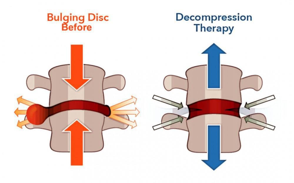
The spinal discs in the spine are flat circular cushions between the spine’s vertebrae and help protect the spine from any injuries. As the body ages naturally, the spinal discs will begin to lose their function through wear and tear. Research studies have shown that when the spinal discs begin to wear and tear with age, it can cause the disc to be dehydrated and cause the cartilage to be stiff. When this happens, the outer layer of the spinal disc will begin to protrude out and will not touch the nerve. If the protruding bulge continues to get worse and starts to crack the outer wall, the inner wall of the spinal disc will begin to come out and touch the spinal nerve root causing pain to the individual. This is known as a herniated disc.
Other research studies have found that when the spine has bulging discs, it is one of the causes of low back pain as about 80% of many individuals suffer from some back pain. Low back pain has many different diagnoses when this happens to the back, and DDD (degenerative disc disease) and disc herniation/bulging are the most common symptoms. These two common symptoms usually go hand in hand and, if not treated, will begin to cause a hindrance to the individual, leaving them with low back pain.
The Symptoms & Factors
The symptoms and factors of disc herniation/bulging vary depending on how severe the pain is. The pain from disc herniation can range from a dull, mild ache on the lower back to a sharp, sudden pain that travels from the lower back down to the leg. Research studies have shown that the primary symptoms of lumbar disc herniation are radicular pain, sensory abnormalities, and weakness in the lumbosacral nerve root. This will increase the pressure on the spinal disc causing the individual to be in pain while sitting. Other research studies have found that other common symptoms and factors that are caused by disc herniation/bulging include:
Excess weight: Can be compressed onto the spinal disc, causing disc herniation
Aging: Can naturally cause wear and tear on the spinal disc
Spinal Decompression Therapy Affecting Bulging Discs- Video
The video above shows how spinal decompression therapy can help alleviate bulging discs by using traction to stretch the spine gently. Spinal decompression is when individuals are lying down on the traction table and are strapped in as their spine is being pulled gently, causing the spinal disc to receive the beneficial nutrients back into the spine and allowing any herniation or bulging disc to go back to normal on the spine. Research studies have shown that utilizing physical therapy and spinal decompression therapy can help many individuals suffering from low back pain, herniated disc, or leg pain. By incorporating spinal decompression therapy into their wellness journey, many individuals will begin to feel relief and be pain-free. If you want to learn more about spinal decompression therapy, this link will explain the benefits of spinal decompression and how it can alleviate low back pain symptoms.
The Effects Of Spinal Decompression On Bulging Disc
Research studies have found that a lumbar herniation/bulging disc on the spine can induce neurological signs that can hinder a person with muscle weakness. One of the many treatments that can help alleviate bulging discs is spinal decompression therapy. Spinal decompression therapy and physical therapy can help stabilize the spine and can help improve muscle strength while providing a gentle stretch on the spine to allow the bulging discs to retreat to the spine, causing instant relief. Other research studies have shown that incorporating the two treatments are effective for many individuals by improving their pain and disability. This will allow their spine to be pain-free and restore their original function in the body.
Conclusion
The spine’s primary function is to make sure that the body is moving around without feeling any pain. As the body naturally ages over time, so does the spine as the spinal disc start to wear and tear, causing them to bulge out of the spine. If they start to press against the spinal nerve root, it can lead to herniation and cause shooting pain down from the lower parts of the body. Luckily some treatments allow the individual to feel relief, which is spinal decompression. Spinal decompression helps the spine by gently stretching it with a traction table, causing the nutrients and fluids to enter the spine and cause instant relief. When spinal decompression is combined with physical therapy, many individuals will notice that they are feeling no pain in their back and can continue their wellness journey.
References
Al Qaraghli, Mustafa I, and Orlando De Jesus. “Lumbar Disc Herniation.” StatPearls [Internet]. Treasure Island (FL), StatPearls Publishing, 30 Aug. 2021, https://www.ncbi.nlm.nih.gov/books/NBK560878/.
Amin, Raj M, et al. “Lumbar Disc Herniation.” Current Reviews in Musculoskeletal Medicine, Springer US, Dec. 2017, https://www.ncbi.nlm.nih.gov/pmc/articles/PMC5685963/.
Choi, Jioun, et al. “Influences of Spinal Decompression Therapy and General Traction Therapy on the Pain, Disability, and Straight Leg Raising of Patients with Intervertebral Disc Herniation.” Journal of Physical Therapy Science, U.S. National Library of Medicine, Feb. 2015, https://pubmed.ncbi.nlm.nih.gov/25729196/.
Medical Professionals, Cleveland Clinic. “Herniated Disk: What It Is, Diagnosis, Treatment & Outlook.” Cleveland Clinic, 1 July 2021, https://my.clevelandclinic.org/health/diseases/12768-herniated-disk.
Shelerud, Randy A. “Bulging Disk vs. Herniated Disk: What’s the Difference?” Mayo Clinic, Mayo Foundation for Medical Education and Research, 23 Apr. 2019, https://www.mayoclinic.org/diseases-conditions/herniated-disk/expert-answers/bulging-disk/faq-20058428.
Sciatica is experienced as lower back pain and pain that radiates down the back of the legs. It is pain caused by compression, irritation, or inflammation of the sciatic nerve. It is generally experienced on one side of the body. Body movements like twisting, bending, sitting, or responses like coughing and sneezing can worsen the pain. Individuals with sciatica also experience muscle weakness, numbness, tingling, or electrical shock-like sensations. Injury Medical Chiropractic and Functional Medicine Clinic offer manual and motorized sciatic nerve decompression to stretch/realign the spine, release the compressed nerves, and relieve pain.
Sciatic Nerve Decompression
The spine consists of 23 spinal discs that are shock absorbers for the body during movement. Each disc consists of a soft inner core of a gel substance and a thick outer layer. Wear and tear of the spinal discs from aging, degenerative disc disease, repetitive physical activities like lifting and bending, obesity, and poor posture are some of the factors that can stress the spine, causing the thick outer layer of the spinal disc to crack/breakdown causing the soft inner core to leak out forming a bulging or herniated disc. This type of injury compresses, pinches, or irritates one or more nerve roots that form the sciatic nerve, triggering sciatica.
Spinal stenosis or the narrowing of the spinal canal.
Spondylolisthesis, or the slipping or dislocation of the spinal vertebrae in the lower part of the spine.
Are also known sciatica causes.
Symptoms
Common compressed nerve symptoms include:
Pain or burning sensations radiating down the leg.
Because branches of the sciatic nerve extend from the lumbar spine through the buttocks and down the leg, pain, burning sensations or dull aching can present along the nerve’s pathway if the nerve gets compressed or irritated.
Weakness in the affected leg.
When walking or moving the legs, the nerves transmit information to the brain, stimulating the muscles to react in specific ways.
A pinched sciatic nerve can cause interference with relaying signals, resulting in weakness.
Numbness.
The compression impedes blood circulation and nerve energy transmission.
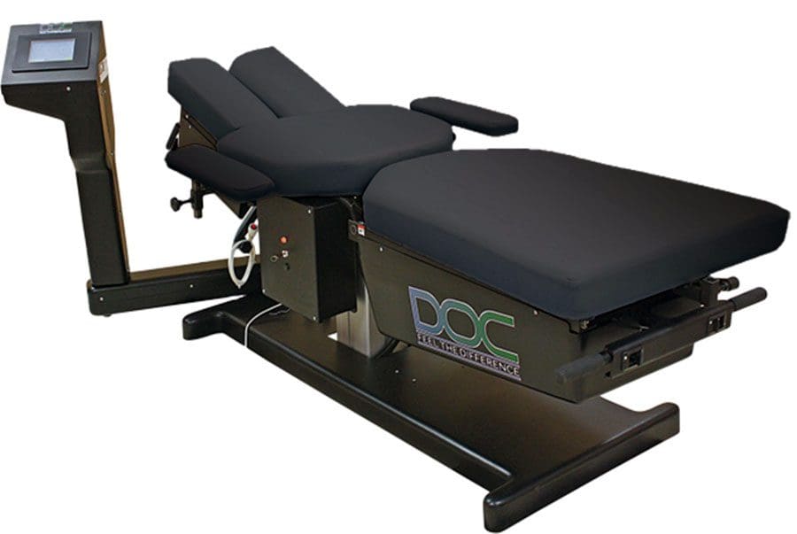
Spinal decompression therapy relieves pressure on the spine by pulling/stretching it in small increments. Non-surgical spinal decompression creates negative pressure within the discs. The negative pressure pulls or vacuums back the disc material that has protruded or herniated and an abundance of nutrients to activate the healing response. The chiropractor, physical therapist, or nurse uses motorized medical equipment with sensors linked to a computer-aided system to perform the procedure. The equipment is designed to adjust the pull force accordingly to prevent muscle resistance. The adjustable table also allows the spine to be stretched at different angles to target the upper or lower back.
The objective of spinal decompression treatment is to relieve the symptoms of sciatica or disc disorders and heal the injured disc. We utilize spinal decompression as an effective tool in treating a vast array of spinal conditions.
DRX9000 Non-Surgical Spinal Decompression
References
Berry, James A et al. “A Review of Lumbar Radiculopathy, Diagnosis, and Treatment.” Cureus vol. 11,10 e5934. 17 Oct. 2019, doi:10.7759/cureus.5934
Davis D, Maini K, Vasudevan A. Sciatica. [Updated 2022 Feb 4]. In: StatPearls [Internet]. Treasure Island (FL): StatPearls Publishing; 2022 Jan-. Available from: https://www.ncbi.nlm.nih.gov/books/NBK507908/
Giuffre BA, Jeanmonod R. Anatomy, Sciatic Nerve. [Updated 2021 Jul 29]. In: StatPearls [Internet]. Treasure Island (FL): StatPearls Publishing; 2022 Jan-. Available from: https://www.ncbi.nlm.nih.gov/books/NBK482431/
National Institutes of Health. (2019.) “Sciatica.” https://medlineplus.gov/sciatica.html
IFM's Find A Practitioner tool is the largest referral network in Functional Medicine, created to help patients locate Functional Medicine practitioners anywhere in the world. IFM Certified Practitioners are listed first in the search results, given their extensive education in Functional Medicine