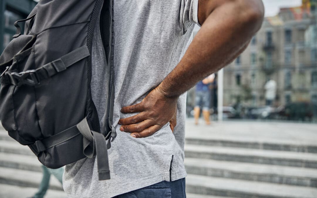
Navigating Car Accident Claims in El Paso, Texas: Pre-Existing Conditions, the Eggshell Skull Rule, and Care at El Paso Back Clinic
The doctor explains an X-ray to the patient and points at the computer screen. The patient wears a cervical collar
Car accidents are common in El Paso, Texas. They can cause new injuries or worsen existing ones. People often wonder if a past health issue, like back pain or arthritis, will block them from getting help after a crash. Texas law offers protection. You can still claim money for injuries even with prior conditions. This article covers the rules, what you can get paid for, and the steps to follow. It highlights the “eggshell skull rule” and why quick medical care is key. In El Paso, El Paso Back Clinic stands out for expert care for auto accidents and worsening conditions.
Understanding the Eggshell Skull Rule
The eggshell skull rule is an important legal concept. It means that if someone causes an accident, they must pay for all resulting damage. This applies even if the injured person had a weakness from a prior condition. It’s like breaking a fragile egg—you can’t blame the thin shell. The rule is also known as the “thin skull rule” or “take your victim as you find them” (Amtz Law, n.d.).
Simply put, the at-fault person takes full responsibility. They can’t use your old health problems to avoid paying. However, the accident must cause new damage or aggravate the existing issue. If your condition had worsened on its own, that might not be covered (Gutierrez Law Firm, n.d.a). For instance, if you had mild back arthritis and the crash resulted in severe pain that required therapy, the at-fault driver is responsible for covering that additional harm.
This rule originated in prior legal cases. It safeguards those who are more vulnerable. In Texas, it’s used in car accident lawsuits to ensure fair compensation (Reyes Law, n.d.).
Applying the Eggshell Skull Rule in Texas and El Paso
Texas fully supports the eggshell skull rule. In El Paso, if a car accident aggravates your pre-existing condition, you can pursue a claim. The law holds the at-fault party liable for all injuries resulting from the crash, including those amplified by prior issues (GDL Firm, n.d.).
El Paso has busy highways, such as I-10, which leads to frequent accidents. Local laws follow Texas standards. For example, if you had an old neck injury and a collision causes whiplash on top of it, the rule helps you recover costs. Insurance companies may argue that your pain stems solely from the prior condition to reduce payments (BHW Law Firm, n.d.). Strong evidence can counter this.
You have two years from the accident date to file in El Paso under the statute of limitations (No Bull Law, n.d.). Act fast to avoid missing out.
Typical Pre-Existing Conditions Impacted: Chronic back pain, sciatica, herniated discs, fibromyalgia, or degenerative disc disease.
Signs of Aggravation: Increased pain, new movement limitations, or the need for additional medical treatment.
El Paso-Specific Risks: Border traffic and dust storms increase crash chances, often affecting backs and necks.
Compensation Options for Aggravated Conditions
When an accident worsens your condition, Texas allows claims for various damages. The eggshell skull rule ensures coverage for the full extent of harm (Siegfried & Jensen, n.d.). This includes bills, lost income, and emotional distress.
Possible compensations include:
Medical Expenses: Costs for new therapies, adjustments, or surgeries due to the aggravation, plus future care.
Wage Loss: Earnings missed from work because of heightened symptoms.
Pain and Suffering: Payment for added physical discomfort and mental strain, such as stress from chronic pain.
Reduced Quality of Life: If daily activities or hobbies become harder.
Long-Term Disability: For permanent worsening, like ongoing sciatica.
Amounts depend on severity. Minor aggravations may yield smaller settlements, while persistent issues, such as the need for regular chiropractic care, may increase them (Reyes Law, n.d.). Age factors in—younger victims may experience greater impacts over time.
In El Paso, solid documentation boosts settlements (Abraham Watkins, n.d.).
Proving Your Case for Compensation
To win, show that the accident directly worsened your condition. Use medical records from before and after to illustrate changes (St. Louis Injury Law, n.d.). This “before-and-after” approach is crucial.
Key steps:
Seek Immediate Care: Visit a doctor soon after. Discuss your history and new symptoms.
Maintain Documentation: Collect bills, notes, and X-ray scans.
Log Daily Effects: Journal pain levels and activity changes.
Expert Testimony: Have a physician explain the connection.
Accident Evidence: Include reports, photos, and statements.
Honesty about your past is vital—concealing it can weaken your claim (Gage Mathers, n.d.). Courts assess if the aggravation ties to the crash or is a natural progression.
Handling Insurance Challenges
Insurers aim to minimize payouts. They may blame your pre-existing condition entirely. They could demand full records to deny claims (Romanow Law Group, n.d.). Avoid broad agreements without advice.
Strategies:
Use Legal Support: Have an attorney negotiate on your behalf.
Reject Low Offers: Initial proposals are often insufficient.
Challenge Rejections: Present evidence linking to the accident.
Recognize Strategies: Beware of their experts minimizing damage.
El Paso attorneys familiar with local rules can help (Ellis & Thomas, n.d.).
Benefits of Specialized Auto Accident Clinics in El Paso
Post-accident, choose a clinic expert in auto injuries. This ensures proper documentation and healing. El Paso Back Clinic excels in this, offering chiropractic care for whiplash, back pain, and aggravated conditions (El Paso Back Clinic, n.d.).
Their approach includes:
Chiropractic Adjustments: To align the spine and ease nerve pressure.
Physical Therapy: To rebuild strength and mobility.
Spinal Decompression: For herniated discs and sciatica.
Functional Medicine: Addressing root causes with nutrition and lifestyle.
Seeing them early helps record aggravations, aiding claims (Your Back in Line Now, n.d.). They coordinate with attorneys for seamless support.
Insights from Dr. Alexander Jimenez at El Paso Back Clinic
Dr. Alexander Jimenez, DC, APRN, FNP-BC, leads El Paso Back Clinic. With dual expertise in chiropractic and nursing, he treats complex cases like auto-aggravated back issues (El Paso Back Clinic, n.d.).
He observes that crashes often intensify conditions like degenerative discs or fibromyalgia. Treatments blend adjustments, acupuncture, and rehab. He emphasizes non-invasive methods, using diagnostic tools such as X-rays to establish links.
Patients praise quick relief. For example, Gale Grijalva recovered from accident-related back pain, resuming activities. Dr. Jimenez’s team offers personalized plans to prevent surgery.
The clinic’s 30,000+ sq ft facility includes gyms and meal prep, supporting full recovery.
Wrapping Up
Dealing with car accidents in El Paso is challenging, especially with pre-existing conditions. Texas’s eggshell skull rule allows compensation for aggravations with proper proof. Seek prompt medical attention, document everything, and consult legal counsel. El Paso Back Clinic, led by Dr. Jimenez, provides top chiropractic and rehab for healing and claims. Contact them at +1-915-850-0900 or visit https://elpasobackclinic.com/ for help.
References
Abraham Watkins. (n.d.). Do Pre-Existing Conditions Disqualify Me From Damages in a Personal Injury Case?Abraham Watkins.
Amtz Law. (n.d.). How Pre-Existing Conditions Affect Your Personal Injury Claim.Amtz Law.
BHW Law Firm. (n.d.). Pre-Existing Injury and Accident in Texas.BHW Law Firm.
Is It Safe to Wear a Backpack? Expert Tips on Spinal Health and Back Pain Prevention in the US and El Paso, TX
A woman walking, wearing a backpack with the recommended weight, and maintaining correct posture to prevent back pain and problems.
Back pain is a big issue for many people in the United States
Up to 80% of adults face low back pain at some point in their lives. This is one of the top reasons for doctor visits and missed workdays. The cost is huge too, with over $100 billion spent on spine problems each year. In El Paso, Texas, where people often have active jobs like industrial work or lots of driving, back pain questions focus on things like sciatica, herniated discs, and spinal stenosis. A common concern across the country, including in places like El Paso, is whether wearing a backpack is safe for the spine. The good news is that it can be safe if you follow some simple rules. This article focuses on backpack safety and then addresses other key questions about managing back pain, treatment options, and daily habits to keep your spine healthy.
Understanding Backpack Safety and Spinal Health
Wearing a backpack is common for carrying things, but if it’s too heavy or worn incorrectly, it can hurt your back. Heavy backpacks can strain muscles and joints in your back, neck, and shoulders. This might lead to pain or bad posture over time. However, backpacks do not cause scoliosis, a spinal curvature that affects about 2% to 3% of people. Scoliosis often starts in teens and is more common in girls, but it’s not linked to backpacks.
Is it safe? Yes, as long as you distribute the weight right and follow the tips to avoid strain. Improper use can cause muscle fatigue, poor posture (such as slouching), and even chronic pain if left unaddressed. In El Paso, where people might carry tools or bags for work, this is especially important to prevent issues such as sciatica, where pain radiates down the leg due to nerve pressure.
Here are some key tips for safe backpack use:
Choose the right backpack: Pick one with wide, padded straps and a padded back. It should fit your body size and have a waist strap for heavy loads. Lightweight materials help too.
Limit the weight: Keep the backpack under 10-15% of your body weight. For example, if you weigh 150 pounds, aim for no more than 15-22.5 pounds.
Distribute weight evenly: Put heavier items at the bottom and close to your back. Use compartments to balance things and stop shifting.
Wear it correctly: Always use both straps. Adjust them so the pack sits in the middle of your back, not sagging low. Bend your knees to lift it.
Make smart choices: Remove extra items often. Use lockers or storage if possible. For very heavy loads, try a rolling backpack or crossbody bag.
These steps help distribute the load across your strong back muscles and keep your spine aligned. If you feel pain, stop and adjust. In places like El Paso, with busy lifestyles, following these can help prevent accidents from becoming long-term back issues.
Common Causes of Back Pain in the US
Back pain affects millions. In the US, about 26% of adults have it at any time, and it’s more common after age 45. Among adults aged 50 and older, up to 45.6% experience it. Causes include muscle strains, ligament injuries, herniated discs (where the disc’s soft center protrudes), arthritis, and spinal stenosis (where the spinal canal narrows). Stress can make it worse by causing muscle spasms. Even factors such as obesity or infections can play a role.
Chronic back pain lasts more than 3 months and affects 8% of adults. It often comes from wear and tear on discs or joints. Poor sleep makes it worse because pain disrupts rest, and lack of sleep raises inflammation. In the US, this results in high costs, such as lost work and medical bills.
Symptoms vary. You might feel an ache in your lower back or sharp pain if it’s sciatica. Numbness, tingling, or weakness in the legs are red flags. Scoliosis, which affects 7 million Americans, can cause symptoms such as uneven shoulders or back pain; most cases are mild.
Muscle or ligament strain: From lifting incorrectly or sudden moves.
Disc problems: Bulges or herniations press on nerves.
Arthritis: Joint wear is common in older people.
Stenosis: Narrowing squeezes nerves, causing leg pain.
Stress and lifestyle: Tension builds up, leading to spasms.
Knowing these helps prevent pain. For example, strengthening your core muscles supports your spine and reduces strain from daily activities like wearing a backpack.
Managing Chronic Back Pain
Chronic back pain needs long-term plans. First, see if it’s new or ongoing. Most cases improve with rest and simple fixes, but if it lasts, get checked. Avoid bed rest; gentle movement helps recovery faster.
Daily habits matter. Exercise like walking or swimming builds strength. Maintain a healthy weight to reduce spinal load. Quit smoking, as it negatively affects spinal tissues and raises surgery risk by up to 50%. Good posture and ergonomic setups at work prevent strain.
In El Paso, with industrial jobs and driving, pain from accidents is common. Recovery focuses on building habits to avoid re-injury.
Stay active: Low-impact exercises like yoga or Pilates.
Watch your diet: Healthy foods reduce inflammation.
Manage stress: Deep breathing or mindfulness helps.
Sleep well: Use pillows to maintain spinal alignment.
Stretch daily: Loosen tight muscles, such as the hamstrings.
These steps reduce pain and improve quality of life.
Treatment Options: Surgery vs. Conservative Care
When pain doesn’t go away, choices include conservative care or surgery. Conservative means non-surgical options such as physical therapy, medications, injections, chiropractic care, or massage. These are tried first for 8-12 weeks. Surgery is indicated for severe cases, such as nerve damage or instability.
Ask your doctor: What causes my pain? What tests do I need? What are the risks and benefits? For surgery, ask about the surgeon’s experience, recovery time, and whether you’ll need help at home. Alternatives like spinal decompression stretch the spine to ease disc pressure.
Chiropractic vs. orthopedic: Chiropractors focus on spinal adjustments to realign the spine and relieve pain without medication. Orthopedists may recommend surgery for significant issues. Both can help, but chiropractic care is well-suited to conservative care.
In El Paso, many choose chiropractic for herniated discs or sciatica. It’s safe and effective for back pain, reducing symptoms by fixing alignment and boosting blood flow.
Spinal Health in El Paso, TX
El Paso has unique needs. Active lives, work injuries, and car accidents lead to questions about sciatica, where nerve pain goes down the leg, or spinal stenosis with leg weakness. Herniated discs are common from lifting or falls.
Lumbar stenosis FAQs: It causes leg pain or numbness when walking. Avoid high-impact exercises like running; try swimming instead. Treatments include therapy or decompression.
Local care often combines chiropractic and orthopedic care. Dr. Alexander Jimenez, a chiropractor in El Paso with over 30 years of experience, notes that integrative care is most effective. He uses adjustments, nutrition, and therapy for root causes. For example, a worker’s back pain improved by 50% within weeks with his plan. He stresses non-surgical options for sciatica and injuries, helping people stay active in El Paso’s environment.
Sciatica: From disc pressure; chiropractic eases it.
Chiropractic: Aligns the spine, safe for all ages.
Dr. Jimenez’s work shows personalized plans reduce pain without surgery.
Daily Habits to Prevent Spinal Injury
Preventing pain starts with habits. Lift by bending knees, not back. Stand every 15 minutes if sitting for long. For driving in El Paso, take breaks to stretch.
Core strength is key. Exercises like planks support your spine. Avoid smoking for better healing. Ergonomics: Screen at eye level, chair with back support.
For backpacks, combine with these: Even weight helps posture.
Lift right: Knees bent, close to body.
Posture: Stand tall, no slouch.
Exercise: Core and back focus.
Weight control: Less strain on the spine.
Breaks: Move often.
These reduce the risk of injury and tie into backpack safety.
Conclusion
Wearing a backpack is safe when done properly, with proper weight distribution and habits. This fits into broader questions about spinal health in the US and El Paso. Manage chronic pain with conservative care first, like chiropractic, and build daily routines to prevent issues. Experts like Dr. Jimenez show that integrative approaches work. Stay active, ask questions, and protect your spine for a better life.
When You Don’t Stretch: Why Muscles Get Stiff, Movement Gets Harder, and Injuries Become More Likely
A patient with chronic back pain does targeted stretches.
If you rarely stretch, your body can start to feel “tight,” which can change how you move. Many people notice they can’t bend, twist, squat, reach overhead, or turn their head as easily as they used to. Over time, this can affect your flexibility, your range of motion (how far a joint can move), and how smooth and efficient your daily movements feel.
At El Paso Back Clinic, Dr. Alexander Jimenez, DC, APRN, FNP-BC, often explains this: when mobility decreases, the body starts to “compensate.” That means you move around a stiff area instead of through it, and those workarounds can build up stress in nearby joints and muscles (Jimenez, n.d.-a). This is one reason people can develop recurring back pain, neck stiffness, hip tightness, or shoulder irritation even without a single big injury.
What “Muscle Stiffness” Really Means
Muscle stiffness usually feels like tightness, soreness, or difficulty moving. It can happen after overuse, after you’ve been still for a long time, or when your muscles stay “stuck” in a more contracted state (Tarantino, 2025). Osmosis
Osmosis notes that stiffness can appear after a long period of minimal motion (such as bed rest or inactivity) or after new exercise that causes temporary muscle cell damage (Tarantino, 2025). Osmosis
Key idea: When your body doesn’t move a joint through its normal range often enough, the muscles and tissues around it can start to feel restricted. That restriction can make normal tasks think harder than they should.
Do Muscles Actually “Shorten” If You Don’t Stretch?
You’ll hear people say, “If you don’t stretch, your muscles will shorten.” That statement is partly true, but it needs context.
Adidas explains that the word “shorten” can be misleading: for most people, it feels like shortening because mobility and flexibility decrease when stretching is skipped, even if the muscle is not literally shrinking in everyday life (Adidas, 2025). adidas
Harvard Health adds an important clarification: without regular stretching, muscles can become tight, and when you need them for activity, they may not extend fully, increasing the risk of joint pain, strains, and muscle damage (Harvard Health Publishing, 2024). Harvard Health
So the practical takeaway is simple:
Skipping stretching often leads to less mobility and flexibility
Tight muscles can reduce how far joints can move
Tight muscles can make injuries more likely when you suddenly “ask more” of your body
How Tight Muscles Reduce Range of Motion
Range of motion (ROM) is the movement around a joint or body part. When ROM is limited, you can’t move that body part through its usual, healthy motion (Jimenez, n.d.-b). El Paso Back Clinic® • 915-850-0900
El Paso Back Clinic explains how tightness—especially in areas like the hips and ankles—can reduce ROM and limit potential for form and strength. When posture and form are compromised, pain and injury risk can rise (Jimenez, n.d.-b). El Paso Back Clinic® • 915-850-0900
What limited ROM can look like in real life
You might notice:
You can’t turn your head fully when driving
You bend from your lower back instead of your hips
You can’t squat without your heels lifting
Your shoulders feel “pinched” when reaching into a cabinet
Your hamstrings feel tight when you try to walk fast
And here’s the tricky part: your body still gets the job done—just with more strain.
Why Stiffness Can Raise Injury Risk
Harvard Health explains that tight muscles may be more easily damaged when they are suddenly stretched during strenuous activity (Harvard Health Publishing, 2024). Harvard Health
That’s why injuries often show up in moments like:
A weekend game after sitting all week
A sudden sprint to catch something
Lifting a heavy box with “cold” hips and hamstrings
A long drive followed by quick unloading or bending
Mayo Clinic also notes that better flexibility can help joints move through full ROM and may decrease injury risk, while emphasizing that stretching must be done correctly (Mayo Clinic Staff, n.d.). Mayo Clinic
Common Reasons People Stop Stretching (And How to Fix Them)
Most people don’t skip stretching because they don’t care. They skip it because it feels confusing, time-consuming, or uncomfortable.
Common barriers
“I don’t have time.”
“Stretching hurts.”
“I’m not flexible, so it doesn’t work for me.”
“I only need stretching if I work out.”
Better, more realistic reframes
You only need 5–10 minutes a few times a week to start seeing benefits (Mayo Clinic Staff, n.d.). Mayo Clinic
Stretching should create tension, not pain (Mayo Clinic Staff, n.d.). Mayo Clinic
Flexibility improves over weeks to months, not days (Harvard Health Publishing, 2024). Harvard Health
Stretching supports everyday movement, not just workouts (Harvard Health Publishing, 2024). Harvard Health
Safe Stretching Basics (So You Don’t Make Things Worse)
This matters: stretching done poorly can backfire.
Mayo Clinic recommends:
Don’t stretch cold muscles—warm up 5–10 minutes first
Don’t bounce
Hold stretches about 30 seconds (longer for problem areas)
Don’t stretch into pain (Mayo Clinic Staff, n.d.). Mayo Clinic
The American Heart Association adds:
Stretch when muscles are warm
Hold 10–30 seconds and repeat 3–5 times
Stretch slowly and smoothly (American Heart Association, 2024). www.heart.org
Quick safety checklist
Warm up first (easy walk, gentle movement)
Move slowly
Breathe
No bouncing
Stop if you feel sharp pain, numbness, or joint pain
A Simple 10-Minute Daily Stretch Routine for Real Life
This is designed for normal adults: busy schedules, stiff hips, tight neck, and lots of sitting.
Step 1: Warm up (1–2 minutes)
Walk around the house
March in place
Gentle arm circles
Step 2: Do these 6 stretches (about 8 minutes total)
1) Hip flexor stretch (1 minute each side) Helps if you sit a lot and feel tight in the front of your hips.
2) Hamstring stretch (1 minute each side) Harvard points out that tight hamstrings from sitting can limit how well you extend your leg and support walking mechanics (Harvard Health Publishing, 2024). Harvard Health
3) Calf stretch (45 seconds each side) Helpful for ankle mobility, walking, and squatting mechanics.
4) Chest opener (45 seconds) Stand in a doorway and gently open the chest to reduce rounded-shoulder posture.
5) Upper back reach (45 seconds) Hug yourself and gently pull your shoulder blades apart.
6) Neck side stretch (30 seconds each side) Gentle only—never crank your neck.
Step 3: Add “micro-mobility” during your day (optional but powerful)
Stand up every hour for 30–60 seconds
Do 5 bodyweight squats to a chair
Do 10 shoulder rolls
Take a 3-minute walk after meals
These small habits often matter as much as one long stretch session.
Stretching After Workouts: What You Should Know
Adidas explains the difference clearly:
Dynamic movement is best before workouts (prepares your body)
Static stretching is typically better after workouts, when you’re warm (Adidas, 2025). adidas
Mayo Clinic also cautions that stretching cold muscles can increase injury risk and notes that some intense activities may not benefit from heavy stretching right before performance (Mayo Clinic Staff, n.d.). Mayo Clinic
A balanced approach
Before exercise: warm up + dynamic mobility
After exercise: gentle static stretching + breathing
On rest days: short, consistent flexibility routine
When Stiffness Is a Sign You Need More Than Stretching
Sometimes the problem is not just “tight muscles.” You may have:
Joint restrictions that block movement
Spine or pelvis alignment issues affecting mechanics
Inflammation around a joint
Pain patterns that keep muscles “guarded”
A nerve-related problem (numbness, tingling, weakness)
El Paso Back Clinic notes that limited ROM in areas like the back, neck, or shoulders can be linked to the body being out of natural alignment, repetitive motions, or wear and tear (Jimenez, n.d.-b). El Paso Back Clinic® • 915-850-0900
If stretching doesn’t help—or makes symptoms worse—it’s smart to get assessed.
The El Paso Back Clinic Approach: Integrative Chiropractic + Nurse Practitioner Support
This is where integrative care can be a game-changer: you’re not only “stretching more,” you’re also finding out why you’re tight and building a plan that fits your body.
What chiropractic care can add
El Paso Back Clinic describes a “restoration” approach that may include:
Soft tissue work (to reduce tightness and improve circulation)
Adjustments (to address misalignments and support mobility)
Nurse practitioners are advanced practice clinicians who assess, diagnose, and treat illnesses and injuries and support chronic condition management (American Nurses Association, n.d.). ANA Healthgrades also describes NPs performing screenings and physical exams, ordering lab work, documenting care, and diagnosing certain conditions (Prosser, 2025). Healthgrades Resources
Why the combo helps stiffness and pain
Together, a chiropractor + NP team can:
Screen for red flags (nerve symptoms, systemic issues)
Decide when imaging or labs are appropriate
Build a movement plan that matches your pain level
Address sleep, stress, inflammation, and recovery habits
Track progress using measurable goals (like ROM improvements)
Dr. Jimenez’s Mobility & Flexibility materials emphasize that “great mobility” supports functional movement without ROM restrictions and that people who don’t stretch often may experience stiffened muscles that reduce effective movement (Jimenez, n.d.-a). El Paso Back Clinic® • 915-850-0900
Red Flags: When to Stop Stretching and Get Checked
Call a clinician promptly if you have:
Numbness, tingling, or weakness in an arm/leg
Loss of balance, clumsiness, or trouble walking
Severe pain that doesn’t improve
Pain after trauma (car accident, fall, sports collision)
Fever, unexplained swelling, or sudden intense stiffness
Muscle stiffness can sometimes be related to underlying medical issues, and diagnosis may require an exam and follow-up testing, depending on the cause (Tarantino, 2025). Osmosis
The Bottom Line
If you don’t stretch regularly, it’s common to feel tighter and less mobile over time. That stiffness can reduce range of motion, make daily tasks harder, and increase your risk of injury when you suddenly push your body. The good news is that you don’t need extreme flexibility. You need consistent, safe mobility work—and when required, professional support to restore movement and reduce pain.
A practical plan usually includes:
Small daily stretching habits
Better warm-ups and recovery routines
Strength + mobility (not stretching alone)
Integrative evaluation when pain, ROM loss, or repeated flare-ups keep returning
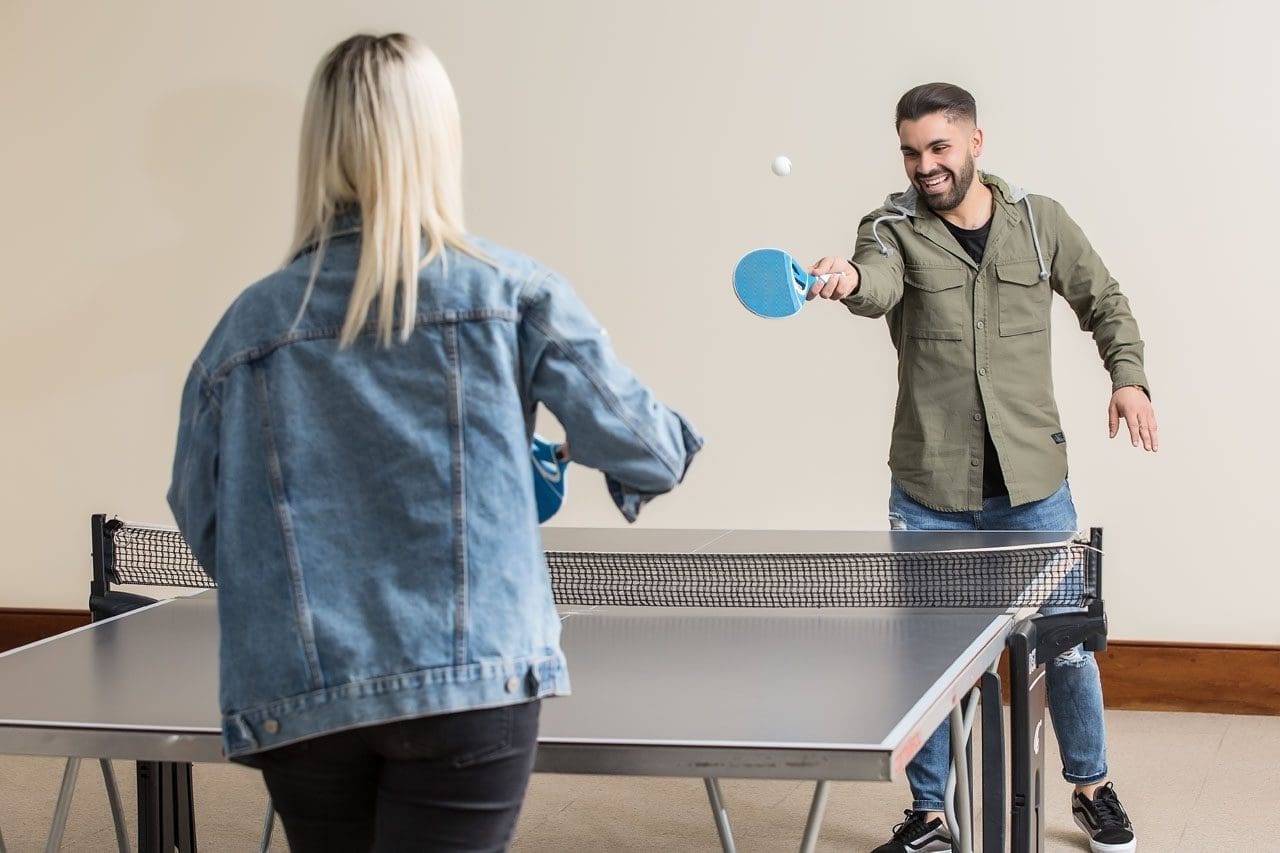
Beat Holiday Stress with Fun Movement and Smart Body Care
A man and a woman play table tennis to ease holiday stress.
The holiday season brings joy, family time, and tasty food, but it can also be stressful. Busy schedules, shopping, travel, and extra tasks can make anyone feel overwhelmed. One great way to feel better is through simple movement and exercise. Physical activity releases endorphins, chemicals in your brain that improve mood and reduce stress (Mayo Clinic, 2023). Even short sessions of fun activities can clear your mind and boost energy.
Many experts agree that almost any form of movement helps manage stress. It acts like a natural reset for your body and brain (Kitsap Physical Therapy, n.d.). Adding some holiday cheer to your routine makes it easier to stick with. This guide shares easy, enjoyable ways to stay active and calm during the holidays.
Why Movement Helps Reduce Holiday Stress
Exercise does more than keep you fit. It pumps up endorphins, boosting a happier feeling, and distracts you from worries. Activities like walking or dancing provide “meditation in motion,” helping you forget daily irritations (Mayo Clinic, 2023). Regular movement also improves sleep, builds confidence, and helps your body better handle stress.
During the holidays, people often move less due to cold weather or busy plans. This can make stress worse. But even one quick workout can lift your mood for hours (Gorman, 2022). Fun, low-pressure activities work best to avoid adding more pressure.
Releases feel-good chemicals to fight anxiety
Clears the mind and improves focus
Boosts energy and helps you sleep better
Builds strength to handle physical holiday demands, like carrying bags
Fun Sports-Inspired Activities to Boost Endorphins
Try activities that feel like play. Sports-inspired moves get your heart pumping and bring smiles.
Jumping rope: A quick cardio blast that raises your heart rate fast. Do it for 10-15 minutes while listening to holiday music (Avec Apartments, n.d.).
Dance breaks: Turn on your favorite songs and dance freely. Join a family dance party or try simple steps. Dancing combines rhythm and fun for great stress relief (NMC Health, n.d.; Triathlete Magazine, n.d.).
Pickup games: Play basketball, tennis, volleyball, or soccer with friends or family. These team sports combine exercise with social time, which further lowers stress (King Chiropractic, n.d.).
Shadowboxing: Punch the air like a boxer. This low-impact move releases tension without needing equipment. It’s perfect for a hotel room or living room (FightCamp, n.d.; Triathlete Magazine, n.d.).
These activities are easy to start and don’t require much space or gear.
Quick and Easy Bodyweight Exercises for Fast Relief
No gym? No problem. These simple moves use only your body and take little time.
Here are some top picks:
High knees: Run in place, lifting knees high. Do it for 1 minute to get your blood flowing (Echelon Fit, n.d.).
Planks: Hold a straight body position on your forearms and toes. Start with 30 seconds of core strength work (Echelon Fit, n.d.).
Bodyweight squats: Lower as if sitting in a chair, then stand up. Great for legs and glutes (Hydrow, n.d.).
Push-ups: Modify on knees if needed. Strengthen your upper body quickly (Hydrow, n.d.).
Jumping jacks: Classic move to warm up and boost mood (Echelon Fit, n.d.).
Try a 20-minute circuit: 30 seconds of each, with short rests in between. Repeat a few times (FightCamp, n.d.). Add holiday twists, like “present pick-up” squats—bend down as if grabbing gifts (Performance Health Academy, n.d.).
Mindful Practices for Calm and Flexibility
For gentler options, try mindful movements that focus on breath and flow.
Yoga flows: Simple poses like downward dog or warrior help stretch and center your mind. A 15-20 minute session reduces tension (Avec Apartments, n.d.; King Chiropractic, n.d.).
Tai Chi: Slow, flowing moves called “meditation in motion.” It improves balance and eases stress without strain (Mind Body Spine, n.d.; FightCamp, n.d.).
These practices calm the nervous system and pair well with busier days.
Outdoor Options: Walks and Hikes for Mind Clearing
Fresh air makes everything better. A brisk walk or hike builds endurance and clears thoughts.
Go for a festive neighborhood walk to see lights. Make it fun with a scavenger hunt for decorations (NMC Health, n.d.).
Hike in nature for extra calm. Being outdoors boosts positive feelings, such as gratitude (Triathlete Magazine, n.d.).
Add active games, such as playing in the yard or stair climbing, between tasks (Muscle MX, n.d.).
Aim for 30 minutes most days. No special gear needed—just good shoes (Club Getaway, n.d.).
Make It Festive: Holiday-Themed Active Fun
Keep things light by tying movement to celebrations.
Dance to holiday tunes or play charades that get everyone moving.
Try “Santa bag throws” or “candy cane curls” with simple weights or air motions (Performance Health Academy, n.d.).
Family games like obstacle courses or mini-golf indoors keep energy high and stress low (NMC Health, n.d.).
These ideas turn exercise into shared joy.
How Integrative Chiropractic Care Fits In
Physical tension from stress often shows up as tight muscles or misalignment. Integrative chiropractic care helps by using gentle adjustments to ease tension and support the nervous system. This improves your body’s stress response and promotes better flexibility (Chiropractic Works Collinsville, n.d.).
Chiropractors may suggest stretches or movements to help maintain alignment. This holistic approach complements exercise for full-body relief. Dr. Alexander Jimenez, a chiropractor and nurse practitioner with over 30 years of experience, notes that spinal health drives overall wellness. His integrative methods combine adjustments with posture exercises and stress management for better mobility and calm (Jimenez, n.d.; Jimenez, 2025a). He often sees that staying active and making adjustments help prevent holiday-related tension and support recovery (Jimenez, 2025b).
Pairing chiropractic visits with daily movement creates a balanced way to enjoy the season.
Tips to Get Started and Stay Consistent
Starting small is key during busy times.
Pick activities you enjoy to make it fun.
Schedule short sessions, like 10-20 minutes.
Involve family or friends for accountability.
Listen to your body—keep it light to avoid extra stress.
Combine with deep breathing for extra calm.
Consistency brings the best results. Even small efforts add up to less stress and more energy (American Fitness Professionals & Associates, n.d.).
By adding these fun movements and mindful care, you can handle holiday demands with ease. Focus on feeling good, not perfect. Your body and mind will thank you.
Avoiding Common Christmas Accidents: Prevention and Recovery at El Paso Back Clinic®
After lying in an awkward position, the woman is suffering from back pain on the couch at home.
The Christmas season fills homes with lights, laughter, and loved ones. But it can also bring unexpected risks. From slips on icy paths to burns in the kitchen, holiday accidents happen more often than you might think. In El Paso, Texas, where winter weather can mix with the festive rush, these issues send many seeking help. Distracted or drunk driving spikes too, making roads risky. At El Paso Back Clinic®, we focus on wellness chiropractic care to help you prevent and heal from these mishaps. This article explains common Christmas accidents, their causes, and tips for prevention. It also shows how our integrative approach, led by Dr. Alexander Jimenez, DC, APRN, FNP-BC, offers holistic recovery. Using spinal adjustments, massage, nutritional guidance, and NP-partnered care, we support your body’s natural healing to help you have a pain-free holiday.
Common Christmas Holiday Accidents at El Paso Back Clinic®
At our clinic in El Paso, TX, we see a rise in holiday-related injuries each year. These range from home mishaps to road incidents. Here’s a list of the most common ones we treat.
Falls: Decorating ladders or icy El Paso sidewalks leads to slips. These cause sprains, fractures, or head trauma. Nationwide, about 160 decorating falls occur daily, accounting for half of decorating injuries. Kids might tumble from unstable trees or during outdoor fun.
Fires: Faulty lights, dry trees, or candles spark fires. In homes across Texas, Christmas tree fires average 155 per year, causing injuries and property damage. We advise checking decorations to avoid these dangers.
Burns: Holiday cooking with hot oil or deep fryers can result in scalds. Touching lit decorations adds risk. Turkey fryers alone cause 5 deaths and 60 injuries annually. Even hot foods like fried treats can burn mouths.
Cuts: Knife slips while wrapping or carving happen often. Broken glass ornaments or toy packaging lead to ER visits – about 6,000 yearly for gift-opening cuts.
Strains: Lifting decorations, gifts, or snow strains muscles. Back issues account for 15% of holiday accidents, and 11,500 ER visits are due to shoveling. In El Paso, our patients often come in after heavy lifting.
Alcohol-Related Incidents: Festive drinks cause falls or “holiday heart” – heart rhythm problems from overdrinking. This leads to dizziness and more.
Food Poisoning: Rushed meals with undercooked food or leftovers breed bacteria. About 48 million cases occur in the U.S. each year, peaking during holidays.
Injuries Related to Toys and Gifts: Choking on small parts injures 251,700 kids yearly. Faulty gifts cause cuts or trips.
Distracted or Drunk Driving: Busy El Paso roads see more crashes from texting or drinking. Drunk driving deaths rose to 1,013 in December 2021.
These issues increase ER visits by 5-12% in the U.S. and by over 80,000 in the UK during festivities. At El Paso Back Clinic®, we help locals recover quickly.
Causes of Holiday Injuries Seen at Our Clinic
Many injuries stem from everyday tasks gone wrong. To stop recurrences, we at El Paso Back Clinic® pinpoint these causes.
Overexertion: Heavy lifting, like trees or bags, strains backs. Bending incorrectly causes 80% of lower back pain. Travel luggage accounts for 72,000 doctor visits each year.
Cooking: Burns from oils or knives in busy kitchens. One in ten child injuries comes from cooking. Grease fires are frequent.
Decorating: Ladder falls, electrical shocks, or ornament cuts. Decorating sends 13,000 to ERs yearly. Cord trips cause 2,000 injuries.
Accidents on the Road or at Home: Distracted driving in El Paso’s traffic or at home. Stress slows reflexes.
Winter sports add 186,000 injuries, though they are less common here. Plants like mistletoe can poison if eaten.
Prevention Tips from El Paso Back Clinic®
Prevent accidents with simple steps. Our team at El Paso Back Clinic® shares these to keep your holidays safe.
For Falls: Use stable ladders and salt icy paths. Get help when climbing.
For Fires and Burns: Inspect wires, water trees, and use LED candles. Watch stoves closely.
For Cuts and Strains: Cut safely and lift with your knees. Team up for heavy items.
For Alcohol and Driving: Designate a driver or use a ride. Drink moderately.
For Food and Toys: Cook thoroughly and chill food fast. Pick safe, age-appropriate toys.
Keep a first aid kit handy and manage stress. Visit us for pre-holiday check-ups.
How Integrative Chiropractic Care at El Paso Back Clinic® Helps
If injured, turn to El Paso Back Clinic® for natural healing. Our integrative chiropractic care, in partnership with NPs, treats the whole person. Dr. Alexander Jimenez, with over 30 years in El Paso, observes that holiday injuries often stem from poor posture or stress, leading to misalignment of the spine. We use non-invasive techniques to ease pain without meds or surgery.
Adjustments for Spinal and Joint Pain: Realign the spine to relieve strain from falls or lifts. This boosts movement and cuts swelling.
Massage and Physiotherapy for Muscle Problems: Ease tension from overwork. Improves circulation for faster recovery.
NP-Led Care for Holistic Wellness: Our NPs manage overall health, including burn care and effects of poisoning, with a natural focus.
Nutrition Guidance: Counter rich holiday foods with diet tips to aid digestion and immunity. Fiber-rich choices help.
Managing Underlying Conditions: Reduce stress hormones for better sleep and mood. Prevents further harm.
Dr. Jimenez’s team uses functional medicine to develop personalized plans that address issues like sciatica from slips. Chiropractic enhances the nervous system for better health during the holidays.
Enjoy a Healthy Holiday with El Paso Back Clinic®
Make Christmas memorable for the right reasons. Know the risks, prevent them, and seek our care if needed. At El Paso Back Clinic®, we’re here for your wellness. Contact us in El Paso, TX, for expert chiropractic support. Happy holidays!
Best Magnesium Supplements for Pain Relief: Types, Benefits, and Chiropractic Insights
A chiropractor and nurse practitioner discuss magnesium supplements for pain relief.
Magnesium is a mineral that your body needs for many tasks. It helps muscles work, nerves send signals, and bones stay strong. Many people do not get enough magnesium from food like nuts, seeds, and greens. This can lead to problems such as muscle pain, fatigue, and stress. Supplements can help fill the gap. In this article, we look at how magnesium eases pain. We focus on forms such as malate, glycinate, and topical. These can help with muscle soreness, nerve pain, and more. Chiropractors often suggest them to boost treatments. We base this on health sites and expert views. Read on to learn which type might work for you.
Pain comes in many forms. It can be sore muscles after a workout or chronic issues like fibromyalgia. Magnesium helps relax muscles and calm nerves. It also cuts down on swelling. Studies show it can lower pain without strong drugs. For example, it supports energy production, helping counter fatigue associated with pain. Different forms absorb in unique ways. Oral pills go through the gut. Topical ones soak into the skin. This matters for how fast they help. Always talk to a doctor before starting supplements. They can check if it’s safe for you.
Understanding Magnesium’s Role in Pain Management
Magnesium plays a big part in how your body handles pain. It blocks pain signals in nerves and helps muscles relax. Low levels can make pain worse. About half of adults in the U.S. lack enough magnesium (Team Red White & Blue, n.d.). This leads to cramps, spasms, and soreness. Supplements fix this by boosting levels.
Here are key ways magnesium helps with pain:
Muscle Relaxation: It controls contractions to stop cramps and tension.
Nerve Calming: It balances signals to reduce nerve pain.
Less Swelling: It fights inflammation that causes discomfort.
Better Recovery: It supports energy for healing after injury.
Chiropractors use magnesium with adjustments. It improves treatment outcomes by loosening tight spots. For acute pain, like after surgery, it cuts down on opioid needs (MedCentral, n.d.). For long-term pain, it eases symptoms in conditions such as migraines and back pain.
Magnesium Malate: Effective for Muscle Soreness and Fatigue in Fibromyalgia
Magnesium malate mixes magnesium with malic acid. This form absorbs well in the gut. It boosts energy by helping make ATP, the body’s fuel (Miye Care, n.d.). That’s why it’s beneficial for fatigue and soreness. People with fibromyalgia often feel worn out and achy. This type can help manage those symptoms.
Benefits include:
Eases Muscle Soreness: Reduces pain after exercise or daily strain.
Fights Fatigue: Supports energy to lessen tiredness in chronic conditions.
Helps with Fibromyalgia: Limited studies show it may lower pain severity (Healthline, n.d.).
Good Absorption: Less likely to cause stomach upset than other forms.
Chiropractors like malate for chronic pain. It supports metabolism and reduces fatigue (Sonoma Sports Chiro, n.d.). Take 200-400 mg a day. Start low to see how your body reacts. It’s often available in pill or powder form.
Magnesium Glycinate: Suitable for Nerve Pain and Relaxation
Magnesium glycinate binds to glycine, an amino acid that calms the brain. This form is easily absorbed and gentle on the stomach (Trace Minerals, n.d.). It’s great for nerve pain and stress. It helps regulate signals to stop overexcitement that causes pain.
Key advantages:
Calms Nerves: Lowers anxiety and eases nerve-related pain.
Relaxes Muscles: Reduces tension and spasms.
Aids Sleep: Promotes rest, which helps pain recovery (NMB Chiro, n.d.).
Fewer Side Effects: No laxative issues like some types.
For chiropractic patients, it cuts inflammation and boosts adjustments (SanTe Chiropractic, n.d.). It’s ideal for back or joint pain. Dose is 300-400 mg daily, often at night.
Topical Magnesium Chloride or Sulfate: Direct Muscle Relief Through Baths or Oils
Topical magnesium goes on the skin. Chloride absorbs well and targets sore spots (Health.com, n.d.). Sulfate, or Epsom salts, is for baths. It soothes muscles without gut processing.
Why choose topical:
Localized Relief: Applies right to the painful areas.
Quick Action: Bypasses digestion for faster help.
No Stomach Issues: Avoids diarrhea from oral forms.
Good for Baths: Epsom salts relax the whole body (Team Red White & Blue, n.d.).
Absorption varies by skin type. Studies are mixed, but many feel relief from soreness (Pierce Chiropractic, n.d.). Use oils or soaks 2-3 times a week.
Selecting the Right Form: Malate for Energy, Glycinate for Nerves, Topical for Localized Pain
Choose based on your pain type. Absorption differs: Oral forms, such as malate and glycinate, are absorbed through the gut; topical forms are absorbed through the skin (Drugs.com, n.d.).
Selection tips:
For Energy and Chronic Pain: Pick malate.
For Nerve Calm: Go with glycinate.
For Spot Relief: Use topical chloride or sulfate.
Consider Absorption: Glycinate is best overall (MN Spine and Sport, n.d.).
Chiropractors’ Preferences: Glycinate and Malate for Pain Management
Chiropractors favor glycinate and malate. Glycinate calms muscles and nerves, aiding adjustments (Everybodys Chiropractic, n.d.). Malate boosts energy for recovery.
How they work together:
Relax Muscles: Lessens tension for better alignment.
Cut Inflammation: Reduces joint swelling.
Boost Nerve Function: Improves signals for less pain.
Support Healing: Speeds recovery after treatments (ChiroCredit, n.d.).
Even phosphate forms help energy and relaxation in care (Edinburgh Chiropractic, n.d.).
Clinical Observations from Dr. Alexander Jimenez
Dr. Alexander Jimenez, DC, APRN, FNP-BC, focuses on integrative pain care. His work stresses non-drug methods for back pain and neuropathy (Jimenez, n.d.). He sees magnesium fitting into plans that mix chiropractic with nutrition. It helps reduce reliance on opioids and boosts recovery. In his clinic, such approaches ease chronic pain by improving mobility and reducing inflammation.
Conclusion
Magnesium offers natural pain relief. Malate helps fight fatigue in fibromyalgia, glycinate calms nerves, and topical forms provide spot relief. Chiropractors use them to enhance care. Pick the right type for your needs. Always check with a health pro. This can lead to less pain and a better life.
Brain Injury Risks in Martial Arts: Understanding Dangers and Recovery Paths
Martial arts, such as mixed martial arts (MMA), combine striking, grappling, and high-energy moves. These sports draw millions of fans and fighters worldwide. But they come with real risks to the brain. Repeated hits to the head can cause short-term problems like dizziness and confusion. Over time, these can lead to bigger issues, such as memory loss or even diseases like chronic traumatic encephalopathy (CTE). This article examines these dangers and how integrative chiropractic care can aid fighters in their recovery. It draws on studies and expert views to demonstrate why early action is crucial.
The rise of MMA has made it one of the fastest-growing sports. Fighters train hard, often taking hundreds of blows in a single session. While gloves and rules help, the brain still takes a hit. Research shows that even light taps can add up, altering how the brain functions (Bernick et al., 2015). Fighters need to know the signs and seek care fast. This knowledge can save careers and lives.
Short-Term Symptoms: What Happens Right After a Hit
When a fighter lands a punch or kick to the head, the brain inside the skull shakes. This jolt can cause a concussion, a type of traumatic brain injury (TBI). Short-term symptoms can develop rapidly and persist for days or weeks.
Vertigo and Dizziness: Fighters often feel the room spin. This comes from the inner ear and brain signals getting mixed up. Balance issues make simple tasks, such as walking, difficult.
Disorientation and Confusion: Right after a blow, a fighter might not know where they are or what just happened. This “fog” can last minutes to hours.
Headaches and Nausea: Sharp pain in the head pairs with an upset stomach. Lights and sounds feel too loud, adding to the stress.
Fatigue and Sleep Changes: Even after rest, fighters often feel exhausted. They might sleep too much or struggle to fall asleep.
These signs show the brain needs time to reset. In MMA, knockouts (KOs) or technical knockouts (TKOs) are common. A study of over 800 UFC fights found 13% ended in KOs and 21% in TKOs, mostly from head strikes (Babić et al., 2014). During a TKO, a fighter takes about 18 head hits in the last 30 seconds. That’s a lot for the brain to handle at once.
Dr. Alexander Jimenez, a chiropractor with over 30 years of experience in sports medicine, frequently sees these symptoms in his clinic. He notes that many fighters push through the pain, thinking it’s just part of training. However, ignoring early signs can exacerbate the situation (Jimenez, 2024a). His patients report quick relief from gentle adjustments that ease neck tension tied to these issues.
Medical teams at fights check pupils and ask basic questions to spot problems. If a fighter blacks out for more than 30 seconds, it’s a red flag. They might need scans to rule out bleeding (Fagan, 2020). Rest is key here—no sparring until cleared.
Long-Term Repercussions: The Hidden Cost of Repeated Hits
The real worry starts after many fights. Each hit, even if it doesn’t knock you out, chips away at brain health. Over the years, this has led to cognitive slowdown and diseases like CTE.
Cognitive Impairment: Memory slips and trouble focusing become normal. Fighters might forget training moves or struggle with decisions in the ring.
Slower Processing Speed: The brain takes longer to react. This shows up in tests where fighters with more bouts score lower (Bernick et al., 2015).
Neurodegenerative Disorders like CTE: CTE builds up from repeated trauma. It causes protein clumps in the brain, leading to mood swings, aggression, and dementia later in life (Meehan et al., 2019).
Studies link exposure to fighting to smaller brain parts, such as the thalamus, which is involved in thinking and movement. One review found 58% to 78% of MMA injuries involve the head, raising CTE odds (Stern et al., 2021). Women might face extra risks due to longer fights and more head strikes per minute (Kavanagh et al., 2022).
Psychological effects grow, too. Anxiety and depression hit hard, with 33% of TBI patients facing major mood issues in the first year (Reis, 2023). Behavioral changes, such as snapping at loved ones, can strain relationships. Physically, tremors and poor balance make daily life tough.
A survey of MMA fighters showed over 60% worry about brain damage. One vet in his 30s noticed stuttering and word loss after years of sparring (Rogers, 2020). CTE cases, like Gary Goodridge’s in 2012, highlight the stakes—no cure exists, only prevention.
Dr. Jimenez observes similar patterns among martial artists. In his practice, he uses functional assessments to spot early decline. He stresses that starting care soon can slow progression (Jimenez, 2024b).
Psychological, Behavioral, and Physiological Effects Over Time
Brain injuries don’t stay in one spot—they spread. Psychological strain increases when fighters begin to doubt their skills. Behavioral shifts, such as increased aggression, can end careers outside the ring.
Psychological Toll: Depression and panic attacks are common. Fighters feel isolated, hiding symptoms to stay competitive.
Behavioral Changes: Impulse control fades, leading to risky choices. Irritability spikes, affecting team dynamics.
Physiological Shifts: Sleep disruption, hormone imbalance, and the body heals more slowly. This cycle feeds more injuries.
These effects worsen with time. A video on concussions notes that most gym coaches miss signs, letting issues grow (Concussions in Combat Sports, 2023). The National Institute of Neurological Disorders and Stroke lists long-term risks like post-traumatic dementia from even mild hits (National Institute of Neurological Disorders and Stroke, 2023).
Fighters report feeling “off” after sessions, with speech changes that fade only after breaks (Rogers, 2020). Physiological changes include less blood flow to the brain, starving cells of oxygen.
Dr. Jimenez incorporates mental health assessments into his treatment plans. His holistic approach, which combines nutrition and therapy, helps patients rebuild their confidence (Jimenez, 2024a).
How Integrative Chiropractic Care Steps In
Integrative chiropractic care provides a comprehensive approach to addressing brain injuries. It goes beyond pain meds, targeting the spine-brain link. Chiropractors, such as Dr. Jimenez, use hands-on methods to realign the body and enhance healing.
This care mixes adjustments, therapy, and lifestyle tips. It’s safe, drug-free, and works in conjunction with doctors for optimal results (Carr Chiropractic Clinic, n.d.). For martial artists, it means a faster return to training without the risk of re-injury.
Symptom Relief: Manipulation cuts headaches and dizziness. Soft tissue work relaxes tight muscles.
Studies back this. Adjustments improve blood flow, key for brain repair (Apex Chiropractic, n.d.). Patients see gains in weeks, not months.
Key Benefits of Chiropractic for Brain Recovery
Chiropractic shines in recovery. It tackles root causes, not just signs. For TBIs, this translates to better long-term outcomes.
Here’s how it helps:
Improved Balance: Neck exercises strengthen stabilizers, reducing the risk of falls. Fighters regain ring control faster.
Increased Cerebrospinal Fluid Circulation: Adjustments clear blockages, flush toxins, and deliver nutrients to the brain.
Stimulation of Brain Neuroplasticity: The brain rewires itself. Gentle pressure sparks new connections, aiding memory and speed.
One clinic reports that patients with concussions experience improved vision and coordination after sessions (Calibration Mansfield, n.d.). Dr. Jimenez utilizes tools such as digital X-rays to track progress, noting quicker healing in athletes (Jimenez, 2024b).
For MMA injuries, care focuses on managing pain and accelerating tissue repair (Turnersville Chiropractic, 2023). It’s holistic—adding diet and exercise for full strength.
Real-World Examples and Expert Insights
Take Paula, a TBI survivor treated at a chiropractic center. After accidents, she battled depression and pain. With adjustments, laser therapy, and balance training, she was able to run half-marathons again (Reis, 2023). Stories like hers show hope.
Dr. Jimenez shares cases of martial artists regaining their agility after injury. His LinkedIn posts highlight non-invasive wins over surgery (Jimenez, 2024b). He teams with therapists for team-based care.
A YouTube doc on fighting concussions stresses protocols. Coaches must identify issues early, and chiropractic care can serve as a first step (Concussions in Combat Sports, 2023).
Prevention Tips for Fighters
Staying safe starts in the gym. Cut heavy sparring and focus on drills. Use better gear and track hits.
Train Smart: Limit head contact. Add brain games, such as puzzles, for protection.
Monitor Symptoms: Log headaches or fog. Rest at the first sign.
Seek Pros Early: Chiropractors identify issues before they become a problem.
Rule changes, such as longer counts after knockdowns, could help (Babić et al., 2014). Fighters own their health—listen to your body.
Why Choose Integrative Care for Lasting Health
Brain risks in martial arts are serious, but recovery is possible. Short-term issues like vertigo typically subside with rest. Long-term threats like CTE require immediate action. Integrative chiropractic bridges the gap, addressing spinal issues and promoting brain repair.
Benefits stack up: better flow, rewiring, and balance. Experts like Dr. Jimenez prove it works for athletes. Don’t wait—start care to fight smarter, not harder.
Bernick, C., Banks, S., Shin, K., & Rao, V. (2015). Repeated head trauma is associated with smaller thalamic volumes and slower processing speed. British Journal of Sports Medicine, 49(15), 1007. https://bjsm.bmj.com/content/49/15/1007
Stern, R. A., et al. (2021). Head injury in mixed martial arts: A review of epidemiology, affected brain structures and risks of cognitive decline. PubMed. https://pubmed.ncbi.nlm.nih.gov/33538222/
IFM's Find A Practitioner tool is the largest referral network in Functional Medicine, created to help patients locate Functional Medicine practitioners anywhere in the world. IFM Certified Practitioners are listed first in the search results, given their extensive education in Functional Medicine