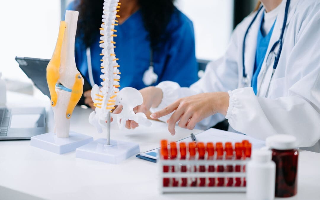
Repairing Your Spine from the Inside Out: How Chiropractic and Regenerative Therapies Work Together
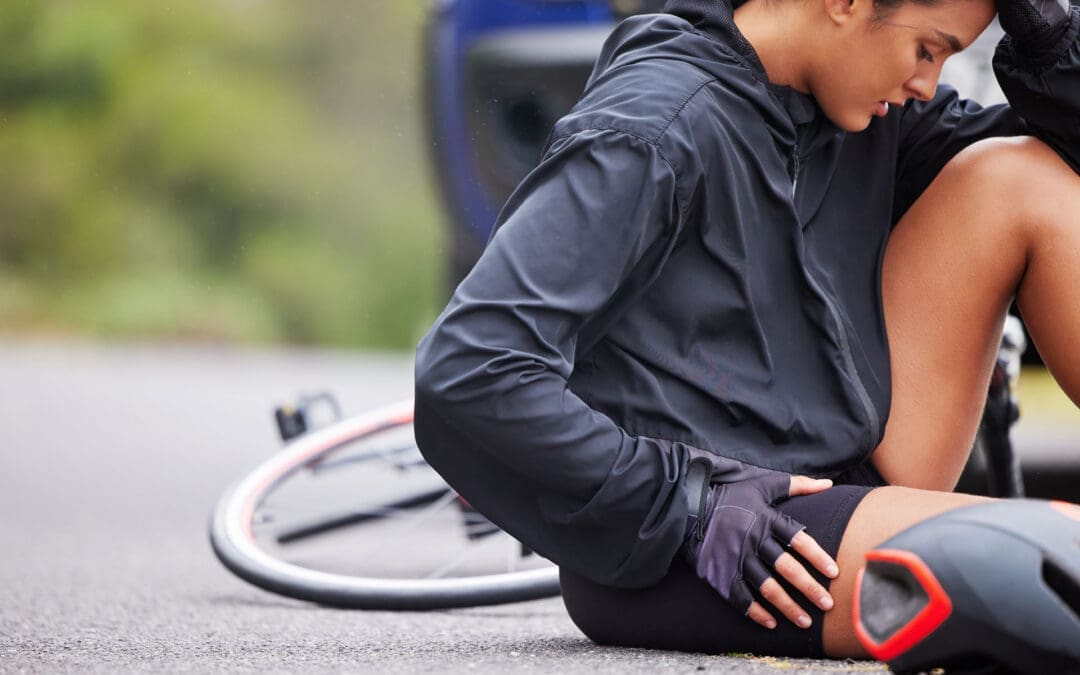
Chronic back or neck pain can make everyday life feel challenging. Many people try rest, pain pills, or physical therapy, but the pain often returns. Surgery is one option, yet it comes with risks and long recovery times. There is a better path for many people. Combining chiropractic care with regenerative therapies helps repair the spine from the inside out. Chiropractic adjustments fix the structure and alignment. Treatments such as PRP injections and shockwave therapy help heal the soft tissues around the bones. Together, they stop chronic pain and help rebuild strength without surgery.
This approach treats the whole picture. It is like fixing a house with problems in both the frame and the interior.
Your Spine Is Like a House
Think of your spine like the wooden frame of a house. The bones (vertebrae) are the main beams. The discs, ligaments, tendons, and muscles are the wood, pipes, and supports that hold everything together.
If the frame leans or shifts, the whole house feels off. Chiropractic adjustments gently move the bones back into better position. This straightens the frame and takes pressure off nerves.
But what if the wood is rotting or the pipes are leaking? Straightening the frame alone will not last. The house will lean again. The same thing happens with the spine. If the soft tissues stay damaged or inflamed, pain and weakness return even after effective adjustments.
Regenerative therapies, laser treatments, and shockwave therapy act like a skilled repair crew. They go into damaged areas, reduce swelling, and help new, healthy tissue grow. Massage and spinal decompression then remove daily stress so the repairs hold strong over time. This comprehensive plan delivers lasting results for many people with ongoing spinal problems.
Chiropractic Care: Fixing the Structural Frame
Chiropractic care focuses on the bones and joints of the spine. A chiropractor uses gentle, hands-on adjustments to correct misalignments. These shifts can happen from poor posture, old injuries, car accidents, or years of sitting and bending.
Adjustments help in simple ways:
They restore normal movement in stiff joints.
They reduce pressure on nerves that cause pain, numbness, or tingling.
They improve blood flow and support the body’s natural healing ability.
They help improve posture so daily activities feel easier.
According to reliable health information, chiropractic adjustments aim to correct alignment problems, ease pain, and support the body’s natural ability to heal itself (MedlinePlus, n.d.). Many people use it for low back pain, neck pain, and headaches. It is a hands-on method that does not rely on drugs or surgery.
When the frame is straight, the rest of the spine can heal better. But adjustments work even better when paired with treatments that fix the soft tissues around the bones.
Regenerative Therapies: The Repair Crew for Soft Tissues
Regenerative therapies use the body’s own materials to heal damaged areas. They target the “rotting wood” parts — inflamed discs, torn ligaments, irritated nerves, and weak tendons. These treatments send growth signals that tell cells to repair and rebuild.
Common options include:
PRP (platelet-rich plasma): A small amount of your blood is spun in a machine to concentrate the healing platelets. These are injected into painful spots. Growth factors signal damaged tissues to calm down and grow stronger (El Paso Back Clinic, n.d.).
Shockwave therapy: Special sound waves reach deep into sore muscles and tendons. They increase blood flow, break up scar tissue, and wake up the body’s repair process (Sleppy Chiropractic, n.d.).
MLS laser therapy: Safe light energy gives cells more power (ATP) to reduce inflammation and speed healing at the deepest level (Sleppy Chiropractic, n.d.).
Spinal decompression: Gentle stretching creates space between vertebrae. This relieves pressure on bulging discs, allows nutrient-rich fluid to flow in, and helps discs rehydrate and repair (Sleppy Chiropractic, n.d.).
These therapies help stop the cycle of chronic pain. They reduce swelling, support new tissue growth, and strengthen the areas that hold the spine in place. Many patients notice reduced pain and improved mobility within weeks when these treatments are used as part of a comprehensive plan.
How Chiropractic and Regenerative Therapies Work Together
The real power comes when the two approaches work together. Chiropractic adjustments create the right alignment so new tissue can form correctly. Regenerative treatments create a healing environment within tissues so that repairs last (El Paso Back Clinic, n.d.).
Here is how the full plan usually flows:
First, a careful exam and imaging find the exact problems.
Chiropractic adjustments straighten the frame and improve motion.
Regenerative injections or shockwave therapy target the damaged soft tissues and nerves.
Spinal decompression and soft-tissue work (such as massage or rehab exercises) reduce daily stress and protect healing areas.
Functional medicine support — nutrition, sleep, and inflammation control — helps the whole body recover.
One clinical observation notes, “When regenerative injections and chiropractic care happen together, the results often last longer. The injections create a better healing environment inside the tissues. The adjustments keep the joints moving correctly so that new tissue forms properly and does not become stressed again” (El Paso Back Clinic, n.d.).
Another insight suggests that a combined approach using chiropractic care, spinal decompression, regenerative injections, and supportive therapies, such as shockwave and laser therapy, often works better. These treatments create both mechanical and biological conditions that help the body heal and maintain better alignment (Personal Injury Doctor Group, 2026).
Patients often report meaningful drops in pain, better strength, and the ability to return to work or activities they enjoy. The goal is not just short-term relief. It is lasting repair that helps people avoid surgery and strong medications.
Expert Multidisciplinary Care in El Paso, Texas
In El Paso, this type of care happens in a true team setting. Dr. Alexander Jimenez, DC, APRN, FNP-BC, CFMP, IFMCP, ATN, CCST, brings over 30 years of chiropractic experience plus his training as a board-certified family nurse practitioner. His clinical observations show that patients with old injuries, car accident damage, sciatica, and posture-related spine pain improve when care targets both tissue repair and nervous system function. He sees people regain mobility, reduce chronic pain, and return to daily life through personalized, non-invasive plans that combine adjustments, regenerative therapies, rehabilitation, and functional medicine support (Dr. Alexander Jimenez, n.d.; Injury Medical Clinic PA, n.d.).
Working alongside him is Dr. Maria Guadalupe Cardenas, MD. She is a board-certified internal medicine physician (NPI #1164426749, Texas MD License #J2933) with over 40 years of experience. She serves as medical director and collaborative physician at Injury Medical Clinic PA. In this multidisciplinary setup — common in integrative and injury care clinics — the MD provides medical oversight, ensures procedural safety, reviews complex health factors, and adds an internal medicine perspective. Dr. Jimenez delivers the hands-on chiropractic and regenerative care. Together with functional medicine, personal injury documentation, and rehabilitation services, the team creates one coordinated plan under one roof.
This collaboration means patients receive complete care. The structural work (chiropractic), the biological repair (regenerative therapies), and the medical guidance all support each other. It is especially helpful for people with personal injuries, sciatica, chronic back pain, or posture problems that have not fully healed with other approaches.
Breaking the Pain Cycle and Rebuilding Strength
Poor posture or old injuries often create a cycle: misalignment stresses soft tissues, tissues become inflamed or torn, pain limits movement, and weakness worsens. The combined approach breaks this cycle at every level.
Chiropractic restores alignment and motion. Regenerative therapies heal and strengthen the supporting tissues. Decompression and soft tissue care protect the repairs from daily stress. Over time, many people feel less pain, stand taller, move more freely, and enjoy activities again.
This path focuses on root causes instead of just masking symptoms. It supports the body’s natural ability to heal while giving it the right tools and environment to succeed.
If ongoing spine pain is limiting your life, learning more about this integrative approach may open new options. Many people in El Paso and surrounding areas have found real relief and lasting improvement through careful, combined care that repairs the spine from the inside out.
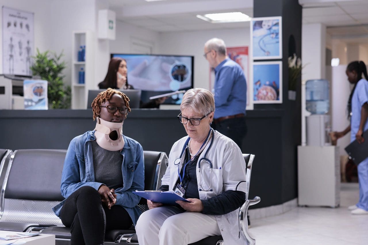
El Paso’s Integrated Injury Clinic: One-Stop Care for Faster Recovery and Strong Legal Support
If you got hurt in a car crash or at work in El Paso, Texas, you know how frustrating it can be. You go to one place for an exam, another for therapy, and still another for special treatments. Papers get lost. Your story gets told many times. Healing slows down. An integrated, multidisciplinary injury clinic solves this problem. Everything happens under one roof. A team of experts works together on your care. They handle medical checks, hands-on therapy, and advanced healing methods. At the same time, they build clear, complete records that help your legal or workers’ compensation case.
This kind of care is different from going to separate offices. You get faster answers, smoother progress, and stronger support for your claim.
Why an Integrated Health System Works Better
Ordinary clinics often focus on one thing. You might get only adjustments or only medications. An integrated clinic brings many experts together in the same place. They share one plan for you. This stops gaps in care and mixed messages.
Here are the main advantages:
One team, one story: Every provider sees your full history and current progress. No one works in the dark.
Faster decisions: If you need a new test or different therapy, the group talks it over quickly.
Better healing: Treatments work in tandem. Chiropractic care improves movement while medical oversight watches your overall health.
Clear records from day one: Everything gets written down in one system. This matters a lot for insurance and legal needs.
Patients who use this model often feel less stressed. They spend less time driving between offices and more time actually getting better.
How the Team Works Together for You
In a true multidisciplinary setup, each expert brings their skills. Nurse practitioners handle full health evaluations. They can order tests, manage medications as needed, and monitor for other health issues that might slow healing.
Physical therapists, massage therapists, and chiropractors team up on your body’s movement. They improve flexibility, strength, and posture. Chiropractic adjustments help the spine and joints work better. Physical therapy builds safe exercises. Massage eases tight muscles. Together, they create a plan that fits your exact injuries.
This is not random care. It is coordinated. Everyone knows what the others are doing. That teamwork often leads to quicker pain relief and better long-term results.
Special Treatments That Help Serious or Lasting Injuries
Some injuries need more than basic care. Car accidents and work injuries can damage deep tissues, nerves, or joints. An integrated clinic offers modern options that directly target the problem.
Here are treatments often used together:
Spinal decompression: A special table gently stretches your spine. This takes pressure off pinched nerves and bulging discs. Many people feel relief from sciatica or radiating leg pain.
MLS laser and shockwave therapy: These use light or sound waves to wake up your body’s healing cells. They lower swelling and help soft tissues repair without drugs or surgery.
Epidural injections: When nerves are very irritated, guided injections can calm the area so you can move and heal.
Regenerative therapies: Treatments such as PRP (platelet-rich plasma), PRF (platelet-rich fibrin), and MFAT (micro-fragmented adipose tissue) use components of your blood or fat. They are placed exactly where tissue is damaged to support natural repair.
These options go beyond what a basic clinic usually offers. They aim at the root of the injury rather than merely masking pain.
Strong Medical-Legal Documentation That Protects Your Case
When your injury comes from a car accident or a job, good records are just as important as good treatment. Insurance companies and lawyers need proof. They want to see what happened, how bad it is, and that the care you received was necessary.
An integrated team creates one complete file. It includes:
Your accident story and first exam findings
Objective tests like range of motion, strength checks, and imaging
Daily notes on how you feel and what treatments you receive
Progress reports that show improvement or ongoing limits
A final summary that explains the lasting effects on your life and work
This kind of documentation helps personal injury lawyers build a stronger case. It shows a clear link between the crash or incident and your injuries. It also proves you followed a real treatment plan.
Many clinics work directly with attorneys. They send updates quickly and often handle cases on a lien basis. This means you can focus on healing while the legal side stays organized.
Expert Care Led by Dr. Alex Jimenez and Dr. Maria Guadalupe Cardenas in El Paso
At Injury Medical Clinic PA in El Paso, this integrated model is led by experienced professionals who understand both health and legal needs.
Dr. Alexander Jimenez, DC, APRN, FNP-BC, CCST, CFMP, IFMCP, ATN, is dual-licensed as a chiropractor and board-certified family nurse practitioner. He has spent decades helping people with car accident injuries, work injuries, whiplash, sciatica, and soft tissue damage. His clinical observations focus on treating the whole person. He looks at root causes, not just symptoms. He stresses careful documentation with clear findings and progress measures, especially when a case involves an auto or work injury claim. His practice combines chiropractic care, functional medicine, rehabilitation, and regenerative options under one roof.
Working alongside him is Dr. Maria Guadalupe Cardenas, MD. She is board-certified in internal medicine with over 40 years of experience. Her NPI is 1164426749, and her Texas MD license is J2933. She serves as Medical Director and Collaborative Physician. In this multidisciplinary setting, she provides medical oversight, reviews overall health, guides advanced procedures, and helps ensure compliance with Texas rules.
Together, they create a powerful team. Chiropractic care from Dr. Jimenez addresses alignment, nerves, and movement. Medical direction from Dr. Cardenas provides safety, diagnostic, and medication management as needed. Functional medicine, personal injury documentation, and rehabilitation services all connect in the same location. This is the kind of collaborative model common in high-quality integrative injury clinics.
Your Simple Path to Recovery in El Paso
Here is what the journey usually looks like:
You call or come in for an evaluation. A nurse practitioner or medical director, along with the chiropractic team, sees you together.
They create one clear plan that may include adjustments, therapy, advanced treatments, or regenerative options.
You receive care in one place. Notes stay organized from the first visit to the last.
Progress is tracked and shared with your lawyer or insurance when needed.
You heal with less stress and stronger support for your claim.
Many patients notice they move better sooner and have less confusion about next steps.
Choose Coordinated Care for Your Injury
If you want care that treats your injury and protects your legal position, an integrated multidisciplinary clinic in El Paso is worth considering. You get medical diagnostics, physical therapy, advanced healing therapies, and solid documentation all in one coordinated system. Dr. Alex Jimenez and Dr. Maria Guadalupe Cardenas lead a team that puts your recovery and your case first.
You do not have to piece your care together alone. One roof, one team, one clear plan can make a real difference in how fast you feel better and how well your case is supported.
Regenerative Therapies Combined with Chiropractic Care Offer New Hope for Sports and Auto Accident Injuries in El Paso
Many people in El Paso deal with ongoing pain and limited movement after sports injuries or car accidents. Simple rest or basic physical therapy often helps at first, but sometimes healing stalls. Tissues stay inflamed, joints feel stiff, and daily life or sports become difficult again. When that happens, more people look for advanced options that work with the body instead of just covering up symptoms.
Regenerative therapies and integrative chiropractic care team up to tackle these tough problems. They focus on real repair at the tissue level while also fixing how the body moves. This combined approach helps many patients get back to feeling better and moving easier without jumping straight to surgery.
Why Standard Treatments Sometimes Fall Short
Injuries from sports collisions or car crashes often damage more than one area. Muscles tear, ligaments stretch, tendons become inflamed, and spinal discs or joints become irritated. Swelling and scar tissue can block normal blood flow and healing signals.
Physical therapy and rest build strength and reduce pain for many people. Yet when progress plateaus, underlying tissue damage or poor joint alignment may still be holding back recovery. That is when patients often seek care that actively supports the body’s repair systems instead of only managing symptoms.
What Regenerative Therapies Actually Do
Regenerative medicine uses materials from your body to kick-start healing. These treatments deliver growth factors and helpful cells directly to the damaged area. The goal is to lower inflammation, encourage new tissue growth, and improve long-term function.
Three main options stand out for musculoskeletal and spinal injuries:
PRP (platelet-rich plasma) comes from a small sample of your blood. The blood is spun in a machine to concentrate platelets, which carry natural growth factors. Doctors inject this concentrated solution into tendons, ligaments, joints, or around nerves. The growth factors signal cells to repair and rebuild.
PFP (platelet-fibrin products) uses protein concentrates from your blood. These capture growth factors and create a stronger, longer-lasting healing signal for tissues that have not responded well to simpler treatments.
MFAT (microfragmented adipose tissue) takes a small amount of your own fat tissue, processes it into tiny fragments, and injects it. The fat contains supportive cells and signaling factors that cushion joints and help repair cartilage, tendons, and soft tissues.
These are called orthobiologics because they come from your biology. They carry a low risk of allergic reactions or rejection since they use your materials.
Epidural injections sometimes join the plan for spine-related pain and nerve irritation. Under careful medical guidance, they reduce inflammation around spinal nerves while the regenerative injections work to repair deeper tissue.
How Chiropractic Care Completes the Picture
Injections alone help tissues heal, but they do not fix how the bones, joints, and muscles line up or move. That is where chiropractic adjustments come in. Gentle, precise realignments improve joint mobility, ease muscle tension, and restore better posture and movement patterns.
When regenerative injections and chiropractic care happen together, the results often last longer. The injections create a better healing environment inside the tissues. The adjustments keep the joints moving correctly so that new tissue forms properly and does not get stressed again. This partnership addresses both the biology of repair and the mechanics of the body.
The Strength of a True Multidisciplinary Team
Patients get the best results when they receive care from a well-established integrative and functional medicine clinic that brings different experts together under one roof. At Injury Medical Clinic PA in El Paso, Texas, the team combines advanced regenerative procedures with chiropractic expertise, functional medicine, rehabilitation, and personal injury support.
Dr. Alexander Jimenez, DC, APRN, FNP-BC, CFMP, IFMCP, ATN, CCST, leads the clinical approach. With decades of experience as a chiropractor and additional training as a board-certified family nurse practitioner, he focuses on whole-person recovery. His clinical observations show that patients with sports trauma or old auto accident injuries often improve when care targets both tissue repair and nervous system function. He uses detailed exams, imaging, and personalized plans that include regenerative injections, adjustments, rehabilitation, and lifestyle support.
Dr. Maria Guadalupe Cardenas, MD, a board-certified internal medicine physician with over 40 years of experience (NPI #1164426749, Texas MD License #J2933), serves as Medical Director and Collaborative Physician. She provides medical oversight for procedures, ensures safety and compliance, manages complex health factors, and brings an internal medicine perspective to every case. This collaboration means patients receive both expert spinal and musculoskeletal care from Dr. Jimenez and broad medical direction from Dr. Cardenas.
This setup is common in high-quality integrative injury clinics. The MD handles medical aspects and procedure oversight while the chiropractor and nurse practitioner team deliver hands-on treatment and functional strategies. Everyone works from the same records and goals, so care stays coordinated and thorough.
Clear Benefits Patients Notice
People who choose this combined path often report several practical improvements:
Noticeable drops in pain and swelling without relying only on medications
Better tissue repair that supports longer-lasting results
Improved joint movement and daily function
Faster return to work, sports, or normal activities when healing had stalled
Lower chance of needing more invasive procedures later
Thorough documentation that helps with insurance and legal needs after personal injury cases
Because the treatments use your own biological materials, side effects stay minimal for most people. Soreness at the injection site usually fades within a few days.
The functional medicine side of care looks at nutrition, inflammation levels, sleep, and stress. These factors influence how well tissues heal. Addressing them alongside the injections and adjustments gives the body every advantage.
What a Typical Care Journey Looks Like
Most patients start with a full evaluation that includes history, physical exam, and any needed imaging. The team identifies exactly which tissues need help and whether alignment issues are slowing progress.
Next comes a customized plan. This may include one or more regenerative injections (PRP, PFP, or MFAT), chiropractic adjustments over several weeks, guided rehabilitation exercises, and supportive therapies such as shockwave treatment when appropriate. Follow-up visits track progress and adjust the plan as tissues respond.
Many people begin to feel meaningful relief within weeks, with continued improvement over the next few months as repair progresses. The team stays involved through the entire process.
Who Benefits Most from This Approach
This type of care often helps adults dealing with:
Lingering pain after sports collisions or overuse injuries
Whiplash, back strain, or nerve irritation from car accidents
Old injuries that never fully settled
Joint or tendon problems that limit activity
It works especially well when conventional treatments have already been tried, and progress has slowed. The focus stays on restoring real function rather than temporary relief.
Moving Forward with Confidence
Healing from serious injuries takes time and the right tools. Regenerative therapies give tissues the biological signals they need. Integrative chiropractic care helps the body use those new repairs by improving movement and alignment. When both occur within a coordinated team that includes medical direction, functional medicine, and personal injury expertise, patients often regain greater comfort and capability than they expected.
If you or someone you know in the El Paso area continues to struggle after sports trauma or an auto accident, consider learning more about these combined options. A thorough evaluation at a clinic experienced in both regenerative procedures and chiropractic care can show whether this path fits your situation. Many people find it opens the door to meaningful, lasting improvement.
El Paso’s 100 Deadliest Days: Teen Driving Risks and Integrative Recovery at El Paso Back Clinic
Summer in El Paso means more time on the road for young drivers heading to work, friends, or trips across town and beyond. But this season also brings greater danger. The stretch from Memorial Day to Labor Day is known as the 100 Deadliest Days because fatal crashes involving young drivers rise sharply. At El Paso Back Clinic, our team sees the real impact when these accidents happen. Many patients come in weeks later with pain that started small but grew because of how the body reacts to sudden trauma. Learning the risks and knowing the right place for complete recovery helps families in El Paso stay safer and heal better if trouble strikes.
What Are the 100 Deadliest Days?
The 100 Deadliest Days run from Memorial Day through Labor Day, about 100 days when the number of deadly crashes with young drivers jumps across the country and right here in El Paso. National numbers show that more than 30 percent of fatal crashes involving a young driver occur during this summer window. On average, eight people die each day in these crashes in summer compared to seven the rest of the year. In 2023, roughly one-third of the yearly total happened in these months alone.
El Paso faces the same spike plus local challenges. Highways like I-10 and Loop 375, busy streets such as Mesa and Montana, and long summer drives to places like White Sands or Ruidoso pose additional risks for drivers who are still gaining experience.
Why Summer Brings Higher Risks for Young Drivers in El Paso
Several things come together once school lets out and young people drive more on their own.
More driving without close supervision. Extra free time means more trips to jobs or social plans. Young drivers often log miles without an adult nearby to remind them to slow down or stay alert.
Extra passengers create distraction. One or two friends in the car can draw attention away from the road by talking or moving. Texas rules for drivers ages 16 and 17 already limit non-family passengers under 21, yet summer plans often test these limits.
Phones and summer plans add distraction. Quick texts or calls happen more when schedules are loose. Even a few seconds of looking away can cause a rear-end crash on busy local roads.
Night driving and longer trips increase fatigue. Low light on I-10 or Loop 375 slows reactions. Heat over 100 degrees can also cause tire trouble that surprises new drivers on long stretches.
Speeding and following too closely. Open roads tempt higher speeds. Tailgating on busy streets like those near Airway or Sunland Park leads to sudden stops and chain-reaction crashes.
These patterns explain why the same careful driver faces greater danger during summer freedom.
Expert Tips to Help Young Drivers Stay Safe
Groups like the National Road Safety Foundation and AAA Texas give simple steps that work. The focus is on cutting distractions and building good habits early.
Buckle up on every single ride. Seat belts greatly lower the chance of serious injury or death.
Keep phones away or turn on do-not-disturb mode while driving. Even one message can lead to a crash.
Limit young passengers. Follow Texas rules that allow only one non-family passenger under 21 for provisional drivers.
Plan routes together before leaving. Review exits, construction, and safe stops on highways like I-10.
Check tires, brakes, and fluids before summer trips. Extreme El Paso heat wears tires faster.
Set clear rules about speed, rest, and no drinking. Parents who drive calmly set the best example.
These habits help turn risky summer miles into safer ones for everyone on El Paso roads.
What Happens When a Crash Occurs?
Even careful drivers can end up in an accident on I-10, at a busy intersection, or in a rear-end on Mesa Street. Right after the crash, adrenaline and endorphins often mask the full extent of the damage. Many people feel okay at the scene, only to notice problems hours or days later. At El Paso Back Clinic, we see patients whose neck stiffness, headaches, or back pain started small but worsened as swelling and inflammation slowly built up in the deeper tissues. Some symptoms even appear weeks later as the body compensates or scar tissue forms.
Common delayed signs include ongoing headaches from neck strain, neck or back stiffness and pain, radiating numbness or tingling into arms or legs, unusual fatigue, brain fog or trouble focusing, dizziness or balance issues, shoulder or hip discomfort, sleep problems, and mood changes. Ignoring these signals can turn a minor issue into long-term pain or changed movement patterns that affect driving, work, and daily life.
That is why prompt, thorough care matters. The right clinic helps the body heal from both the direct physical trauma and the whole-system stress the crash creates.
How El Paso Back Clinic Supports Integrative Recovery
At El Paso Back Clinic, we specialize in helping car accident victims recover fully, especially when pain shows up later. Our integrative approach treats the musculoskeletal injuries and the broader effects on inflammation, nerve function, sleep, and tissue repair. This combination often leads to faster relief, better movement, and fewer long-term problems.
Dr. Alexander Jimenez, DC, APRN, FNP-BC, leads the team with years of experience in personal injury and spinal trauma. His clinical observations show that patients with delayed symptoms improve significantly when care targets spinal alignment early and supports the body’s natural repair processes. Gentle chiropractic adjustments restore joint movement, relieve nerve pressure, and reduce muscle guarding. Myofascial release loosens tight tissues so the body stops compensating in ways that create new pain.
We also offer advanced options when deeper support is needed. Regenerative injections such as platelet-rich plasma (PRP) use the patient’s own concentrated platelets to release growth factors that help build collagen, improve blood flow, and repair ligaments, tendons, and muscles. Spinal decompression gently stretches the spine to ease pressure on discs and nerves, helping with radiating pain or sciatica-like symptoms. Ultrasound and shockwave therapy boost circulation and calm inflammation without surgery. Rehabilitation exercises rebuild strength and stability so patients return to normal activities with lower risk of setbacks.
Working alongside Dr. Jimenez is Dr. Maria Guadalupe Cardenas, MD. She is board-certified in internal medicine with over 40 years of experience. Her NPI number is 1164426749, and her Texas medical license is J2933. As Medical Director and Collaborative Physician at the clinic, she provides medical oversight, reviews overall health, guides complex cases, and ensures everything stays safe and compliant. This multidisciplinary setup, common in strong injury clinics, means chiropractic care, functional support, and medical direction happen in one place with consistent records.
One of the biggest benefits for El Paso families is the detailed documentation we create. Clear notes link the crash to the injuries, record objective measures like range of motion and strength, track daily limitations such as driving or working, and show steady progress. These records help insurance claims move smoothly and give personal injury attorneys the credible timeline they need for fair settlements. Many patients appreciate that everything from the first exam to final recovery notes stays in one location, reducing stress during an already difficult time.
Our team focuses on whole-person healing so the body can repair at the cellular level. Early attention prevents small problems from becoming chronic pain or altered posture that lasts for years. Patients often report less ongoing discomfort, easier movement, and a quicker return to family life and work.
Taking the Next Step Toward Safety and Healing
The 100 Deadliest Days remind us that summer driving in El Paso carries real risks for young drivers. More freedom, extra passengers, phones, and longer trips on local highways all raise the chances of trouble. Simple habits like buckling up, limiting distractions, and planning routes can prevent many crashes.
When an accident does happen, know that delayed pain is common and can be treated. At El Paso Back Clinic, we provide integrative care that addresses both visible injuries and hidden stress on the body. With Dr. Alex Jimenez’s expertise in spinal trauma and delayed symptoms, Dr. Maria Guadalupe Cardenas’s medical oversight, and a full range of chiropractic, regenerative, and rehabilitation services, patients receive complete support and strong documentation for insurance or legal needs.
Summer should bring cherished memories, not lasting pain. Understanding the risks and choosing thorough recovery care at El Paso Back Clinic helps young drivers and their families in El Paso move forward with confidence.
If you or someone you care about was in a summer car accident and is now feeling delayed pain or stiffness, contact our team today. Call 915-850-0900 or visit elpasobackclinic.com to schedule a consultation. We are here to help you heal fully and get back to living, loving, and thriving.
Unlocking Cellular Healing: The Power of Advanced Laser Therapy in Integrative Care
Abstract
As a clinician with a diverse background spanning chiropractic, advanced practice nursing, and functional medicine, my primary goal is to offer patients the most effective, evidence-based treatments available. In this educational post, I will take you on a journey into the world of Multiwave Locked System (MLS) Laser Therapy, a cutting-edge technology that is transforming how we manage pain and inflammation. We will explore the science behind this therapy, moving beyond surface-level explanations to understand its profound effects on cellular biology, including its impact on mitochondria and the inflammatory cascade. I will share insights from leading researchers and demonstrate how we apply this technology in clinical settings, particularly for conditions such as low back pain and joint issues. Furthermore, I will explain how MLS Laser Therapy integrates seamlessly into a comprehensive care model like ours at Injury Medical Clinic, where we combine chiropractic adjustments, physical rehabilitation, and advanced medical oversight from our Medical Director, Dr. Maria Guadalupe Cardenas, MD, to optimize patient outcomes. This post will detail specific treatment protocols, the importance of energy density, and how this therapy can augment other regenerative treatments, such as Platelet-Rich Plasma (PRP), offering a multifaceted approach to true healing.
A New Frontier in Healing at Injury Medical Clinic
Hello, I’m Dr. Alex Jimenez. With my credentials as a Doctor of Chiropractic (DC) and Advanced Practice Registered Nurse (APRN), and my certifications in functional and integrative medicine (CFMP, IFMCP), my passion has always been to bridge gaps between healing disciplines. At Injury Medical Clinic PA, we have built a practice on this very principle: a truly integrative approach to patient wellness.
A cornerstone of our collaborative model is my partnership with Dr. Maria Guadalupe Cardenas, MD. Dr. Cardenas is Board Certified in Internal Medicine and serves as our esteemed Medical Director and Collaborative Physician. With over 40 years of invaluable experience, she provides essential medical oversight, ensuring our patients receive safe, comprehensive, and well-rounded care. This multidisciplinary structure allows us to blend the best of chiropractic and physical rehabilitation with the diagnostic and medical expertise of internal medicine. Our team works in synergy, designing treatment plans that address not just the symptoms but the underlying physiological dysfunction. Whether a patient is recovering from a personal injury, managing a chronic condition, or seeking to optimize their overall health, our integrated team provides a holistic, evidence-based pathway to recovery.
Navigating Low Back Pain with MLS Laser Therapy
One of the most common ailments we see is chronic low back pain. Today, we have a patient, John, who is experiencing persistent joint pain and stiffness in his lumbar spine, specifically around the L4-L5 facet joints, with some discomfort radiating down his right side. This is a classic presentation that responds exceptionally well to a targeted, multimodal approach.
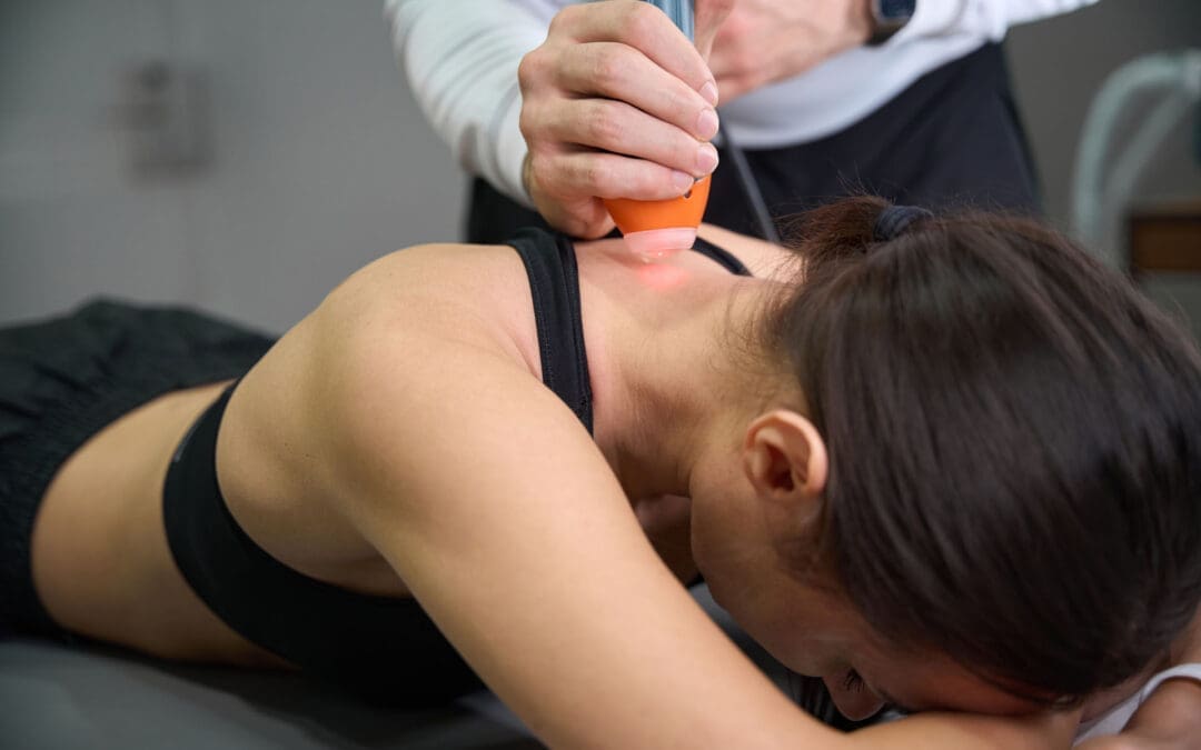
For John, we are utilizing the M6 Robotic MLS Laser. The first priority is always patient comfort. When using a robotic system, it’s critical that the patient remains still, as the laser is programmed to treat a precise area. We position the patient face down to allow direct access to the skin over the lumbar spine, as the laser energy must be delivered without the barrier of clothing.
The Clinical Multimodal Approach: More Than Just the “Spot of Pain”
Once John is comfortable, we begin the setup. The robotic laser interface is remarkably sophisticated yet user-friendly.
Targeting the Ailment: I select the “Joint Pain and Stiffness” protocol for the back.
Centering the Treatment: I zero out the X and Y axes on the control panel. This temporarily stops the robotic arm’s movement, allowing me to manually position the guiding red light directly over the primary source of John’s discomfort—the L4-L5 region he indicated.
Expanding the Field: This is where our clinical multimodal approach comes into play. Instead of just treating the single spot of pain, I expand the treatment area using the X and Y controls. This creates a larger therapeutic field that covers not only the symptomatic facet joints but also the surrounding connective tissue, muscles, and nerve roots. We aren’t just chasing pain; we are treating the entire functional unit to address the source of the dysfunction and support the interconnected biological systems.
The laser head is positioned at a precise distance from the skin—about six inches—using a provided ruler. This is crucial because the MLS laser beam is collimated, meaning the light rays are parallel. The focal point is engineered to be most effective at this distance, ensuring the therapeutic energy penetrates deep into the tissues rather than dissipating at the surface.
The Science of Healing: How MLS Laser Therapy Works
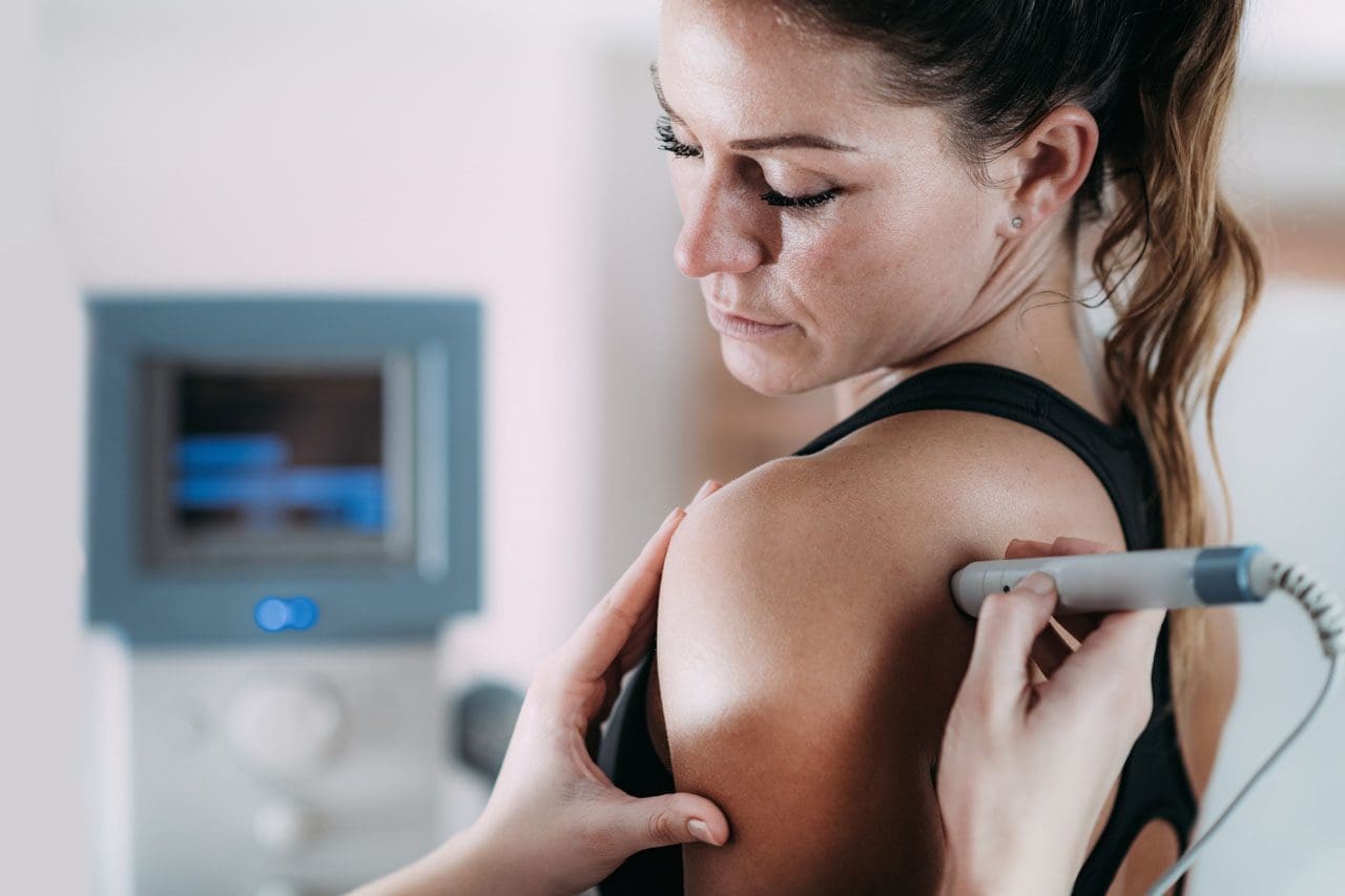
With the treatment underway—an eight-minute session for John’s low back—let’s dive into what’s happening at a cellular level. It’s common for patients to ask if they will feel anything. Most feel nothing at all, though some may notice a gentle warmth or tingling. This lack of intense heat is a hallmark of the MLS system’s advanced design.
The device combines two specific wavelengths of light: an 808-nanometer (nm) continuous-wave and a 905-nanometer (nm) pulsed-wave.
The 808 nm wavelength works more superficially to reduce inflammation and edema. It enhances blood circulation to the area, which helps clear out inflammatory byproducts and deliver oxygen and nutrients.
The 905 nm wavelength, delivered in powerful, short pulses, penetrates much deeper, reaching tissues such as muscle, nerve, and even the joint capsule. This pulsed energy is what provides the powerful analgesic (pain-relieving) effect.
These two wavelengths are synchronized, creating the patented “MLS pulse.” This enables delivery of very high peak power (up to 50 watts) in extremely short bursts (nanoseconds). This high-intensity “punch” of energy stimulates the cells without generating heat. A period of rest follows each pulse, allowing the tissue to absorb the energy efficiently. If a laser produces significant heat at the skin’s surface, it often means the energy isn’t being absorbed properly by the target tissues. The MLS system maintains tissue temperature at a constant level, ensuring optimal therapeutic delivery.
Seeing the Invisible: A Window into the Treatment
A fascinating demonstration of this technology involves using a smartphone camera. While the red aiming light is visible to the naked eye, the therapeutic infrared laser light is not. However, a camera’s sensor can detect it. If you were to look at John’s back through a phone camera during treatment, you would see a distinct triangle of light—this is the 808 nm wavelength at work, covering a significant area and illustrating how comprehensively we are treating the region.
Energy Density: The Key to Effective Dosing
A critical concept in laser therapy is energy density, measured in joules per centimeter squared (J/cm²). This is more important than the total number of joules delivered. Think of it like watering a plant: you need to provide the right amount of water for the pot’s size. Too little has no effect; too much drowns it. Similarly, our goal is to deliver a precise dose of light energy to the target tissue.
The World Association for Laser Therapy (WALT) and a large body of research support an optimal therapeutic window of 4-10 J/cm².
For John’s condition, the protocol is set to deliver approximately 6 J/cm². The laser’s software automatically calculates the treatment time required to achieve this density over the selected area. If I were to make the treatment area smaller or larger, the software would instantly recalibrate the time to ensure the correct dose is delivered.
This concept also relates to the Arndt-Schultz Law, a pharmacological principle stating that low doses stimulate, moderate doses inhibit, and high doses are toxic. With laser therapy, if you “overcook” an area with too much energy, you risk a bioinhibitory effect, in which the treatment becomes less effective or even counterproductive. The body’s cells can only absorb so much energy at once. This is why our protocols focus on precise energy density and, if more treatment is needed, we target different areas (e.g., an anterior and posterior approach for a knee) rather than just increasing the time on one spot.
Integrating Modalities for Superior Results
While the robotic laser treats the broader lumbar region, I can simultaneously use a handheld MLS laser applicator. This handpiece allows for more focused treatment on specific points, such as trigger points or “knots” in the muscle. I often use the “cooked meat” versus “raw meat” analogy that a physical therapist once taught me. Healthy, relaxed muscle feels like raw meat, while a tight, knotted trigger point feels firm, like cooked meat. The handheld applicator is perfect for treating these punctual spots.
The robot and the handpiece operate on two separate channels, allowing us to perform this dual treatment. This is a perfect example of our integrative philosophy in action:
Chiropractic Care: Before or after the laser session, I can perform specific chiropractic adjustments to restore proper motion to the L4-L5 facet joints and relieve mechanical stress.
Physical Rehabilitation: Our team can guide John through exercises to strengthen his core musculature and improve spinal stability.
MLS Laser Therapy: The laser works at the cellular level to reduce pain and inflammation that may be hindering his ability to engage in rehabilitation, thereby accelerating healing.
This combination addresses the structural, functional, and biochemical aspects of his condition simultaneously.
Advanced Applications: Augmenting Regenerative Medicine
The conversation around healing is increasingly turning toward orthobiologics, such as Platelet-Rich Plasma (PRP) injections. This is where MLS Laser Therapy shows even more remarkable potential. A common question arises: if PRP induces a beneficial pro-inflammatory phase to kickstart healing, won’t an anti-inflammatory laser treatment counteract it?
The answer is no. In fact, the laser augments the process. The data and our clinical observations show that using laser therapy in conjunction with PRP can improve outcomes by an estimated 15-20%.
Here is the progressive protocol we often recommend:
Pre-Injection Priming (2-3 treatments): In the weeks leading up to the PRP injection, we use the laser to “prepare the soil.” These sessions are designed to increase local blood circulation, reduce baseline chronic inflammation, and optimize the cellular environment, making the tissue more receptive to the growth factors in the PRP.
Day of Injection (1 treatment): A treatment on the day of the procedure can further enhance the effects.
Post-Injection Support (6+ treatments): Following the injection, a series of laser treatments helps manage pain and supports the regenerative cascade initiated by the PRP. The laser enhances mitochondrial function, which is critical for providing the cellular energy (ATP) needed for tissue repair.
The Cascade of Healing: From Acute Relief to Chronic Repair
How does a single modality address both acute pain and chronic conditions? The effects occur in a cascade.
Immediate Effect (Acute Phase): The initial pain relief often comes from the laser’s effect on small, unmyelinated nerve fibers (C-fibers) that transmit pain signals. The energy can temporarily block these signals, providing rapid relief. This is the analgesic effect.
Subsequent Effect (Inflammatory Modulation): Over the next few hours and days, the anti-inflammatory effect takes hold. The laser energy modulates the immune response, reducing pro-inflammatory cytokines and promoting the resolution of inflammation and edema.
Long-Term Effect (Biostimulation and Chronic Repair): With a series of treatments, we get to the core of cellular repair. Light energy is absorbed by cytochrome c oxidase in the mitochondria, the powerhouses of our cells. This significantly increases ATP (adenosine triphosphate) production, the body’s primary energy currency. This surge in available energy fuels all cellular repair processes, from protein synthesis to cell replication, promoting true, long-term tissue healing.
This mitochondrial boost is especially relevant in today’s world, where many common medications, such as statins, can impair mitochondrial function. By enhancing mitochondrial biogenesis and efficiency, laser therapy can help overcome these hurdles and optimize the body’s innate healing capacity. This is why we also discuss nutritional and lifestyle factors—such as CoQ10 supplementation to support mitochondrial function—as part of a truly comprehensive functional medicine approach.
Treatment Frequency and The Cumulative Effect
Healing is a process, not an event. The effects of MLS Laser Therapy are cumulative. We recommend a series of treatments to achieve lasting results.
Acute Conditions: Typically, a course of 6 treatments is effective.
Chronic Conditions: A more intensive course of 12 treatments is often needed.
Ideally, treatments are scheduled close together (e.g., Monday, Wednesday, Friday) to build therapeutic momentum. It is crucial for patients to complete the full course. Many start feeling significantly better after just 3-4 sessions and are tempted to stop. However, completing the entire protocol ensures deeper cellular repair, leading to more durable outcomes.
At Injury Medical Clinic, our mission is to empower your body’s own ability to heal. By integrating the best of chiropractic, medical oversight, and groundbreaking technologies like MLS Laser Therapy, we offer a path to recovery that is not only faster but also more complete.
World Association for Laser Therapy. (n.d.). WALT Recommended Treatment Doses for LLLT. WALT. Retrieved from https://waltza.co.za/wp-content/uploads/2012/08/Dose_table_780-860nm_for_Low_Level_Laser_Therapy_WALT-2010.pdf
Memorial Day Weekend Rear-End Car Accidents: Common Causes, Injuries, and How Integrative Chiropractic Care Can Help
Memorial Day weekend marks the unofficial start of summer for many families. Roads fill up fast as people head out for beach trips, barbecues, and long drives to visit loved ones. With millions of cars on the highway at once, traffic slows to a crawl on major routes. This heavy congestion sets the stage for one of the most frequent crashes during holiday weekends: rear-end collisions.
These accidents happen when one vehicle slams into the back of another. They often create chain-reaction pileups because traffic stops suddenly. Even at low speeds, the impact can jolt the body hard. In this article, you will learn why rear-end crashes spike during Memorial Day travel, what distractions play a role, how these crashes injure the neck and spine, and why seeing a chiropractor soon after makes a big difference. The journey from crash to recovery is clearer when you understand the steps.
Why Rear-End Collisions Spike During Memorial Day Weekend
Heavy traffic turns busy highways into parking lots. Drivers brake suddenly for slow traffic ahead. The car behind may not have time to stop safely. According to safety data, rear-end crashes make up about 23 percent of all car accidents in the United States each year.
Holiday weekends like Memorial Day see extra travel volume. More cars mean more stops and starts. Chain-reaction incidents become common when one car hits another, and the force pushes forward through several vehicles.
Congestion on key routes: Interstates and major roads fill quickly with vacationers.
Abrupt halts: Traffic lights, construction zones, or accidents ahead force sudden stops.
Longer drives: Tired drivers on extended trips react more slowly.
These factors turn a relaxing weekend trip into a stressful situation.
Common Causes: Distractions Behind the Wheel
Driver distraction is a leading cause of rear-end crashes. When traffic moves in fits and starts, even a few seconds of lost focus can cause trouble. Common distractions during holiday drives include:
Adjusting a GPS or phone map for the next exit.
Checking mobile devices for texts, calls, or traffic updates.
Attending to passengers—kids asking questions, pets moving around, or family conversations.
Other causes include tailgating (following too closely) and speeding for the conditions. Distracted driving was linked to hundreds of serious crashes in recent state reports. Even hands-free phone use pulls attention from the road.
Simple rule: Keep eyes forward, hands on the wheel, and mind on traffic. A quick glance at a phone can turn a safe gap into a collision.
What Happens to Your Body in a Rear-End Crash
Picture this: Your car sits stopped in traffic. The vehicle behind hits you. Your body snaps backward, then forward, in a split second. This whip-like motion—called whiplash—puts sudden force on the neck and spine.
The head weighs about 10 to 12 pounds. That quick jerk multiplies the stress on soft tissues and bones. Even a 5-mile-per-hour bump can create enough force to stretch or tear ligaments and muscles.
Rear-end impacts affect the cervical (neck) and lumbar (lower back) areas most. The spine tries to absorb the shock, but it often cannot do so without sustaining damage.
Common Injuries from Rear-End Collisions
Rear-end crashes frequently lead to specific injuries because of the forceful jerking. Soft tissues take the biggest hit, but bones and nerves can suffer too. Here are the most reported issues:
Soft tissue sprains and strains: Ligaments and muscles stretch or tear. This causes pain, swelling, and stiffness in the neck and back.
Whiplash: The rapid back-and-forth motion strains neck muscles, tendons, and ligaments. Symptoms include neck pain, headaches starting at the skull base, and limited movement.
Herniated or bulging discs: The force pushes spinal discs out of place. Disc material can press on nerves.
Muscular spasms: Muscles tighten suddenly to protect the area, leading to painful knots and reduced motion.
Nerve impingement: Pinched nerves cause tingling, numbness, or shooting pain down the arms or legs.
These injuries often affect the whole upper body. Shoulders, upper back, and even jaw muscles can ache from the impact.
Many people feel okay right after the crash because adrenaline masks the pain. But stiffness or headaches can show up hours or days later.
Why Symptoms May Appear Later—and Why Early Evaluation Matters
The body’s natural response hides problems at first. Adrenaline surges during the scare, dulling pain signals. Once it fades, inflammation builds, and tissues swell.
A minor headache today might become constant neck pain tomorrow. Small sprains can become chronic issues if left untreated. Experts stress that a full check-up soon after any accident is smart—even if you feel fine. Waiting too long can allow scar tissue to form or cause a posture change for the worse.
Florida law, for example, encourages care within 14 days to protect insurance benefits. The same idea applies everywhere: early action speeds healing.
Integrative Chiropractic Care: Natural Healing for Accident Injuries
Integrative chiropractic care focuses on helping the body heal itself without heavy reliance on drugs or surgery. It targets both the skeleton (bones and joints) and soft tissues (muscles, ligaments, tendons).
Chiropractors use gentle spinal adjustments to realign vertebrae. This takes pressure off nerves and restores normal movement. Soft tissue therapies like massage, trigger-point work, and myofascial release loosen tight muscles and break up scar tissue.
Other helpful tools include:
Therapeutic exercises to strengthen weak areas and improve posture.
Ultrasound or heat/ice therapy to reduce swelling and boost blood flow.
Lifestyle tips on ergonomics, sleep positions, and daily movement.
These methods work together for whole-body recovery. Patients often report less pain, better range of motion, and improved energy after a few sessions.
Chiropractic care shines for whiplash and back sprains because it addresses the root cause—misalignments and muscle imbalances—rather than merely masking symptoms.
Clinical Observations from Dr. Alexander Jimenez
Dr. Alexander Jimenez, DC, APRN, FNP-BC, brings a unique blend of chiropractic expertise and advanced nursing practice to auto accident care. As the founder of Injury Medical Clinic in El Paso, Texas, he specializes in personal injury and multidisciplinary recovery.
Dr. Jimenez observes that many patients arrive weeks or months after a crash, still dealing with lingering neck, back, and shoulder pain. He notes that injuries often affect more than just the spine—they impact joints, nerves, soft tissue, mobility, sleep, and even stress levels. His clinical approach emphasizes natural healing through integrative methods.
He combines traditional chiropractic adjustments with functional medicine, regenerative therapies such as platelet-rich plasma (PRP), nutritional guidance, and rehabilitation exercises. This team-based care helps patients recover faster and avoid long-term complications. Dr. Jimenez stresses thorough evaluations, including imaging when needed, to catch hidden issues early. His patients frequently share stories of regaining mobility and returning to daily life pain-free after following personalized plans.
His work shows that even old or “minor” accident injuries can improve dramatically with the right holistic support.
Steps to Take After a Memorial Day Crash
If you are involved in a rear-end collision this holiday weekend, follow these simple steps:
Check for immediate safety and call for help if needed.
Exchange information and document the scene with photos.
Seek a full medical evaluation right away—even without obvious pain.
Consider integrative chiropractic care as part of your recovery team.
Follow through with recommended therapies and exercises.
Most people recover well when they act early and stay consistent with care.
Safe Driving Tips for Holiday Travel
Prevention beats treatment every time. Keep these habits in mind:
Leave extra space between cars in heavy traffic.
Put phones away and use voice commands only if necessary.
Take breaks on long drives to stay alert.
Watch for sudden braking ahead.
A calm, focused drive keeps everyone safer on the road.
Memorial Day weekend brings fun and family together, but extra traffic raises the risk of rear-end collisions. Understanding the causes—congestion and distractions—helps you stay alert. Knowing how these crashes jolt the neck and spine explains why whiplash, sprains, herniated discs, spasms, and nerve issues are so common. Because symptoms can sneak up later, a prompt check-up is key. Integrative chiropractic care offers a natural path to healing by realigning the body, easing soft-tissue damage, and restoring posture and movement.
Dr. Alexander Jimenez and similar specialists show that combining chiropractic techniques with supportive therapies delivers real results for accident victims. Whether your crash happened this weekend or years ago, relief is possible. Listen to your body, seek care early, and give yourself the best chance at a full, pain-free recovery. Drive safely, enjoy the holiday, and remember—your health comes first after any bump on the road.
El Paso PRP Therapy for Faster Pain Relief and Healing
Hello, I’m Dr. Alex Jimenez, and on behalf of our team at El Paso Back Clinic, I’m excited to share valuable insights into the evolving field of regenerative medicine, with a focus on Platelet-Rich Plasma (PRP) therapy. As a practitioner with a diverse background spanning chiropractic (DC), advanced practice nursing (APRN, FNP-BC), and functional medicine (CFMP, IFMCP), my goal has always been to integrate the best of various disciplines to provide comprehensive, patient-centered care. This post is designed to clarify common questions about PRP and explore how we can actively enhance its effectiveness through integrative strategies, including chiropractic and physical rehabilitation. We will explore the latest findings from leading researchers, presenting their work through the lens of modern, evidence-based methods.
Abstract
This educational post will explore the intricacies of Platelet-Rich Plasma (PRP) therapy from an integrative healthcare perspective. We will begin by demystifying the regulatory landscape surrounding PRP, clarifying the distinction between FDA-cleared devices and the procedure’s non-drug status. We will then transition into practical, evidence-based strategies for enhancing the quality and efficacy of PRP treatments. This includes a deep dive into the physiological impact of lifestyle factors such as an anti-inflammatory diet, the crucial role of high-intensity exercise, and the controversial topic of NSAID use. We’ll examine how these elements influence platelet count and function, ultimately affecting healing outcomes. Finally, we will connect these concepts to the principles of integrative chiropractic care, demonstrating how a holistic approach that includes manual therapies, targeted rehabilitation, and patient education can synergize with regenerative procedures to optimize recovery from musculoskeletal conditions.
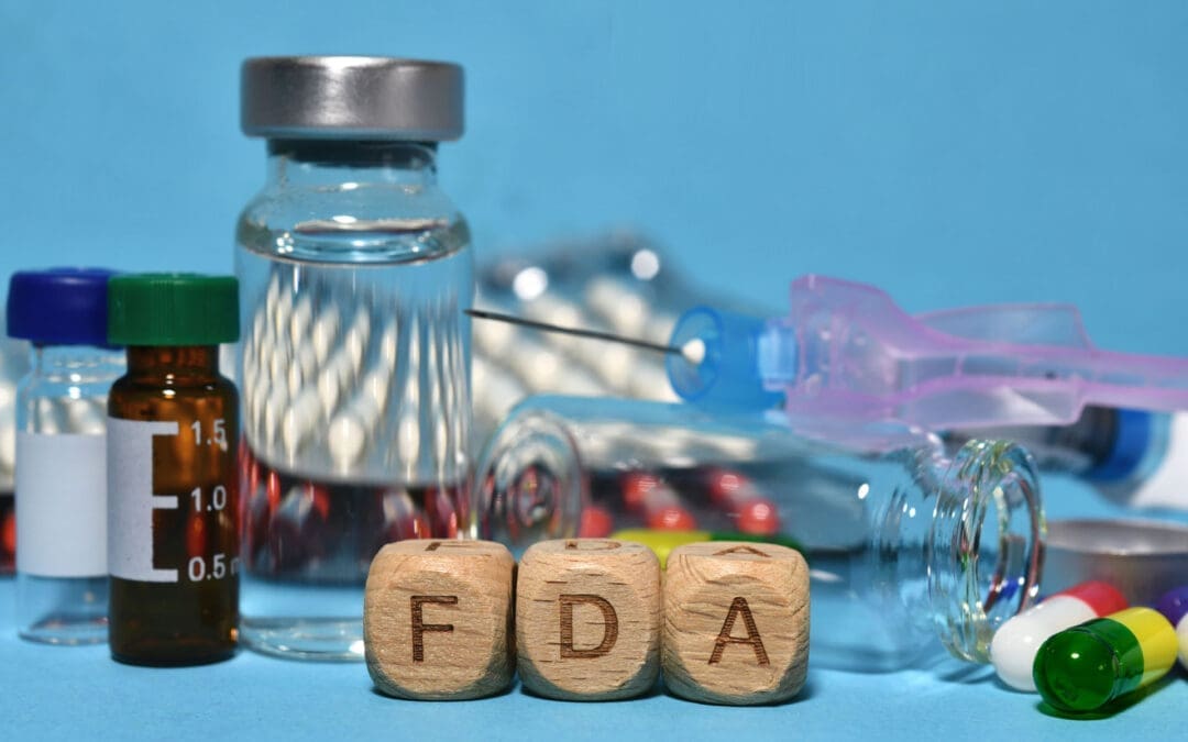
Understanding PRP and FDA Regulations: A Guide for Patients
One of the most frequent conversations I have with patients considering PRP therapy revolves around its regulatory status. Questions like, “Is it FDA-approved?” are common and completely understandable. It’s crucial for patients to feel confident and informed. Let’s break this down to provide some clarity.
The Device vs. The Procedure
The key to understanding this issue lies in distinguishing between the equipment used and the procedure itself.
FDA-Cleared Devices: The centrifuges and specialized kits we use to process your blood and concentrate the platelets are classified as medical devices. These devices undergo a regulatory process with the U.S. Food and Drug Administration (FDA) and may receive 510(k) clearance. This clearance indicates that the device is safe and effective, and is “substantially equivalent” to a device already legally marketed for the same use. So, when we perform PRP, we are using FDA-cleared technology.
PRP is a Procedure, Not a Drug: This is the most critical point. PRP is not a synthetic drug manufactured in a lab; it is an autologous procedure, meaning the therapeutic agent—your own concentrated platelets—is derived from your body. Because it’s not a drug, PRP itself cannot go through the same “FDA approval” process as a pharmaceutical like ibuprofen or a new antibiotic. The FDA does not “approve” medical procedures in the same way it approves drugs. Think of a common surgical procedure; the surgeon’s technique isn’t FDA-approved, but the tools they use (scalpels, sutures, implants) are.
Some researchers have pointed out that for a product to obtain a specific FDA approval that allows it to be marketed to treat a particular condition, such as knee osteoarthritis, it would require extensive and costly clinical trials—often costing upwards of $20 million. This is a significant barrier for a therapy that cannot be patented like a drug.
Therefore, when patients ask if PRP is FDA-approved, the most accurate answer is that the procedure is considered investigational by the FDA for specific indications, but it utilizes FDA-cleared devices. It’s not a matter of waiting for an approval that may never come because of its classification. Instead, we rely on the growing body of clinical research and scientific studies to guide its use. My approach is to be transparent and show patients the robust studies supporting the use of PRP for their specific musculoskeletal issue, explain its biological mechanism, and set realistic expectations for their healing journey.
Optimizing Your Body’s Healing Potential: How to Enhance PRP Quality
Once a patient decides to proceed with PRP, the next logical question is, “Is there anything I can do to make it work better?” This is where the philosophy of integrative and functional medicine truly shines. The quality of your PRP is a direct reflection of your health. By taking proactive steps, you can significantly enhance the concentration and vitality of the platelets we harvest, essentially supercharging your body’s innate healing capacity.
This is a core tenet at El Paso Back Clinic. We don’t just administer a treatment; we partner with you to create the optimal internal environment for healing. Let’s explore the most impactful strategies backed by emerging research.
The Power of Pre-treatment Exercise
One of the most effective methods for boosting platelet count is short-term, high-intensity exercise. Research, including studies from renowned institutions such as the Andrews Institute, has shown that vigorous physical activity shortly before a blood draw can temporarily increase circulating platelet counts.
Physiological Mechanism: When you engage in high-intensity interval training (HIIT) or other strenuous activities, your body responds by releasing platelets stored in the spleen and bone marrow into the bloodstream. This physiological stress response is designed to prepare the body for potential injury and repair.
Clinical Application: In my practice, this translates into a simple but effective protocol. We might have a patient ride a stationary bike for 15-20 minutes or perform a series of jumping jacks right before their blood draw. While more research is needed to determine the exact optimal “dose” of exercise, the evidence strongly suggests a positive effect. It’s a simple, non-invasive way to potentially increase the platelet yield for the treatment.
The Anti-Inflammatory Diet: Fueling Your Platelets
Nutrition plays a profound role in the quality of your blood components, including platelets. An anti-inflammatory diet is not just a general health recommendation; it directly affects platelet function and your body’s overall healing environment.
What is an Anti-Inflammatory Diet? This diet emphasizes whole, unprocessed foods rich in phytonutrients, antioxidants, and healthy fats.
Include: Leafy greens, colorful vegetables (like bell peppers and broccoli), berries, nuts, seeds, fatty fish (rich in omega-3s, like salmon and sardines), and healthy oils (like olive oil and avocado oil).
Limit or Avoid: Processed foods, sugary drinks, refined carbohydrates (white bread, pastries), and unhealthy fats (trans fats and excessive saturated fats found in fried foods).
Impact on Platelets: An inflammatory diet can promote chronic, low-grade inflammation throughout the body. This can make platelets “sticky” and hyperactive in a non-productive way. Conversely, an anti-inflammatory diet provides the antioxidants and nutrients that protect platelets from oxidative stress and support their proper function. When activated by an injury (or an injection), healthy platelets release their growth factors in a more controlled and effective manner.
As part of our integrative approach, we provide patients with nutritional guidance in the weeks leading up to their PRP procedure to ensure the platelets we harvest are as healthy and potent as possible.
The NSAID Controversy: To Take or Not to Take?
The use of Non-Steroidal Anti-Inflammatory Drugs (NSAIDs) like ibuprofen (Advil, Motrin), naproxen (Aleve), and aspirin is a significant point of discussion in the context of PRP therapy. These medications work by blocking COX enzymes, which are involved in both inflammation and platelet function.
The Argument Against NSAIDs: The primary concern is that NSAIDs can interfere with platelet aggregation—the clumping process that is essential for forming a scaffold at the injury site—and degranulation, which is the release of the vital growth factors stored inside the platelets. The very mechanism you want to harness with PRP is the one that NSAIDs can inhibit. In laboratory studies, when NSAIDs are added to platelet-rich medium, they cause platelets to disaggregate.
Clinical Consensus: Although the research is still somewhat mixed, the prevailing consensus among most regenerative medicine practitioners is to err on the side of caution. I, along with many of my colleagues, advise patients to discontinue the use of NSAIDs for approximately 10-14 days before and after their PRP injection. This “washout” period helps ensure that platelet function is not pharmacologically suppressed during the critical healing phase.
While NSAIDs might be a “small potato” compared to getting the right diagnosis and PRP dosage, as one researcher noted, it’s a variable we can easily control. Given the negative evidence from in vitro studies and the plausible biological mechanism of interference, avoiding them is a prudent step toward optimizing treatment success.
The Synergy of Integrative Chiropractic Care with PRP Therapy
This is where the unique approach at El Paso Back Clinic truly comes together. PRP therapy is a powerful tool, but it is not a magic bullet. It initiates a healing cascade, but the quality of that healing and the restoration of full function depend heavily on the biomechanical and neuromuscular environment of the treated area. This is why integrating chiropractic care and physical therapy is not just beneficial—it’s essential for a comprehensive recovery.
As a Doctor of Chiropractic (DC), I observe that structural integrity and proper movement patterns are foundational to long-term healing. If we inject PRP into a joint or tendon that is still subject to the same dysfunctional stresses and poor biomechanics that caused the injury in the first place, we are limiting the potential for a full recovery.
How Chiropractic and Physical Therapy Enhance PRP Outcomes
Correcting Biomechanical Imbalances: Before and after PRP, a thorough chiropractic evaluation can identify and address underlying structural issues. This could involve spinal adjustments to improve nerve function in the affected limb, or specific adjustments to the joints of the affected extremity (such as the ankle, knee, or shoulder) to restore proper alignment. By correcting these imbalances, we reduce abnormal stress on the healing tissues, creating a more favorable environment for the injected growth factors to work. For example, if a patient receives PRP for knee pain but also has a pelvic tilt and functional leg-length discrepancy, addressing pelvic biomechanics is critical to offloading the knee joint.
Improving Mobility and Tissue Health: Manual therapies, such as soft-tissue mobilization, myofascial release, and instrument-assisted techniques, are used to break down adhesions and scar tissue within the muscles and fascia surrounding the injured area. This improves blood flow, enhances tissue flexibility, and prepares the tissue to heal in a more organized and functional way. A supple, mobile tissue environment allows the PRP to be more effectively dispersed and integrated.
Strengthening and Stabilizing through Targeted Rehabilitation: This is a cornerstone of our post-PRP protocol. Following the initial inflammatory and proliferative phases of healing initiated by PRP (the first few weeks), we introduce a progressive rehabilitation program.
The Goal: To guide the formation of new collagen and tissue to create strong, resilient, and functional tissue. Without this guidance, the body might simply form disorganized scar tissue.
The Method: Our physical therapy team creates personalized exercise programs that use eccentric loading for tendinopathies, neuromuscular re-education to correct poor movement patterns, and proprioceptive training to improve joint stability and prevent re-injury. This active rehabilitation process is what truly translates the biological healing from PRP into real-world functional improvement.
Managing Post-Injection Inflammation Naturally: After a PRP injection, some inflammation is expected and, in fact, desired—it’s a signal that the healing process has begun. Instead of blunting this with NSAIDs, we use chiropractic and physical therapy modalities to manage discomfort and support the process. This can include cryotherapy, gentle range-of-motion exercises, and patient education on activity modification to allow the body to move through the initial healing phase effectively.
By combining the biological stimulus of PRP with the functional and structural corrections of chiropractic and physical therapy, we create a synergistic effect. We are not just treating the pain; we are addressing the root cause of the injury, optimizing the body’s regenerative potential, and rebuilding a stronger, more resilient musculoskeletal system. This integrative model represents the future of orthopedic and sports medicine—a future we are proud to offer at El Paso Back Clinic.
References
Andrews, J. R., et al. (Year).Title of Study on Blood Flow Restriction and PRP. Journal Name, Volume(Issue), pages. [Link to Article]
Andrews, J. R., et al. (Year).Title of Study on Exercise and Platelet Counts. Journal Name, Volume(Issue), pages. [Link to Article]
Researcher, A. A. (Year).Title of Study on NSAID Effect on Platelet Aggregation. Journal Name, Volume(Issue), pages. [Link to Article]
IFM's Find A Practitioner tool is the largest referral network in Functional Medicine, created to help patients locate Functional Medicine practitioners anywhere in the world. IFM Certified Practitioners are listed first in the search results, given their extensive education in Functional Medicine