Chiropractic Benefits: If you have ever had a migraine before then you know that it is much more than a simple headache. The symptoms of a migraine can be debilitating, lasting hours and even days. According to the Migraine Research Foundation, it is the eighth most disabling disease in the world. It is estimated that 38 million people in the United States alone suffer from migraine headaches. That�s around one in every ten people.
According to the Migraine Research Foundation, migraine headaches are extremely difficult to treat and even more difficult to control. This is mainly due to the fact that doctors still don�t know exactly what causes it. This leaves it undiagnosed in many patients and often terribly under treated in those with a diagnosis.
The best many doctors seem to be able to do is prescribe pain medication that has undesirable side effects in an effort to manage the symptoms. However, chiropractic has been shown in several studies to not only effectively manage the pain of migraines, it also helps stop and prevent them.
Anatomy Of A Migraine Headache
There are two types of migraines, those with an aura and those without an aura. An aura can appear up to an hour before the onset of a migraine. It is a warning sign that usually presents as a disturbance that is either visual or olfactory. The person may see flashes of light or smell particular odors before the headache begins. About one in six migraines are preceded by an aura.
Once the migraine itself begins, the pain is typically on one side of the head, although this is not always the case. Other symptoms may include nausea, vomiting, sensitivity to noise, sensitivity to light, and sensitivity to smell. Some patients experience an inability to concentrate, hot or cold flashes, stiffness in neck or shoulders, slurred speech, loss of coordination, and in rare cases, loss of consciousness.
The migraine can last several minutes, hours, or even days. Afterwards the patient may feel fatigued or washed out. They may be unable to concentrate and either lethargic or extremely energetic.
Studies Show: Chiropractic As A Migraine Treatment
There have been several clinical studies on chiropractic as a treatment for migraine headaches. The results of one study reported that 22 percent of patients who received chiropractic treatment for their migraines reported that their attacks were reduced by more than 90 percent. Additionally, 49 percent reported that the intensity of their migraines was significantly reduced.
Another study randomly assigned people with migraine headaches several different treatments. One group was given Elavil, a daily medication, another group was given chiropractic treatment and a third group received a combination of the two treatments. The results showed that chiropractic was as effective in reducing migraines as the medication and it had fewer side effects. Other studies have also found that chiropractic is as effective as medication for the treatment and prevention of migraine or tension headaches.
Chiropractic Benefits For Migraines Headaches
Spinal adjustments are very effective as a treatment for migraines. The whole body approach of chiropractic also utilizes dietary recommendations, including foods to avoid, as well as lifestyle changes.
The patient may be counseled on managing stress, advised to engage in exercise, and given supplements. The treatments may be used to reduce the pain and severity of a migraine once it begins or it can be used to prevent migraines and reduce their frequency.
Chiropractic benefits everyone and is a safer treatment with fewer side effects than�prescription medications. Chiropractic is quickly becoming the treatment of choice for many migraine sufferers. As the studies show, it works! So if you or a loved one suffer from migraines, give us a call. Our Doctor of Chiropractic is here to help!
Feet are important. When you consider what your feet go through, taking 8,000 steps over the course of a day, according to the Illinois Podiatric Medical Association (IPMA), it�s easy to see how 75 percent of all Americans will have some type of foot pain at some point in their lives. Plantar fasciitis is a common and very painful foot condition that can become chronic if not treated. It is also a condition that responds very well to chiropractic care.
Plantar Fasciitis Explained
Plantar fasciitis is caused when the ligament that connects your toes to your heel (the plantar fascia) becomes inflamed, swollen, and weak. This causes the bottom of your foot or heel to hurt when you walk or stand, especially when you first wake or after sitting for a long time.
The pain tends to be sharp and stabbing. It is the most common cause of heel pain and is more prevalent among middle aged people. However, anyone can get it at any age, especially people who spend a lot of time on their feet.
How Chiropractic Helps Plantar Fasciitis
Chiropractic care is a very effective treatment for plantar fasciitis as well as the pain that is caused by the condition. Chiropractic for plantar fasciitis involves a very precise technique that involves adjustments to the feet and ankles as well as spinal alignment. This provides several benefits.
Reduces Stress in the Plantar Fascia � When a ligament is inflamed or stressed the tissue can develop very small tears that cause the pain of plantar fasciitis. Chiropractic adjustments made to the heel and foot take the pressure off of the plantar fascia, allowing it to relax.
Promotes Healing � When the stress on the plantar fascia is reduced through these chiropractic adjustments, the foot can begin to heal. The chiropractor may also recommend specific exercises that stretch the ligament and help it heal. They may also advise the patient of lifestyle changes as well as nutritional adjustments that can help with the pain and condition.
Provides Effective Pain Management � Chiropractic is a very effective way to manage pain throughout the body. Spinal adjustments allow better communication between the brain and nerves, allowing the central nervous system to function more effectively. Condition specific adjustments speak to the root of the problem, not just the symptoms. This means a more effective form of pain management that is longer lasting.
Reduces the Risk of Further Injury � When a person has a condition like plantar fasciitis, they will often adjust their gait in an effort to avoid the pain. This puts stress on other parts of the body and can lead to back pain, sore joints, strained muscles, and other problems. Chiropractic�s whole body approach helps the person realign their body properly so that they stand and walk properly. This helps them avoid further injury and discomfort.
Chiropractic Complements Other Treatments
While chiropractic care can be an effective treatment for plantar fasciitis on its own, it is also a very good complement to other treatments for the condition. Patients may use chiropractic in conjunction with physical therapy, massage, and even injections to manage the pain and treat the condition. It can also help with speeding healing and helping to provide better mobility.
Plantar fasciitis can take several months to heal, but by adding chiropractic treatments to your recovery plan, you can feel better faster while more effectively managing your pain. Regular chiropractic treatments can also keep the condition from becoming chronic. By working with your chiropractor and following their recommendations you can reduce your pain and shorten your healing time.
Injury Medical Chiropractic Clinic & Chronic Pain & Treatments
Sandra Rubio talks about personal injury rehabilitation, including stretches and exercises, which are provided by Dr. Alex Jimenez and his staff, aside from chiropractic care. Dr. Jimenez offers the services of personal trainers and physical therapists to help improve the patient’s strength, mobility and flexibility to prevent further injury as well as to help speed up the patient’s recovery process.
Personal Injury Rehabilitation
Physical therapy (PT), also known as physiotherapy, is one of the allied health professions which, by utilizing mechanical force and motions (bio-mechanics or kinesiology), manual therapy, exercise therapy, and electrotherapy, helps treat injuries and conditions by promoting proper function. Physical therapy is used to improve a patient’s quality of life through examination, diagnosis, prediction and physical intervention. It’s generally performed by physical therapists (called physiotherapists in several nations).
A personal injury doctor or chiropractor is an effective, alternative treatment option for a variety of injuries surrounding the spine. When an individual has suffered an injury as a result of an auto accident, work accident or home accident a personal injury doctor can positively influence the proper progress of their rehabilitation, helping the individual obtain the fair compensation they need and deserve for their injuries. An experienced and well-trained personal injury doctor can provide quality treatment as well as support patients throughout their injury claim procedures.
We want you to live a life that is fulfilled with more energy, positive attitude, better sleep, less pain, proper body weight and educated on how to maintain this way of life. I have made a life of taking care of each and every one of my patients.
I assure you, I will only accept the best for you�
God Bless You & Your Health�?
If you have enjoyed this video and/or we have helped you in any way please feel free to subscribe and share us.
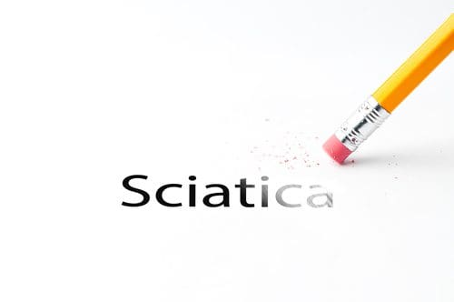
Chiropractic Therapy: Sandra Rubio discusses how Dr. Alex Jimenez and his staff can help relieve your sciatica symptoms. Chiropractic care can improve pain and discomfort as well as reduce irritation and inflammation caused by sciatica. In addition, a chiropractor like Dr. Jimenez can also provide nutritional and fitness advice for sciatic nerve pain. Other treatment methods, like deep-tissue massage, can help relieve sciatica symptoms. Dr. Alex Jimenez is the homeopathic, non surgical choice for sciatic nerve pain and its associated symptoms.
Chiropractic Therapy For Sciatica Pain
Sciatica is generally caused by the compression of lumbar or sacral nerves or by compression of the sciatic nerve. When sciatica is caused by compression of a dorsal nerve root, it’s known as lumbar radiculopathy. This can occur because of a spinal disk bulge or spinal disk herniation (a herniated intervertebral disc), or by roughening, enlarging, or misalignment (spondylolisthesis) of the fascia, or as a consequence of degenerated discs which can reduce the diameter of the lateral foramen by which nerve roots exit the spine.
Our objective is to alleviate your pain and restore freedom, in�treating��our�sciatica�sufferers. Employing state of the art x-ray/fluoroscopic and ultrasound helps our experts pinpoint what�s going on. For treating sciatica we believe in the ability of medicine. By stimulating the body�s natural healing response, these procedures let us naturally provide long-term and deep pain relief.
We want you to live a life that is fulfilled with more energy, positive attitude, better sleep, less pain, proper body weight and educated on how to maintain this way of life. I have made a life of taking care of each and every one of my patients.
I assure you, I will only accept the best for you�
God Bless You & Your Health�?
If you have enjoyed this video and/or we have helped you in any way please feel free to subscribe and share us.
Sciatica Sufferers:Sciatica is a common back ailment that affects approximately 1 in 10 adults in the United States. It is most prevalent in people between the ages of 25 and 45. Sciatica is characterized by a shooting pain that originates in the lower back and travels down through the hip, buttock and back of leg. The pain can be so severe that it inhibits mobility and can prevent people from working, taking care of their home, or just enjoying their life. Traditionally, doctors have treated the condition with medications and some invasive therapies, but chiropractic treatments have been found to be extremely effective in alleviating the pain and curing the condition.
What Is Sciatica?
Sciatica is a condition that affects the lower back, specifically pain that travels along the sciatic nerve path. This path originates in the lower back and extends down each side of the hip, buttocks, and down the leg to the feet. Usually, only one side is affected during a case of sciatica.
Sciatica commonly occurs when there is a spinal condition, such as a bone spur on the spine, a herniated disk, or when a condition like spinal stenosis (narrowing of the spine) compresses the nerve. The result is inflammation, numbness, pain, and stiffness in the leg that is affected.
While sciatica pain can be severe, the majority of the time the condition is resolved in a matter of weeks without surgical intervention. Some doctors may suggest surgery if the patient experiences extreme weakness in the leg or has problems with their bowel or bladder. Most of the time doctors will prescribe medication to treat the pain and relax the muscles that surround the sciatic path in an effort to provide the patient with some relief.
How Chiropractic Treatments Help Sciatica Sufferers
Chiropractic has been shown to be very effective in treating sciatica by helping the body heal itself. It is non-invasive and does not use medications, making it an optimal choice for many patients. There are various treatments that a chiropractor may use or recommend. They may be used alone or in conjunction with other therapies.
Adjustments. Spinal manipulation, or adjustments, is the core of chiropractic treatment. It helps to realign the spine, freeing restricted movement and helps bring the body back into its proper alignment so that it functions better and more effectively. It also helps to reduce the pain that is associated with nerve inflammation, particularly that associated with sciatica.
Ultrasound. A very mild heat that is created by sound waves at a frequency that is beyond human hearing providing deep tissue penetration. This therapy stimulates circulation and aids in reducing muscle spasms, stiffness, swelling, pain, and cramping.
Ice or Cold Therapy. This therapy helps to reduce and control the pain of sciatica, as well as reduce inflammation.
TENS. Transcutaneous electrical nerve stimulation is a therapy that employs a TENS unit, a small battery powered, portable box that stimulates the muscles. It uses electrical currents at variable intensities to help control pain and reduce the occurrence of muscle spasms. Some chiropractors and physical therapists use larger versions of this device in their offices, but many use the portable units because they are more convenient.
A chiropractor may incorporate exercises and nutritional recommendations into their treatment for sciatica sufferers. This often depends on the patient�s individual needs, the severity of the condition, and their lifestyle and habits. A variety of chiropractic techniques may also be used to treat sciatica. Since each patient is different, the chiropractor will talk with the patient to better understand what they do on a day-to-day basis and what may be causing the sciatic pain to occur. From there they will treat the problem from the patient�s perspective, seeking the best, most effective approach.
If you or a loved one is suffering from this condition, please give us a call. Our Doctor of Chiropractic is here to help!
Chiropractic treatment is a nonsurgical option that can help reduce neck pain and related symptoms. Below are some of the different types of neck (cervical) conditions that Doctors of Chiropractic (DC’s) treat:
Chiropractors also use manual therapies to treat neck pain:
Cervical intervertebral disc injuries that don�t require surgery
Cervical sprain injuries
Degenerative joint syndrome of the neck (eg, facet joints)
A chiropractor evaluates the spine as a whole because other regions of the neck (cervical), mid back (thoracic) and low back (lumbar) can be affected as well. Along with treating the spine as a whole, chiropractic medicine treats the entire person and not just a specific symptom/s. Chiropractors may educate on nutrition, stress management, and lifestyle goals in addition to treating neck pain.
A chiropractor will do a thorough examination to diagnose the specific cause of the neck pain before deciding on which approach/technique to use.
They will determine any areas of restricted movement and will look at a walking cycle along with posture and spinal alignment. Doing these things can help the chiropractor understand the body’s mechanics.
In addition to the physical exam, a chiropractor will want to go over past medical history, and they may order imaging tests (eg, an x-ray or MRI) to help them diagnose the exact cause of the neck pain.
All these steps in the diagnostic process will give a chiropractor more information about the neck pain, which will help the� chiropractor create a customized treatment plan for the individual patient.
A chiropractor will rule out neck pain conditions that require surgery. If they believe surgery is the best treatment for the neck pain, then the patient will be referred to a spine surgeon.
Chiropractic Treatment: Neck Pain
A chiropractor may use a combination of spinal manipulation, manual therapy, and other techniques as part of the treatment plan.
Spinal Manipulation Techniques Used:
Flexion-Distraction Technique:�Gentle hands-on spinal manipulation that involves a pumping action on the intervertebral disc rather than direct force.
Instrument Assisted Manipulation:�Uses hand-held instruments, which allow the chiropractor to apply force without thrusting into the spine.
Specific Spinal Manipulation:�Restores joint movement with a gentle thrusting technique.
Chiropractors also use manual therapies to treat neck pain.
Instrument Assisted Soft Tissue Therapy: uses special instruments to diagnose and treat muscle tension.
Trigger Point Therapy is used to relieve tight, painful points on a muscle.
Other therapies used to ease neck pain symptoms.
Inferential Electrical Stimulation:�Is a low frequency electrical current used to stimulate neck muscles.
Ultrasound:�Sound waves travel into the muscle tissues to help stiffness and pain in the neck.
Therapeutic Exercises:�Helps improve overall range of motion in the neck and prevent neck pain from progressing.
The treatments listed are examples of possible chiropractic treatment for neck pain; The actual treatment plan will depend on the diagnosis. A chiropractor will thoroughly explain the treatment options available along with the actual customized treatment for the individual patient.
Chiropractic Clinic Extra: Neck Pain Care & Treatments
If you are pregnant and have back pain, you are not alone. An estimated 50 to 70 percent of women who are pregnant experience back pain, according to the American Pregnancy Association. While pregnancy and childbirth is one of the most incredible experiences a woman can have, it is also very hard on her body. There are many dramatic changes that take place during that 9 to 10 month of gestation so it is understandable that she is going to feel some aches and pains along the way.
There are a number of reasons why a pregnant woman may experience back pain including:
Natural changes to her body such as softening of ligaments and loosening of joints as her body prepares to give birth
A shift in her center of gravity as her girth increases.
Weight gain.
Position of the baby.
Her posture.
Stress, exhaustion, and worry.
Is Chiropractic Care Safe During Pregnancy?
Chiropractic care has long been held as a viable method for relieving back pain in pregnant women. Historically, midwives and other natural or alternative practitioners were the ones advocating its many benefits. This resulted in minimal data from clinical studies existing on the topic.
However, in the last decade or so, researchers have been looking closer at chiropractic and its many benefits. In one study of pregnant women and chiropractic, 94 percent of the participants experienced dramatic improvement in their pain in just 5 days.
Today many doctors and obstetricians are sending their pregnant patients to chiropractors to help them manage their back and joint pain. It is perfectly safe for both mother and baby � and both can benefit from it.
Benefits Of Chiropractic Care During Pregnancy
While chiropractic care during pregnancy can be used as a safe, non-invasive, and drug free method of pain relief, women may also enjoy other benefits which include:
A healthier, happier pregnancy.
Improved mood and less anxiety.
More mobility.
Decreased morning sickness and nausea.
Easier, faster labor and delivery.
Better flexibility.
In some cases, prevent cesarean delivery.
Improved sleep.
Faster recovery time.
Relief of pain in the back, joints, and neck.
By keeping the body in proper alignment, chiropractic care can help a woman have a healthier, happier pregnancy. She can enjoy the many benefits and experience less pain so that she can better focus on the joy of pregnancy and the wonder of bringing a new life into the world.
Why You Should Have Chiropractic Care During Pregnancy
Pregnancy brings about many changes in a woman�s body. Hormonal changes as well as physiological ones occur at rapid speeds as her body creates and maintains a perfect environment where her baby will develop and grow. These changes can cause the spine or joints to become misaligned. When this occurs, painful conditions can be created, including:
Increased curvature of the back.
Pelvic changes.
Protruding abdomen that puts pressure on the back.
Changes in posture.
Keeping the pelvis and lower back well balanced and aligned is integral to preventing lower back pain during pregnancy. What�s more, when the pelvis and spine are not in alignment, it can limit the amount of room the baby has in the womb. This condition is called intrauterine constraint. This can also inhibit the baby�s ability to get in an optimal position for delivery.
Keeping the body, including the spine, in proper alignment is vital to mobility, flexibility, and overall wellness of the body even when it is not pregnant. However, pregnancy puts specific stress on the body, creating certain needs that chiropractic care can meet. It is safe, it is effective, it is fast, and it works.
Chiropractic Clinic Extra: Stress Management Care & Treatments
IFM's Find A Practitioner tool is the largest referral network in Functional Medicine, created to help patients locate Functional Medicine practitioners anywhere in the world. IFM Certified Practitioners are listed first in the search results, given their extensive education in Functional Medicine


 While
While 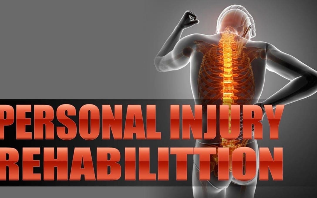
 A personal injury doctor or chiropractor is an effective, alternative treatment option for a variety of injuries surrounding the spine. When an individual has suffered an injury as a result of an auto accident, work accident or home accident a personal injury doctor can positively influence the proper progress of their rehabilitation, helping the individual obtain the fair compensation they need and deserve for their injuries. An experienced and well-trained personal injury doctor can provide quality treatment as well as support patients throughout their injury claim procedures.
 Our objective is to alleviate your pain and restore freedom, in�treating��our�sciatica�sufferers. Employing state of the art x-ray/fluoroscopic and ultrasound helps our experts pinpoint what�s going on. For treating sciatica we believe in the ability of medicine. By stimulating the body�s natural healing response, these procedures let us naturally provide long-term and deep pain relief.

 A chiropractor may use a combination of spinal manipulation, manual therapy, and other techniques as part of the treatment plan.