PRP Therapy in El Paso for Back Pain Relief and Joint Healing
Abstract
As a clinician dedicated to integrative and evidence-based care, I am constantly exploring the latest advancements that can help my patients heal more effectively. This post explores the science behind Platelet-Rich Plasma (PRP), a powerful regenerative therapy. We will journey into the microscopic world of platelets, exploring their crucial role in orchestrating the body’s natural healing processes. You will learn about the specific growth factors and signaling molecules released by platelets, how they reduce inflammation, and how we can concentrate this healing potential to treat various musculoskeletal conditions. We will also discuss how PRP, as a cornerstone of orthobiologic therapy, integrates seamlessly with chiropractic care and physical rehabilitation to create a comprehensive, synergistic treatment plan that accelerates your return to a pain-free, active life.
Hello, I’m Dr. Alexander Jimenez. With my extensive background in both chiropractic and advanced practice nursing, coupled with certifications in functional and integrative medicine, my primary mission has always been to offer my patients the most effective, evidence-based pathways to wellness. At our El Paso clinic, we are passionate about harnessing the body’s innate ability to heal itself. One of the most exciting fields that allows us to do this is orthobiologics, and a cornerstone of this approach is Platelet-Rich Plasma, or PRP.
Today, I want to take you on a journey—not into a complex scientific lecture, but into an easy-to-understand exploration of your body’s remarkable healing capabilities. We’re going to look at the latest findings from leading researchers and see how this science translates into real-world results for conditions such as chronic back pain, joint injuries, and soft-tissue damage.
The Orchestra Within: Understanding the Power of Platelets
When you think of platelets, you probably think of blood clotting. If you get a cut, platelets rush to the scene to form a plug and stop the bleeding. While this is a critical function, it’s only the beginning of their story. Platelets are not just simple plugs; they are sophisticated, mobile storage units packed with powerful biological instructions.
Think of your platelets as the first-response commanders at an injury site. Once they arrive, they don’t just patch the hole; they release a cascade of potent signaling molecules—growth factors, cytokines, and chemokines—that direct a complex healing orchestra. It’s this biological symphony that truly drives tissue repair and regeneration.
PRP therapy is based on a simple yet profound concept: what if we could concentrate these healing commanders and deliver them directly to an area of chronic injury or degeneration? By doing so, we can amplify the body’s natural healing signals, telling it to repair tissue that it may have otherwise “given up” on.
Inside the Platelet: The Granules That Drive Healing
To truly appreciate PRP, we need to look inside the platelet itself. A single platelet contains several types of tiny packets, or granules, each with a specific job.
Alpha Granules: These are the most important for regenerative medicine. Each platelet contains about 50 to 80 alpha granules, which house hundreds of different proteins, including the essential growth factors that orchestrate tissue repair. When platelets are activated at an injury site, they undergo a process called degranulation, releasing the contents of these alpha granules into the surrounding environment. This is the moment the healing cascade truly begins.
Dense Granules: These granules release smaller molecules that are crucial for amplifying the initial response. They help recruit more platelets (platelet aggregation), signal blood vessels to constrict to limit bleeding, and modulate the initial immune response.
Lysosomes: These act as the cleanup crew. They release enzymes that help break down damaged tissue, clear cellular debris, and exert antimicrobial effects, essentially preparing the site for new, healthy tissue to form.
In our clinical practice, we’ve observed that the effectiveness of PRP is directly tied to the concentration and quality of these platelets. Newer research highlights the importance of reticulated platelets—younger, denser platelets recently released from the bone marrow. These platelets are richer in alpha granules and, therefore, contain a higher payload of growth factors. Our advanced processing techniques are designed to capture these highly potent platelets, ensuring that the PRP we administer has the maximum regenerative potential. This concentration is key; by increasing platelet count, we dramatically increase the number of biological signals delivered to the injured area.
The Key Players: Growth Factors and Their Roles
When the alpha granules release their contents, a variety of growth factors become active. While it’s a complex interaction among hundreds of proteins, let’s focus on a few of the star players and their specific roles in healing.
Platelet-Derived Growth Factor (PDGF)
As its name suggests, PDGF was one of the first growth factors discovered in platelets. Think of PDGF as the “beacon.” Its primary role is to attract other healing cells to the injury site. It sends out a powerful chemical signal that recruits mesenchymal stem cells (MSCs)—the body’s master repair cells—as well as other cells necessary for tissue repair.
A Crucial Note on Stem Cells: PRP itself does not contain stem cells. However, it is a powerful signaling therapy. PDGF effectively awakens and recruits the local stem cells that are already present but dormant in your tissues, directing them to the site of injury, where they can begin their work of repair and regeneration.
The Power of PDGF-BB: Researchers have identified PDGF-BB as the most biologically active and important isoform. It is a potent stimulator of cell replication and is vital for initiating the entire repair process.
Transforming Growth Factor-Beta (TGF-β)
TGF-β is the master architect of tissue reconstruction. Once cells have been recruited to the area, TGF-β provides them with their building instructions.
Collagen Synthesis: It strongly promotes the synthesis of type I collagen, which is the primary structural protein in tendons, ligaments, and cartilage. This is crucial for restoring the strength and integrity of injured tissues.
Angiogenesis: In coordination with other growth factors, TGF-β stimulates angiogenesis, the formation of new blood vessels. This is a critical step because new blood vessels bring a fresh supply of oxygen and nutrients to the healing area, fueling the repair process and removing waste products.
Vascular Endothelial Growth Factor (VEGF)
VEGF works hand in hand with TGF-β to build this new blood supply. It specifically enhances endothelial cell proliferation (the cells that line blood vessels), promotes the sprouting of new capillaries, and is essential for neovascularization. Research has shown that platelet concentration is a significant factor in this effect. Studies suggest that a PRP concentration of approximately 1.5 billion platelets per milliliter is optimal for robust angiogenesis, a key target in our preparation protocols.
Fibroblast Growth Factor (FGF)
FGF is a powerful “mitogen,” meaning it stimulates cell division and proliferation. It acts on a wide variety of cells, including MSCs recruited by PDGF, as well as fibroblasts (which produce collagen) and osteoblasts (which build bone). FGF helps to ensure that a sufficient number of builder cells are available to carry out the repairs directed by the other growth factors.
Beyond Building: The Anti-Inflammatory Power of PRP
Chronic pain is often driven by chronic inflammation. An injury that never fully heals can get stuck in a persistent inflammatory state, causing ongoing pain and tissue degradation. One of the most profound benefits of PRP therapy is its ability to break this cycle.
While the initial response to an injury involves inflammation (a necessary step to clear damage), PRP helps guide the process toward resolution and healing. It does this in several ways:
Modulating Macrophages: PRP influences the behavior of immune cells called macrophages. These cells can exist in an inflammatory state (M1) or an anti-inflammatory, pro-healing state (M2). PRP promotes a shift from the M1 to the M2 phenotype, effectively flipping the switch from “inflammation” to “repair.”
Leukocyte Interaction: Platelets in PRP can interact with white blood cells (leukocytes) at the injury site, prompting them to release anti-inflammatory cytokines. This helps to quiet the inflammatory storm.
Preventing Cell Death: The chemokines released by platelets also act as survival factors for monocytes (which become macrophages), preventing their premature death and allowing them to complete their transition to the healing M2 state.
From my clinical observations, this powerful anti-inflammatory effect is often the first thing patients notice. Many report a significant reduction in pain and swelling within weeks of treatment as the chronic inflammatory environment begins to normalize, paving the way for long-term tissue repair.
The Synergy of Integrative Care: PRP, Chiropractic, and Physical Therapy
At the El Paso Back Clinic, we firmly believe that no single therapy is a magic bullet. True healing comes from a comprehensive, integrative approach. This is where PRP, chiropractic care, and physical therapy come together to create a powerful synergy.
Imagine a patient with chronic low back pain due to a degenerated disc and facet joint arthritis. The underlying problem is both biochemical (inflammation, tissue decay) and biomechanical (spinal misalignment, muscle imbalance, faulty movement patterns).
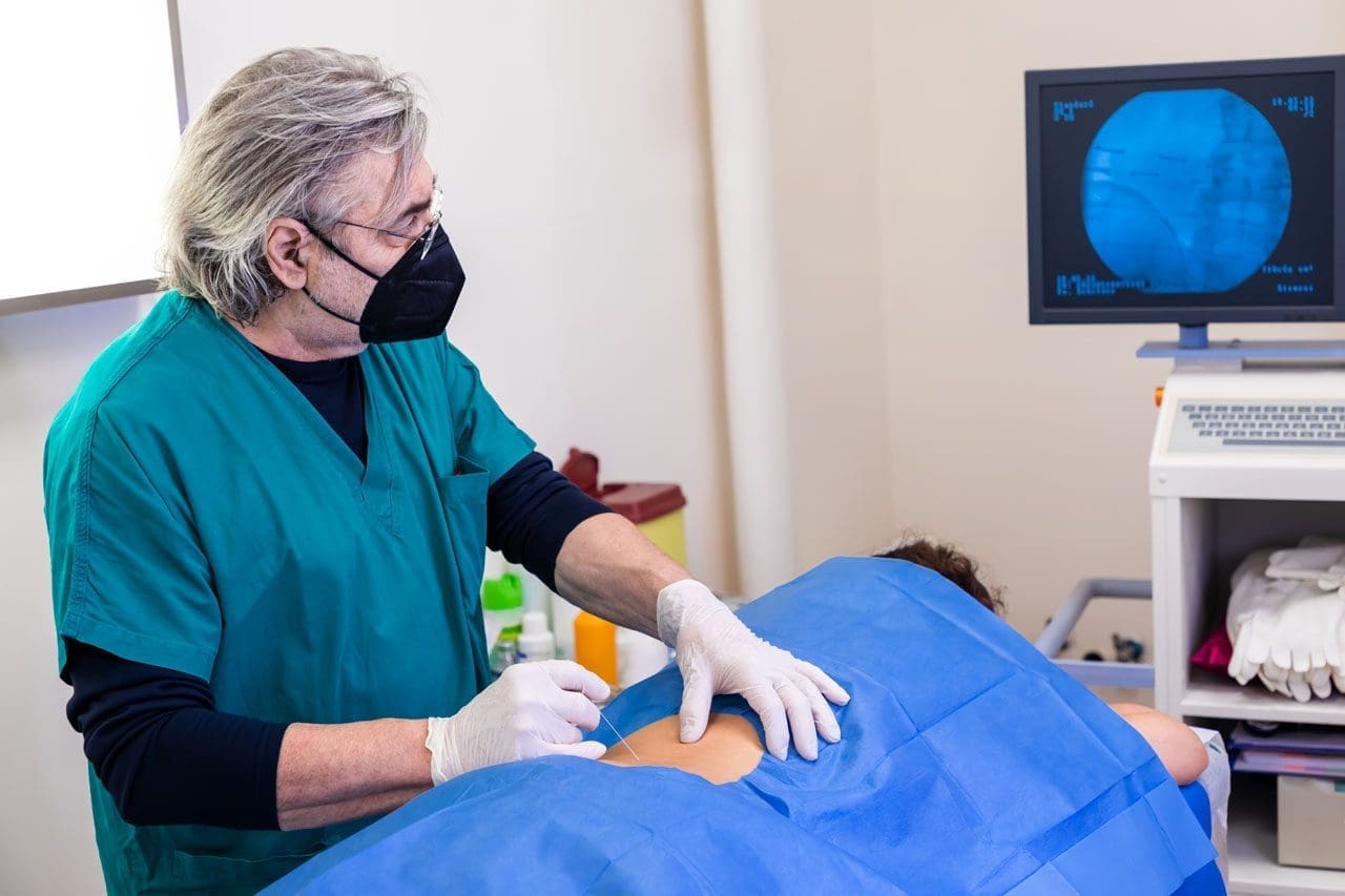
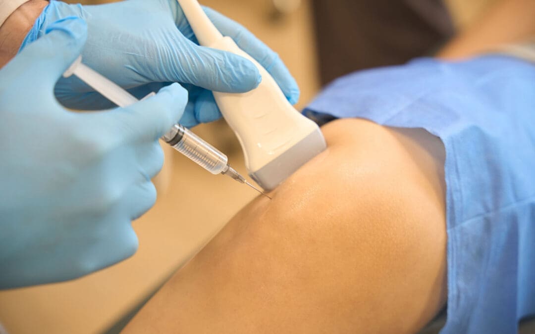
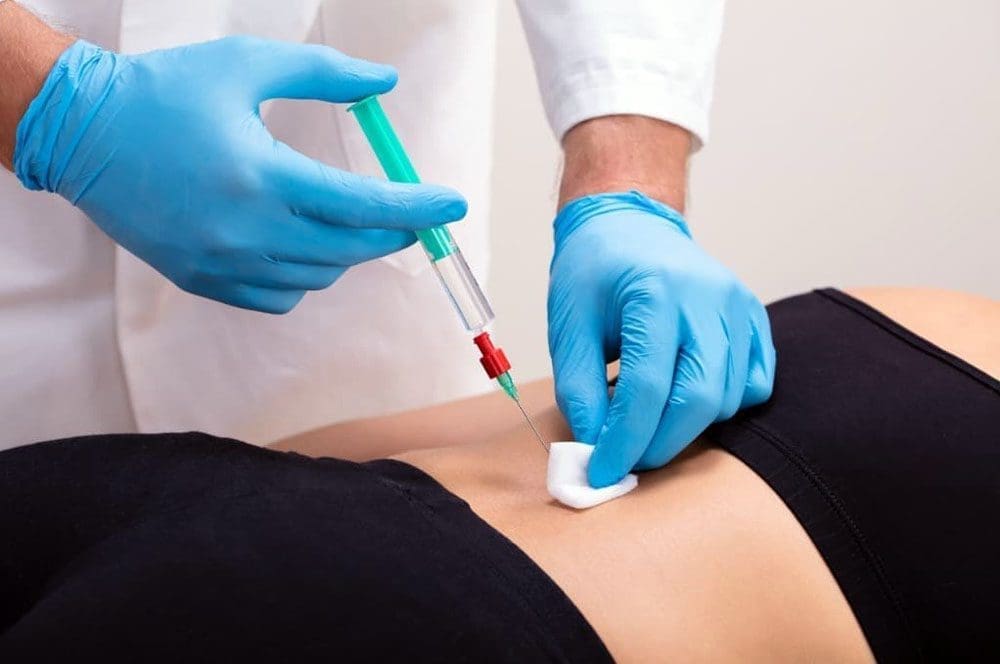
PRP Injections to Reboot Healing: We first use ultrasound guidance to precisely inject PRP into the degenerated disc space and the arthritic facet joints. This delivers a high concentration of growth factors directly to the source of pain, reducing inflammation and initiating biological repair of damaged cartilage and connective tissue. The PRP effectively “reboots” the local healing environment.
Chiropractic Care to Restore Function: While PRP works at the cellular level, a dysfunctional joint will remain dysfunctional unless its mechanics are addressed. This is the crucial role of chiropractic adjustments. Through specific, gentle manipulations, we restore proper motion to the spinal segments. This not only alleviates pain by decompressing nerves but also improves the flow of nutrients to healing tissues and ensures that the new collagen formed by PRP is laid down in an organized, functional way. Correcting the biomechanics prevents the joint from being repeatedly re-injured, allowing the PRP-stimulated healing to take hold.
Physical Therapy to Rebuild and Stabilize: Once the pain is reduced and joint mechanics are improved, physical therapy and rehabilitation become essential. Our customized exercise programs focus on strengthening the deep core and spinal stabilizing muscles. This creates a “muscular corset” that supports the spine, offloads the healing joints, and corrects the poor movement patterns that contributed to the injury in the first place. This phase ensures that PRP and chiropractic care achieve results that are not just temporary but are sustained for the long term.
This three-pronged approach addresses the injury from every angle: PRP promotes biochemical repair, chiropractic care corrects structural and biomechanical dysfunction, and physical therapy provides functional stabilization for lasting recovery. Each therapy enhances the effects of the others, leading to faster, more complete, and more durable healing than any single approach could achieve on its own.
Summary: A New Era in Healing
PRP therapy represents a paradigm shift in how we treat musculoskeletal injuries. Instead of just masking symptoms with medications or resorting to invasive surgery, we can now harness the body’s sophisticated biological toolkit to promote true healing and regeneration.
The main takeaway is that PRP provides a powerful, short-term biological “dose” of instructions. It doesn’t do all the work itself; rather, it acts as the director of the orchestra, calling in the body’s own repair cells and guiding them to reduce inflammation, rebuild damaged tissue, and restore function. When combined with an integrative framework of expert chiropractic care and targeted physical therapy, PRP becomes a transformative tool that can help our patients break free from chronic pain and get back to living their lives to the fullest.
References
The following resources provide a deeper look into the science of platelet-rich plasma and its applications.
PRP Therapy for Sports Injuries: How It May Speed Healing Without Surgery
Sports injuries can slow life down fast. A sore tendon, a strained ligament, or a muscle tear can make it difficult to train, work, sleep, or even walk comfortably. That is one reason Platelet-Rich Plasma, or PRP, has gained attention in sports medicine. PRP is made from a patient’s own blood and then injected into an injured area to support healing. Medical centers such as Yale Medicine, Penn Medicine, Johns Hopkins Medicine, and Temple Health describe PRP as a biologic or regenerative treatment that may help repair tissue, lower pain, and improve function in certain musculoskeletal injuries. It is often used for tendon, ligament, muscle, cartilage, and joint problems, including some cases of osteoarthritis. (Johns Hopkins Medicine, n.d.; Penn Medicine, 2025; Yale Medicine, n.d.).
PRP is appealing because it is non-surgical and uses the body’s own healing tools. Still, it is not a miracle fix for every athlete or every injury. Research shows promising results in many cases, but outcomes can vary depending on the tissue involved, how long the injury has been present, how the PRP is prepared, and whether the person also follows a successful rehab plan. In other words, PRP works best as part of a comprehensive care strategy rather than a stand-alone shot. (Saini et al., 2021; Jimenez, n.d.).
What PRP Therapy Is
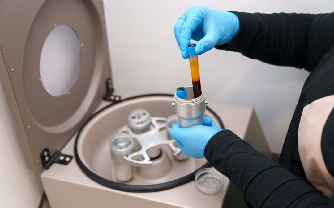
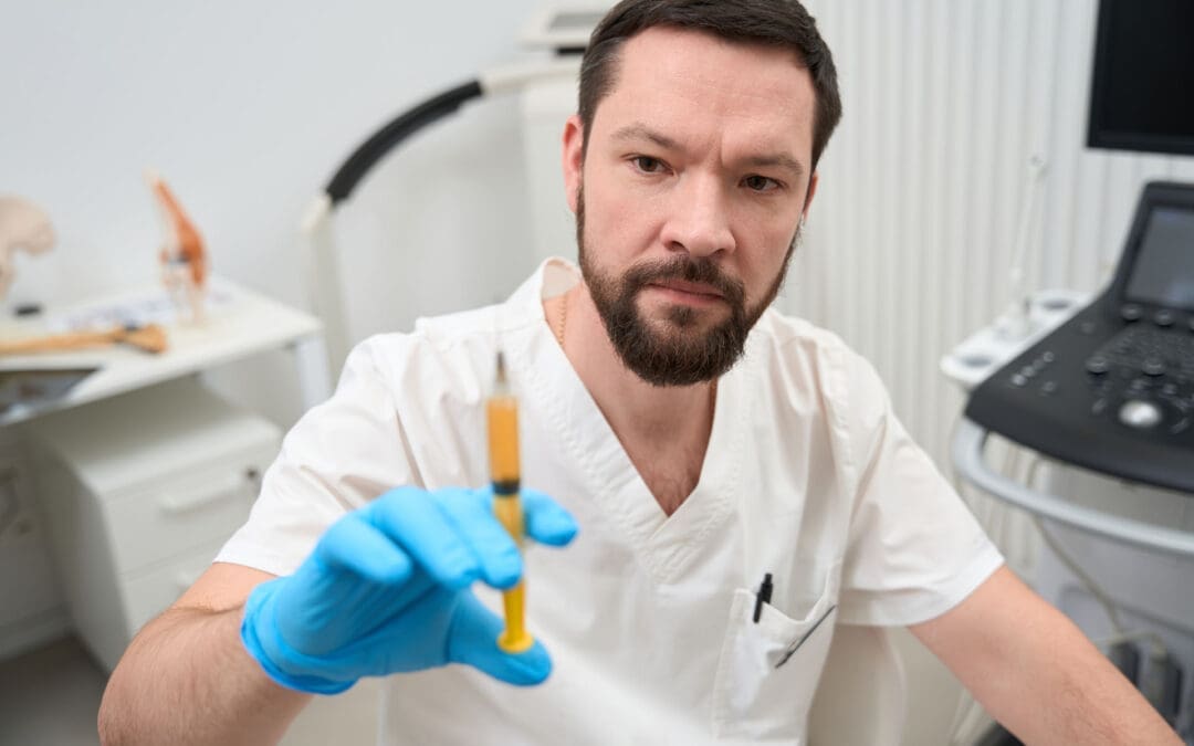
PRP stands for Platelet-Rich Plasma. Plasma is the liquid part of blood, and platelets are blood components best known for their role in clotting. However, platelets also carry growth factors and signaling molecules that help tissue repair. To make PRP, a clinician draws a small amount of blood, spins it in a centrifuge, and separates out a platelet-rich portion. That concentrated solution is then placed into the injured area. The goal is to increase healing signals directly at the site of tissue damage. (Johns Hopkins Medicine, n.d.; Yale Medicine, n.d.; HSS, n.d.; Penn Medicine, 2025).
A simple way to think about PRP is this: it does not just try to numb pain. It tries to support the body’s repair response. Hospital for Special Surgery describes PRP as a form of regenerative medicine that amplifies natural growth factors in blood cells to help damaged tissue heal. Johns Hopkins Medicine similarly explains that the concentrated growth factors in PRP may stimulate tissue regeneration and speed healing in the treated area. (HSS, n.d.; Johns Hopkins Medicine, n.d.).
What the procedure usually includes
A small blood draw from the patient
Processing the sample in a centrifuge
Preparing the platelet-rich portion
Injecting the PRP into the injured tissue
In some cases, using ultrasound to guide the injection
A visit that often takes less than an hour
This basic process is described by major medical centers, including Penn Medicine, Yale Medicine, and Johns Hopkins Medicine. (Johns Hopkins Medicine, n.d.; Penn Medicine, 2025; Yale Medicine, n.d.).
How PRP May Help Sports Injuries Heal
When tissue is injured, the body sends platelets to the area early in the healing process. Temple Health explains that platelets contain growth factors that help promote cell growth, repair tissue, and reduce inflammation. Yale Medicine notes that PRP contains concentrated platelets, cytokines, and growth factors with anti-inflammatory properties. This is why PRP is often used for injuries that have been slow to heal on their own. (Temple Health, 2021; Yale Medicine, n.d.).
PRP may be especially useful in tissues that do not receive a strong blood supply. The 2021 review in the Indian Journal of Orthopaedics notes that tendons heal more slowly than many other tissues because of their poor vascularity. That same review also explains that PRP has been studied in tendon disorders such as Achilles tendinopathy, rotator cuff tendinitis, and epicondylitis, as well as in muscle strains and osteoarthritis. (Saini et al., 2021).
For athletes, this matters because many sports injuries are overuse or repetitive-stress injuries. If a tendon stays irritated for months, or a ligament strain never fully calms down, the body may need extra support to restart a healthier repair process. Some research suggests earlier PRP use in select injuries may help guide inflammation toward recovery and restore tissue balance. Even so, researchers also note there is no universal PRP formula or perfect protocol yet, so treatment must be individualized. (Saini et al., 2021).
Common Sports Injuries PRP Is Used For
Medical centers and sports medicine sources commonly describe PRP for the following problems:
Chronic tendinitis or tendinopathy
Tennis elbow
Patellar tendinopathy or “jumper’s knee”
Achilles tendon problems
Ligament strains
Muscle strains and some muscle tears
Cartilage irritation
Osteoarthritis in active adults
These uses are repeatedly listed by Penn Medicine, Yale Medicine, Temple Health, and HSS. (Penn Medicine, 2025; Temple Health, 2021; Yale Medicine, n.d.; HSS, n.d.).
Temple Health highlights tennis elbow and jumper’s knee as common orthopedic conditions that may benefit from PRP. In its overview, Penn Medicine also lists structures such as the Achilles tendon, ACL, hamstring, patellar tendon, and cartilage as areas in sports medicine where PRP is used. Yale Medicine adds tendon, ligament, and muscle conditions, as well as degenerative joint conditions, to that list. (Penn Medicine, 2025; Temple Health, 2021; Yale Medicine, n.d.).
There is also supportive evidence for muscle injury care when injections are placed carefully. A 2014 study in Blood Transfusion reported that athletes with grade II muscle lesions who received ultrasound-guided PRP showed full healing on ultrasound, pain resolution, and return to sport, with only one relapse reported a year later. That does not prove PRP is right for every muscle injury, but it does show why sports clinicians remain interested in it. (Borrione et al., 2014).
What Recovery Feels Like After PRP
One important point for patients is that PRP can cause short-term soreness. Yale Medicine says the most common side effects are discomfort, pain, and stiffness at the injection site. Penn Medicine also notes that mild soreness, swelling, or stiffness is common for the first few days. Johns Hopkins Medicine adds that some people notice soreness and bruising after the procedure. In most cases, these effects are temporary. (Johns Hopkins Medicine, n.d.; Penn Medicine, 2025; Yale Medicine, n.d.).
Patients also need realistic expectations. PRP is not usually an instant pain reliever. Penn Medicine says improvement may take a few weeks to become noticeable, with fuller benefits developing over months. Yale Medicine reports that some people notice pain improvement in four to six weeks, with continued progress for up to a year. (Penn Medicine, 2025; Yale Medicine, n.d.).
Aftercare often includes
Resting the area for a short time
Avoiding hard exercise right away
Using a guided rehab plan
Following instructions about pain control
Avoiding some anti-inflammatory medicines when advised
Penn Medicine and HSS both note that anti-inflammatory medicines may interfere with the early healing response that PRP is meant to support, so patients should follow their treating clinician’s advice. (HSS, n.d.; Penn Medicine, 2025).
Why Ultrasound-Guided PRP Matters
Not every injection needs the same level of precision, but many sports injuries benefit from careful image guidance. Both Johns Hopkins Medicine and Yale Medicine acknowledge the use of ultrasound during PRP procedures. Research in athletes also supports this approach. The 2014 study on muscle injuries emphasized that ultrasound was important for both locating the lesion and guiding the needle accurately into it. The 2021 sports injury review similarly reported that ultrasound-guided injections improve accuracy, particularly for musculoskeletal conditions. (Johns Hopkins Medicine, n.d.; Yale Medicine, n.d.; Borrione et al., 2014; Saini et al., 2021).
On Dr. Alexander Jimenez’s public clinical website, one recent educational article describes ultrasound-guided intra-articular hip PRP as a precision-focused procedure in which ultrasound helps the clinician visualize anatomy, confirm correct placement, and improve safety. That same article stresses that biologic injections work best when they are combined with rehabilitation and movement-based recovery rather than used alone. (Jimenez, n.d.).
Dr. Alexander Jimenez’s Clinical Observations and the Value of Integrated Care
Dr. Alexander Jimenez, DC, APRN, FNP-BC, describes his El Paso practice as a multidisciplinary and integrative model that combines chiropractic care, functional medicine thinking, sports medicine principles, rehabilitation, and regenerative strategies. His website presents regenerative medicine as a natural, non-surgical option designed not only to reduce pain but also to improve structure, movement, and function. (Jimenez, n.d.).
That point matters in sports injury care. A tendon or muscle may not stay healthy if the athlete still has poor joint mechanics, weak stabilizers, incorrect loading patterns, or nutrition and recovery habits that slow healing. Dr. Jimenez’s site repeatedly frames recovery as a full process that includes a detailed history, physical evaluation, attention to biomechanics, regenerative options when appropriate, chiropractic care to improve motion, rehab planning, and follow-up focused on function. (Jimenez, n.d.).
In a comprehensive clinic model, that means PRP can be paired with structural care, progressive rehabilitation, and functional medicine support. The injection may help the tissue biologically, while rehab helps the athlete move better and reduce repeated stress on the injured area. This combined approach aligns with the broader message from both sports medicine research and Dr. Jimenez’s clinical content: better recovery usually comes from treating the tissue and the movement pattern together. (Borrione et al., 2014; Jimenez, n.d.; Saini et al., 2021).
Benefits and Limits of PRP
Possible benefits
Uses the patient’s own blood
Minimally invasive
May reduce pain and improve function
May help some chronic tendon, ligament, muscle, and joint problems
Can be part of a non-surgical recovery plan
Can be combined with rehab and other supportive care
These benefits are commonly described by Yale Medicine, Penn Medicine, Johns Hopkins Medicine, and HSS. (HSS, n.d.; Johns Hopkins Medicine, n.d.; Penn Medicine, 2025; Yale Medicine, n.d.).
Important limits
Results vary from person to person
Some injuries still need surgery or other procedures
Relief may take weeks or months, not days
PRP preparation methods are not fully standardized
Some tissues have stronger evidence than others
Those limits are important because proper medicine depends on the right treatment for the right injury at the right time. PRP may be a strong option, but it should be chosen carefully after a full exam and diagnosis. (Saini et al., 2021; Penn Medicine, 2025).
Final Thoughts
PRP therapy offers a promising non-surgical option for sports injuries because it delivers a concentrated dose of the patient’s own platelets to damaged tissue, where growth factors may support repair, reduce inflammation, and improve recovery. It is commonly used for chronic tendinopathy, ligament strain, muscle injury, and some joint conditions. Short-term soreness at the injection site can happen, but serious side effects are uncommon. The best results usually come when PRP is matched to the right injury and combined with smart rehabilitation, movement correction, and careful follow-up. (Johns Hopkins Medicine, n.d.; Penn Medicine, 2025; Yale Medicine, n.d.; Jimenez, n.d.).
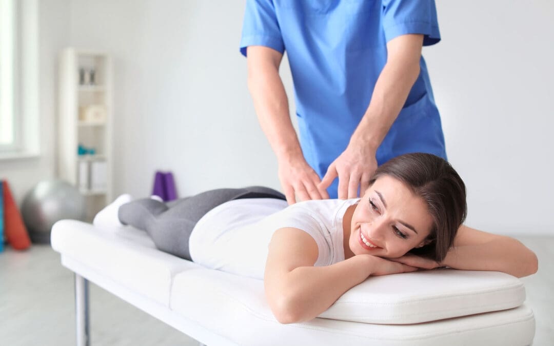
Integrative Chiropractic Care at El Paso Back Clinic: Natural Recovery Without Surgery
Many people struggle with back pain, joint stiffness, or injuries from daily life, work, or accidents. They look for lasting relief that helps them move freely again. At El Paso Back Clinic, integrative chiropractic care stands out as a natural, effective way to address these issues. Led by Dr. Alexander Jimenez, the clinic focuses on fixing the root causes of pain through structural chiropractic adjustments and supportive therapies. This approach restores proper alignment, improves movement, and accelerates the body’s natural healing without the need for surgery or heavy medications.
The team at El Paso Back Clinic believes in treating the whole person. They combine hands-on chiropractic care with physical therapy and other non-invasive methods to create lasting results. By focusing on structure and function, patients often avoid surgery and return to active, pain-free lives. This integrative style has helped countless individuals in El Paso recover from personal injuries, auto accidents, and chronic back problems.
What Makes Integrative Chiropractic Care Different?
Integrative chiropractic care at El Paso Back Clinic goes beyond quick fixes. It looks at how the spine, nerves, muscles, and joints work together. When the spine is out of alignment, it can press on nerves and cause pain, weakness, or limited motion. Chiropractic adjustments gently realign the body to free up those nerves and restore normal function.
Unlike traditional care, which might only mask symptoms, this method treats the root cause. Structural chiropractic adjustments correct posture issues, ease muscle tension, and improve overall body mechanics. When paired with physical therapy exercises, patients build strength and flexibility that lasts.
Here are the main benefits of this approach:
It uses natural techniques to reduce inflammation and promote better blood flow.
It restores functional movement so everyday tasks feel easier.
It helps prevent future injuries by fixing poor alignment early.
It fits perfectly with the body’s own repair systems for long-term wellness.
Dr. Jimenez and his team emphasize that true healing starts with proper structure. Their clinical observations show that patients who receive consistent chiropractic care often report faster recovery and greater confidence in their bodies. (Jimenez, n.d.-c)
How Supportive Therapies Enhance Chiropractic Results
While structural chiropractic care forms the foundation, El Paso Back Clinic sometimes uses supportive therapies to further enhance healing. These non-surgical options work in the background to stimulate the body’s natural processes. They include concentrated healing cells from a patient’s own blood or fat, along with signaling molecules like peptides. These tools act as gentle stimulants that help repair damaged tissues and lower swelling.
For example, platelet-rich plasma (PRP) and similar options can support tissue repair after chiropractic adjustments have created better alignment. Shockwave therapy is another tool that pairs well with chiropractic care. It sends sound waves to increase blood flow and break down scar tissue, making adjustments more effective and recovery quicker.
The clinic’s integrative practice keeps these supportive methods secondary to the main chiropractic focus. The goal remains the same: fix the root problem and restore normal movement. This combination helps patients with back pain, sciatica, or soft tissue injuries heal faster without invasive procedures.
Key ways these supportive tools work alongside chiropractic care include:
They speed up the body’s natural repair after adjustments open up better nerve pathways.
They reduce inflammation so patients feel relief sooner during physical therapy sessions.
They support long-term tissue strength, helping chiropractic corrections last longer.
They fit into a holistic plan that avoids surgery and heavy reliance on pain pills.
This balanced method has shown strong results in personal injury and sports-related cases. (StemWave, 2024; El Paso Chiropractic, n.d.)
Dr. Alexander Jimenez’s Integrative Approach at El Paso Back Clinic
Dr. Alexander Jimenez, DC, APRN, FNP-BC, leads the clinical team at El Paso Back Clinic with more than 30 years of experience. As a chiropractor first, he specializes in structural care that restores spinal alignment and functional movement. His dual background allows him to blend chiropractic adjustments with advanced rehabilitation techniques for complete recovery.
At the clinic, Dr. Jimenez focuses on finding and treating the true source of pain. He uses gentle adjustments, spinal decompression, and targeted exercises to resolve issues like herniated discs, sciatica, and scoliosis. Supportive regenerative options stay in the background as beneficial additions that enhance the primary chiropractic work.
His clinical observations highlight how this integrative style helps patients recover from trauma with greater strength and confidence. Many who visit El Paso Back Clinic after car accidents or work injuries see big improvements in mobility and daily function. Dr. Jimenez often notes that addressing structure first sets the stage for the body to heal naturally. (Personal Injury Doctor Group, 2026)
What patients can expect at the clinic includes:
Thorough exams that spot hidden alignment problems or nerve pressure.
Customized chiropractic plans that include physical therapy and movement training.
Supportive therapies are used only when needed to enhance overall outcomes.
Focus on nutrition and lifestyle tips to keep the body strong between visits.
The clinic’s multidisciplinary team of chiropractors and physical therapists works together under Dr. Jimenez’s guidance. This team approach ensures every patient receives care tailored to their needs. (Jimenez, n.d.-a)
Real Results for Personal Injuries and Everyday Back Problems
Life can bring sudden injuries from auto accidents, sports injuries, or repetitive work strain. These issues often lead to back pain, stiff joints, or limited motion. At El Paso Back Clinic, integrative chiropractic care shines in these cases by correcting structure and supporting natural recovery.
For auto accident victims, chiropractic adjustments help with whiplash and spinal misalignment that can cause long-term discomfort. Physical therapy builds strength, while supportive therapies in the background reduce swelling and speed tissue repair. Sports injuries, such as strains or tendon problems, also respond well. Athletes regain a full range of motion and return to play with less risk of re-injury.
Patients often notice these advantages:
Faster return to work or favorite activities, with less downtime.
Reduced need for pain medications that can have side effects.
Stronger, more stable joints thanks to proper alignment and support.
Overall, a better quality of life with less daily discomfort.
One review of integrative care found that patients with chronic back issues experienced steady progress and avoided surgery when chiropractic was the primary focus. (Ortho Edge El Paso, n.d.; West Texas Pain, n.d.)
The clinic’s location in El Paso makes it convenient for local families and workers seeking natural solutions. Many patients report feeling renewed energy after a few sessions of structured chiropractic care.
Why This Chiropractic-First Method Promotes Lasting Wellness
Traditional treatments sometimes rely on temporary relief or major operations. Integrative chiropractic care at El Paso Back Clinic takes a smarter path. It works with the body’s design by correcting alignment and supporting its natural repair abilities.
Younger bodies heal quickly on their own, but aging or repeated stress can slow the process. Chiropractic adjustments keep the spine and joints in proper position so healing happens efficiently. Supportive therapies like shockwave therapy or concentrated healing cells remain in the background to provide an extra nudge when needed.
This non-surgical style offers clear advantages:
No scars or infection risks that come with operations.
Better long-term mobility and fewer flare-ups.
A focus on prevention ensures problems do not become big ones.
Improved posture and movement that benefit overall health.
Experts agree that fixing the root cause leads to the best recovery. When chiropractic care leads the way, patients often experience lasting relief and greater confidence in their bodies. (New Regen Ortho, n.d.; Serenity Health Care Center, n.d.)
At El Paso Back Clinic, the emphasis remains on empowering patients through structure and function. Dr. Jimenez’s team helps people of all ages live more active, pain-free lives.
Moving Forward With Natural, Effective Care
Integrative chiropractic care at El Paso Back Clinic provides a clear path for anyone dealing with back pain or injury. Structural adjustments form the core, restoring alignment and functional movement. Supportive therapies work quietly in the background to stimulate the body’s natural healing without surgery or strong drugs.
This holistic method addresses the root causes of problems and helps patients recover faster from personal injuries, auto accidents, and sports injuries. Under Dr. Alexander Jimenez’s guidance, the clinic delivers care that fits real life and delivers real results.
If back pain or limited motion holds you back, consider the integrative chiropractic approach at El Paso Back Clinic. It proves that sometimes the best way forward is to work with the body’s own systems through skilled, hands-on care.
Restore Flexibility and Mobility with Integrative Chiropractic Care and Shockwave Therapy at El Paso Back Clinic
Many El Paso residents wake up with stiff joints or tight muscles, making simple daily tasks feel hard. Reaching overhead, bending down, or walking for long stretches can become painful or limited. At El Paso Back Clinic, integrative chiropractic care combined with Extracorporeal Shockwave Therapy (ESWT) offers a natural solution. This approach restores proper joint alignment, reduces muscle tension, and resolves soft-tissue restrictions, allowing patients to move freely again. Led by Dr. Alexander Jimenez, DC, APRN, FNP-BC, the clinic’s team uses gentle adjustments, stretching, exercises, and advanced shockwave treatments to help people regain flexibility and enjoy life in El Paso.
What Integrative Chiropractic Care Does for Flexibility at El Paso Back Clinic
Integrative chiropractic care at El Paso Back Clinic treats the whole body instead of just one problem area. It corrects small misalignments, called subluxations, in the spine and joints. These misalignments put pressure on nerves and tighten muscles. Regular adjustments gently move everything back into place. This restores proper joint alignment, eases tension, and lets the nervous system send clearer signals to the muscles.
When joints line up correctly, range of motion improves right away. Stiffness fades, and daily movements become smoother and more efficient. Patients at the clinic often say they feel looser and more energetic after just a few visits. (Gentle Chiro, n.d.) The care also includes stretching and therapeutic exercises to maintain gains over time. Muscles and joints start working together as a team, building resilience that lasts.
How Chiropractic Adjustments Restore Joint Alignment and Reduce Stiffness
Adjustments form the core of care at El Paso Back Clinic. The team uses precise, gentle pressure to correct subluxations. This simple step brings clear benefits that patients notice quickly:
Better range of motion, so joints glide freely without catching
Less muscle tension around the back, neck, and limbs
Improved nervous system function for better balance and coordination
Smoother daily activities like turning your head while driving or reaching for groceries
Lower risk of future stiffness because proper alignment trains the body to stay balanced
Many people in El Paso report that these changes make physical activities feel easier and less tiring. (Rodgers Stein Chiropractic, n.d.) The adjustments help the body move more efficiently without pain, supporting an active lifestyle.
Adding Stretching and Therapeutic Exercises for Long-Term Results
Adjustments open the door to better movement, but stretching and exercises keep it open. At El Paso Back Clinic, the rehabilitation team creates simple home programs that match each patient’s needs. Dynamic stretches warm up the body before activity. Static stretches hold the new mobility after adjustments. Therapeutic exercises strengthen the muscles that support the joints.
These steps build endurance and agility. Patients find they can stay active longer without soreness. The clinic’s sports medicine approach helps people return to hiking in the Franklin Mountains, playing with family, or working without the same old limitations. (Chiropractic Fitness, n.d.) Consistent practice turns short-term gains into lasting flexibility.
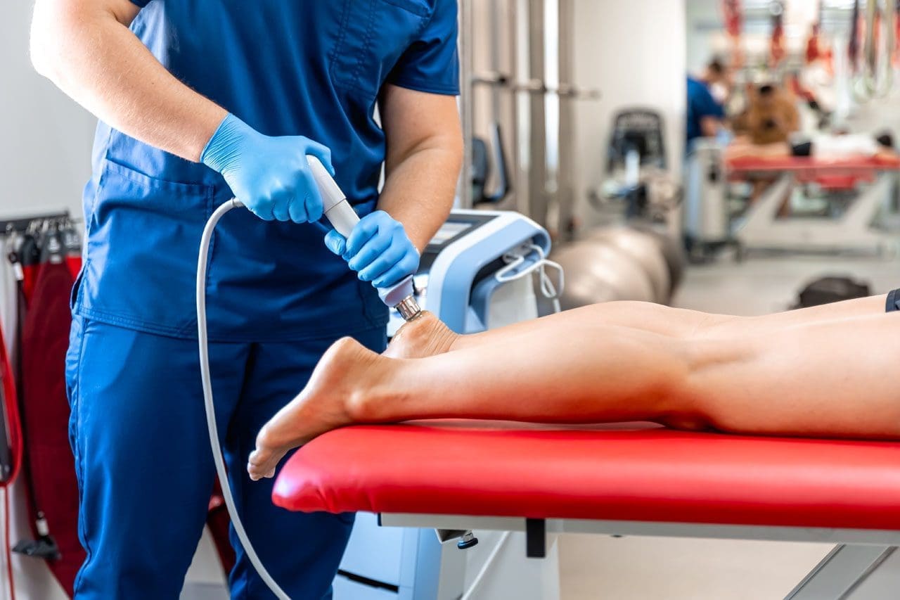
Introducing Extracorporeal Shockwave Therapy (ESWT) at El Paso Back Clinic
ESWT uses focused sound waves to reach deep into muscles, tendons, and ligaments. The waves create tiny pulses that restart healing in areas stuck with scar tissue or chronic tightness. This noninvasive treatment increases blood flow, breaks down old buildup, and reduces inflammation. At El Paso Back Clinic, ESWT is available as a key component of advanced care plans for patients who need additional support for soft tissue problems.
Why Combining Chiropractic Care and ESWT Delivers Stronger Flexibility Gains
The real power at El Paso Back Clinic comes from pairing chiropractic adjustments with ESWT. Adjustments fix the mechanical side—joint position and nerve signals—while ESWT handles the soft-tissue side—scar tissue, poor circulation, and stubborn tension. Together, they create faster, longer-lasting results than either method alone.
This dual approach works in several key ways:
Chiropractic restores spinal and joint mobility
ESWT breaks down scar tissue and releases tight fascia
The pair reduces inflammation and collagen cross-linking that causes stiffness
Blood flow improves, helping muscles and tendons heal
Patients regain a greater range of motion because both structure and tissue health get better at once
Clinic reports show that this combination can significantly improve outcomes compared with standard care. Many El Paso patients with ongoing tightness notice a real return of freedom of movement.
Common Conditions That Benefit from This Integrated Approach
El Paso Back Clinic uses this combined approach to treat several conditions that rob people of flexibility. Here are some of the most common:
Frozen shoulder – Adjustments free stuck joints while ESWT dissolves scar tissue and calcium deposits. Patients often regain full arm motion without pain.
Achilles tendinopathy – Chiropractic realigns the lower body to ease strain. Shockwave therapy stimulates the growth of new blood vessels and clears chronic buildup, so walking and running feel normal again.
General chronic muscle tension – Tightness in the back, neck, or legs from stress, work, or old injuries—responds well. The therapies release trigger points and restore smooth movement.
Post-injury stiffness from car accidents or sports – The clinic specializes in personal injury care. The combination speeds recovery and safely rebuilds mobility.
Other issues, such as plantar fasciitis and tennis elbow, also improve because the care addresses both alignment and tissue damage. (Bend Total Body Chiropractic, n.d.)
Clinical Insights from Dr. Alexander Jimenez at El Paso Back Clinic
Dr. Alexander Jimenez, DC, APRN, FNP-BC, leads El Paso Back Clinic with more than 30 years of experience. As both a Doctor of Chiropractic and a board-certified Family Nurse Practitioner, he brings a unique integrative perspective to every patient. In his clinical work in El Paso, Dr. Jimenez sees how chiropractic adjustments correct subluxations and improve nervous system function, thereby boosting flexibility and range of motion. When combined with ESWT, the results are even stronger for soft tissue injuries from accidents or overuse.
Dr. Jimenez often notes that this teamwork helps patients break down scar tissue, reduce inflammation, and restore proper movement patterns faster than traditional methods alone. His approach includes personalized functional medicine, nutritional support, and rehabilitation exercises to help patients build lasting resilience. At the clinic’s convenient El Paso locations, patients receive complete care that addresses the root causes of stiffness and helps them return to daily life and favorite activities with confidence.
Tips to Get the Most from Care at El Paso Back Clinic
Start with a full evaluation so the team can build a plan that fits your body and lifestyle. Attend regular adjustments and ESWT sessions as recommended. Follow the simple stretching and exercise routine at home every day. Support your progress with good posture, daily walks, proper hydration, and enough rest. The friendly staff at El Paso Back Clinic makes the process easy and supportive. Many patients see big improvements in flexibility within just a few weeks when they stay consistent.
A Natural Path to a More Flexible, Resilient Life in El Paso
Integrative chiropractic care and ESWT at El Paso Back Clinic offer a powerful, drug-free way to fight stiffness and reclaim natural movement. By correcting joint alignment, releasing muscle tension, and healing soft tissues, this approach makes daily life and physical activity feel effortless again. Muscles and joints work in harmony, the nervous system functions smoothly, and the body stays strong through the years.
Whether you deal with occasional tightness or a specific injury, the experienced team at El Paso Back Clinic can help. Contact the clinic today to schedule an evaluation and discover how these natural tools can work for you. With the right plan, better flexibility and mobility are well within reach for El Paso residents.
Sciatic Nerve Health and Sciatica Relief: An Integrative Chiropractic Approach at El Paso Back Clinic
The sciatic nerve should work like a clear, pain-free communication line between the lower spine and the lower body. When it is healthy, it carries nerve signals smoothly from the lower back through the hips, buttocks, legs, and feet. This allows comfortable walking, bending, standing, climbing, and turning. It also helps the body perceive touch, pressure, and position in the lower leg and foot. In simple terms, optimal sciatic nerve function means you can move well, feel normal sensation, and stay steady on your feet without burning, tingling, weakness, or pain traveling down the leg (Cleveland Clinic, 2026; Health.com, 2024; MedlinePlus, 2024).
The sciatic nerve is the longest and widest single nerve in the body. It is formed from spinal nerve roots L4 through S3 and travels from the lower spine through the pelvis, under the buttock area, down the back of the thigh, and toward the lower leg and foot. Because it is so long, irritation in the lower back, pelvis, or deep hip area can create symptoms that run down the leg. That is why sciatica often feels like more than just back pain. It can affect movement, balance, comfort, and daily function from the low back all the way to the foot (TeachMeAnatomy, 2025; Cleveland Clinic, 2026).
Why the Sciatic Nerve Matters So Much
The sciatic nerve has both motor and sensory jobs. On the motor side, it helps control the hamstrings and, through its branches, many muscles in the lower leg and foot. That means it plays a major role in bending the knee, moving the ankle, controlling the foot, and helping the body walk with stability. On the sensory side, it helps carry feeling from much of the lower leg and foot. Without normal sciatic nerve function, movement may feel weak or awkward, and sensation may feel dull, numb, sharp, or irritated (TeachMeAnatomy, 2025; NCBI Bookshelf, 2023).
When the sciatic nerve is functioning well, people often do not think about it at all. That is actually a positive sign. The nerve is quietly doing its job, helping the lower body move smoothly and respond to its environment.
Healthy sciatic nerve function supports:
Comfortable walking and standing
Smooth bending and lifting
Stable balance and coordination
Normal sensation in the lower leg and foot
A fuller, less painful range of motion
Better confidence in everyday movement
When any part of that nerve pathway becomes irritated, compressed, or inflamed, the result may be sciatica. Sciatica is not a separate disease by itself. It is a symptom pattern that usually happens when the sciatic nerve or the nerve roots that form it become irritated (Cleveland Clinic, 2026; Mayo Clinic, 2025).
What Can Interfere With Sciatic Nerve Function?
The sciatic nerve works best when signals can move freely without obstruction. Problems begin when pressure, inflammation, or mechanical strain affects the nerve roots or the nerve itself. One of the most common reasons is a herniated lumbar disc. Other causes include spinal stenosis, bone spurs, spondylolisthesis, muscle imbalance, piriformis syndrome, postural strain, and movement patterns that keep irritating the nerve (Mayo Clinic, 2025; MedlinePlus, 2024; Health.com, 2024).
People with sciatica may notice:
Sharp, shooting, or burning pain down one leg
Tingling or “pins and needles”
Numbness in part of the leg or foot
Weakness when walking or climbing stairs
Pain that worsens with long sitting
Tightness or pulling in the buttocks and thighs
Trouble standing up straight or moving normally
Sciatica can range from mild to severe. Some people feel a dull ache. Others feel intense nerve pain that makes simple movement difficult. Symptoms often get worse with prolonged sitting, repeated bending, lifting, twisting, or sudden spikes in activity (MedlinePlus, 2024; Hinge Health, 2025).
What Healthy Sciatic Function Feels Like
When the sciatic nerve is healthy, the lower body usually feels freer and more responsive. The hips and legs move with less guarding. Walking feels smoother. The foot responds normally. Stretching and changing position do not trigger a wave of pain down the leg. Good sciatic function also supports better posture and more efficient movement because the muscles and sensory pathways are working together the way they should (TeachMeAnatomy, 2025; Cleveland Clinic, 2026).
A healthy sciatic nerve should allow:
Nerve signals travel freely from the lower back to the foot
Stronger and more coordinated leg movement
Better lower-body flexibility
Comfortable daily activity with less compensation
Less irritation during sitting, standing, and walking
How an Integrative Chiropractic Clinic Can Help
At El Paso Back Clinic, sciatica care fits into a broader multidisciplinary model. The clinic website highlights chiropractic care, sciatica treatment, mobility and flexibility science, rehabilitation, exams and imaging diagnostics, injury care, and integrative wellness services as part of its approach to musculoskeletal recovery and function
That matters because sciatica is often more than a simple pain complaint. It can involve the spine, discs, joints, muscles, fascia, movement patterns, posture, and sometimes broader health and recovery factors. A more complete evaluation can help uncover why the nerve is irritated, rather than just covering up symptoms.
An integrative chiropractic clinic may help by focusing on:
Spinal alignment and joint motion
Disc stress and nerve root irritation
Muscle tightness and soft tissue tension
Hip and pelvic imbalance
Poor posture and repetitive strain
Weakness in the core, hips, and lower body
Mobility limits that keep the nerve irritated
When these issues are addressed together, the goal is to reduce pressure on the irritated nerve, improve motion, and help the body function better without relying only on pain medication.
Conservative, Non-Surgical Support for Sciatica
Many people with sciatica improve with conservative care. A non-surgical approach may include chiropractic adjustments, mobilization, soft tissue work, guided exercise, stretching, walking progression, posture correction, and activity modification. NICE guidance states that manual therapy, such as spinal manipulation, mobilization, or massage, may be considered as part of a treatment package that includes exercise for low back pain with or without sciatica (National Institute for Health and Care Excellence [NICE], 2016).
That kind of combined care can be helpful because the nerve usually responds best when the surrounding body is also improving. If the spine moves better, the soft tissues calm down, the hips become more balanced, and the core becomes stronger, then the lower back and nerve pathway may be under less stress.
Conservative sciatica care may include:
Chiropractic spinal adjustments or mobilization
Soft tissue therapy for the low back, gluteal area, and hips
Stretching for tight muscles that may affect nerve movement
Core and hip strengthening
Walking and mobility drills
Ergonomic and posture coaching
Recovery strategies that reduce repeated flare-ups
Cleveland Clinic also notes that stretching, light movement, and exercise can help relieve pressure, build strength, and support recovery in many cases of sciatica (Cleveland Clinic, 2026).
Clinical Observations from Dr. Alexander Jimenez
Dr. Alexander Jimenez, DC, APRN, FNP-BC, describes sciatica care as a root-cause process that should look beyond pain alone to identify why the nerve is being irritated. On his clinical and professional platforms, he emphasizes integrative, personalized treatment plans designed to improve mobility, reduce nerve irritation, and support long-term healing rather than only temporary symptom control
His published clinical perspective also supports a broader model of care. That includes chiropractic treatment, rehabilitation strategies, movement assessment, posture evaluation, and, when needed, more advanced diagnostic thinking. Because of his dual licensure as a chiropractor and nurse practitioner, Dr. Jimenez often frames sciatic pain as something that benefits from both structural and clinical evaluation, especially in more complex cases involving severe pain, weakness, chronic recurrence, or injury-related nerve irritation
That style fits the El Paso Back Clinic platform well. The site presents itself as a multidisciplinary clinic focused on severe pain, mobility, flexibility, injury recovery, rehabilitation, and advanced diagnostics, all of which are highly relevant when dealing with sciatica or nerve-related lower back pain
Restoring Mobility, Flexibility, and Daily Function
A major goal in sciatica care is not just pain relief. It is restoring function. Many people with sciatic irritation stop moving normally. They sit, stand, and walk differently, and avoid bending, lifting, or exercising. That can create a cycle where stiffness, weakness, fear of movement, and poor mechanics keep the problem going.
An integrative chiropractic approach tries to break that cycle. Early care may focus on calming pain, reducing guarding, and improving tolerance for basic movement. Later care often shifts toward strengthening, posture correction, improved movement habits, and prevention of new flare-ups.
That functional recovery may include:
Improving walking tolerance
Restoring hip and lower back mobility
Building core support
Relearning safer lifting and bending
Reducing repeated postural strain
Improving flexibility without overstretching the nerve
Helping patients return to work, exercise, and normal daily life
Ohio State Wexner Medical Center and Hinge Health both emphasize prevention strategies, such as regular movement, posture awareness, exercise, and limiting long periods of sitting, to reduce the risk of sciatic flare-ups (Hinge Health, 2025; Ohio State Wexner Medical Center, n.d.).
Why Medication Alone Is Not the Full Answer
Pain medication may sometimes help control symptoms, especially during a severe flare. But medication alone usually does not correct the mechanical or functional issue that keeps the nerve irritated. If the body still has poor spinal motion, muscle imbalance, repeated compression, or weak support systems, the symptoms may return.
That is why a more complete plan often works better for long-term progress. A patient may still need medical guidance, but the strongest long-term gains usually come from improving how the body moves, supports itself, and protects the irritated nerve pathway (NICE, 2016; Cleveland Clinic, 2026).
When Sciatica Needs Urgent Medical Attention
Even though many cases respond well to conservative care, some symptoms should be treated as urgent. Mayo Clinic advises prompt medical attention for sudden severe weakness, numbness, bowel or bladder control changes, or pain after major trauma. Those symptoms may point to a more serious problem and should not be ignored (Mayo Clinic, 2025).
Red flags include:
Sudden leg weakness
Loss of bowel or bladder control
Numbness in the groin or saddle area
Severe pain after a fall or crash
Rapidly worsening symptoms
When conservative care is appropriate, a good integrative clinic should recognize the need for referral, imaging, or urgent medical evaluation.
Conclusion
For optimal health, the sciatic nerve should function as a pain-free, unobstructed pathway for nerve signals between the lower spine and lower body. It should help the legs move with strength and coordination while providing sensory feedback that supports balance, movement, and comfort. Because it is the largest and longest nerve in the body, irritation anywhere along its pathway can significantly affect daily life, leading to symptoms such as pain, numbness, or weakness in the legs, which can hinder mobility and overall quality of life.
At El Paso Back Clinic, the sciatica model presented across the site supports a broader view of recovery that includes chiropractic care, rehabilitation, mobility work, injury support, diagnostics, and integrative wellness services. That kind of approach is useful because sciatica often involves more than pain alone. It may involve disc stress, joint restriction, muscle imbalance, posture, weakness, reduced flexibility, and repeated mechanical strain.
When care focuses on identifying and correcting underlying issues, patients may experience improved mobility, greater flexibility, reduced nerve irritation, and less dependence on medication alone. In that way, integrative chiropractic care can support not just temporary relief but also stronger long-term function and better lower-body movement.
That “Reset Pain” After You Sit or Hold a Weird Position: What It Is and How El Paso Back Clinic Approaches It
Have you ever held your body in an awkward position—like slouching on a couch, twisting in a chair, leaning on one hip, or sleeping with your neck turned—then you stand up and feel a sharp ache, tightness, or a “catch”? Sometimes it feels like a joint or muscle has to “reset” before you feel normal again. You might even feel clumsy for a minute, then things settle down.
At El Paso Back Clinic, this pattern is commonly discussed as a mix of postural strain, muscle guarding, myofascial tightness (trigger points), and sometimes joint restriction—especially when movement has been limited for too long or posture has been stressing the same tissues over and over.
This article explains what that “reset” feeling usually means, why it happens, and how integrative chiropractic care—like the approach described at El Paso Back Clinic—can help restore smoother motion and reduce the chances of it happening again.
What Do You Call This “Reset” Feeling?
There isn’t one single official name that covers every case, because different tissues can create the same sensation. But the most common clinical labels include:
Postural strain (tissues overloaded by a sustained position)
Muscle stiffness (tightness and reduced ease of motion)
Muscle guarding (protective tension driven by the nervous system)
Myofascial trigger points (irritable “knots” in muscle/fascia)
Joint restriction / joint dysfunction (a joint that temporarily doesn’t glide well)
Many people casually call it a “stuck joint” or “something out of place.” In reality, it’s often less dramatic than it feels—more like a temporary movement problem plus a protective muscle response.
Why It Often Hurts When You Return to Neutral (Not While You’re Sitting)
This surprises many people: “If the posture was the problem, why didn’t it hurt until I moved?”
Because your body adapts to the position you hold. While you’re still:
Your muscles settle into a holding pattern
Your joints move less
Your fascia (connective tissue) can get less “slippery” with inactivity or repeated stress
Your nervous system may “turn down” certain signals until movement starts again
Then you stand, rotate, or straighten up—and your tissues have to slide, load, and coordinate again. That’s when you feel the catch, the sting, or the awkward “reset” moment.
What’s Actually Happening: 5 Common Mechanisms Behind the “Reset”
Most cases are a combo, not just one thing.
Postural Strain: You Overloaded a Region
When you hold a position that isn’t friendly to your body—like forward head posture, slumped sitting, or a rotated spine—you can stress:
muscles
ligaments
joint capsules
fascia
Over time, those tissues complain when you ask them to move again. El Paso Back Clinic describes how repetitive positions and mechanical issues can contribute to stiffness and restriction patterns.
Muscle Guarding: Your System “Braces” for Safety
Muscle guarding is your nervous system’s way of saying, “I’m not sure this movement is safe, so I’m going to tighten things up.” It can feel like:
locked
braced
hard to relax
stiff even when you try to stretch
El Paso Back Clinic notes that pain patterns can keep muscles guarded and that stiffness may involve more than “tight muscles.”
Trigger Points: The “Knot” That Bites When You Move
Trigger points are sensitive spots in tight muscle bands. When you change position, those fibers stretch and can cause sharp, deep, or referred pain.
Fascia health is closely tied to this, because fascia surrounds muscle and helps movement feel smooth. Johns Hopkins Medicine explains that fascia can become “gummy,” stiff, and painful with limited movement, repetitive movement, or trauma.
Fascial Stiffness: The “Gummy Tissue” Effect
Fascia is like a body-wide web. When you don’t move much or repeat the same posture all day, fascia can get less elastic and less hydrated. That can make motion feel “sticky.”
Johns Hopkins Medicine specifically lists limited activity, repetitive movement, and trauma as factors that can contribute to fascia adhesions and stiffness.
Joint Cavitation: The Pop or Release
Sometimes the reset comes with a pop. A well-known imaging study found evidence that joint cracking is linked to cavity formation in the joint fluid (not bones grinding).
A pop isn’t automatically “good” or “bad.” What matters more is:
Do you move more easily afterward?
Does pain decrease?
Or does pain increase and function drop?
Why You Feel Awkward for a Bit After the “Reset”
That lingering weirdness—seconds to minutes—is often your body downshifting from protection back into normal movement.
Common reasons include:
muscles slowly letting go of guarding
irritated tissue calming down
fascia rehydrating and sliding better with movement
your brain re-mapping posture and balance (proprioception “recalibration”)
This is one reason many people feel better after a short walk post-sitting.
A Quick Self-Check: Is This Normal Stiffness or Something More?
Muscle stiffness is common and often improves with gentle movement and better posture habits. The Cleveland Clinic notes that stiffness often improves without medical treatment, but it should be taken more seriously if it comes with concerning symptoms such as fever, weakness, swelling, or persistent worsening.
Consider getting evaluated if you notice:
pain that’s getting worse over days/weeks
tingling, numbness, or weakness
pain that wakes you up repeatedly
symptoms after a significant fall or crash
the “reset pain” keeps happening in the exact same spot
What You Can Do Right Away (Safe, Simple, and Usually Helpful)
The 2–3 minute “reset without forcing it”
Stand up and walk 30–90 seconds
Do small, slow movements in a pain-free range
Try a long exhale breathing pattern (relaxes guarding)
Use gentle heat if it helps you relax
Simple posture habits that reduce repeat episodes
Change position every 30–60 minutes
Avoid “camping” in end-range posture (deep slouch, deep twist)
Use a supportive setup for workstations when possible
Build basic endurance in the muscles that hold posture (core, glutes, upper back)
How El Paso Back Clinic Approaches This Pattern (Integrative Chiropractic Style)
El Paso Back Clinic describes an integrative model that blends chiropractic care with rehab-style strategies and multidisciplinary support for spine and soft tissue problems.
Identify what’s actually driving the “reset”
Sometimes stiffness isn’t just “tight muscles.” It may involve:
joint restrictions
spine or pelvis mechanics
inflammation around a joint
pain patterns that keep muscles guarded
nerve-related problems
That’s why an exam matters—so the plan matches the cause.
Restore motion with chiropractic adjustments or mobilization
A chiropractic adjustment is a controlled force applied to a spinal joint to improve motion and movement ability.
When a joint isn’t moving well, nearby muscles often overwork and tighten. Improving joint motion can reduce the need for your body to “force” a painful reset.
Address myofascial tightness (muscle + fascia)
Because fascia can become stiff due to limited movement or repetitive strain, integrative care often includes hands-on work and guided movement to improve tissue glide.
Stabilize the area so it doesn’t keep “getting stuck”
If a joint repeatedly feels like it “locks,” the missing piece is often:
strength
endurance
timing/control
movement habits
El Paso Back Clinic frequently emphasizes rehabilitation and conditioning alongside chiropractic care to restore normal function after spine and soft-tissue issues.
A “Stop the Reset Cycle” Plan (2–3 Weeks)
These are general strategies that many patients tolerate well. Keep it gentle and pain-free.
Daily (2–5 minutes, 1–2 times/day)
1 minute easy walking
5 slow neck turns each side (easy range)
8 shoulder blade squeezes (2–3 sec hold)
8 hip hinges (small, smooth)
3 slow breaths with long exhale
During the day (30–60 seconds every hour)
stand up
10–20 steps
reset your sitting position (hips back, chest relaxed, neck tall)
3 days/week (10–15 minutes)
core stability (dead bug / modified plank)
glute strength (bridges / step-ups)
upper back endurance (band rows)
If stretching makes symptoms worse, or if stiffness keeps returning the same way, that’s a good reason to get assessed—El Paso Back Clinic even notes that persistent stiffness may signal joint restrictions or mechanics issues beyond “tight muscles.”
When to Reach Out to El Paso Back Clinic
If your “reset pain” is frequent, sharp, or starting to change your daily routine, it’s reasonable to get an evaluation—especially if you suspect joint restriction, posture-related mechanics, or muscle guarding patterns.
El Paso Back Clinic lists multiple El Paso locations and a main phone line for help and questions.
Phone: (915) 850-0900
Location (example listing): 11860 Vista Del Sol, Ste 128, El Paso, TX 79936
Key Takeaway
The experience of “I held a posture → now it hurts → then it resets” usually indicates that your body is showing a predictable pattern:
posture overloads tissues
fascia and muscle tension increase
a joint may move less smoothly
the nervous system guards
returning to neutral triggers a brief recalibration
The goal isn’t to chase pops or force releases. The goal is to restore smooth motion + stable control, so your body doesn’t keep needing that painful “reset.”
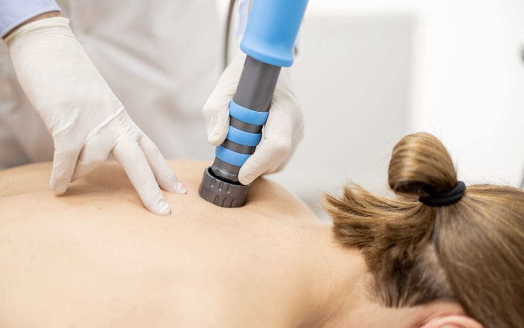
El Paso Back Clinic Shockwave Therapy: A Non-Surgical Option for Chronic Pain
Why Real ESWT Matters for Deep Healing at an Integrative El Paso Back Clinic
When people hear the term shockwave therapy, they often assume every machine is the same. It is not.
Some devices are true medical Extracorporeal Shockwave Therapy (ESWT) systems. Other devices are weaker radial pressure wave tools that are sometimes marketed as shockwave devices, even though they work differently. That difference matters if your goal is real tissue healing, not just short-term soreness relief. Mayo Clinic explains that focused shockwave (FSW) and radial pressure wave (RPW) are distinct waveforms, and only FSW is considered a “true shockwave” in a strict physical sense.
For a clinic like El Paso Back Clinic, where patients often come in with chronic pain, sports injuries, auto injuries, soft-tissue damage, and complex back conditions, the type of device and the treatment plan can make a big difference. The clinic’s site emphasizes multidisciplinary care, non-surgical recovery, and an integrative model that includes chiropractic, rehab, and functional medicine support.
This article explains, in plain language, what “real” shockwave therapy is, why focused shockwave is different from weaker devices, and how it fits into a complete recovery program in an integrative chiropractic setting.
What Is Real Shockwave Therapy?
Extracorporeal Shockwave Therapy (ESWT) is a non-invasive treatment that sends acoustic energy (sound waves) into injured tissue from outside the body. It is used in musculoskeletal care to help reduce pain and support healing in stubborn injuries. UCHealth describes ESWT as a noninvasive option for people who have not responded well to more conventional treatments, noting that it delivers high-energy acoustic waves to injured areas.
Mayo Clinic also describes shockwave therapy as a growing tool in physical medicine and sports medicine, especially for tendon and fascia problems.
In simple terms
Shockwave therapy is used to help the body “restart” healing in tissue that has been painful or stuck for a long time, such as:
tendons
fascia
ligaments
some chronic soft-tissue injuries
certain bone healing problems (in selected cases)
Mayo Clinic lists many musculoskeletal uses, including plantar fasciitis, Achilles tendinopathy, patellar tendinopathy, and lateral epicondylitis (tennis elbow).
Not All “Shockwave” Machines Are the Same
This is the most important part of the topic.
Many clinics use the word shockwave, but there are two main categories of devices used in musculoskeletal care:
Focused Shockwave (FSW / F-ESWT)
Radial Pressure Wave (RPW / radial therapy)
Mayo Clinic clearly explains that these are different technologies and should not be treated as identical. In fact, Mayo states that only focused shockwave generates a true shockwave, while radial devices generate a radial pressure wave.
Why that matters
The difference is not just marketing. It affects:
how deep the energy goes
how precise the treatment is
how much energy reaches the target tissue
what conditions may respond best
If a patient has a deep tendon problem, scar tissue, or a stubborn chronic injury, the provider should know exactly what machine is being used and why.
Focused Shockwave vs. Radial Pressure Wave
Here is the practical difference in plain language.
Focused Shockwave (FSW)
Focused shockwave is designed to deliver energy to a specific target depth. It is more precise and is often the better choice when the provider wants to treat a deeper structure or a smaller, more exact area. Mayo Clinic notes that focused shockwave has different physical properties and can be used alone or in combination with radial treatment, depending on the condition.
Radial Pressure Wave (RPW)
Radial therapy spreads energy more broadly and is often more surface-level. Mayo Clinic explains that radial devices generate pressure waves and notes tissue penetration of about 4 to 5 cm in its 2022 discussion of radial ESWT.
That does not mean radial is “bad.” It means it is different. In many cases, radial therapy remains helpful. But if a clinic claims “shockwave” and the patient expects high-energy focused treatment, the patient should ask which device is being used.
Quick comparison
Focused shockwave
More precise targeting
True shockwave physics
Often used for deeper or more exact lesions
Better fit for some regenerative goals
Radial pressure wave
Broader spread
Pressure-wave technology
Often, more superficial or diffuse treatment
Can still be useful in the right case
Why Energy Dose Matters
Real ESWT is not just “machine on, machine off.” It is dosed.
One of the main ways clinicians describe ESWT dose is Energy Flux Density (EFD), and the standard unit is mJ/mm² (millijoules per square millimeter). A PubMed Central review explains that EFD is the professional parameter used to describe shockwave energy flow through tissue, and specifically notes the unit of measurement as mJ/mm².
This is important because:
stronger energy is not always better
tissue type matters
the diagnosis matters
different injuries need different treatment settings
A quality clinic should be able to explain the treatment plan in a way that matches your condition, rather than using the same approach for every patient.
Does Shockwave Therapy Create “Microtrauma”?
Many people explain shockwave therapy by saying it creates “microtrauma” that triggers healing. That is a common explanation, and Mayo Clinic Sports Medicine uses this language in a patient-friendly way, noting that acoustic waves can create microtrauma to help reinitiate a healing response in tendons.
That said, many experts also describe the process in a more modern way as mechanotransduction—meaning the waves create a mechanical signal that helps cells activate repair pathways. Mayo Clinic’s 2025 article also highlights mechanotransduction and regenerative effects like cellular signaling and neovascular changes.
A simple way to think about it
Shockwave therapy helps by:
stimulating local tissue response
improving healing signaling
reducing pain pathways over time
helping stubborn tissue become more “active” in repair
So the short answer is:
Yes, “microtrauma” is a common way to explain it.
But the bigger idea is that the shockwave creates a healing signal, not uncontrolled tissue damage.
FDA Regulation and Why It Matters
Another reason patients should ask questions is that regulatory status matters.
The FDA has approved/cleared specific extracorporeal shockwave devices for specific uses. For example, the FDA PMA listing for the OrthoSpec Extracorporeal Shock Wave Therapy device states that it is indicated for adults with proximal plantar fasciitis (with or without a heel spur) who have had symptoms for 6 months or more and have failed conservative treatment.
That helps patients understand two important points:
real ESWT is a recognized medical technology
device claims should match actual indications and training
If a clinic says “shockwave,” it is fair to ask:
What exact device is this?
Is it focused or radial?
Is it FDA-cleared/approved for a musculoskeletal indication?
These are smart questions, not rude questions.
Why Real ESWT Is Useful in an Integrative Chiropractic Clinic
Shockwave therapy can be very effective, but it works best when the diagnosis is correct, and the rest of the care plan supports healing.
That is where an integrative clinic model is helpful.
The El Paso Back Clinic describes on its website a multidisciplinary, non-surgical, and functional recovery approach that includes chiropractic care, rehab, and broader wellness support. It also describes care for back, auto, and sports injuries, tendinopathy-related issues, and chronic pain.
Why this pairing makes sense
Shockwave therapy targets soft tissue and the healing response.
Chiropractic and rehab help restore:
joint motion
spinal alignment
posture
movement control
load tolerance
When these are combined, the patient gets a more complete plan.
Example of an integrative recovery setup
A patient with chronic Achilles pain, plantar fasciitis, or post-accident scar tissue restriction may benefit from:
Focused shockwave or radial therapy (depending on the tissue depth and goal)
Chiropractic adjustments to improve joint mechanics
Mobility work to reduce compensation patterns
Strength training/rehab exercise to improve tissue tolerance
Lifestyle support (sleep, inflammation control, nutrition)
This is especially important for back and soft-tissue injuries, as pain often has multiple causes. The tissue may be irritated, but there may also be a movement issue, posture problem, or old compensation pattern keeping it from healing.
Clinical Observations in Dr. Alexander Jimenez’s Integrative Model
Public information on dralexjimenez.com and El Paso Back Clinic describes Dr. Alexander Jimenez as a Doctor of Chiropractic and board-certified Family Nurse Practitioner (DC, APRN, FNP-BC) who uses a multidisciplinary, integrative approach focused on non-surgical recovery, diagnostics, and personalized care.
His El Paso Back Clinic content also emphasizes:
advanced injury rehabilitation
chronic pain care
sports injury care
auto injury care
functional medicine support
team-based recovery planning
These clinic observations support the idea that shockwave therapy should not be used as a stand-alone “gadget” treatment. Instead, it fits best within a broader care plan that includes biomechanics, rehab, and whole-person recovery.
Why dual training matters in this setting
In a clinic model that blends chiropractic and nurse practitioner perspectives, the provider can often look at a case more completely, including:
musculoskeletal pain drivers
nerve irritation patterns
inflammation
healing delays
activity limitations
overall recovery readiness
That type of clinical reasoning is helpful when deciding whether a patient should receive:
focused shockwave
radial therapy
chiropractic and rehab only
imaging first
referral or co-management
What Conditions Often Respond to Shockwave Therapy?
Shockwave therapy is often used for chronic injuries that have not improved enough with standard care.
Mayo Clinic and UCHealth commonly describe these types of cases:
Plantar fasciitis
Tennis elbow (lateral epicondylitis)
Achilles tendinopathy
Patellar tendinopathy
Shoulder tendinopathy
Other chronic tendon or fascia pain problems
Mayo’s clinical articles also note that ESWT has roles in treating tendons, ligaments, fascia, and even in selected bone-healing situations.
It may be especially helpful when:
pain has lasted for months
the patient plateaued in regular therapy
surgery is being considered, but not yet desired
the injury is painful with loading (walking, running, lifting, gripping)
the provider wants a non-invasive option
How to Tell if a Clinic Is Offering “Real” Shockwave Therapy
Because the market uses confusing language, patients should ask direct questions before paying for treatment.
Ask these questions
Is this focused shockwave (FSW) or radial pressure wave (RPW)?
What condition are you treating, and why is this device the right choice?
How do you set the energy dose (EFD/mJ/mm2)?
How many sessions are usually recommended for my condition?
Will I also get rehab or movement treatment?
If my pain is deep, how will you target it?
Is the device FDA-cleared/approved for musculoskeletal use?
A strong clinic should be comfortable answering these questions in simple language.
Why Device Hype Alone Is Not Enough
Some clinics advertise shockwave therapy as a miracle treatment. That is not the best way to present it.
Shockwave therapy can be a powerful tool, but results depend on:
Even the best technology will not work well if the diagnosis is wrong or if the patient returns to the same harmful movement pattern right away.
This is one reason integrated care models, like the one described at El Paso Back Clinic and Dr. Jimenez’s clinical sites, can be so useful for complex injuries: patients receive more than one treatment option and more than one clinical lens.
Bottom Line: Focused ESWT Is the Better Choice for True Regenerative Shockwave Goals
If your goal is real regenerative shockwave therapy, focused shockwave (FSW/F-ESWT) is usually the benchmark because it is the true shockwave form and offers more precise targeting. Mayo Clinic makes this distinction very clearly.
Radial devices can still be helpful in many cases, but they are not the same technology. Patients should not be told they are identical.
For patients in El Paso dealing with:
chronic tendon pain
back-related soft tissue problems
sports injuries
accident-related soft tissue injury
stubborn pain that has not improved
An integrative clinic model like El Paso Back Clinic can be a strong fit because it combines:
non-invasive care
structural assessment
chiropractic and rehab
broader healing support
multidisciplinary planning
That is often what it takes to move from “temporary pain relief” to true recovery.
IFM's Find A Practitioner tool is the largest referral network in Functional Medicine, created to help patients locate Functional Medicine practitioners anywhere in the world. IFM Certified Practitioners are listed first in the search results, given their extensive education in Functional Medicine