An annular fissure is caused by age-related changes to the spine, which often do not cause symptoms but can cause back pain. Can understanding the causes help individuals manage lower back pain and help healthcare providers develop an effective treatment program?
Annular Fissure
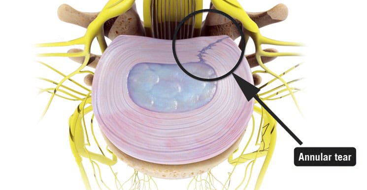
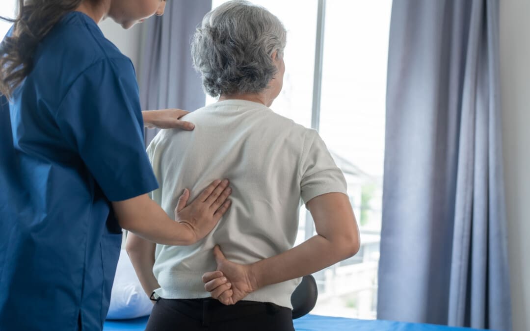
An annular fissure is a discogenic condition that affects the spine and can cause lower back pain. Also called an annular tear, it’s usually a wear-and-tear condition that happens over time rather than a condition caused by trauma. It usually happens when the fibers that make up the annulus or the tough outer covering of the intervertebral disc break or separate. To manage it, healthcare providers may recommend:
Making lifestyle changes.
Staying aware of how you go about daily activities and take steps to make adjustments, such as being mindful of unhealthy posture.
Start doing exercises that help make the back stronger.
Medical care if pain and other symptoms need to be managed.
Symptoms
Lower back pain may be a sign of an annular fissure, or there may be no symptoms. Symptoms can include:
Pain
Weakness
Numbness
Electrical sensations travel down one leg or arm if a cervical/neck tear is present.
Numbness and weakness may be caused by the nerves getting irritated or compressed near an annular tear. (Stadnik, T. W. et al., 1998)
These symptoms can also be similar to a herniated disc, which can be a complication of an annular fissure.
However, studies have shown that annular tears and herniated discs often go unnoticed because they have few obvious symptoms. (Jarvik, J. G. et al., 2005)
Annulus Function
The annulus comprises several layers of tough fibers/fibrocartilage that surround, contain, and protect the soft, liquid nucleus inside the disc. The layers of the annulus fibrosus crisscross to provide support. The nucleus is a shock absorber cushions the body’s weight on the spinal joints when sitting, standing, or moving. Its strength also allows the disc to buffer the jolts and jars it experiences. It also helps maintain the integrity of the intervertebral joint by supporting the space between the two vertebrae. When an annular fissure occurs, the fibers separate or tear off from insertion on the nearby spinal bone. A fissure can also be a break in the fibers of one or more layers. (Jarvik, J. G. et al., 2005)
Causes
An annular tear is not the standard term medical professionals use to describe or diagnose a fissure because the word tear suggests that trauma has led to the separation or break in the fibers. While an injury can cause an annular fissure, it’s usually caused by long-term wear and tear. (Guterl, C. C. et al., 2013) The tears are typically caused by age-related degenerative changes in the disc, which can also lead to degeneration in other areas of the spine. Wear and tear are caused by annular fissures due to an individual’s daily living habits, such as sitting, standing, walking, climbing stairs, and performing other routine movements.
Treatment
While a large annular fissure is not likely to improve without treatment, a small one could heal independently. However, once an area has torn, it becomes more likely to continue tearing. (Virginia Spine Institute, N.D.) Conservative treatment is usually enough to control pain and symptoms. Physical therapy and anti-inflammatory medication are the first line of treatment. (Cheng, J. et al., 2019) Medication can be over-the-counter or prescription. Physical therapy treatment includes exercises, traction, and other therapies. If these do not help with the symptoms, the provider may suggest a steroid injection to reduce inflammation and pain. It can take three to six months to recover from degenerative disc problems if doing a standard treatment plan that includes rest, low-impact therapy exercises, and anti-inflammatory treatments. (Cheng, J. et al., 2019)
In severe cases, surgery may be recommended, including disc replacement surgery. An annular tear is not a reason to have disc replacement surgery alone; it is only when there are degenerative changes in the vertebral disc that surgery might be necessary. (Yue, J. J. et al., 2012)
Improving Body Alignment
Not paying attention and being aware of how the body performs everyday activities can, over time, set the stage for an annular fissure and other musculoskeletal injuries. However, fixing daily movement and posture habits to prevent injuries can be done through simple adjustments. For example, strengthening the core and back muscles can reduce pressure on the spine and help prevent injuries. (Camp, C. L. et al., 2016) The idea is to improve joint and overall body alignment. Activities can include:
Strength training
Walking
Pilates classes
Yoga
Tai chi
Somatic exercises
These activities help with muscle balance and joint alignment, which are recommended prevention strategies that physical therapists use when working with individuals who need help with spinal problems.
Visiting a chiropractic and physical therapy team can help treat injuries and chronic pain syndromes, relieve pain, resolve musculoskeletal issues, and prevent future symptoms. Injury Medical Chiropractic and Functional Medicine Clinic works with primary healthcare providers and specialists to develop a personalized care program for each patient through an integrated approach to treating injuries, improving flexibility, mobility, and agility to help return to normal and optimal function. If other treatments are needed, Dr. Jimenez has teamed up with top surgeons, clinical specialists, medical researchers, and rehabilitation providers to provide the most effective treatments.
Back Pain Specialist
References
Stadnik, T. W., Lee, R. R., Coen, H. L., Neirynck, E. C., Buisseret, T. S., & Osteaux, M. J. (1998). Annular tears and disk herniation: prevalence and contrast enhancement on MR images in the absence of low back pain or sciatica. Radiology, 206(1), 49–55. https://doi.org/10.1148/radiology.206.1.9423651
Jarvik, J. G., Hollingworth, W., Heagerty, P. J., Haynor, D. R., Boyko, E. J., & Deyo, R. A. (2005). Three-year incidence of low back pain in an initially asymptomatic cohort: clinical and imaging risk factors. Spine, 30(13), 1541–1549. https://doi.org/10.1097/01.brs.0000167536.60002.87
Guterl, C. C., See, E. Y., Blanquer, S. B., Pandit, A., Ferguson, S. J., Benneker, L. M., Grijpma, D. W., Sakai, D., Eglin, D., Alini, M., Iatridis, J. C., & Grad, S. (2013). Challenges and strategies in the repair of ruptured annulus fibrosus. European cells & materials, 25, 1–21. https://doi.org/10.22203/ecm.v025a01
Virginia Spine Institute. (N.D.). Annular disc tear Understanding the Symptoms, Causes, and Treatments. https://www.spinemd.com/conditions/annular-disc-tear/
Cheng, J., Santiago, K. A., Nguyen, J. T., Solomon, J. L., & Lutz, G. E. (2019). Treatment of symptomatic degenerative intervertebral discs with autologous platelet-rich plasma: follow-up at 5-9 years. Regenerative medicine, 14(9), 831–840. https://doi.org/10.2217/rme-2019-0040
Yue, J. J., Telles, C., Schlösser, T. P., Hermenau, S., Ramachandran, R., & Long, W. D., 3rd (2012). Do presence and location of annular tear influence clinical outcome after lumbar total disc arthroplasty? A prospective 1-year follow-up study. International journal of spine surgery, 6, 13–17. https://doi.org/10.1016/j.ijsp.2011.09.001
Camp, C. L., Conti, M. S., Sgroi, T., Cammisa, F. P., & Dines, J. S. (2016). Epidemiology, Treatment, and Prevention of Lumbar Spine Injuries in Major League Baseball Players. American journal of orthopedics (Belle Mead, N.J.), 45(3), 137–143.
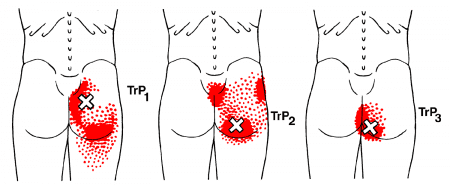
Individuals dealing with pain in the buttocks can make life difficult to sit, walk, or perform simple, everyday tasks. Can understanding the anatomy, location, and function of the gluteus maximus muscle help in muscle rehabilitation and avoid potential injuries?
Gluteus Maximus
The gluteus maximus is the largest human body muscle responsible for hip extension, external rotation, adduction, and abduction, as well as the ability to stand upright. The primary muscle extends laterally and keeps the body upright by supporting the bony pelvis and trunk. (Neto W. K. et al., 2020) When the gluteus maximus is strained, injured, or weak, it can lead to pain and inflammation.
Common symptoms can include:
Stiffness in the buttock
Discomfort while sitting
Difficulty standing up from sitting
Difficulty bending over
Pain when walking, especially upstairs or on a hill
Pain in the lower back and/or tailbone
Anatomy and Structure
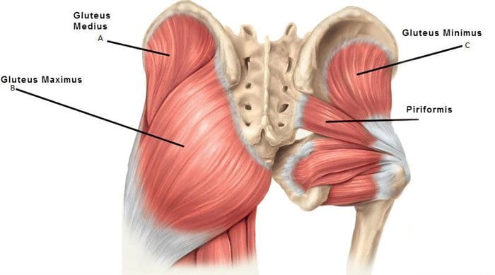
The muscles that comprise the glutes are the gluteus maximus, gluteus medius, and gluteus minimus. The gluteus medius runs underneath the gluteus maximus, and the gluteus minimus is underneath the gluteus medius.
The gluteus maximus is one of the strongest muscles. Fibers from the muscle connect to different body parts, including the femur/thighbone and the iliotibial band, which comprises connective tissue that runs up the thigh. The superior gluteal artery transports blood from the heart to the glutes.
Nerve Supply
The inferior gluteal nerve, part of the sacral plexus branch, innervates the maximus muscle. The sacral plexus nerves support motor and sensory function in the thighs, lower legs, feet, and pelvis. The sciatic nerve runs under the gluteus maximus, from the lower back down to the leg, and is often the cause of nerve pain in and around the area. (Carro L. P. et al., 2016) The main nerve of the perineum is the pudendal nerve, which runs under the gluteus maximus muscle.
Location
The gluteus maximus muscle defines the buttocks. It can be called a superficial muscle, sometimes referred to muscles that help provide shape. The origin of the gluteus maximus connects to the sacrum, the ilium, or the large upper part of the hip bone, the thoracolumbar fascia tissue, and the sacrotuberous ligaments attached to the posterior superior iliac spine. The gluteus maximus has a 45-degree angle from the pelvis to the buttocks and then inserts at the gluteal tuberosity of the femur and the iliotibial tract.
Variations
Sometimes, a duplicate muscle may originate from the gluteus maximus muscle in rare cases. However, it is more common that the gluteus maximus muscle fibers may be inserted into different body parts than where they are typically inserted. (Taylor, V. G., Geoffrey & Reeves, Rustin. 2015) This can cause a condition called greater trochanteric pain syndrome or GTPS. Inflammation of the gluteus medius, minimum tendons, and bursa inflammation can also cause GTPS. Individuals with GTPS will have tenderness or a pulsing feeling on the outer side of the hip and thigh when lying on the side, along with other symptoms.
Function
The gluteus maximus extends and externally rotates the hip joint, stabilizing the body. It is highly engaged during running and hiking activities. Regular walking does not typically target gluteus maximus strength training. However, the gluteus maximus promotes balance when walking and other activities by helping keep the pelvis and posture upright.
Conditions
The most common condition associated with the gluteus maximus is muscle strain, and deep gluteus maximus syndrome is another condition that can cause pain and involves the muscles.
Muscle Strain
A muscle strain can result from stretching and working the muscle too much that it becomes overstretched or tears. (Falótico G. G. et al., 2015) This can happen from not warming up or cooling down properly, repetitive use injury, and over-exercising. Alternatively, not exercising and not utilizing your gluteus maximus can weaken it, leading to lower back pain, hip pain, and stability and posture issues. (Jeong U. C. et al., 2015)
Deep Gluteus Maximus Syndrome
This syndrome causes pain in the buttocks when the sciatic nerve becomes entrapped. (Martin, H. D. et al., 2015) The location of the pain can help healthcare providers determine where the nerve is trapped. Those with deep gluteus maximus syndrome may experience various types of discomfort, including (Martin, H. D. et al., 2015)
Numbness and tingling in the leg
Pain when sitting
Pain when walking
Pain that radiates down the back and hips and into the thigh
To diagnose the condition, a healthcare provider may perform a physical examination and various tests to rule out other conditions that can cause similar symptoms.
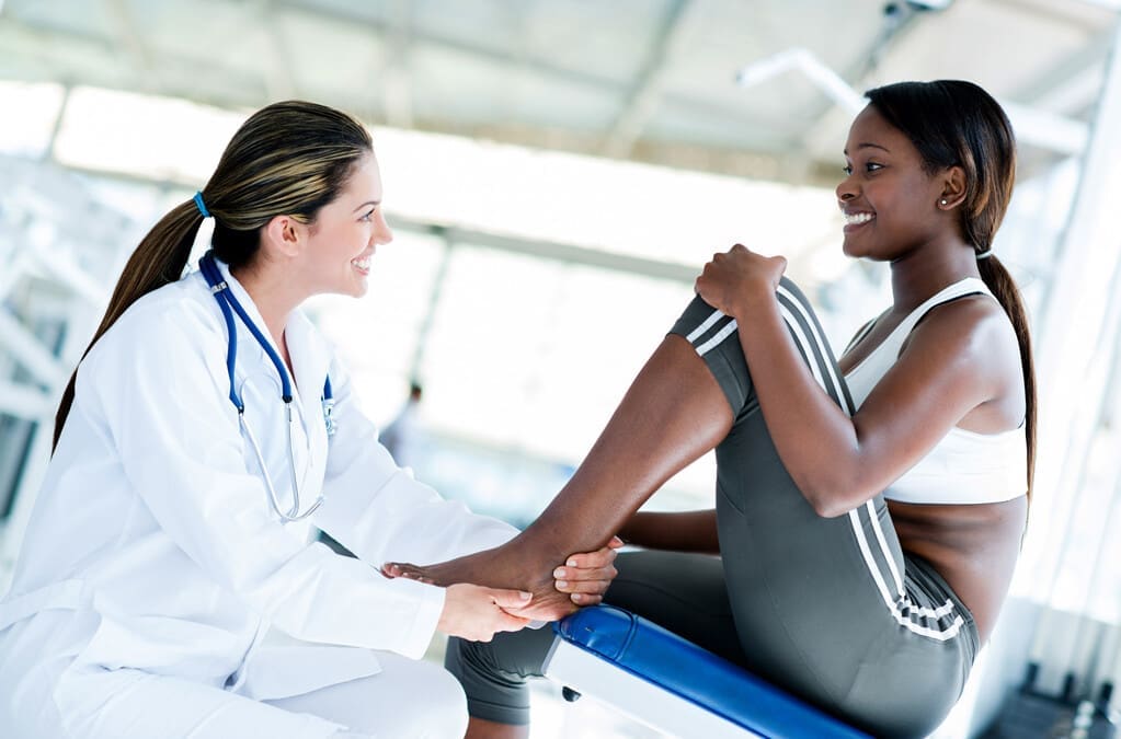
Treatment and Rehabilitation
If there is pain in the buttocks and/or lower extremities, it’s important to consult a primary doctor, chiropractor, or physical therapist. They will evaluate the strength of the gluteus muscles to diagnose any strain or weakness. From there, they will develop a personalized treatment plan to help heal the injury, strengthen the muscles, and restore function. Treatment will include stretches once the strain is rested and improves.
Recommendations can include taking a few days off to rest the muscle or, at the very least, stop performing the work or activity that caused the strain.
Ice and over-the-counter medication like ibuprofen can help reduce inflammation.
For weak gluteus maximus, a physical therapist will strengthen and retrain the muscle with a tailored program of exercises. (Jeong U. C. et al., 2015)
Treatment for deep gluteus maximus syndrome may include conservative treatment, such as chiropractic decompression and realignment, physical therapy, medications for pain and inflammation, and injections.
If conservative treatments do not relieve the pain, a primary healthcare provider may recommend surgery. (Martin, H. D. et al., 2015)
Working with a chiropractic physical therapy team can help individuals return to normal function and expedite healing. Injury Medical Chiropractic and Functional Medicine Clinic works with primary healthcare providers and specialists to develop a customized treatment program through an integrated approach to treating injuries and chronic pain syndromes, improving flexibility, mobility, and agility to relieve pain and help individuals return to normal activities. If other treatments are needed, Dr. Jimenez has teamed up with top surgeons, clinical specialists, medical researchers, and rehabilitation providers to provide the most effective treatments.
The Science of Motion and Chiropractic Care
References
Neto, W. K., Soares, E. G., Vieira, T. L., Aguiar, R., Chola, T. A., Sampaio, V. L., & Gama, E. F. (2020). Gluteus Maximus Activation during Common Strength and Hypertrophy Exercises: A Systematic Review. Journal of sports science & medicine, 19(1), 195–203.
Carro, L. P., Hernando, M. F., Cerezal, L., Navarro, I. S., Fernandez, A. A., & Castillo, A. O. (2016). Deep gluteal space problems: piriformis syndrome, ischiofemoral impingement and sciatic nerve release. Muscles, ligaments and tendons journal, 6(3), 384–396. https://doi.org/10.11138/mltj/2016.6.3.384
Taylor, Victor & Guttmann, Geoffrey & Reeves, Rustin. (2015). A variant accessory muscle of the gluteus maximus. International Journal of Anatomical Variations. 8. 10-11.
Falótico, G. G., Torquato, D. F., Roim, T. C., Takata, E. T., de Castro Pochini, A., & Ejnisman, B. (2015). Gluteal pain in athletes: how should it be investigated and treated?. Revista brasileira de ortopedia, 50(4), 462–468. https://doi.org/10.1016/j.rboe.2015.07.002
Jeong, U. C., Sim, J. H., Kim, C. Y., Hwang-Bo, G., & Nam, C. W. (2015). The effects of gluteus muscle strengthening exercise and lumbar stabilization exercise on lumbar muscle strength and balance in chronic low back pain patients. Journal of physical therapy science, 27(12), 3813–3816. https://doi.org/10.1589/jpts.27.3813
Martin, H. D., Reddy, M., & Gómez-Hoyos, J. (2015). Deep gluteal syndrome. Journal of hip preservation surgery, 2(2), 99–107. https://doi.org/10.1093/jhps/hnv029
For individuals wanting to try Pilates for back pain and exercise, can learning how to find their neutral spine help improve flexibility and increase the range of motion in the joints?
Pilates Neutral Spine
Pilates is a functional exercise modality emphasizing core stability, which is fundamental to developing a balanced body. The exercises strengthen the muscles, improve flexibility, and increase the range of motion in the joints. (Kloubec J. 2011) It is considered a functional fitness method because its principles work to establish more graceful, efficient movements from everyday life, such as improving posture. Pilates has shown its effectiveness in that it is often used in physical therapy and rehabilitation settings. (Byrnes, K., Wu, P. J., and Whillier, S. 2018) However, knowing how to find the neutral spine is essential for performing various Pilates exercises correctly. (Barbosa, A. C. et al., 2018) This subtle adjustment during practice may help prevent injury and increase overall performance. A neutral spine is the natural position of the spine when all three curves:
Cervical (neck)
Thoracic (middle)
Lumbar (lower)
Are active and in healthy alignment.
This is the strongest position for the spine when standing or sitting, allowing the body to move more naturally.
Alignment
The following exercise can help find the Pilates neutral spine.
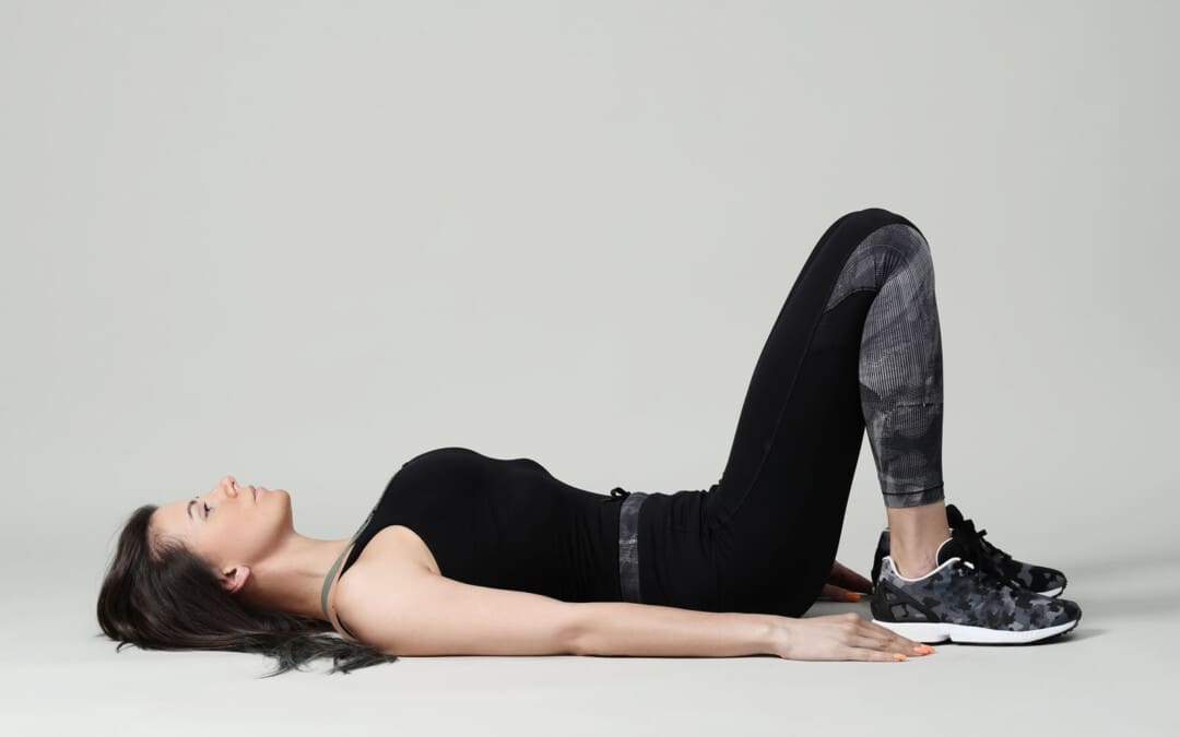
Basic Position
Lie on the back with knees bent and feet flat on the floor.
Ensure the legs are parallel to the hips, knees, heels, and toes.
Let the arms rest at your sides.
Relax
Relax the body, including the shoulders, neck, and jaw.
Allow the back to melt into the floor.
The rib cage will drop when the lower ribs are released to the floor.
Breathe Deep
Inhale all the way into the body, allowing it to move into the back and sides of the rib cage and all the way to the pelvis.
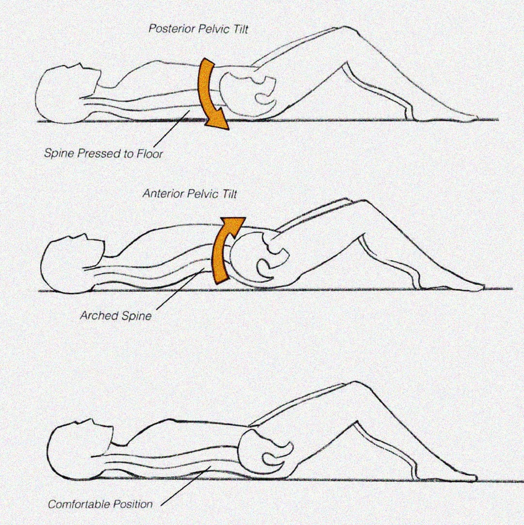
Pelvic Tilt
Exhale and use the abdominals to press the lower spine into the floor in a pelvic tuck. (Eickmeyer S. M. 2017)
Inhale to release.
Exhale and pull the lower spine off the floor, creating a pelvic tilt.
Inhale to release.
Pilates exercises don’t use excess energy or tension. Proper alignment and a neutral spine position can ensure that tension is released and excess energy is not exerted. (Byrnes, K., Wu, P. J., and Whillier, S. 2018) When performing the exercise, ensure that the shoulders, neck, and legs are relaxed and not involved in the movement.
When Exercising
Once a neutral spine is achieved, the goal is to maintain this spinal position during the exercises and when changing positions.
Start by lifting the right leg and placing it back down without letting the hips move.
Then, repeat the motion with the left leg.
Engage the abdominal muscles to help stabilize the pelvis, keeping it from moving and maintaining a neutral spine.
Repeat this process with each leg.
Once each leg can be lifted easily, test with both legs.
Exhale deeply and lift the legs while keeping the core and pelvis stable.
Then, lower them back down.
When performing this progression, there may be a want to release the abs and let the back arch.
This will cause a tuck and tilt position away from the neutral spine position.
If this progression is difficult, keep practicing until you can maintain a neutral spine.
Once this basic progression feels easy, try additional progressions and positioning.
Visualization Tips
Most people have their spines in one of two positions: tucked or tilted. A neutral spine requires individuals to be in between, with the lower abdominals flat and the lower spine’s natural curve slightly off the floor. The following visualization can help establish a neutral spine.
Balanced Pelvic Placement
Imagine a cup of water sitting on the lower abdomen, a couple of inches below the belly button.
Allow the abdominal muscles to drop toward the spine, flattening the belly.
You don’t want the water to spill, so the pelvis cannot be tipped forward or tucked under.
Body Scan Meditation
Once the body is relaxed with a balanced alignment on the floor.
Allow breathing to become deep and full and the abdominals to drop toward the floor.
The natural neck and lower spine curves should be off the floor.
Ensure the lower spine is not pressed into the floor, as this indicates a pelvic tilt.
If there is any discomfort or pain when working to increase endurance, seek advice from a healthcare professional. Injury Medical Chiropractic and Functional Medicine Clinic uses an integrated approach to treating injuries and chronic pain syndromes. It offers personalized care plans that improve ability through flexibility, mobility, and agility programs to relieve pain. Our providers use an integrated approach to create personalized care plans for each patient, including Functional Medicine, Acupuncture, Electro-Acupuncture, and Sports Medicine principles. Our goal is to relieve pain naturally by restoring health and function to the body. If other treatment is needed, Dr. Jimenez has teamed up with top surgeons, clinical specialists, medical researchers, and rehabilitation providers to provide the most effective treatments.
Is Motion Key to Healing?
References
Kloubec J. (2011). Pilates: how does it work and who needs it?. Muscles, ligaments and tendons journal, 1(2), 61–66.
Byrnes, K., Wu, P. J., & Whillier, S. (2018). Is Pilates an effective rehabilitation tool? A systematic review. Journal of bodywork and movement therapies, 22(1), 192–202. https://doi.org/10.1016/j.jbmt.2017.04.008
Barbosa, A. C., Vieira, E. R., Silva, A. F., Coelho, A. C., Martins, F. M., Fonseca, D. S., Barbosa, M. A., & Bordachar, D. (2018). Pilates experience vs. muscle activation during abdominal drawing-in maneuver. Journal of bodywork and movement therapies, 22(2), 467–470. https://doi.org/10.1016/j.jbmt.2017.05.002
Eickmeyer S. M. (2017). Anatomy and Physiology of the Pelvic Floor. Physical medicine and rehabilitation clinics of North America, 28(3), 455–460. https://doi.org/10.1016/j.pmr.2017.03.003
Can understanding the nucleus pulposus help in body positioning and prevention for individuals wanting to practice spinal hygiene and protect their discs from injury?
Nucleus Pulposus
The spinal discs are located between the spine’s vertebrae and are the body’s natural impact and shock absorbers. Within the disc is the nucleus pulposus, which plays a major role in providing the spine with shock absorption during movement. (Zhou Z. et al., 2014) The discs have a tough outer portion and a soft inner core. They are the:
It forms the tough circular exterior and comprises concentric sheets of collagen fibers or lamellae surrounding the inner core.
It has cartilaginous endplates that firmly attach to the vertebrae above and below.
Nucleus Pulposus
The nucleus pulposus is the inner core soft filling of the discs.
It contains a network of fibers suspended in a mucoprotein gel with a water base to maintain strength and pliability.
The near-liquid consistency makes it responsive to movement to handle the body’s axial load.
It helps maintain spinal suspension to prevent pressure on the bones and prevent bone-to-bone contact, reducing the potential for injuries and pain.
Shock Absorber
Each intervertebral disc is a shock-absorbing cushion, with the nucleus pulposus providing shock-absorbing properties (Zhou Z. et al., 2014). The intervertebral discs move as the body moves. For example, when arching the back, the disc moves forward slightly, and when twisting, the disc twists as well.
Spinal Action
The intervertebral disc supports spinal movements. When bending, twisting, arching, or tilting the spine, the nucleus pulposus swivels to accommodate these actions. These repeated spinal actions, which occur throughout the day and night, contribute to shifting positions while sitting, working, playing sports, carrying groceries, performing house chores, etc. An example is bending forward to pick something up. This action involves forward spinal flexion, which is bending the spine forward, flattening, or rounding. When bending using flexion, the spinal bones come closer together, pushing the nucleus pulposus toward the back.
Injuries
The disc can be pushed too far back with persistent or excessive spinal flexion. If the fibers of the annulus fibrosus become weak, they can tear, causing the nucleus pulposus to leak out and disc herniation. Generally, the nucleus pulposus will leak to the side and back; however, this corresponds to the location of the very sensitive nerve root/s with which it can come into contact, causing pain and other symptoms. The most common causes of disc herniation are degenerative wear and tear changes of the disc and trauma. Disc degeneration occurs as the body ages; it weakens the annulus fibers, allowing the nucleus pulposus to distend, bulge, or herniate.
Aging
Disc degeneration occurs with age but can also occur with injuries to the area. In young individuals, the nucleus pulposus is mostly water. For this age group, a herniation from trauma is more likely than in older individuals. (Ucar, D. et al., 2021) But as the body ages, the discs, especially the nucleus pulposus, begin to dry out. This dehydration leads to a significant loss of disc height. (UCLA Health, 2024) By age 60 or 70, the discs may be composed entirely of fiber, which can cause the shock absorption function not to work and disappear.
Chiropractic therapy is among the more conservative treatment options for a herniated disc and may be tried first before proceeding with more invasive treatments. Injury Medical Chiropractic and Functional Medicine Clinic works with primary healthcare providers and specialists to develop an optimal health and wellness solution that fully benefits the individual to get back to normal.
The Science of Functional Healing
References
Zhou, Z., Gao, M., Wei, F., Liang, J., Deng, W., Dai, X., Zhou, G., & Zou, X. (2014). Shock absorbing function study on denucleated intervertebral disc with or without hydrogel injection through static and dynamic biomechanical tests in vitro. BioMed research international, 2014, 461724. https://doi.org/10.1155/2014/461724
Nosikova, Y. S., Santerre, J. P., Grynpas, M., Gibson, G., & Kandel, R. A. (2012). Characterization of the annulus fibrosus-vertebral body interface: identification of new structural features. Journal of anatomy, 221(6), 577–589. https://doi.org/10.1111/j.1469-7580.2012.01537.x
Ucar, D., Duman, S., Bayram, Y., & Ucar, B. Y. (2021). Extruded disc herniations are experienced earlier by inactive young people in the high-tech gaming era. Journal of medicine and life, 14(3), 402–407. https://doi.org/10.25122/jml-2021-1059
Can individuals with Ehlers-Danlos syndrome find relief through various non-surgical treatments to reduce joint instability?
Introduction
The joints and ligaments surrounding the musculoskeletal system allow the upper and lower extremities to stabilize the body and be mobile. The various muscles and soft connective tissues that surround the joints help protect them from injuries. When environmental factors or disorders start to affect the body, many people develop issues that cause overlapping risk profiles, which then affect the stability of the joints. One of the disorders that affect the joints and connective tissue is EDS or Ehlers-Danlos syndrome. This connective tissue disorder can cause the joints in the body to be hypermobile. It can cause joint instability in the upper and lower extremities, thus leaving the individual to be in constant pain. Today’s article focuses on Ehlers-Danlos syndrome and its symptoms and how there are non-surgical ways to manage this connective tissue disorder. We discuss with certified medical providers who consolidate our patients’ information to assess how Ehlers-Danlos syndrome can correlate with other musculoskeletal disorders. We also inform and guide patients on how various non-surgical treatments can help reduce pain-like symptoms and manage Ehlers-Danlos syndrome. We also encourage our patients to ask their associated medical providers many intricate and important questions about incorporating various non-surgical therapies as part of their daily routine to manage the effects of Ehlers-Danlos syndrome. Dr. Jimenez, D.C., includes this information as an academic service. Disclaimer.
What Is Ehlers-Danlos Syndrome?
Do you often feel extremely tired throughout the day, even after a full night of sleep? Do you bruise easily and wonder where these bruises are coming from? Or have you noticed that you have an increased range in your joints? Many of these issues are often correlated with a disorder known as Ehlers-Danlos syndrome or EDS that affects their joints and connective tissue. EDS affects the connective tissues in the body. The connective tissues in the body help provide strength and elasticity to the skin, joints, as well as blood vessel walls, so when a person is dealing with EDS, it can cause a significant disruption to the musculoskeletal system. EDS is largely diagnosed clinically, and many doctors have identified that the gene coding of the collagen and proteins that interact in the body can help determine what type of EDS affects the individual. (Miklovic & Sieg, 2024)
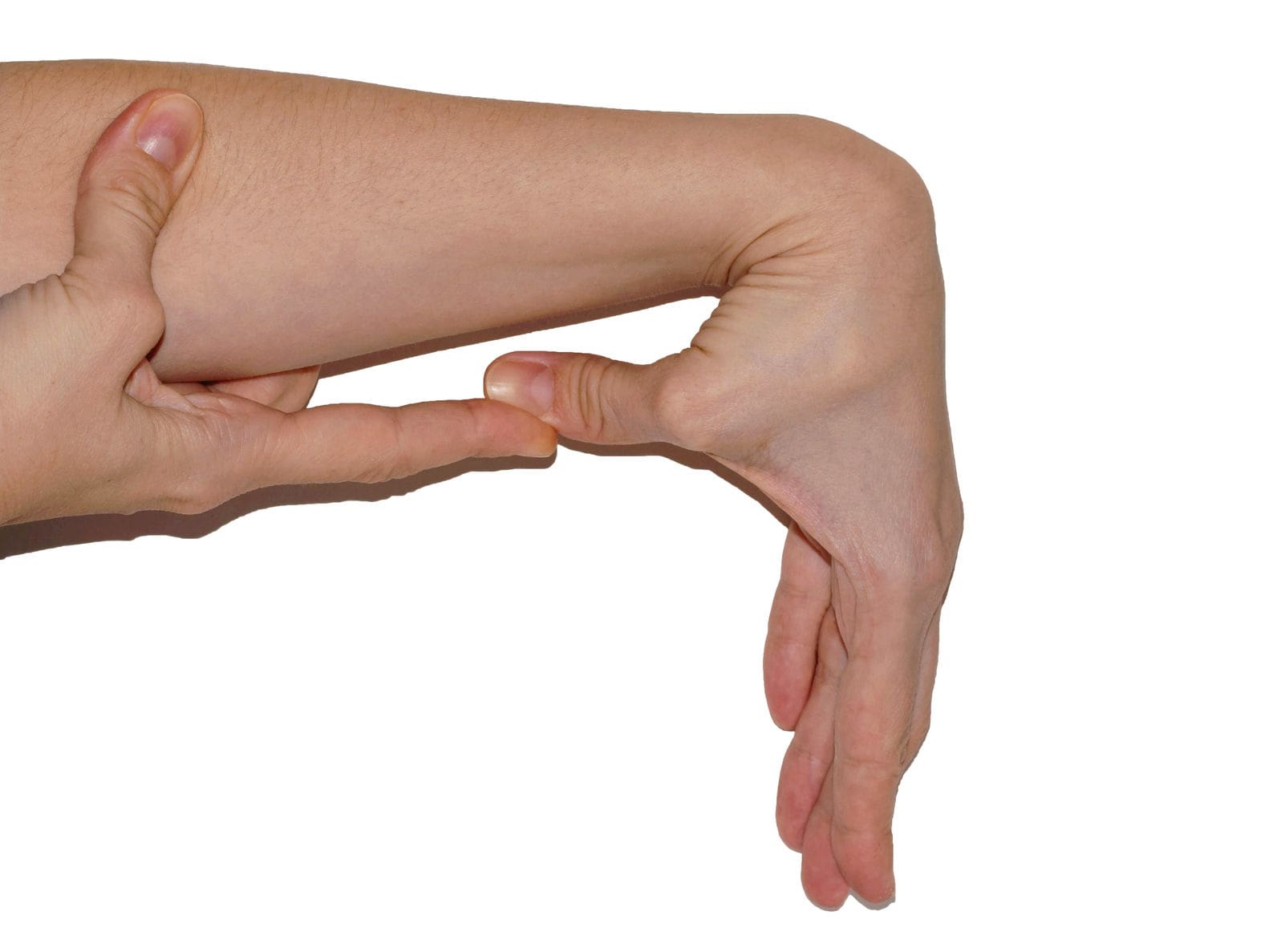
The Symptoms
When understanding EDS, it is essential to know the complexities of this connective tissue disorder. EDS is classified into numerous types with distinct features and challenges that vary depending on the severity. One of the most common types of EDS is hypermobile Ehlers-Danlos syndrome. This type of EDS is characterized by general joint hypermobility, joint instability, and pain. Some of the symptoms that are associated with hypermobile EDS include subluxation, dislocations, and soft tissue injuries that are common and may occur spontaneously or with minimal trauma. (Hakim, 1993) This can often cause acute pain to the joints in the upper and lower extremities. With its broad range of symptoms and the personal nature of the condition itself, many often don’t realize that joint hypermobility is common in the general population and may present no complications that indicate that it is a connective tissue disorder. (Gensemer et al., 2021) Additionally, hypermobile EDS can lead to spinal deformity due to the hyperextensibility of the skin, joints, and various tissue fragility. The pathophysiology of spinal deformity associated with hypermobile EDS is primarily due to muscle hypotonia and ligament laxity. (Uehara et al., 2023) This causes many people to reduce their quality of life and daily living activities significantly. However, there are ways to manage EDS and its correlating symptoms to reduce joint instability.
Movement Medicine: Chiropractic Care-Video
Ways To Manage EDS
When it comes to looking for ways to manage EDS to reduce pain and joint instability, non-surgical treatments can help address the physical and emotional aspects of the condition. Non-surgical treatments for individuals with EDS commonly focus on optimizing the body’s physical function while improving muscular strength and joint stabilization. (Buryk-Iggers et al., 2022) Many individuals with EDS will try to incorporate pain management techniques and physical therapy anduse braces and assistive devices to reduce the effects of EDS and improve their quality of life.
Non-surgical Treatments For EDS
Various non-surgical treatments like MET (muscle energy technique), electrotherapy, light physical therapy, chiropractic care, and massages can help strengthen while toning the surrounding muscles around the joints, provide sufficient pain relief, and limit long-term dependence on medications. (Broida et al., 2021) Additionally, individuals dealing with EDS aim to strengthen the affected muscles, stabilize the joints, and improve proprioception. Non-surgical treatments allow the individual to have a customized treatment plan for the severity of EDS symptoms and help reduce the pain associated with the condition. Many individuals, when going through their treatment plan consecutively to manage their EDS and reduce the pain-like symptoms, will notice improvement in symptomatic discomfort. (Khokhar et al., 2023) This means that non-surgical treatments allow individuals to be more mindful of their bodies and reduce the pain-like effects of EDS, thus allowing many individuals with EDS to lead fuller, more comfortable lives without feeling pain and discomfort.
References
Broida, S. E., Sweeney, A. P., Gottschalk, M. B., & Wagner, E. R. (2021). Management of shoulder instability in hypermobility-type Ehlers-Danlos syndrome. JSES Rev Rep Tech, 1(3), 155-164. https://doi.org/10.1016/j.xrrt.2021.03.002
Buryk-Iggers, S., Mittal, N., Santa Mina, D., Adams, S. C., Englesakis, M., Rachinsky, M., Lopez-Hernandez, L., Hussey, L., McGillis, L., McLean, L., Laflamme, C., Rozenberg, D., & Clarke, H. (2022). Exercise and Rehabilitation in People With Ehlers-Danlos Syndrome: A Systematic Review. Arch Rehabil Res Clin Transl, 4(2), 100189. https://doi.org/10.1016/j.arrct.2022.100189
Gensemer, C., Burks, R., Kautz, S., Judge, D. P., Lavallee, M., & Norris, R. A. (2021). Hypermobile Ehlers-Danlos syndromes: Complex phenotypes, challenging diagnoses, and poorly understood causes. Dev Dyn, 250(3), 318-344. https://doi.org/10.1002/dvdy.220
Hakim, A. (1993). Hypermobile Ehlers-Danlos Syndrome. In M. P. Adam, J. Feldman, G. M. Mirzaa, R. A. Pagon, S. E. Wallace, L. J. H. Bean, K. W. Gripp, & A. Amemiya (Eds.), GeneReviews((R)). https://www.ncbi.nlm.nih.gov/pubmed/20301456
Khokhar, D., Powers, B., Yamani, M., & Edwards, M. A. (2023). The Benefits of Osteopathic Manipulative Treatment on a Patient With Ehlers-Danlos Syndrome. Cureus, 15(5), e38698. https://doi.org/10.7759/cureus.38698
For individuals who have exhausted all other treatment options for low back pain and nerve root compression, can laser spine surgery help alleviate nerve compression and provide long-lasting pain relief?
Laser Spine Surgery
Laser spine surgery is a minimally invasive surgical procedure that uses a laser to cut through and remove spinal structures that are compressing nerves and causing intense pain. The minimally invasive procedure often results in less pain, tissue damage, and faster recovery than more extensive surgeries.
How It Works
Minimally invasive procedures result in less scarring and damage to surrounding structures, often reducing pain symptoms and a shorter recovery time. (Stern, J. 2009) Small incisions are made to access spinal column structures. With open-back surgery, a large incision is made down the back to access the spine. The surgery differs from other surgeries in that a laser beam, rather than other surgical instruments, is used to cut structures in the spine. However, the initial incision through the skin is made with a surgical scalpel. Laser is an acronym for Light Amplification Stimulated by Emission of Radiation. A laser can generate intense heat to cut through soft tissues, especially those with a high water content, like spinal column discs. (Stern, J. 2009) For many spine surgeries, the laser cannot be used to cut through bone as it generates instant sparks that can damage surrounding structures. Rather, laser spine surgery is primarily used to perform a discectomy, which is a surgical technique that removes a portion of a bulging or herniated disc that is pushing against the surrounding nerve roots, causing nerve compression and sciatic pain. (Stern, J. 2009)
Surgical Risks
Laser spine surgery may help resolve the cause of nerve root compression, but there is an increased risk of damage to nearby structures. Associated risks include: (Brouwer, P. A. et al., 2015)
Infection
Bleeding
Blood clots
Remaining symptoms
Returning symptoms
Further nerve damage
Damage to the membrane around the spinal cord.
Need for additional surgery
A laser beam is not precise like other surgical tools and requires practiced mastery and control to avoid damage to the spinal cord and nerve roots. (Stern, J. 2009) Because lasers cannot cut through bone, other surgical instruments are often used around corners and at different angles because they are more efficient and allow greater accuracy. (Atlantic Brain and Spine, 2022)
Purpose
Laser spine surgery is performed to remove structures that are causing nerve root compression. Nerve root compression is associated with the following conditions (Cleveland Clinic. 2018)
Bulging discs
Herniated discs
Sciatica
Spinal stenosis
Spinal cord tumors
Nerve roots that are injured or damaged and constantly send chronic pain signals can be ablated with laser surgery, known as nerve ablation. The laser burns and destroys the nerve fibers. (Stern, J. 2009) Because laser spine surgery is limited in treating certain spinal disorders, most minimally invasive spine procedures do not use a laser. (Atlantic Brain and Spine. 2022)
Preparation
The surgical team will provide more detailed instructions on what to do in the days and hours before surgery. To promote optimal healing and a smooth recovery, it is recommended that the patient stay active, eat a healthy diet, and stop smoking prior to the operation. Individuals may need to stop taking certain medications to prevent excess bleeding or interaction with anesthesia during the operation. Inform the healthcare provider about all prescriptions, over-the-counter drugs, and supplements being taken.
Laser spine surgery is an outpatient procedure at a hospital or outpatient surgical center. The patient will likely go home on the same day of the operation. (Cleveland Clinic. 2018) Patients cannot drive to or from the hospital before or after their surgery, so arrange for family or friends to provide transportation. Minimizing stress and prioritizing healthy mental and emotional well-being is important to lowering inflammation and aiding recovery. The healthier the patient goes into surgery, the easier the recovery and rehabilitation will be.
Expectations
The surgery will be decided by the patient and healthcare provider and scheduled at a hospital or outpatient surgical center. Arrange for a friend or family member to drive to the surgery and home.
Before Surgery
The patient will be taken to a pre-operative room and asked to change into a gown.
The patient will undergo a brief physical examination and answer questions about medical history.
The patient lies on a hospital bed, and a nurse inserts an IV to deliver medication and fluids.
The surgical team will use the hospital bed to transport the patient in and out of the operating room.
The surgical team will assist the patient in getting onto the operating table, and the patient will be administered anesthesia.
The patient may receive general anesthesia, which will cause the patient to sleep for the surgery, or regional anesthesia, injected into the spine to numb the affected area. (Cleveland Clinic. 2018)
The surgical team will sterilize the skin where the incision will be made.
An antiseptic solution will be used to kill bacteria and prevent the risk of infection.
Once sanitized, the body will be covered with sterilized linens to keep the surgical site clean.
During Surgery
For a discectomy, the surgeon will make a small incision less than one inch in length with a scalpel along the spine to access the nerve roots.
A surgical tool called an endoscope is a camera inserted into the incision to view the spine. (Brouwer, P. A. et al., 2015)
Once the problematic disc portion causing the compression is located, the laser is inserted to cut through it.
The cut disc portion is removed, and the incision site is sutured.
After Surgery
After surgery, the patient is brought to a recovery room, where vital signs are monitored as the effects of the anesthesia wear off.
Once stabilized, the patient can usually go home one or two hours after the operation.
The surgeon will determine when the individual is clear to resume driving.
Recovery
Following a discectomy, the individual can return to work within a few days to a few weeks, depending on the severity, but it can take up to three months to return to normal activities. Length of recovery can range from two to four weeks or less to resume a sedentary job or eight to 12 weeks for a more physically demanding job that requires heavy lifting. (University of Wisconsin School of Medicine and Public Health, 2021) During the first two weeks, the patient will be given restrictions to facilitate the spine’s healing until it becomes more stable. Restrictions can include: (University of Wisconsin School of Medicine and Public Health, 2021)
No bending, twisting, or lifting.
No strenuous physical activity, including exercise, housework, yard work, and sex.
No alcohol in the initial stage of recovery or while taking narcotic pain medications.
No driving or operating a motor vehicle until discussed with the surgeon.
The healthcare provider may recommend physical therapy to relax, strengthen, and maintain musculoskeletal health. Physical therapy may be two to three times weekly for four to six weeks.
Process
Optimal recovery recommendations include:
Getting enough sleep, at least seven to eight hours.
Maintaining a positive attitude and learning how to cope and manage stress.
Maintaining body hydration.
Following the exercise program as prescribed by the physical therapist.
Practicing healthy posture with sitting, standing, walking, and sleeping.
Staying active and limiting the amount of time spent sitting. Try to get up and walk every one to two hours during the day to stay active and prevent blood clots. Gradually increase the amount of time or distance as recovery progresses.
Do not push to do too much too soon. Overexertion can increase pain and delay recovery.
Learning correct lifting techniques to utilize the core and leg muscles to prevent increased pressure on the spine.
Discuss treatment options for managing symptoms with a healthcare provider or specialist to determine if laser spine surgery is appropriate. Injury Medical Chiropractic and Functional Medicine Clinic care plans and clinical services are specialized and focused on injuries and the complete recovery process. Dr. Jimenez has teamed with the top surgeons, clinical specialists, medical researchers, therapists, trainers, and premiere rehabilitation providers. We focus on restoring normal body functions after trauma and soft tissue injuries using Specialized Chiropractic Protocols, Wellness Programs, Functional and integrative Nutrition, Agility and mobility Fitness Training, and Rehabilitation Systems for all ages. Our areas of practice include Wellness & Nutrition, Chronic Pain, Personal Injury, Auto Accident Care, Work Injuries, Back Injury, Low Back Pain, Neck Pain, Migraine Headaches, Sports Injuries, Severe Sciatica, Scoliosis, Complex Herniated Discs, Fibromyalgia, Chronic Pain, Complex Injuries, Stress Management, Functional Medicine Treatments, and in-scope care protocols.
The Non-Surgical Approach
References
Stern, J. SpineLine. (2009). Lasers in Spine Surgery: A Review. Current Concepts, 17-23. https://www.spine.org/Portals/0/assets/downloads/KnowYourBack/LaserSurgery.pdf
Brouwer, P. A., Brand, R., van den Akker-van Marle, M. E., Jacobs, W. C., Schenk, B., van den Berg-Huijsmans, A. A., Koes, B. W., van Buchem, M. A., Arts, M. P., & Peul, W. C. (2015). Percutaneous laser disc decompression versus conventional microdiscectomy in sciatica: a randomized controlled trial. The spine journal : official journal of the North American Spine Society, 15(5), 857–865. https://doi.org/10.1016/j.spinee.2015.01.020
Atlantic Brain and Spine. (2022). The Truth About Laser Spine Surgery [2022 Update]. Atlantic Brain and Spine Blog. https://www.brainspinesurgery.com/blog/the-truth-about-laser-spine-surgery-2022-update?rq=Laser%20Spine%20Surgery
Cleveland Clinic. (2018). Can Laser Spine Surgery Fix Your Back Pain? https://health.clevelandclinic.org/can-laser-spine-surgery-fix-your-back-pain/
University of Wisconsin School of Medicine and Public Health. (2021). Home Care Instructions after Lumbar Laminectomy, Decompression or Discectomy Surgery. https://patient.uwhealth.org/healthfacts/4466
Spinal stenosis is the term used to describe a narrowing spine. Treatments vary because everybody’s case is different. Some individuals experience mild symptoms, while others experience severe symptoms. Can knowing treatment options help the patient and healthcare team customize and personalize a treatment plan to the individual’s condition?
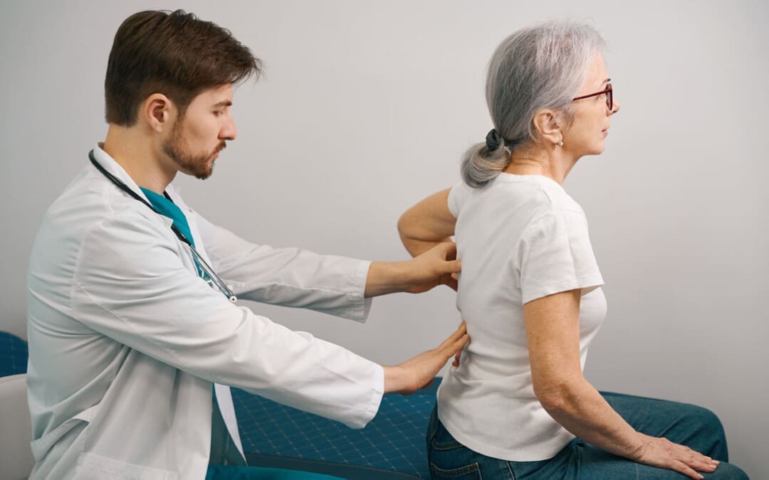
Spinal Stenosis Treatments
Spaces within the spine can become narrower than they’re supposed to be, which can cause pressure on nerve roots and the spinal cord. Anywhere along the spine can be affected. The narrowing can cause pain, burning, and/or aching in the back and weakness in the legs and feet. Spinal stenosis has several primary treatments. When working through spinal stenosis treatments, a healthcare provider will assess symptoms and start treatment with first-line therapy, such as pain medication and/or physical therapy. These are often the first among individuals with the disease.
Medication
Chronic pain is one of the main symptoms. The first-line treatment often involves using pain-relieving medication/s. Commonly prescribed medications are nonsteroidal anti-inflammatories or NSAIDs. These medications reduce pain and inflammation. However, NSAIDs are not recommended for long-term use, and other medications may need to be used to relieve pain that includes: (Sudhir Diwan et al., 2019)
Tylenol – acetaminophen
Gabapentin
Pregabalin
Opioids for severe cases
Exercise
Exercise can reduce spinal stenosis symptoms by taking pressure off the nerves, which can reduce pain and improve mobility. (Andrée-Anne Marchand et al., 2021) Healthcare providers will recommend the most effective exercises for the individual. Examples include:
Another primary spinal stenosis treatment is physical therapy, which is often used alongside pain medications. Typically, individuals undergo six to eight weeks of physical therapy, with sessions two to three times a week. Utilizing physical therapy has been shown to (Sudhir Diwan et al., 2019)
Reduce pain
Increase mobility
Reduce pain medications.
Reduce mental health symptoms like anger, depression, and mood changes.
For severe cases, physical therapy following surgery can reduce recovery times.
Back Braces
Back braces can help reduce movement and pressure on the spine. This is helpful because even small spinal movements can lead to nerve irritation, pain, and worsening symptoms. Over time, the bracing can lead to a positive increase in mobility. (Carlo Ammendolia et al., 2019)
Injections
Epidural steroid injections may be recommended to relieve severe symptoms. Steroids act as anti-inflammatories to reduce pain and swelling caused by inflammation and irritation of the spinal nerves. They are considered nonsurgical medical procedures. According to research, injections can effectively manage pain for two weeks and up to six months, and some research has found that after a spinal injection, relief can last 24 months. (Sudhir Diwan et al., 2019)
Thickened Ligaments Decompression Procedure
Some individuals may be recommended to undergo a decompression procedure. This procedure involves using a thin needle tool inserted into the back. The thickened ligament tissue is removed to reduce the pressure on the spine and nerves. Research has found that the procedure can reduce symptoms and the need for more invasive surgery. (Nagy Mekhail et al., 2021)
Alternative Treatments
In addition to first-line treatments, individuals may be referred to alternative therapies for symptom management, including:
Acupuncture
This involves the insertion of thin-tipped needles into various acupoints to relieve symptoms.
Some research has found that acupuncture may be more effective at reducing symptoms than physical therapy alone. Both options are viable and can improve mobility and pain. (Hiroyuki Oka et al., 2018)
Chiropractic
This therapy reduces pressure on nerves, maintains spinal alignment, and helps to improve mobility.
Massage
Massage helps to increase circulation, relax the muscles, and reduce pain and stiffness.
New Treatment Options
As spinal stenosis research continues, new therapies are emerging to help relieve and manage symptoms in individuals who don’t respond to traditional medicine or cannot partake in conventional therapies for various reasons. However, some evidence presented is promising; medical insurers may consider them experimental and not offer coverage until their safety has been proven. Some new treatments include:
Acupotomy
Acupotomy is a form of acupuncture that uses thin needles with a small, flat, scalpel-type tip to relieve tension in painful areas. Research on its effects is still limited, but preliminary data shows it could be an effective complementary treatment. (Ji Hoon Han et al., 2021)
Stem Cell Therapy
Stem cells are the cells from which all other cells originate. They act as the raw material for the body to create specialized cells with specific functions. (National Institutes of Health. 2016)
Individuals with spinal stenosis can develop soft tissue damage.
Stem cell therapy uses stem cells to help repair injured or diseased tissues.
Stem cell therapy can help repair or improve the damaged areas and provide symptom relief.
Clinical studies for spinal stenosis report that it could be a viable treatment option for some.
However, more research is needed to confirm whether the therapy is effective enough to be widely used. (Hideki Sudo et al., 2023)
Dynamic Stabilization Devices
LimiFlex is a medical device undergoing research and analysis for its ability to restore mobility and stability in the spine. It is implanted into the back through a surgical procedure. According to research, individuals with spinal stenosis who receive the LimiFlex often experience a higher reduction in pain and symptoms than with other forms of treatment. (T Jansen et al., 2015)
Lumbar Interspinous Distraction Decompression
Lumbar interspinous distraction decompression is another surgical procedure for spinal stenosis. The surgery is performed with an incision above the spine and places a device between two vertebrae to create space. This reduces movement and pressure on the nerves. Preliminary results show positive short-term relief from symptoms; long-term data is not yet available as it is a relatively new spinal stenosis treatment option. (UK National Health Service, 2022)
Surgical Procedures
There are several surgical procedures are available for spinal stenosis. Some include: (NYU Langone Health. 2024) Surgery for spinal stenosis is often reserved for individuals with severe symptoms, like numbness in the arms or legs. When these symptoms develop, it indicates a more notable compression of the spinal nerves and the need for a more invasive treatment. (NYU Langone Health. 2024)
Laminectomy
A laminectomy removes part or all of the lamina, the vertebral bone covering the spinal canal.
The procedure is designed to reduce pressure on nerves and the spinal cord.
Laminotomy and Foraminotomy
Both surgeries are used if an individual’s spinal stenosis negatively affects an opening in the vertebral foramen.
Ligaments, cartilage, or other tissues that constrict the nerves are removed.
Both reduce pressure on the nerves traveling through the foramen.
Laminoplasty
A laminoplasty relieves pressure on the spinal cord by removing parts of the spinal canal’s lamina.
This surgical procedure involves removing herniated or bulging discs that are placing pressure on the spinal cord and nerves.
Spinal fusion
Spinal fusion involves joining two vertebrae using metal pieces like rods and screws.
The vertebrae are more stable because the rods and screws act as a brace.
Which Treatment Is The Right One?
Because all treatment plans differ, determining the most effective is best suited for a healthcare provider. Each approach will be personalized to the individual. To decide what therapy is best, healthcare providers will assess: (National Institute of Arthritis and Musculoskeletal and Skin Diseases. 2023)
The severity of symptoms.
The current level of overall health.
The level of damage that’s occurring in the spine.
The level of disability and how mobility and quality of life are affected.
Injury Medical Chiropractic and Functional Medicine Clinic will work with an individual’s primary healthcare provider and/or specialists to help determine the best treatment options and concerns regarding medications or other forms of treatment.
Unlocking Wellness
References
Diwan, S., Sayed, D., Deer, T. R., Salomons, A., & Liang, K. (2019). An Algorithmic Approach to Treating Lumbar Spinal Stenosis: An Evidenced-Based Approach. Pain medicine (Malden, Mass.), 20(Suppl 2), S23–S31. https://doi.org/10.1093/pm/pnz133
Marchand, A. A., Houle, M., O’Shaughnessy, J., Châtillon, C. É., Cantin, V., & Descarreaux, M. (2021). Effectiveness of an exercise-based prehabilitation program for patients awaiting surgery for lumbar spinal stenosis: a randomized clinical trial. Scientific reports, 11(1), 11080. https://doi.org/10.1038/s41598-021-90537-4
Ammendolia, C., Rampersaud, Y. R., Southerst, D., Ahmed, A., Schneider, M., Hawker, G., Bombardier, C., & Côté, P. (2019). Effect of a prototype lumbar spinal stenosis belt versus a lumbar support on walking capacity in lumbar spinal stenosis: a randomized controlled trial. The spine journal : official journal of the North American Spine Society, 19(3), 386–394. https://doi.org/10.1016/j.spinee.2018.07.012
Mekhail, N., Costandi, S., Nageeb, G., Ekladios, C., & Saied, O. (2021). The durability of minimally invasive lumbar decompression procedure in patients with symptomatic lumbar spinal stenosis: Long-term follow-up. Pain practice : the official journal of World Institute of Pain, 21(8), 826–835. https://doi.org/10.1111/papr.13020
Oka, H., Matsudaira, K., Takano, Y., Kasuya, D., Niiya, M., Tonosu, J., Fukushima, M., Oshima, Y., Fujii, T., Tanaka, S., & Inanami, H. (2018). A comparative study of three conservative treatments in patients with lumbar spinal stenosis: lumbar spinal stenosis with acupuncture and physical therapy study (LAP study). BMC complementary and alternative medicine, 18(1), 19. https://doi.org/10.1186/s12906-018-2087-y
Han, J. H., Lee, H. J., Woo, S. H., Park, Y. K., Choi, G. Y., Heo, E. S., Kim, J. S., Lee, J. H., Park, C. A., Lee, W. D., Yang, C. S., Kim, A. R., & Han, C. H. (2021). Effectiveness and safety of acupotomy on lumbar spinal stenosis: A pragmatic randomized, controlled, pilot clinical trial: A study protocol. Medicine, 100(51), e28175. https://doi.org/10.1097/MD.0000000000028175
Sudo, H., Miyakoshi, T., Watanabe, Y., Ito, Y. M., Kahata, K., Tha, K. K., Yokota, N., Kato, H., Terada, T., Iwasaki, N., Arato, T., Sato, N., & Isoe, T. (2023). Protocol for treating lumbar spinal canal stenosis with a combination of ultrapurified, allogenic bone marrow-derived mesenchymal stem cells and in situ-forming gel: a multicentre, prospective, double-blind randomised controlled trial. BMJ open, 13(2), e065476. https://doi.org/10.1136/bmjopen-2022-065476
National Institutes of Health. (2016). Stem cell basics. U.S. Department of Health and Human Services. Retrieved from https://stemcells.nih.gov/info/basics/stc-basics
Jansen, T., Bornemann, R., Otten, L., Sander, K., Wirtz, D., & Pflugmacher, R. (2015). Vergleich dorsaler Dekompression nicht stabilisiert und dynamisch stabilisiert mit LimiFlex™ [A Comparison of Dorsal Decompression and Dorsal Decompression Combined with the Dynamic Stabilisation Device LimiFlex™]. Zeitschrift fur Orthopadie und Unfallchirurgie, 153(4), 415–422. https://doi.org/10.1055/s-0035-1545990
UK National Health Service. (2022). Lumbar decompression surgery: How It’s performed. https://www.nhs.uk/conditions/lumbar-decompression-surgery/what-happens/
NYU Langone Health. (2024). Surgery for spinal stenosis. https://nyulangone.org/conditions/spinal-stenosis/treatments/surgery-for-spinal-stenosis
Columbia Neurosurgery. (2024). Cervical laminoplasty procedure. https://www.neurosurgery.columbia.edu/patient-care/treatments/cervical-laminoplasty
National Institute of Arthritis and Musculoskeletal and Skin Diseases. (2023). Spinal stenosis: Diagnosis, treatment and steps to take. Retrieved from https://www.niams.nih.gov/health-topics/spinal-stenosis/diagnosis-treatment-and-steps-to-take
IFM's Find A Practitioner tool is the largest referral network in Functional Medicine, created to help patients locate Functional Medicine practitioners anywhere in the world. IFM Certified Practitioners are listed first in the search results, given their extensive education in Functional Medicine