When You Don’t Stretch: Why Muscles Get Stiff, Movement Gets Harder, and Injuries Become More Likely
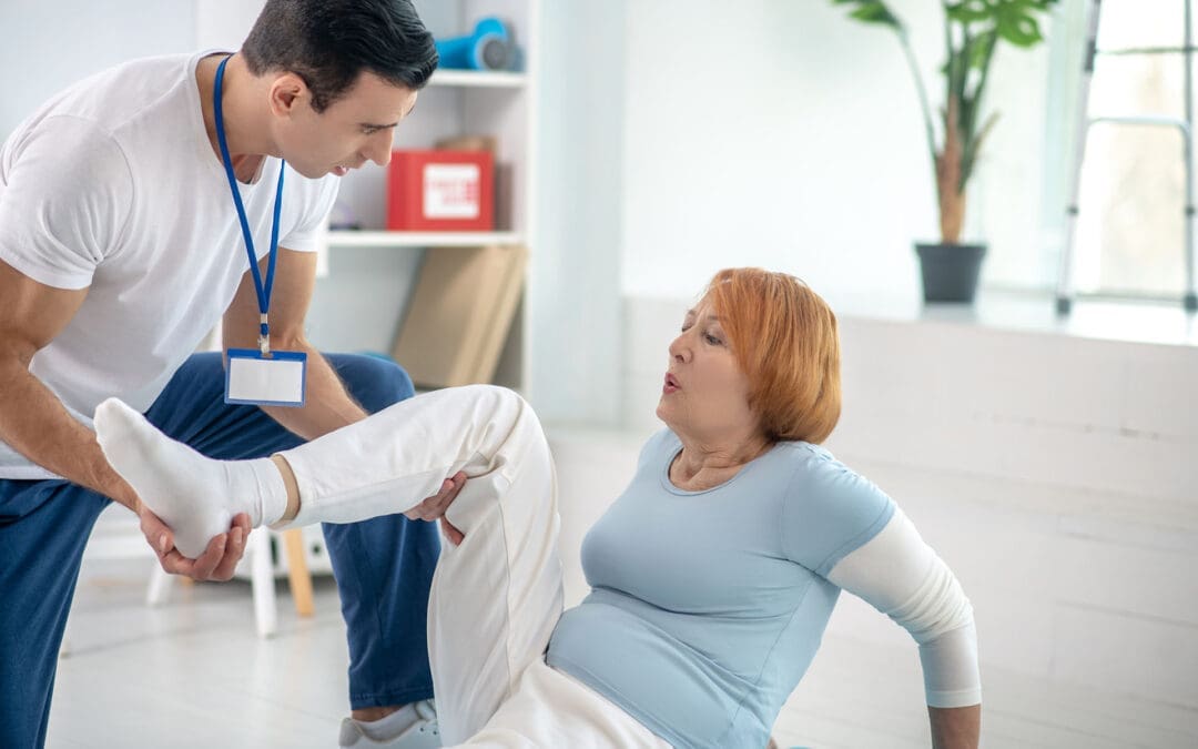
A patient with chronic back pain does targeted stretches.
If you rarely stretch, your body can start to feel “tight,” which can change how you move. Many people notice they can’t bend, twist, squat, reach overhead, or turn their head as easily as they used to. Over time, this can affect your flexibility, your range of motion (how far a joint can move), and how smooth and efficient your daily movements feel.
At El Paso Back Clinic, Dr. Alexander Jimenez, DC, APRN, FNP-BC, often explains this: when mobility decreases, the body starts to “compensate.” That means you move around a stiff area instead of through it, and those workarounds can build up stress in nearby joints and muscles (Jimenez, n.d.-a). This is one reason people can develop recurring back pain, neck stiffness, hip tightness, or shoulder irritation even without a single big injury.
What “Muscle Stiffness” Really Means
Muscle stiffness usually feels like tightness, soreness, or difficulty moving. It can happen after overuse, after you’ve been still for a long time, or when your muscles stay “stuck” in a more contracted state (Tarantino, 2025). Osmosis
Osmosis notes that stiffness can appear after a long period of minimal motion (such as bed rest or inactivity) or after new exercise that causes temporary muscle cell damage (Tarantino, 2025). Osmosis
Key idea: When your body doesn’t move a joint through its normal range often enough, the muscles and tissues around it can start to feel restricted. That restriction can make normal tasks think harder than they should.
Do Muscles Actually “Shorten” If You Don’t Stretch?
You’ll hear people say, “If you don’t stretch, your muscles will shorten.” That statement is partly true, but it needs context.
Adidas explains that the word “shorten” can be misleading: for most people, it feels like shortening because mobility and flexibility decrease when stretching is skipped, even if the muscle is not literally shrinking in everyday life (Adidas, 2025). adidas
Harvard Health adds an important clarification: without regular stretching, muscles can become tight, and when you need them for activity, they may not extend fully, increasing the risk of joint pain, strains, and muscle damage (Harvard Health Publishing, 2024). Harvard Health
So the practical takeaway is simple:
Skipping stretching often leads to less mobility and flexibility
Tight muscles can reduce how far joints can move
Tight muscles can make injuries more likely when you suddenly “ask more” of your body
How Tight Muscles Reduce Range of Motion
Range of motion (ROM) is the movement around a joint or body part. When ROM is limited, you can’t move that body part through its usual, healthy motion (Jimenez, n.d.-b). El Paso Back Clinic® • 915-850-0900
El Paso Back Clinic explains how tightness—especially in areas like the hips and ankles—can reduce ROM and limit potential for form and strength. When posture and form are compromised, pain and injury risk can rise (Jimenez, n.d.-b). El Paso Back Clinic® • 915-850-0900
What limited ROM can look like in real life
You might notice:
You can’t turn your head fully when driving
You bend from your lower back instead of your hips
You can’t squat without your heels lifting
Your shoulders feel “pinched” when reaching into a cabinet
Your hamstrings feel tight when you try to walk fast
And here’s the tricky part: your body still gets the job done—just with more strain.
Why Stiffness Can Raise Injury Risk
Harvard Health explains that tight muscles may be more easily damaged when they are suddenly stretched during strenuous activity (Harvard Health Publishing, 2024). Harvard Health
That’s why injuries often show up in moments like:
A weekend game after sitting all week
A sudden sprint to catch something
Lifting a heavy box with “cold” hips and hamstrings
A long drive followed by quick unloading or bending
Mayo Clinic also notes that better flexibility can help joints move through full ROM and may decrease injury risk, while emphasizing that stretching must be done correctly (Mayo Clinic Staff, n.d.). Mayo Clinic
Common Reasons People Stop Stretching (And How to Fix Them)
Most people don’t skip stretching because they don’t care. They skip it because it feels confusing, time-consuming, or uncomfortable.
Common barriers
“I don’t have time.”
“Stretching hurts.”
“I’m not flexible, so it doesn’t work for me.”
“I only need stretching if I work out.”
Better, more realistic reframes
You only need 5–10 minutes a few times a week to start seeing benefits (Mayo Clinic Staff, n.d.). Mayo Clinic
Stretching should create tension, not pain (Mayo Clinic Staff, n.d.). Mayo Clinic
Flexibility improves over weeks to months, not days (Harvard Health Publishing, 2024). Harvard Health
Stretching supports everyday movement, not just workouts (Harvard Health Publishing, 2024). Harvard Health
Safe Stretching Basics (So You Don’t Make Things Worse)
This matters: stretching done poorly can backfire.
Mayo Clinic recommends:
Don’t stretch cold muscles—warm up 5–10 minutes first
Don’t bounce
Hold stretches about 30 seconds (longer for problem areas)
Don’t stretch into pain (Mayo Clinic Staff, n.d.). Mayo Clinic
The American Heart Association adds:
Stretch when muscles are warm
Hold 10–30 seconds and repeat 3–5 times
Stretch slowly and smoothly (American Heart Association, 2024). www.heart.org
Quick safety checklist
Warm up first (easy walk, gentle movement)
Move slowly
Breathe
No bouncing
Stop if you feel sharp pain, numbness, or joint pain
A Simple 10-Minute Daily Stretch Routine for Real Life
This is designed for normal adults: busy schedules, stiff hips, tight neck, and lots of sitting.
Step 1: Warm up (1–2 minutes)
Walk around the house
March in place
Gentle arm circles
Step 2: Do these 6 stretches (about 8 minutes total)
1) Hip flexor stretch (1 minute each side) Helps if you sit a lot and feel tight in the front of your hips.
2) Hamstring stretch (1 minute each side) Harvard points out that tight hamstrings from sitting can limit how well you extend your leg and support walking mechanics (Harvard Health Publishing, 2024). Harvard Health
3) Calf stretch (45 seconds each side) Helpful for ankle mobility, walking, and squatting mechanics.
4) Chest opener (45 seconds) Stand in a doorway and gently open the chest to reduce rounded-shoulder posture.
5) Upper back reach (45 seconds) Hug yourself and gently pull your shoulder blades apart.
6) Neck side stretch (30 seconds each side) Gentle only—never crank your neck.
Step 3: Add “micro-mobility” during your day (optional but powerful)
Stand up every hour for 30–60 seconds
Do 5 bodyweight squats to a chair
Do 10 shoulder rolls
Take a 3-minute walk after meals
These small habits often matter as much as one long stretch session.
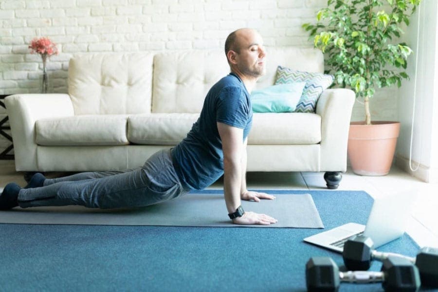
Stretching After Workouts: What You Should Know
Adidas explains the difference clearly:
Dynamic movement is best before workouts (prepares your body)
Static stretching is typically better after workouts, when you’re warm (Adidas, 2025). adidas
Mayo Clinic also cautions that stretching cold muscles can increase injury risk and notes that some intense activities may not benefit from heavy stretching right before performance (Mayo Clinic Staff, n.d.). Mayo Clinic
A balanced approach
Before exercise: warm up + dynamic mobility
After exercise: gentle static stretching + breathing
On rest days: short, consistent flexibility routine
When Stiffness Is a Sign You Need More Than Stretching
Sometimes the problem is not just “tight muscles.” You may have:
Joint restrictions that block movement
Spine or pelvis alignment issues affecting mechanics
Inflammation around a joint
Pain patterns that keep muscles “guarded”
A nerve-related problem (numbness, tingling, weakness)
El Paso Back Clinic notes that limited ROM in areas like the back, neck, or shoulders can be linked to the body being out of natural alignment, repetitive motions, or wear and tear (Jimenez, n.d.-b). El Paso Back Clinic® • 915-850-0900
If stretching doesn’t help—or makes symptoms worse—it’s smart to get assessed.
The El Paso Back Clinic Approach: Integrative Chiropractic + Nurse Practitioner Support
This is where integrative care can be a game-changer: you’re not only “stretching more,” you’re also finding out why you’re tight and building a plan that fits your body.
What chiropractic care can add
El Paso Back Clinic describes a “restoration” approach that may include:
Soft tissue work (to reduce tightness and improve circulation)
Adjustments (to address misalignments and support mobility)
Nurse practitioners are advanced practice clinicians who assess, diagnose, and treat illnesses and injuries and support chronic condition management (American Nurses Association, n.d.). ANA Healthgrades also describes NPs performing screenings and physical exams, ordering lab work, documenting care, and diagnosing certain conditions (Prosser, 2025). Healthgrades Resources
Why the combo helps stiffness and pain
Together, a chiropractor + NP team can:
Screen for red flags (nerve symptoms, systemic issues)
Decide when imaging or labs are appropriate
Build a movement plan that matches your pain level
Address sleep, stress, inflammation, and recovery habits
Track progress using measurable goals (like ROM improvements)
Dr. Jimenez’s Mobility & Flexibility materials emphasize that “great mobility” supports functional movement without ROM restrictions and that people who don’t stretch often may experience stiffened muscles that reduce effective movement (Jimenez, n.d.-a). El Paso Back Clinic® • 915-850-0900
Red Flags: When to Stop Stretching and Get Checked
Call a clinician promptly if you have:
Numbness, tingling, or weakness in an arm/leg
Loss of balance, clumsiness, or trouble walking
Severe pain that doesn’t improve
Pain after trauma (car accident, fall, sports collision)
Fever, unexplained swelling, or sudden intense stiffness
Muscle stiffness can sometimes be related to underlying medical issues, and diagnosis may require an exam and follow-up testing, depending on the cause (Tarantino, 2025). Osmosis
The Bottom Line
If you don’t stretch regularly, it’s common to feel tighter and less mobile over time. That stiffness can reduce range of motion, make daily tasks harder, and increase your risk of injury when you suddenly push your body. The good news is that you don’t need extreme flexibility. You need consistent, safe mobility work—and when required, professional support to restore movement and reduce pain.
A practical plan usually includes:
Small daily stretching habits
Better warm-ups and recovery routines
Strength + mobility (not stretching alone)
Integrative evaluation when pain, ROM loss, or repeated flare-ups keep returning
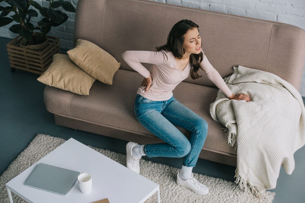
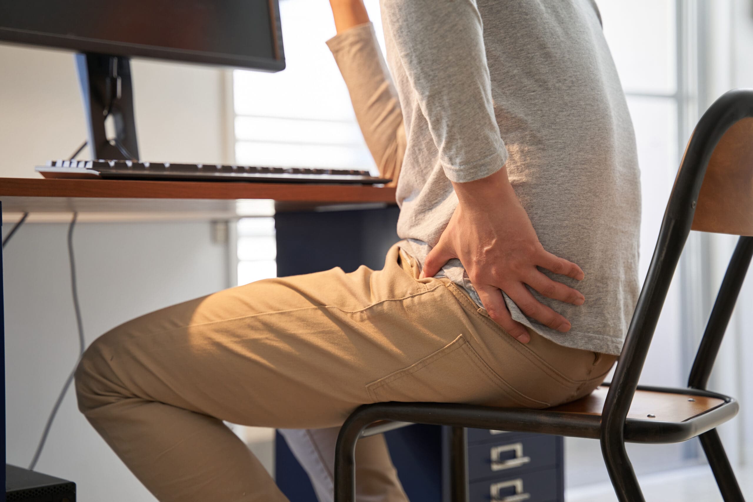
Avoiding Common Christmas Accidents: Prevention and Recovery at El Paso Back Clinic®
After lying in an awkward position, the woman is suffering from back pain on the couch at home.
The Christmas season fills homes with lights, laughter, and loved ones. But it can also bring unexpected risks. From slips on icy paths to burns in the kitchen, holiday accidents happen more often than you might think. In El Paso, Texas, where winter weather can mix with the festive rush, these issues send many seeking help. Distracted or drunk driving spikes too, making roads risky. At El Paso Back Clinic®, we focus on wellness chiropractic care to help you prevent and heal from these mishaps. This article explains common Christmas accidents, their causes, and tips for prevention. It also shows how our integrative approach, led by Dr. Alexander Jimenez, DC, APRN, FNP-BC, offers holistic recovery. Using spinal adjustments, massage, nutritional guidance, and NP-partnered care, we support your body’s natural healing to help you have a pain-free holiday.
Common Christmas Holiday Accidents at El Paso Back Clinic®
At our clinic in El Paso, TX, we see a rise in holiday-related injuries each year. These range from home mishaps to road incidents. Here’s a list of the most common ones we treat.
Falls: Decorating ladders or icy El Paso sidewalks leads to slips. These cause sprains, fractures, or head trauma. Nationwide, about 160 decorating falls occur daily, accounting for half of decorating injuries. Kids might tumble from unstable trees or during outdoor fun.
Fires: Faulty lights, dry trees, or candles spark fires. In homes across Texas, Christmas tree fires average 155 per year, causing injuries and property damage. We advise checking decorations to avoid these dangers.
Burns: Holiday cooking with hot oil or deep fryers can result in scalds. Touching lit decorations adds risk. Turkey fryers alone cause 5 deaths and 60 injuries annually. Even hot foods like fried treats can burn mouths.
Cuts: Knife slips while wrapping or carving happen often. Broken glass ornaments or toy packaging lead to ER visits – about 6,000 yearly for gift-opening cuts.
Strains: Lifting decorations, gifts, or snow strains muscles. Back issues account for 15% of holiday accidents, and 11,500 ER visits are due to shoveling. In El Paso, our patients often come in after heavy lifting.
Alcohol-Related Incidents: Festive drinks cause falls or “holiday heart” – heart rhythm problems from overdrinking. This leads to dizziness and more.
Food Poisoning: Rushed meals with undercooked food or leftovers breed bacteria. About 48 million cases occur in the U.S. each year, peaking during holidays.
Injuries Related to Toys and Gifts: Choking on small parts injures 251,700 kids yearly. Faulty gifts cause cuts or trips.
Distracted or Drunk Driving: Busy El Paso roads see more crashes from texting or drinking. Drunk driving deaths rose to 1,013 in December 2021.
These issues increase ER visits by 5-12% in the U.S. and by over 80,000 in the UK during festivities. At El Paso Back Clinic®, we help locals recover quickly.
Causes of Holiday Injuries Seen at Our Clinic
Many injuries stem from everyday tasks gone wrong. To stop recurrences, we at El Paso Back Clinic® pinpoint these causes.
Overexertion: Heavy lifting, like trees or bags, strains backs. Bending incorrectly causes 80% of lower back pain. Travel luggage accounts for 72,000 doctor visits each year.
Cooking: Burns from oils or knives in busy kitchens. One in ten child injuries comes from cooking. Grease fires are frequent.
Decorating: Ladder falls, electrical shocks, or ornament cuts. Decorating sends 13,000 to ERs yearly. Cord trips cause 2,000 injuries.
Accidents on the Road or at Home: Distracted driving in El Paso’s traffic or at home. Stress slows reflexes.
Winter sports add 186,000 injuries, though they are less common here. Plants like mistletoe can poison if eaten.
Prevention Tips from El Paso Back Clinic®
Prevent accidents with simple steps. Our team at El Paso Back Clinic® shares these to keep your holidays safe.
For Falls: Use stable ladders and salt icy paths. Get help when climbing.
For Fires and Burns: Inspect wires, water trees, and use LED candles. Watch stoves closely.
For Cuts and Strains: Cut safely and lift with your knees. Team up for heavy items.
For Alcohol and Driving: Designate a driver or use a ride. Drink moderately.
For Food and Toys: Cook thoroughly and chill food fast. Pick safe, age-appropriate toys.
Keep a first aid kit handy and manage stress. Visit us for pre-holiday check-ups.
How Integrative Chiropractic Care at El Paso Back Clinic® Helps
If injured, turn to El Paso Back Clinic® for natural healing. Our integrative chiropractic care, in partnership with NPs, treats the whole person. Dr. Alexander Jimenez, with over 30 years in El Paso, observes that holiday injuries often stem from poor posture or stress, leading to misalignment of the spine. We use non-invasive techniques to ease pain without meds or surgery.
Adjustments for Spinal and Joint Pain: Realign the spine to relieve strain from falls or lifts. This boosts movement and cuts swelling.
Massage and Physiotherapy for Muscle Problems: Ease tension from overwork. Improves circulation for faster recovery.
NP-Led Care for Holistic Wellness: Our NPs manage overall health, including burn care and effects of poisoning, with a natural focus.
Nutrition Guidance: Counter rich holiday foods with diet tips to aid digestion and immunity. Fiber-rich choices help.
Managing Underlying Conditions: Reduce stress hormones for better sleep and mood. Prevents further harm.
Dr. Jimenez’s team uses functional medicine to develop personalized plans that address issues like sciatica from slips. Chiropractic enhances the nervous system for better health during the holidays.
Enjoy a Healthy Holiday with El Paso Back Clinic®
Make Christmas memorable for the right reasons. Know the risks, prevent them, and seek our care if needed. At El Paso Back Clinic®, we’re here for your wellness. Contact us in El Paso, TX, for expert chiropractic support. Happy holidays!
Best Magnesium Supplements for Pain Relief: Types, Benefits, and Chiropractic Insights
A chiropractor and nurse practitioner discuss magnesium supplements for pain relief.
Magnesium is a mineral that your body needs for many tasks. It helps muscles work, nerves send signals, and bones stay strong. Many people do not get enough magnesium from food like nuts, seeds, and greens. This can lead to problems such as muscle pain, fatigue, and stress. Supplements can help fill the gap. In this article, we look at how magnesium eases pain. We focus on forms such as malate, glycinate, and topical. These can help with muscle soreness, nerve pain, and more. Chiropractors often suggest them to boost treatments. We base this on health sites and expert views. Read on to learn which type might work for you.
Pain comes in many forms. It can be sore muscles after a workout or chronic issues like fibromyalgia. Magnesium helps relax muscles and calm nerves. It also cuts down on swelling. Studies show it can lower pain without strong drugs. For example, it supports energy production, helping counter fatigue associated with pain. Different forms absorb in unique ways. Oral pills go through the gut. Topical ones soak into the skin. This matters for how fast they help. Always talk to a doctor before starting supplements. They can check if it’s safe for you.
Understanding Magnesium’s Role in Pain Management
Magnesium plays a big part in how your body handles pain. It blocks pain signals in nerves and helps muscles relax. Low levels can make pain worse. About half of adults in the U.S. lack enough magnesium (Team Red White & Blue, n.d.). This leads to cramps, spasms, and soreness. Supplements fix this by boosting levels.
Here are key ways magnesium helps with pain:
Muscle Relaxation: It controls contractions to stop cramps and tension.
Nerve Calming: It balances signals to reduce nerve pain.
Less Swelling: It fights inflammation that causes discomfort.
Better Recovery: It supports energy for healing after injury.
Chiropractors use magnesium with adjustments. It improves treatment outcomes by loosening tight spots. For acute pain, like after surgery, it cuts down on opioid needs (MedCentral, n.d.). For long-term pain, it eases symptoms in conditions such as migraines and back pain.
Magnesium Malate: Effective for Muscle Soreness and Fatigue in Fibromyalgia
Magnesium malate mixes magnesium with malic acid. This form absorbs well in the gut. It boosts energy by helping make ATP, the body’s fuel (Miye Care, n.d.). That’s why it’s beneficial for fatigue and soreness. People with fibromyalgia often feel worn out and achy. This type can help manage those symptoms.
Benefits include:
Eases Muscle Soreness: Reduces pain after exercise or daily strain.
Fights Fatigue: Supports energy to lessen tiredness in chronic conditions.
Helps with Fibromyalgia: Limited studies show it may lower pain severity (Healthline, n.d.).
Good Absorption: Less likely to cause stomach upset than other forms.
Chiropractors like malate for chronic pain. It supports metabolism and reduces fatigue (Sonoma Sports Chiro, n.d.). Take 200-400 mg a day. Start low to see how your body reacts. It’s often available in pill or powder form.
Magnesium Glycinate: Suitable for Nerve Pain and Relaxation
Magnesium glycinate binds to glycine, an amino acid that calms the brain. This form is easily absorbed and gentle on the stomach (Trace Minerals, n.d.). It’s great for nerve pain and stress. It helps regulate signals to stop overexcitement that causes pain.
Key advantages:
Calms Nerves: Lowers anxiety and eases nerve-related pain.
Relaxes Muscles: Reduces tension and spasms.
Aids Sleep: Promotes rest, which helps pain recovery (NMB Chiro, n.d.).
Fewer Side Effects: No laxative issues like some types.
For chiropractic patients, it cuts inflammation and boosts adjustments (SanTe Chiropractic, n.d.). It’s ideal for back or joint pain. Dose is 300-400 mg daily, often at night.
Topical Magnesium Chloride or Sulfate: Direct Muscle Relief Through Baths or Oils
Topical magnesium goes on the skin. Chloride absorbs well and targets sore spots (Health.com, n.d.). Sulfate, or Epsom salts, is for baths. It soothes muscles without gut processing.
Why choose topical:
Localized Relief: Applies right to the painful areas.
Quick Action: Bypasses digestion for faster help.
No Stomach Issues: Avoids diarrhea from oral forms.
Good for Baths: Epsom salts relax the whole body (Team Red White & Blue, n.d.).
Absorption varies by skin type. Studies are mixed, but many feel relief from soreness (Pierce Chiropractic, n.d.). Use oils or soaks 2-3 times a week.
Selecting the Right Form: Malate for Energy, Glycinate for Nerves, Topical for Localized Pain
Choose based on your pain type. Absorption differs: Oral forms, such as malate and glycinate, are absorbed through the gut; topical forms are absorbed through the skin (Drugs.com, n.d.).
Selection tips:
For Energy and Chronic Pain: Pick malate.
For Nerve Calm: Go with glycinate.
For Spot Relief: Use topical chloride or sulfate.
Consider Absorption: Glycinate is best overall (MN Spine and Sport, n.d.).
Chiropractors’ Preferences: Glycinate and Malate for Pain Management
Chiropractors favor glycinate and malate. Glycinate calms muscles and nerves, aiding adjustments (Everybodys Chiropractic, n.d.). Malate boosts energy for recovery.
How they work together:
Relax Muscles: Lessens tension for better alignment.
Cut Inflammation: Reduces joint swelling.
Boost Nerve Function: Improves signals for less pain.
Support Healing: Speeds recovery after treatments (ChiroCredit, n.d.).
Even phosphate forms help energy and relaxation in care (Edinburgh Chiropractic, n.d.).
Clinical Observations from Dr. Alexander Jimenez
Dr. Alexander Jimenez, DC, APRN, FNP-BC, focuses on integrative pain care. His work stresses non-drug methods for back pain and neuropathy (Jimenez, n.d.). He sees magnesium fitting into plans that mix chiropractic with nutrition. It helps reduce reliance on opioids and boosts recovery. In his clinic, such approaches ease chronic pain by improving mobility and reducing inflammation.
Conclusion
Magnesium offers natural pain relief. Malate helps fight fatigue in fibromyalgia, glycinate calms nerves, and topical forms provide spot relief. Chiropractors use them to enhance care. Pick the right type for your needs. Always check with a health pro. This can lead to less pain and a better life.
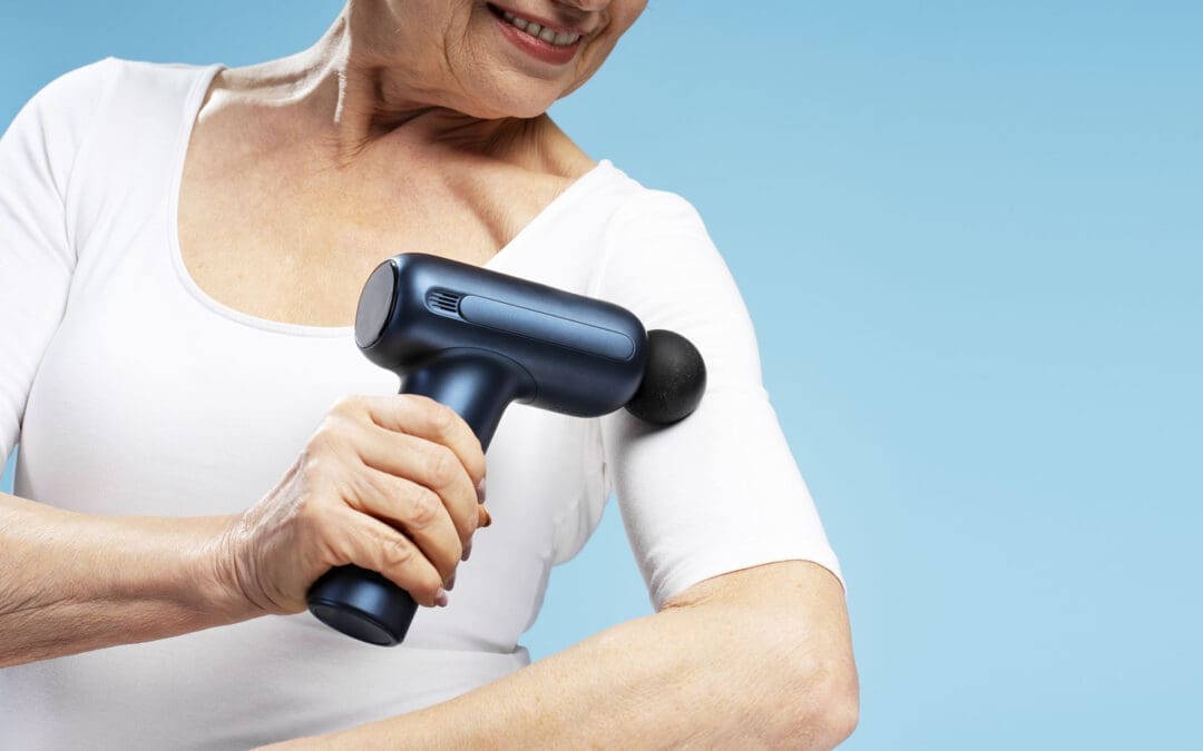
Self-Massage Tools That Support Your Care at El Paso Back Clinic
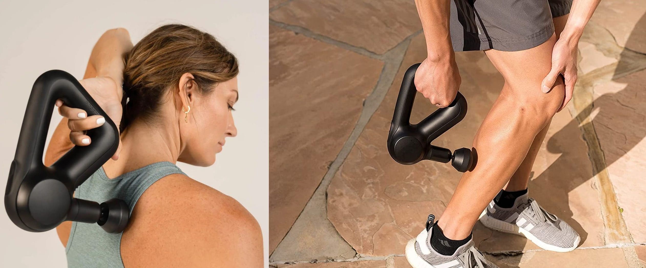
Using A Percussive Massager Correctly: El Paso Back Clinic
At El Paso Back Clinic, patients do not just get an adjustment and leave. The team, led by Dr. Alexander Jimenez, DC, APRN, FNP-BC, focuses on full-body recovery, including what you do at home between visits. El Paso Back Clinic® • 915-850-0900+1
One of the simplest ways to support your spine, joints, and muscles is with self-massage tools. When used correctly and with guidance, tools like foam rollers, massage balls, massage guns, and trigger point devices can:
Ease muscle tension
Improve circulation and tissue recovery
Help your adjustments “hold” longer
Support better posture and movement
However, not every tool is right for every person. The doctors, nurse practitioners, and rehab team at El Paso Back Clinic help patients decide which devices are safe for their bodies and how to use them without causing harm. El Paso Back Clinic® • 915-850-0900+1
Integrative Chiropractic Care at El Paso Back Clinic
Because Dr. Jimenez is both a chiropractor and a family nurse practitioner, he views your body from both structural and medical perspectives. This dual training helps him safely combine: El Paso, TX Doctor Of Chiropractic+1
Spine and joint alignment
Muscle and fascia recovery
Nerve health
Whole-person wellness, including nutrition and lifestyle
Self-massage devices fit into this model as home-care tools that extend the benefits of what happens in the clinic.
Why Self-Massage Tools Help Your Spine and Muscles
Most self-massage tools work by applying controlled pressure to muscles and fascia (the thin connective tissue around muscles). This pressure can:
Loosen tight areas that restrict movement
Improve local blood flow
Help your body remove waste products after activity
In simple terms, self-massage tools can help your body feel “less stuck” and more able to move. When your muscles and fascia move more freely, your joints can do the same, which supports your chiropractic adjustments.
Foam Rollers: A Core Tool for El Paso Back Clinic Patients
Foam rollers are one of the most recommended self-massage tools in chiropractic and rehab settings. They are firm foam cylinders you use under your back, hips, or legs as you slowly roll over them.
What Foam Rolling Does
Chiropractic and rehab sources describe foam rolling as a type of self-myofascial release that can:
Improve circulation and tissue oxygenation
Reduce muscle tightness and soreness
Support better posture by opening the chest and upper back
A chiropractic clinic article notes that foam rollers, when used properly, can enhance circulation and “support preventive chiropractic treatment,” while also helping with posture and movement. King Chiropractic Hand & Foot
Important: Foam rollers are usually not rolled directly over the lower back for patients with certain spine problems unless a provider has shown a safe method.
Basic Foam Rolling Tips
Your El Paso Back Clinic team may teach you:
Go slow. Roll slowly along the muscle, pausing on tender spots for 20–30 seconds.
Breathe. Relax your breathing instead of tensing up.
Control pressure. Use your arms and opposite leg to reduce weight if it is too intense.
Aim for “good discomfort.” If the pain is sharp, electric, or burning, stop and tell your provider.
Short sessions—5–10 minutes a day—can be enough to make a difference when done consistently.
Massage Balls and Spheres: Targeting the Tough Spots
Massage balls (such as lacrosse balls, rubber balls, or specialized therapy balls) deliver more precise pressure than a foam roller. They are very helpful for small or hard-to-reach areas. Articles on self-massage tools note that balls are especially useful for the feet, hips, and muscles around the spine. RAD Roller+3High Amplitude Health Chiropractic+3IDEA Health & Fitness Association+3
Areas Where Massage Balls Shine
Between the shoulder blades
Back of the shoulders and rotator cuff
Glutes and deep hip muscles
Bottom of the feet (plantar fascia)
Small tight spots along the spine (used carefully)
How Your Chiropractor Might Have You Use Them
Examples your provider might show you:
Wall technique:
Place the ball between your upper back and a wall.
Gently lean into it and roll up, down, or side-to-side until you find a tight spot.
Floor technique (hips):
Sit or lie with the ball under one buttock.
Slowly shift your weight until you feel a trigger point, then hold and breathe.
Foot rolling:
Stand or sit and roll the ball under your foot from heel to toes.
Use light to moderate pressure, not sharp pain.
Because these points can be very sensitive, Dr. Jimenez and his team usually suggest short, frequent sessions rather than long, aggressive work—especially in people with nerve irritation or high pain sensitivity. El Paso Back Clinic® • 915-850-0900+1
Percussion Massage Guns: High-Tech Help for Sore Muscles
Percussion massage guns use rapid pulses to work into muscle tissue. Articles reviewing these devices note that they can improve local blood flow, reduce muscle soreness, and assist recovery when used properly. Allure+3BarBend+3BarBend+3
Massage Guns vs. Foam Rollers
Fitness and recovery experts have compared massage guns with foam rollers: BarBend+1
Massage guns
More targeted
Easier to use while standing or sitting
Adjustable speeds and attachments
Can be very intense if used on high settings
Foam rollers
Broader, more gentle pressure
Less expensive
Great for overall mobility and posture work
At El Paso Back Clinic, a massage gun may be recommended for:
Large muscle groups like the quadriceps, hamstrings, and glutes
Athletes or highly active patients who need a quick recovery
Patients who struggle to get on and off the floor to use a foam roller
Safe Use Tips for Massage Guns
Based on physical therapy and recovery guidance: BarBend+1
Start with the lowest speed.
Move slowly over the muscle, not the bones.
Limit each area to about 1–2 minutes.
Avoid the front of the neck, directly over the spine, or areas with swelling or bruising.
Do not use directly over recent injuries or unhealed surgical sites, or if you have vascular conditions, unless your provider clears it.
The team at El Paso Back Clinic may show you which muscles are safe to massage with a massage gun and which areas to avoid.
Manual Trigger Point Tools and Massage Sticks
Manual tools like massage sticks, canes, and handheld knobs are popular because they let you apply deep pressure without overworking your hands. Chiropractic and massage supply companies offer many options, including neck supports, rollers, and trigger-point tools. Redison Tech LLC+3ScripHessco+3RAD Roller+3
Common Manual Tools
Massage sticks: Rolled along muscles in the legs and back
Trigger point canes: The Hooked shape allows you to press knots between the shoulder blades
Handheld knobs: Designed to mimic a therapist’s thumb or elbow
When Dr. Jimenez Might Suggest These
Long-standing muscle knots that flare between visits
Old injuries with scar tissue
Posture correction programs that need focused daily soft-tissue work
Often, these tools are paired with corrective exercises right after use. For example:
Use a trigger point cane on the upper back
Then do posture drills, band work, or thoracic mobility exercises
Many patients ask about back massager chairs, cushions, or handheld units for home use. Consumer guides and chiropractic associations discuss how these devices can provide gentle, hands-free relief for general muscle tension. The Spruce+2ACA Today+2
Possible Benefits
Soothing end-of-day relaxation
Heat plus massage to ease stiffness
Helpful for people who sit long hours or drive frequently around El Paso
However, these devices do not replace a full evaluation at El Paso Back Clinic, especially if you have:
Radiating pain, numbness, or tingling down the arms or legs
Known disc herniations, spinal stenosis, or severe arthritis
Recent injuries from car accidents, sports, or falls
In those cases, the clinic team may only clear gentle back massagers after imaging, testing, and a clear plan.
Myofascial Release and Why Guidance Matters
The deeper goal behind many of these tools is myofascial release—loosening tight fascia and muscle layers so they can move freely again. Educational articles on myofascial release stress that: Spine & Health Co+2Spine & Health Co+2
Fascia can become tight due to injury, overuse, or prolonged sitting.
Skilled manual therapy can teach you how to extend these techniques at home safely.
Poor technique or excessive pressure can irritate tissues and sometimes worsen pain.
That is why the El Paso Back Clinic team often:
Demonstrates tool use in the office
Gives written or video instructions
Uses telemedicine follow-ups to review technique
Adjust your plan if your symptoms change
Guided self-massage is much safer and more effective than guessing on your own.
When to Be Careful or Avoid Self-Massage Tools
Self-massage tools are not for everyone, nor for every situation. Always speak with your chiropractor, nurse practitioner, or medical provider first if you have:
Recent fractures or major sprains
Recent surgery
Active infection, fever, or unexplained weight loss
History of blood clots or bleeding disorders
Cancer, especially in bone
Severe osteoporosis
Stop and call your provider or seek emergency care if you notice:
Sudden, sharp, or electric pain
New numbness or weakness in arms or legs
Loss of bowel or bladder control
Also, avoid using tools directly over:
Joints and bony areas
Open wounds or rashes
Areas with obvious swelling or strong bruising
The El Paso Back Clinic team will clearly explain what is safe for your specific diagnosis.
Simple Self-Massage Routines for El Paso Patients
Below are example routines that Dr. Jimenez and the team might customize for different patient groups. These are not medical advice; they show how tools can be used when approved by your provider.
1. Desk and Driver Routine
Goal: Reduce neck and upper-back tension from screens and driving.
Tools: Foam roller, massage ball
3–5 minutes foam rolling mid-back against the floor or wall
2 minutes lying lengthwise on the roller to open the chest
2–3 minutes with a massage ball against the wall between the shoulder blades
Follow with simple chin tucks and shoulder blade squeezes
2. Post-Workout Recovery Routine
Goal: Help muscles recover after sports or gym workouts.
5–10 minutes foam rolling quads, hamstrings, glutes, and calves
1–2 minutes per muscle group with a massage gun on low speed
3–5 minutes of light stretching and mobility drills after using the tools
3. Gentle Routine for Chronic Back Pain
Goal: Support mobility without overloading sensitive tissues.
Tools: Soft foam roller, massage ball, possibly a gentle back cushion
2–3 minutes foam rolling glutes and upper back (avoiding painful low back areas)
2 minutes of gentle ball work for glutes and hips
Short session with a low-intensity back cushion, if cleared
Follow with core stability exercises prescribed by the clinic
4. Mobility and Posture Routine
Goal: Improve posture and spinal mobility for daily life.
Tools: Foam roller, trigger point cane
3–5 minutes of foam rolling the upper back and sides of the rib cage
3–5 minutes using a trigger point cane on knots between the shoulder blades
Then, posture drills, band pulls, and breathing exercises are prescribed
These routines are most powerful when combined with the chiropractic adjustments, rehab exercises, and nutrition plans created for you at El Paso Back Clinic. El Paso Back Clinic® • 915-850-0900+1
Telemedicine visits for follow-up and problem-solving
Integration with exercises, nutrition support, and lifestyle changes
The goal is simple:
Make home care safe, effective, and easy to follow so your body keeps healing between visits.
If you are a current or new patient in the El Paso area and want to know which self-massage tools are right for you, contact El Paso Back Clinic® (915-850-0900) to schedule an in-person or telemedicine consultation and get a plan that matches your spine, lifestyle, and goals. El Paso Back Clinic® • 915-850-0900+1
How Telemedicine Can Assist in the Management of Sciatica (with Integrative Chiropractic Care)
A man at home consults a chiropractor via telemedicine for back pain and sciatica.
Sciatica can make even simple tasks—like getting out of bed, sitting at a desk, or driving—feel almost impossible. When pain shoots down your leg or feels like burning, stabbing, or tingling, the idea of driving across town to sit in a waiting room can be overwhelming.
Telemedicine offers a way to get expert help for sciatica without leaving home. Telemedicine can significantly improve the quality of life for many individuals experiencing limited mobility or frequent flare-ups of pain. Spine specialists and integrative chiropractic teams now use secure video visits to evaluate symptoms, design treatment plans, and follow patients through recovery. UT Southwestern Medical Center+1
Dr. Alexander Jimenez, DC, APRN, FNP-BC, is a dual-licensed chiropractor and nurse practitioner in El Paso, Texas. His integrative model combines medical decision-making (such as imaging and prescriptions) with chiropractic and functional medicine. This blended approach fits perfectly with telemedicine because it allows him to assess nerve pain, guide movement, and adjust treatment plans over time—even when the patient is at home. El Paso, TX Doctor Of Chiropractic
What Is Sciatica?
Sciatica is not a disease by itself. It is a pattern of symptoms caused by irritation or compression of the sciatic nerve. This nerve starts in the lower back, runs through the hips and buttocks, and travels down each leg.
Common symptoms include:
Sharp or burning pain in the lower back, buttocks, and legs
Numbness, tingling, or “pins and needles” in the leg or foot
Weakness when trying to stand, walk, or lift the leg
Pain that worsens with sitting, coughing, or bending
Sciatica is usually caused by:
Herniated or bulging discs pressing on a nerve root
Spinal stenosis (narrowing of the spinal canal)
Degenerative disc disease
Muscle or joint dysfunction in the pelvis and lower back
Less commonly, tumors, infections, or serious conditions
Because sciatica can have many causes, proper evaluation and treatment planning are very important—this is where telemedicine can help you start sooner and stay on track.
What Is Telemedicine and How Does It Work for Back and Nerve Pain?
Telemedicine (also called telehealth) is health care delivered via secure video or phone rather than an in-person visit. You use a smartphone, tablet, or computer to speak with your provider, similar to a video call with family or friends.
Clinics that treat spine and nerve problems have made telemedicine a core part of their care model. They use it for first visits, follow-ups, second opinions, and surgical planning, especially for conditions like back pain, neck pain, and sciatica. UT Southwestern Medical Center+1
During a typical telemedicine visit for sciatica, your provider can:
Ask detailed questions about your pain pattern
Watch how you move on camera
Guide simple movement and strength tests
Review MRI, X-ray, or CT results
Explain treatment options, including chiropractic, physical therapy, injections, or surgery if needed
Many clinics report that they can accurately diagnose spine issues through video visits and that most telemedicine-based surgical plans do not require major changes after in-person exams. UT Southwestern Medical Center
Why Telemedicine Is Especially Helpful for Sciatica
People with sciatica often have trouble sitting, driving, or walking long distances. Telemedicine meets them where they are—literally.
Key benefits for sciatica patients
Less travel and less pain getting to care
No long car rides or sitting in waiting rooms
Easier for patients who have mobility issues or rely on others for transportation Southeast Texas Spine+1
Faster access to evaluation and treatment
Many clinics can schedule telemedicine visits sooner than in-person visits
You can start treatment earlier instead of waiting weeks to be seen
Better continuity of care
Telemedicine makes it easier to attend follow-ups, especially during long recovery plans
Providers can adjust medications, exercises, and activity limits in real time Southeast Texas Spine+1
Home-based evaluation of your real environment
Your provider can see your work setup, couch, bed, or home office
Straight-leg raise or seated leg raise while on camera
Heel and toe walking to assess nerve strength
Balance and gait observation
Imaging and tests
Your nurse practitioner or physician can order MRI, X-rays, or CT scans when needed
They may also recommend nerve tests (EMG/NCS) through in-person referrals
Spine centers and orthopedic clinics report that telemedicine visits can help determine when conservative care is sufficient and when urgent in-person care or surgery is needed. UT Southwestern Medical Center+1
Integrative Chiropractic Telemedicine for Sciatica
Integrative chiropractic telemedicine combines:
Medical care—history, diagnosis, imaging orders, prescriptions, and referrals
Chiropractic care—movement analysis, spinal and pelvic mechanics, and guided home-based therapies
Dr. Jimenez’s dual-scope role as a chiropractor and nurse practitioner is a strong example of this model. In his practice, he uses telemedicine to:
Review MRI and other imaging results with patients
Coordinate conservative care (chiropractic, physical therapy, massage, acupuncture, and functional medicine)
Monitor nerve symptoms and red flags that require fast in-person intervention
Looks for patterns of dysfunction in the lower back, pelvis, and hips
Guides you through gentle tests and movements
Designs a home exercise and stretching plan
Educates you about ergonomics, sleep positions, and movement habits
Even without hands-on adjustments, chiropractic expertise is used to understand mechanics and guide safe self-care at home. Evolve Chiropractic+2HealthCentral+2
Telemedicine and Medication Management for Sciatica
Telemedicine is also useful for medication oversight and pain management. Virtual pain management services can:
Review current medications and supplements
Start or adjust anti-inflammatory drugs, muscle relaxers, or nerve pain medications when appropriate
Help taper short-term medications to avoid long-term dependence
Coordinate with other therapies like physical therapy and chiropractic care Everlywell+1
This is important because the goal is not just to reduce pain for a few days but to manage it safely while addressing the underlying cause.
Guided Home Exercises and Self-Care for Sciatica via Telemedicine
A large part of sciatica management involves what you do every day at home. Telemedicine allows your integrative provider to coach you in real time.
Types of exercises a provider may guide over video
Always follow your own provider’s instructions. The list below is for education, not a personal prescription.
An integrative chiropractor, such as Dr. Jimenez, will often blend chiropractic reasoning (how joints and muscles are moving) with physical therapy-style exercise progressions to build strength and reduce nerve irritation over time. Integrative Medical of DFW+1
Telemedicine and Physical Therapy for Sciatica
Physical therapy is a key part of long-term sciatica care. Telemedicine makes it easier for your team to coordinate and supervise this care.
An NP–chiropractor team can:
Refer you to in-person physical therapy when you need hands-on manual work
Work with therapists to align goals: pain reduction, nerve mobility, strength, and posture
Review PT progress notes with you by video
Add or modify home exercises between in-person therapy visits
Modern integrative clinics describe physical therapy as treatment focused on your goals, your function, and your time—whether you are recovering from an acute episode of sciatica or managing long-term spine issues. Integrative Medical of DFW+1
Telemedicine for Office Workers and Remote Workers with Sciatica
Many people with sciatica sit for long periods at desks or work remotely at kitchen tables, couches, or beds. Poor ergonomics can worsen nerve pain.
Telemedicine allows providers to see your real work setup and give specific advice.
They may help you:
Adjust chair height, screen level, and keyboard position
Chiropractic-based telemedicine visits for office workers often focus on spinal alignment, hip position, and load sharing between joints — even if the provider cannot physically adjust the spine during the visit, they can teach you how to move better and reduce pressure on the sciatic nerve. tigardchiropracticautoinjury.com+1
How to Prepare for a Telemedicine Visit for Sciatica
Preparing well can make your telemedicine visit smoother and more helpful.
Before your appointment
Check your technology
Test your camera, microphone, and internet connection
Charge your device and have a backup (like a phone) ready
Choose your space
Find a quiet, private room
Make sure you have enough room to stand, walk, and lie down if needed
Gather information
List your current medications and supplements
Have your medical history and imaging reports handy
Dr. Jimenez’s clinical experience shows that when patients feel seen and supported—through regular check-ins, education, and coordinated care—they are more likely to stay consistent with their home program and achieve better long-term outcomes. El Paso, TX Doctor Of Chiropractic+1
Practical Tips for Getting the Most from Telemedicine for Sciatica
Here are some simple strategies to make telemedicine work for you:
Treat the visit like an in-person appointment
Show up on time and minimize distractions
Have a notebook handy for instructions
Be specific about your goals
“I want to sit for 30 minutes without pain”
“I want to walk around the block again”
Clear goals help your provider design better plans
Use photos or videos
Take a short video of how you walk or how you get out of a chair during painful times
Share this with your provider if their platform allows
Stay consistent with home exercises
Put reminders in your phone
Tie exercises to habits (after brushing teeth, after lunch, etc.)
Ask for a written or emailed summary
Many clinics send a visit summary through the patient portal
This can include your diagnosis, exercise plan, and red-flag symptoms
The Future: Telemedicine, Sciatica, and Integrative Care
Telemedicine is no longer just an emergency backup plan—it is a core part of modern spine and pain care. Spine centers, pain clinics, and integrative practices across the country use telemedicine to: UT Southwestern Medical Center+2NJ Spine & Orthopedic+2
Speed up diagnosis and treatment
Improve convenience for patients in pain
Coordinate care between specialists, therapists, and primary providers
Support long-term recovery with flexible follow-ups
For people with sciatica, this means you can:
Get expert guidance without leaving your home
Partner with an integrative chiropractor and nurse practitioner who can see both the nerve problem and the whole person
Combine remote consultations, at-home exercises, and lifestyle changes into a comprehensive plan
Under the care of a dual-licensed provider like Dr. Alexander Jimenez, telemedicine becomes more than a video call. It becomes a bridge between medical science, chiropractic biomechanics, and day-to-day life—helping you move from intense nerve pain toward safer movement, better function, and long-term relief. El Paso, TX Doctor Of Chiropractic+2Evolve Chiropractic+2
How Head Trauma Can Trigger Sciatica: The Hidden Link and Ways to Heal
A doctor of chiropractic explains to an automobile accident patient how a head injury can cause sciatica and lower back problems.
Head injuries can occur in car crashes, sports-related falls, or everyday slips. They shake the brain and body in ways you might not expect. One surprising outcome? Sciatica. That’s the sharp pain shooting down your leg from a pinched sciatic nerve. Many people don’t connect a bump on the head to that nagging leg ache. However, science reveals a clear connection between the two. This article breaks it down simply. We’ll explore how head trauma messes with your spine and nerves. We’ll also cover how gentle chiropractic care can help ease pain and speed up recovery. If you’ve had a head injury and now feel leg pain, this could explain why—and what to do next.
What Is Head Trauma and How Does It Relate to Sciatica?
Head trauma means any blow to the skull that jars the brain. It ranges from mild concussions to severe traumatic brain injuries (TBI). A concussion might leave you dizzy for days. A serious TBI could mean hospital stays and long-term changes. These injuries don’t just affect thinking. They ripple through the whole body.
Sciatica is a type of pain caused by the sciatic nerve. This nerve starts in your lower back and runs down each leg. It’s the longest nerve in your body. When irritated, it causes burning, tingling, or shooting pain from the butt to the foot. Common causes include herniated discs or tight muscles. But head trauma adds a twist. It can trigger changes deep within your nervous system that lead to nerve trouble.
Studies show that up to 78% of TBI survivors deal with ongoing pain. That includes back and leg aches, such as sciatica. Why? The brain controls everything, including how your spine moves. A head hit disrupts that control.
Dr. Alexander Jimenez, a chiropractor in El Paso, Texas, frequently observes this phenomenon in his clinic. As a Doctor of Chiropractic and Nurse Practitioner, he treats patients after accidents. He notes that head trauma often hides as simple bumps but leads to widespread pain. In his observations, many patients come in with leg pain that they attribute to old falls or crashes. His team uses functional medicine to trace the issue back to the brain-spine connection.
How Head Trauma Alters Brain Control Over Spinal Muscles
Your brain is like a boss for your muscles. It sends signals down the spinal cord to maintain balance. Head trauma throws that off. A TBI damages brain areas that regulate movement. This leads to spasticity—tight, jerky muscles in the legs and back.
Think of it this way: Normally, your brain tells spinal muscles to relax and stretch smoothly. After a head injury, those signals glitch. Muscles in the lower back get out of sync. They pull unevenly on the spine. Over time, this puts strain on the sciatic nerve roots as they exit the lower back.
One study found that mild TBIs cause extra sensitivity in the legs. It’s as if the brain amplifies pain signals through chemicals called chemokines. These build up in the spinal cord, making nerves fire too easily. For sciatica, this means even small movements cause more pain.
Dr. Jimenez observes this in athletes after concussions. “Patients tell me their legs feel heavy, like they’re fighting their own body,” he shares in his wellness podcasts. His clinic uses nerve tests to spot these glitches early. By addressing them, they prevent the pain from becoming chronic.
This muscle chaos doesn’t stop at the back. It can weaken core support, leading to poor posture. Slouching adds pressure on the sciatic nerve. It’s a slow build, but real.
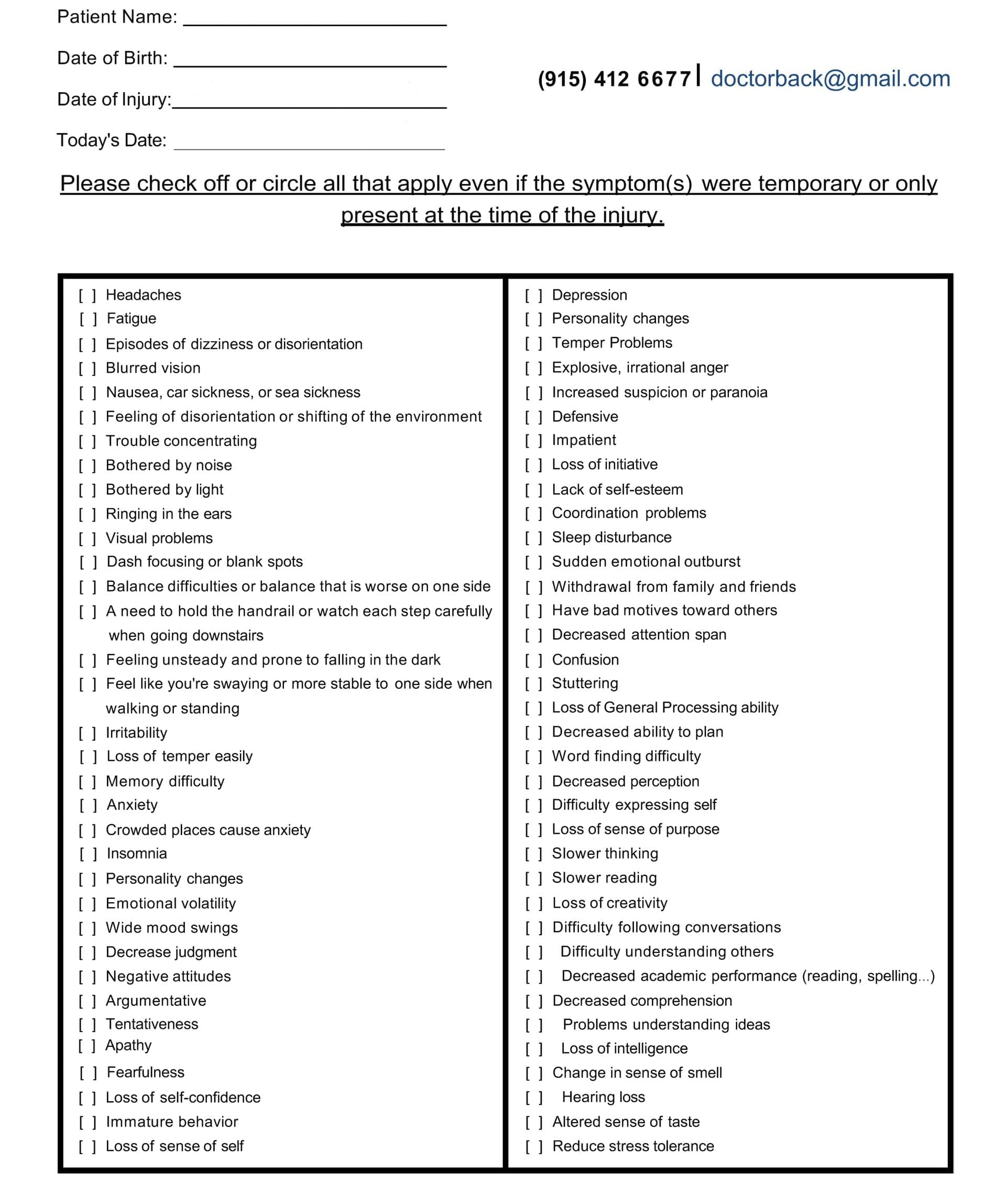
Head Injury/TBI Symptom Questionnaire:
Head Injury/TBI Symptom Questionnaire
Misalignment and Muscle Impairment: Irritating the Sciatic Nerve
Head trauma often hits the neck hard. The force whips the head forward and back—like in a car crash. This misaligns the upper spine, particularly the top vertebrae, known as the atlas and axis. That misalignment travels down like a domino fall.
Impaired muscles from brain signals exacerbate the condition. Tight neck muscles pull the spine off-kilter. In the lower back, this squeezes discs and nerves. The sciatic nerve can become pinched between bones or become inflamed. Result? That classic leg zap.
Research backs this. Up to 8% of severe TBI cases come with spine injuries. Even mild ones raise the risk. A study on 180 patients showed that older folks or those with low consciousness scores face higher odds. The neck shift stresses the whole chain, irritating the sciatic nerve.
Concussions alone can spark lower back pain. The brain’s balance center gets knocked. Muscles overwork to compensate, tiring the back. Dr. Jimenez refers to this as the “cascade effect” in his LinkedIn posts. He treats it with targeted adjustments to reset muscle tone.
Raising the Risk of Further Spinal Damage
Head trauma doesn’t just irritate—it invites more trouble. A damaged brain means slower reflexes. You might stumble more easily, leading to falls that jar the spine again. Plus, inflammation from TBI spreads. It swells the tissues around the spine, causing the discs to bulge and the nerves to become vulnerable.
One key risk: Concomitant injuries. That’s when head and spine hits happen together. In motor vehicle crashes—the top TBI cause—neck strains often tag along. This doubles the chance of disc slips that pinch the sciatic nerve.
Dr. Jimenez observes this in patients involved in car accidents. “A rear-end collision jars the brain and twists the lumbar spine,” he explains in his functional medicine series. His observations show early chiropractic checks cut re-injury risks by improving stability.
The Role of Swelling and Heterotopic Ossification in Nerve Crushing
TBI triggers swelling fast. Brain tissue bruises, and fluids build up. This chaos can spread to the body. In rare but serious cases, it leads to heterotopic ossification (HO). That’s when bone grows in soft tissues—like muscles or around nerves.
Around the sciatic nerve, HO is sneaky. It starts after hip or pelvic trauma, tied to the head hit. Scar tissue hardens into bone, encasing the nerve. Over months, this crushes it. Symptoms creep in: Numbness, weakness, foot drop.
A case report described a young man following traumatic brain injury (TBI). Seventeen months later, bilateral sciatic entrapment from hip HO caused severe pain. Surgery freed the nerve, but prevention is key. Anti-inflammatory drugs or radiation cut HO risks.
Dr. Jimenez warns of this in his injury recovery blogs. He uses imaging to identify early signs of HO in TBI patients with leg pain. His integrative plans include nutrition to fight inflammation and slow bone overgrowth.
Upper Neck Misalignment: Starting a Pain Cascade to the Sciatic Nerve
The upper neck is ground zero for many head traumas. Whiplash from falls or sports bends it unnaturally. This throws off the atlas—the top bone. It shifts pressure down the spine.
The cascade? Misaligned neck pinches nerves there. Signals to the lower back get scrambled. Muscles tighten unevenly, pulling on the lumbar joints. This stresses the sciatic nerve roots, causing inflammation and pain.
Inflammation plays a big role. Concussion swelling in the neck disrupts blood flow and nerve signals, causing significant complications. It causes the brain to misread pain, amplifying the sensation of hurt in the leg.
Dr. Jimenez’s clinical notes highlight this in veterans with whiplash-TBI. “Neck shifts create a domino pain chain,” he says. His team uses precise X-rays to map it, then adjusts to break the cycle.
Integrative Chiropractic: A Path to Relief and Recovery
Integrative chiropractic blends hands-on care with wellness tools. It’s perfect for post-head injury sciatica. No drugs or surgery—just realignment and support.
First, it realigns the spine. Gentle adjustments fix neck and back shifts. This eases nerve pressure fast. For sciatica, lumbar tweaks reduce the disc bulge on the nerve.
Second, it boosts nervous system function. Adjustments reduce interference, allowing brain signals to flow more smoothly. This calms spastic muscles and dials down pain sensitivity.
Third, it fights inflammation. Soft tissue work, like massage, releases tight spots. Add nutrition advice, including anti-inflammatory foods, and use swelling drops.
Finally, it restores cerebrospinal fluid (CSF) flow. CSF cushions the brain and spine. Trauma clogs it, raising pressure. Craniosacral therapy—light touches on the skull and sacrum—clears the path. Patients report clearer heads and less pain.
Dr. Jimenez integrates all this. His clinic mixes adjustments with functional tests. “We trace sciatica back to the head hit, then rebuild from there,” he observes. Patients who have been in accidents often experience mobility gains within weeks. One testimonial: A crash survivor ditched leg braces after targeted care.
Studies agree. Chiropractic reduces TBI pain by 50% in some individuals. For post-concussion, it eases dizziness and back aches.
Real-Life Stories and Expert Tips
Take Sarah, a soccer player Dr. Jimenez treated. A header caused a concussion and later sciatica. Adjustments realigned her neck, easing leg pain. Now she plays pain-free.
Tips from experts: Start care early. Get imaging if pain lingers post-injury. Pair chiropractic care with rest and omega-3 fatty acids for managing inflammation.
Wrapping It Up: Take Control of Your Recovery
Head trauma to sciatica seems far-fetched, but the links are strong. From brain glitches to bone growth, it stresses the sciatic nerve. Integrative chiropractic offers hope—realigning, calming, and healing.
Don’t ignore the signs. See a pro like Dr. Jimenez for a check. Your body can bounce back stronger.
Overcoming Sciatic Nerve Pain: Expert Insights from El Paso Back Clinic® in El Paso, TX
At El Paso Back Clinic®, located in the heart of El Paso, Texas, we specialize in helping people get back to their active lives without the constant burden of pain. Led by Dr. Alexander Jimenez, DC, APRN, FNP-BC, our team combines chiropractic care with modern wellness strategies to treat conditions like sciatic nerve issues. Whether you’re dealing with sharp leg pain from a work injury or numbness after a car accident, our clinic offers personalized plans to ease your symptoms and promote long-term health. We focus on non-invasive methods that address the root causes, not just the signs. If you’re in El Paso and searching for effective sciatica relief, our integrated approach could be the key to feeling better.
Sciatica isn’t just a back problem—it’s a nerve issue that can disrupt your daily routine. The sciatic nerve, which is like a thick cable running from your lower spine down each leg, gets irritated or squeezed, causing discomfort that travels far. At our clinic, we’ve seen how this affects everyone from athletes to office workers. In this guide, we’ll dive into what sciatica really is, why it happens, and how our team at El Paso Back Clinic® uses proven techniques to help. We’ll cover the physical side of nerve damage, common triggers, signs to watch for, and recovery steps. Plus, we’ll share how our chiropractic integrative care stands out in treating these issues right here in El Paso.
The Basics of the Sciatic Nerve and How Pressure Affects It
The sciatic nerve is your body’s main pathway for signals between the brain and legs. It begins at the lower back, where several nerve roots join, then branches through the buttocks and down to the feet. This nerve handles movement in your hamstrings, calves, and feet, as well as sensation in those areas. When something, like a slipped disk or tight muscle, presses on it, problems start.
When the sciatic nerve is compressed, pinched, or crushed, it suffers physical damage that disrupts its ability to transmit signals, leading to pain, numbness, and muscle weakness. The severity of the physical changes depends on the nature and duration of the pressure. Inside the nerve, axons carry messages, protected by myelin sheaths for fast travel. Pressure squishes these, causing swelling and blocking blood flow, which starves cells of oxygen—a state called ischemia (Verywell Health, 2023). In mild squeezes, like poor posture, the myelin gets worn but regrows. But harder crushes break axons, triggering degeneration where the nerve falls apart below the spot (Menorca et al., 2013).
Think of it like a garden hose: a light kink slows water, but a stomp cuts it off. Short pressure might cause temporary numbness, but ongoing force leads to scarring and chronic issues. At El Paso Back Clinic®, we use advanced checks to spot these changes early, helping prevent lasting harm.
Recognizing the Signs of Sciatic Nerve Trouble
Sciatica shows up in ways that can sneak up on you. The classic sign is radiating pain—starting in the lower back and zipping down one leg like an electric shock. It might burn, tingle, or feel sharp, worse when you sit, stand, or sneeze (Penn Medicine, n.d.). Numbness follows, making parts of your leg feel asleep or prickly. Weakness hits muscles, causing limps or trouble with stairs (Align Wellness Center, n.d.).
In our El Paso clinic, patients often describe it as a “leg giving out” or a constant ache. Severe cases bring muscle shrinking or even bladder issues if nerves are badly pinched (ADR Spine, n.d.). We see this in folks from all walks—drivers with long hauls, athletes pushing limits, or those in desk jobs. Early signs? Pay attention to one-sided pain that doesn’t fade with rest.
What Causes Sciatic Nerve Compression in Everyday Life
Life in El Paso means active days, but that can lead to sciatica triggers. A herniated disk, where the cushy part bulges and presses on the spinal cord roots, is common from lifting heavy boxes at work (Mayo Clinic, 2023). Aging narrows the spine in stenosis, squeezing nerves (Physio Pretoria, n.d.). Tight piriformis muscles in the butt can pinch the sciatic nerve, too, especially in runners or cyclists.
Accidents amp it up—car crashes crush nerves directly (MedStar Health, n.d.). Bone growths from arthritis add pressure, and extra weight strains everything (Advanced Orthopaedics & Sports Medicine, n.d.). Double crush? That’s when two spots squeeze, such as the back and leg, worsening the condition (Southwest Regional Wound Care Center, n.d.). At our clinic, we trace these in locals from border commutes to sports fields.
How We Diagnose Sciatica at El Paso Back Clinic®
Getting the right diagnosis is step one to relief. At our El Paso locations, we start with a full chat about your history—when the pain started and what makes it worse. Then, during hands-on exams, we test reflexes, strength, and perform the leg raise to pinpoint nerve irritation (Penn Medicine, n.d.).
We use top tools like MRI for disk views, EMG for signal checks, and X-rays for bones (ICliniq, 2023). Dr. Jimenez’s dual training as a chiropractor and nurse practitioner allows for a dual-scope diagnosis—blending medical tests with chiropractic insights for a full picture (Jimenez, n.d.). This helps link injuries to symptoms accurately, especially in complex cases from MVAs or sports.
Physical Damage from Compression: A Closer Look
Diving deeper, compression physically alters the nerve. Mild pressure causes neurapraxia: myelin dents, slowing signals without axon breaks. You feel weak but recover fast (Menorca et al., 2013). Stronger pinches lead to axonotmesis: axons snap, walls degenerate, and swelling builds. Healing takes time as new growth crawls along (Horton Mendez, n.d.).
Crushes bring neurotmesis: full sever, with scars blocking regrowth (Bhatia, 2023). Pressure cuts off blood, causing ischemia and cell death (Verywell Health, 2023). Chronic? Fibrosis hardens tissue (Mackinnon, 1998). Our clinic spots these via imaging, guiding treatments to reduce pressure and aid repair.
Integrative Chiropractic Care: Our Approach at El Paso Back Clinic®
We believe in whole-body healing. Our chiropractic integrative care combines spinal adjustments, soft tissue work, and exercises to correct alignment, enhance muscle function, and improve nerve signals. Adjustments ease pressure on the sciatic nerve, while massage loosens tight spots (AMTA, n.d.). Rehab builds strength to prevent repeats (Byington, n.d.).
This holistic method tackles pain now and builds stability for tomorrow. We add nutrition and stress tips, as gut health and emotions affect recovery. For El Paso folks, it’s about getting back to hikes or family time pain-free.
Dr. Alexander Jimenez’s Clinical Observations and Clinic Breakdown
With over 30 years, Dr. Jimenez brings unique insights. His dual credentials allow clinical correlations: linking injuries to assessments via advanced imaging like MRI for neuromusculoskeletal views (LinkedIn, n.d.). We treat work strains, sports sprains, personal slips, and MVAs with tailored plans.
Our process: Dual-scope diagnosis merges chiropractic and medical for thorough checks. Treatments include adjustments, non-surgical decompression, acupuncture, and functional medicine. We handle legal documents for accident claims, ensuring smooth care (Jimenez, n.d.). Observations? Stress worsens posture, and gut issues slow healing—we address all.
Recovery and Prevention Tips from Our El Paso Team
Recovery varies: Mild cases heal in weeks with rest and therapy, while severe cases require months (ADR Spine, n.d.). We guide with exercises, avoiding surgery when possible.
Prevent? Good posture, regular moves, and weight control. At our clinic, we teach these in sessions.
In El Paso, sciatica doesn’t have to hold you back. El Paso Back Clinic® offers expert, local care to restore your life.
IFM's Find A Practitioner tool is the largest referral network in Functional Medicine, created to help patients locate Functional Medicine practitioners anywhere in the world. IFM Certified Practitioners are listed first in the search results, given their extensive education in Functional Medicine