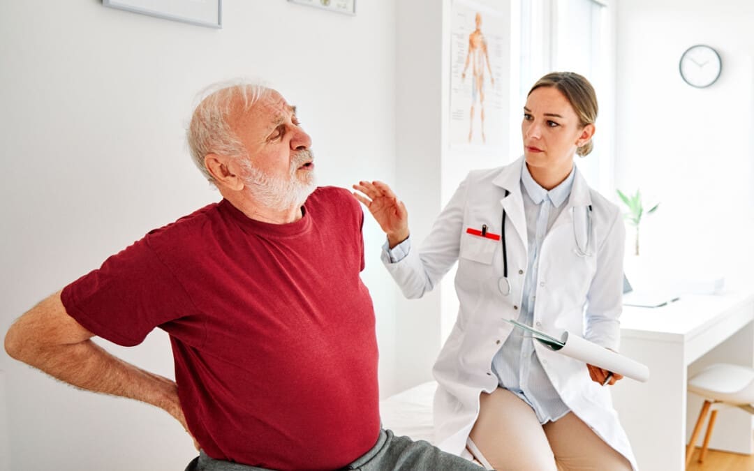
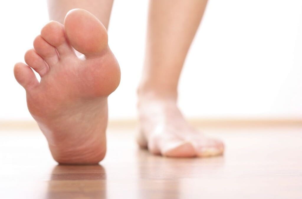
For individuals considering acupuncture for sciatica relief and management, can knowing how it works and what to expect during a session help in making the decision?
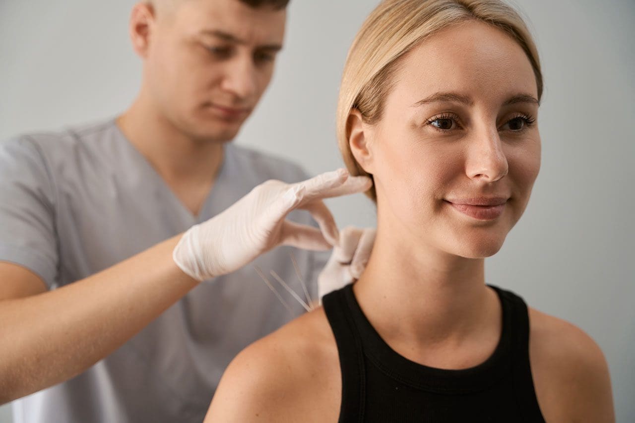
Acupuncture Sciatica Treatment Session
Acupuncture for sciatica is a safe and effective medical treatment to relieve and manage pain symptoms. Studies suggest it is as effective as other treatment strategies and causes fewer side effects. (Zhihui Zhang et al., 2023) The frequency of acupuncture to relieve sciatica pain depends on the severity of the condition and injury, but many report improvement within two to three weeks. (Fang-Ting Yu et al., 2022)
Needle Placement
Circulation problems can cause the body’s energy to stagnate in one or more meridians/channels, leading to pain in and around the surrounding area. (Wei-Bo Zhang et al., 2018)
The objective of acupuncture is to restore optimal circulation by stimulating specific points in the body called acupoints.
Thin, sterile needles stimulate the acupoints to activate the body’s natural healing abilities and relieve pain. (Heming Zhu 2014)
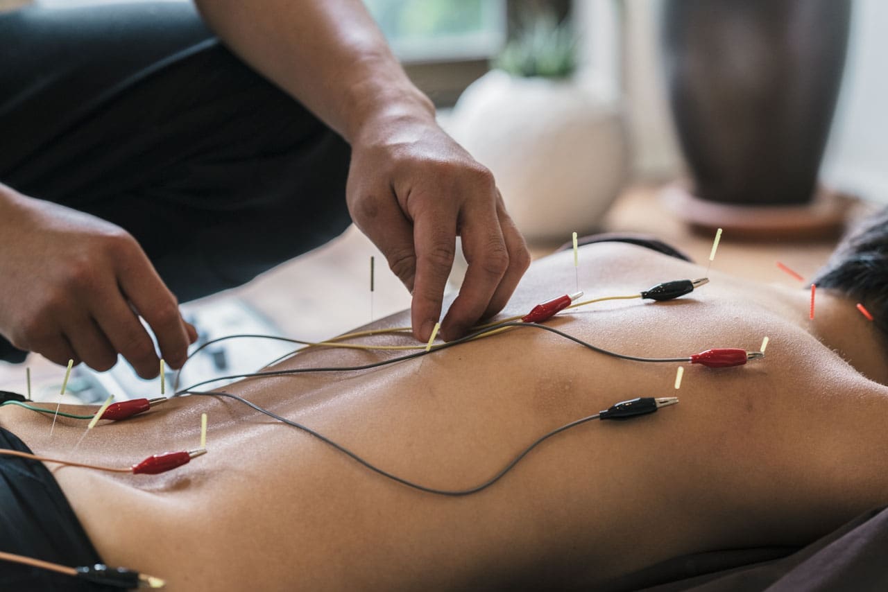
Some practitioners use electroacupuncture – a gentle, mild electrical current is applied to the needles and passes through the tissues to activate the nervous system. (Ruixin Zhang et al., 2014)
Acupoints
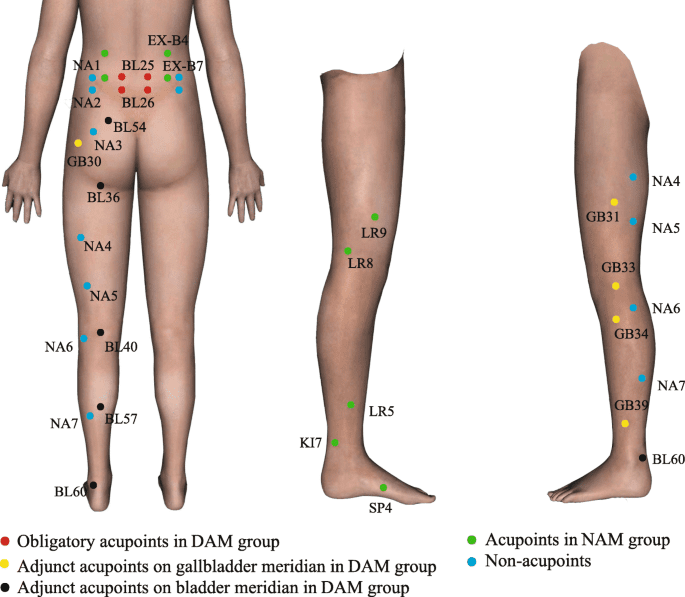
Acupuncture sciatica treatment involves specific acupoints along the bladder and gallbladder meridians.
Bladder Meridian – BL
The bladder meridian/BL runs down the back along the spine, hips, and legs. The acupoints within the meridian for sciatica include: (Fang-Ting Yu et al., 2022)
BL 23 -Shenshu – Location on the lower back, near the kidney.
BL 25 – Dachangshu – Location on the lower back.
BL 36 – Chengfu – Location on the back of the thigh, just below the buttocks.
GB 30 – Huantiao – Location on the back, where the buttocks meet the hips.
GB 34 – Yanglingquan – Location on the outside of the leg, below the knee.
GB 33 – Xiyangguan – Location lateral to the knee, on the side.
Stimulating acupoints in these meridians increases blood flow to the area, reduces inflammation, and releases endorphins and other pain-relieving neurochemicals to relieve symptoms. (Ningcen Li et al., 2021) The specific acupoints vary depending on symptoms and the root cause. (Tiaw-Kee Lim et al., 2018)
Example Patient
An example of acupuncture sciatica treatment session: A patient with persistent shooting pain extending down the back and side of the leg. A standard treatment consists of the following:
The acupuncturist thoroughly goes over the patient’s medical history and symptoms and has the patient point to where the pain is located.
Then, they palpate on and around the area to find where the pain worsens and lessens, communicating with the patient as they go along.
Depending on the site and severity, they may start placing needles at the lower back, focusing on the site of the injury.
Sometimes, the sacrum is involved, so the acupuncturist will place needles on those acupoints.
They then move to the back of the leg and insert needles.
The needles are retained for 20-30 minutes.
The acupuncturist leaves the room or treatment area but regularly checks in.
The patient may feel a warmth, tingling, or mild heaviness, which is a normal response. This is where patients report a calming effect. (Shilpadevi Patil et al., 2016)
The needles are carefully removed.
The patient may feel deeply relaxed and will be advised to get up slowly to avoid dizziness.
There may be soreness, redness, or bruising at the needle insertion site, which is normal and should resolve quickly.
The patient will be given recommendations as to avoiding strenuous activity, properly hydrating, and performing gentle stretches.
Acupuncture Benefits
Acupuncture has been shown to be a complementary therapy for pain relief and management. The benefits of acupuncture:
Improves Circulation
Acupuncture stimulates blood circulation, which nourishes damaged or irritated nerves and promotes healing.
This helps relieve sciatica symptoms, like numbness, tingling, and pain. (Song-Yi Kim et al., 2016)
Releases Endorphins
Acupuncture triggers the release of endorphins and other natural pain-relieving chemicals, which help relieve pain. (Shilpadevi Patil et al., 2016)
Regulates the Nervous System
Acupuncture rebalances the sympathetic and parasympathetic responses, which reduces stress, tension, and pain. (Xin Ma et al., 2022)
Relaxes the Muscles
Nerve pain often accompanies muscle tension and spasms.
Acupuncture relaxes tight muscles, reducing pressure and providing relief. (Zhihui Zhang et al., 2023)
From Symptoms to Solutions
References
Zhang, Z., Hu, T., Huang, P., Yang, M., Huang, Z., Xia, Y., Zhang, X., Zhang, X., & Ni, G. (2023). The efficacy and safety of acupuncture therapy for sciatica: A systematic review and meta-analysis of randomized controlled trails. Frontiers in neuroscience, 17, 1097830. https://doi.org/10.3389/fnins.2023.1097830
Yu, F. T., Liu, C. Z., Ni, G. X., Cai, G. W., Liu, Z. S., Zhou, X. Q., Ma, C. Y., Meng, X. L., Tu, J. F., Li, H. W., Yang, J. W., Yan, S. Y., Fu, H. Y., Xu, W. T., Li, J., Xiang, H. C., Sun, T. H., Zhang, B., Li, M. H., Wan, W. J., … Wang, L. Q. (2022). Acupuncture for chronic sciatica: protocol for a multicenter randomised controlled trial. BMJ open, 12(5), e054566. https://doi.org/10.1136/bmjopen-2021-054566
Zhang, W. B., Jia, D. X., Li, H. Y., Wei, Y. L., Yan, H., Zhao, P. N., Gu, F. F., Wang, G. J., & Wang, Y. P. (2018). Understanding Qi Running in the Meridians as Interstitial Fluid Flowing via Interstitial Space of Low Hydraulic Resistance. Chinese journal of integrative medicine, 24(4), 304–307. https://doi.org/10.1007/s11655-017-2791-3
Zhu H. (2014). Acupoints Initiate the Healing Process. Medical acupuncture, 26(5), 264–270. https://doi.org/10.1089/acu.2014.1057
Zhang, R., Lao, L., Ren, K., & Berman, B. M. (2014). Mechanisms of acupuncture-electroacupuncture on persistent pain. Anesthesiology, 120(2), 482–503. https://doi.org/10.1097/ALN.0000000000000101
Perreault, T., Fernández-de-Las-Peñas, C., Cummings, M., & Gendron, B. C. (2021). Needling Interventions for Sciatica: Choosing Methods Based on Neuropathic Pain Mechanisms-A Scoping Review. Journal of clinical medicine, 10(10), 2189. https://doi.org/10.3390/jcm10102189
Li, N., Guo, Y., Gong, Y., Zhang, Y., Fan, W., Yao, K., Chen, Z., Dou, B., Lin, X., Chen, B., Chen, Z., Xu, Z., & Lyu, Z. (2021). The Anti-Inflammatory Actions and Mechanisms of Acupuncture from Acupoint to Target Organs via Neuro-Immune Regulation. Journal of inflammation research, 14, 7191–7224. https://doi.org/10.2147/JIR.S341581
Lim, T. K., Ma, Y., Berger, F., & Litscher, G. (2018). Acupuncture and Neural Mechanism in the Management of Low Back Pain-An Update. Medicines (Basel, Switzerland), 5(3), 63. https://doi.org/10.3390/medicines5030063
Kim, S. Y., Min, S., Lee, H., Cheon, S., Zhang, X., Park, J. Y., Song, T. J., & Park, H. J. (2016). Changes of Local Blood Flow in Response to Acupuncture Stimulation: A Systematic Review. Evidence-based complementary and alternative medicine : eCAM, 2016, 9874207. https://doi.org/10.1155/2016/9874207
Patil, S., Sen, S., Bral, M., Reddy, S., Bradley, K. K., Cornett, E. M., Fox, C. J., & Kaye, A. D. (2016). The Role of Acupuncture in Pain Management. Current pain and headache reports, 20(4), 22. https://doi.org/10.1007/s11916-016-0552-1
Ma, X., Chen, W., Yang, N. N., Wang, L., Hao, X. W., Tan, C. X., Li, H. P., & Liu, C. Z. (2022). Potential mechanisms of acupuncture for neuropathic pain based on somatosensory system. Frontiers in neuroscience, 16, 940343. https://doi.org/10.3389/fnins.2022.940343
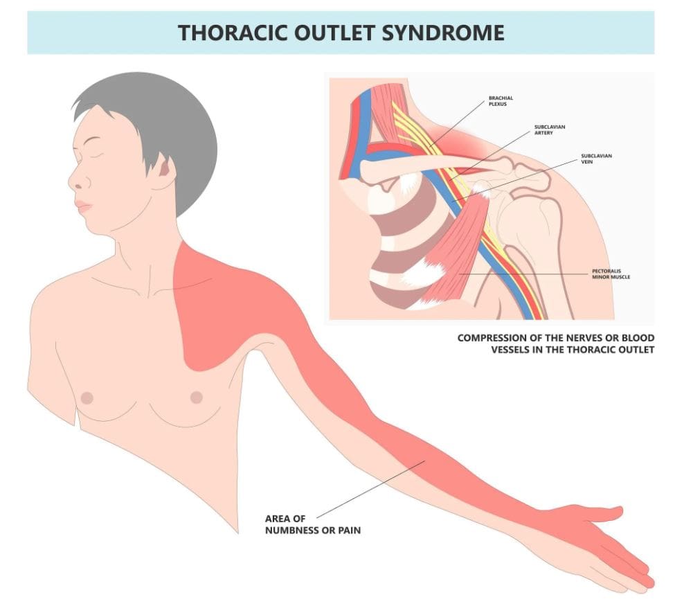
Can individuals with thoracic outlet syndrome incorporate electroacupuncture to reduce neck pain and restore proper posture?
Introduction
More times throughout the world, many individuals have experienced pain around their necks, which can lead to pain and discomfort. Many environmental factors, like being in a hunched position while looking at the computer or phone, traumatic injuries, poor posture, or spinal issues, can cause pain-like symptoms and complications to the body. Since neck pain is a common complaint many people suffer, symptoms like tingling, numbness, or muscle weakness in the upper extremities can lead to comorbidities. When this happens, it can lead to the development of a complex condition known as thoracic outlet syndrome or TOS. Today’s article looks at the link between thoracic outlet syndrome and neck pain, how to manage TOS while alleviating neck pain, and how electroacupuncture can help with TOS. We talk with certified medical providers who consolidate our patients’ information to assess how to minimize the effects of TOS while reducing neck pain. We also inform and guide patients on how electroacupuncture can help manage TOS. We encourage our patients to ask their associated medical providers intricate and important questions about incorporating electroacupuncture to alleviate TOS associated with the neck. Dr. Jimenez, D.C., includes this information as an academic service. Disclaimer.
The Link Between Thoracic Outlet Syndrome & Neck Pain
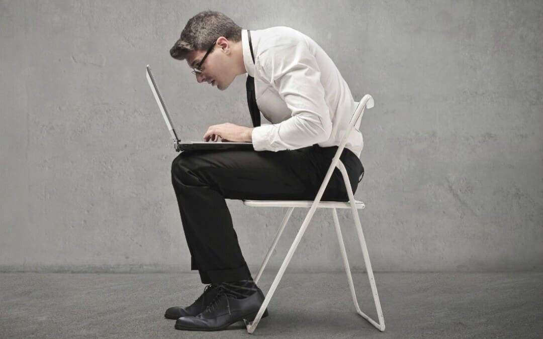
Have you been noticing how you are hunched over more than usual? Do you experience symptoms of tingling or numbness down from your arms to your hands? Or do you feel muscle tension in your neck? Thoracic outlet syndrome, or TOS, is a challenging condition resulting in the compression of neurovascular structures between the clavicle and the first rib. (Masocatto et al., 2019) These neurovascular structures are near the neck and shoulders. When environmental structures affect the upper extremities, it can lead to referred neck pain, which can cause overlapping risk profiles. Some of the factors that TOS can contribute to neck pain include:
Atomical variations
Poor posture
Repetitive motions
Traumatic injuries
At the same time, people with neck pain can develop TOS, as neck pain is a multifactorial musculoskeletal condition that can be associated with overlapping risk profiles that contribute to TOS. (Kazeminasab et al., 2022) As stated earlier, factors like poor posture can overstretch the neck muscles and the neurovascular structures, leading to neuropathic pain symptoms that can cause deep aching referred pain to the neck and muscle weakness. (Childress & Stuek, 2020) When this happens, many people will begin to feel miserable and start to seek treatment to not only reduce TOS but also alleviate neck pain.
What Is Thoracic Outlet Syndrome- Video
Managing TOS & Alleviating Neck Pain
When it comes to treating TOS, especially when neck pain is a significant component, many individuals will try to seek out non-surgical treatments to reduce the symptoms. Many individuals may try physical therapy to stretch and strengthen their shoulder, chest, and neck muscles to relieve compression. Others might try a manual treatment that is joint-oriented for the neck while neural-tissue-oriented for TOS to improve mobilization on the upper extremities and even improve poor posture. (Kuligowski et al., 2021) Additionally, non-surgical treatments can be combined with other therapies to reduce the chances of TOS from returning as they can further increase sensory-motor function back to the neck and upper extremities. (Borrella-Andres et al., 2021)
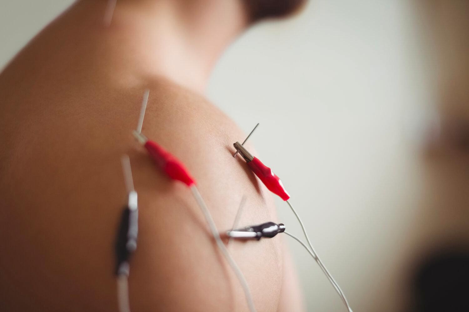
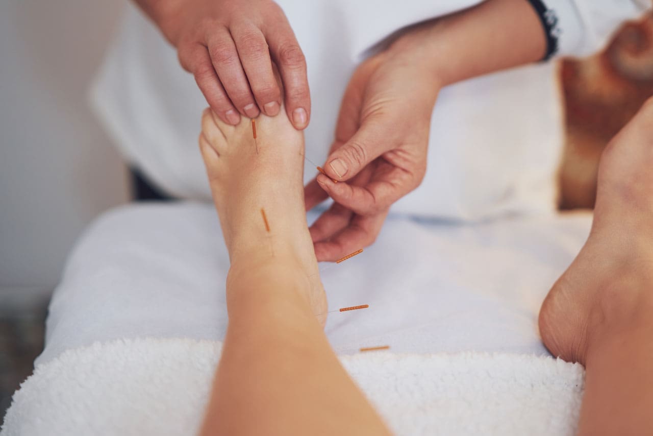
How Electroacupuncture Can Help With TOS
Electroacupuncture is a modern form of traditional acupuncture that is part of the non-surgical treatments that can help manage TOS while alleviating neck pain. Electroacupuncture is a modification of inserting needles into the body’s acupoints while incorporating electric stimulation to deliver a pulsed electrical current to the affected area gently. (Zhang et al., 2022) Some of the beneficial properties that electrostimulation can provide for TOS include:
Pain reduction by stimulating the release of endorphins to decrease inflammation.
Help relax the affected muscles in the chest and neck to alleviate the pressure on the nerves of the thoracic outlet.
Help enhance the blood flow to reduce vascular compression of TOS.
Help stimulate the nerve pathway to promote healthy nerve function and reduce pain-like symptoms.
By incorporating electroacupuncture and non-surgical treatments to reduce TOS, many individuals can make modifications to their lifestyle habits and prevent issues from affecting their upper body extremities. By utilizing these treatments, many people can listen to their bodies and focus on their health and well-being by addressing the pain-like symptoms they are experiencing from TOS correlating with neck pain. At the same time, they have a positive relationship with their primary doctors to develop a personalized treatment plan that can manage their TOS symptoms to the best outcomes.
References
Borrella-Andres, S., Marques-Garcia, I., Lucha-Lopez, M. O., Fanlo-Mazas, P., Hernandez-Secorun, M., Perez-Bellmunt, A., Tricas-Moreno, J. M., & Hidalgo-Garcia, C. (2021). Manual Therapy as a Management of Cervical Radiculopathy: A Systematic Review. Biomed Res Int, 2021, 9936981. https://doi.org/10.1155/2021/9936981
Kazeminasab, S., Nejadghaderi, S. A., Amiri, P., Pourfathi, H., Araj-Khodaei, M., Sullman, M. J. M., Kolahi, A. A., & Safiri, S. (2022). Neck pain: global epidemiology, trends and risk factors. BMC Musculoskelet Disord, 23(1), 26. https://doi.org/10.1186/s12891-021-04957-4
Kuligowski, T., Skrzek, A., & Cieslik, B. (2021). Manual Therapy in Cervical and Lumbar Radiculopathy: A Systematic Review of the Literature. Int J Environ Res Public Health, 18(11). https://doi.org/10.3390/ijerph18116176
Masocatto, N. O., Da-Matta, T., Prozzo, T. G., Couto, W. J., & Porfirio, G. (2019). Thoracic outlet syndrome: a narrative review. Rev Col Bras Cir, 46(5), e20192243. https://doi.org/10.1590/0100-6991e-20192243 (Sindrome do desfiladeiro toracico: uma revisao narrativa.)
Zhang, B., Shi, H., Cao, S., Xie, L., Ren, P., Wang, J., & Shi, B. (2022). Revealing the magic of acupuncture based on biological mechanisms: A literature review. Biosci Trends, 16(1), 73-90. https://doi.org/10.5582/bst.2022.01039
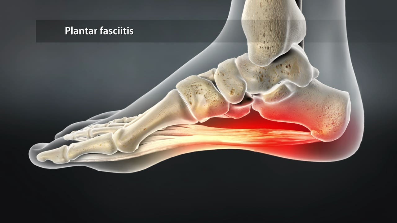
Can plantar fasciitis patients incorporate non-surgical treatments to reduce hip pain and restore mobility?
Introduction
Everyone is on their feet constantly as it helps people stay mobile and allows them to go from one location to another. Many people are constantly on their feet from childhood to adulthood. This is because the feet are part of the lower musculoskeletal extremities that stabilize the hips and allow sensory-motor function to the legs, thighs, and calves. The feet also have various muscles, tendons, and ligaments surrounding the skeletal structure to prevent pain and discomfort. However, when repetitive motions or injuries start to affect the feet, it can lead to plantar fasciitis and, over time, cause overlapping risk profiles that lead to hip pain. When people are experiencing these pain-like conditions, it can significantly affect their daily activities and overall quality of life. When this happens, many people seek various treatments to reduce the pain-like symptoms caused by plantar fasciitis and restore hip mobility. Today’s article looks at how plantar fasciitis correlates with hip pain, the connection between the feet and the hips, and how there are non-surgical solutions to reduce plantar fasciitis. We talk with certified medical providers who consolidate our patients’ information to assess how to mitigate plantar fasciitis and restore hip mobility. We also inform and guide patients on how numerous non-surgical treatments can help strengthen weak muscles associated with plantar fasciitis and help with restoring stabilization from hip pain. We encourage our patients to ask their associated medical providers intricate and important questions about incorporating small changes to reduce the pain-like effects caused by plantar fasciitis. Dr. Jimenez, D.C., includes this information as an academic service. Disclaimer.
How Plantar Fasciitis Correlates With Hip Pain
Do you experience pain in your heels constantly after a long walk? Do you feel stiffness in your hips when stretching? Or do you feel your shoes are causing tension and pain in your feet and calves? Often, many of these pain-like scenarios are due to people dealing with plantar fasciitis, characterized by heel pain due to inflammation or degenerative irritation of the plantar fascia, a band of thick tissues is running across the bottom of the foot and connecting to the heel bone to the toes in the lower extremities. This band of tissues plays an essential role in the body, providing normal biomechanics to the foot while supporting the arch and helping with shock absorption. (Buchanan et al., 2024) Plantar fasciitis can affect the stability of the lower extremities since the pain affects the feet and causes hip pain.
So, how would plantar fasciitis correlate with hip pain? With plantar fasciitis, many people are experiencing pain in their feet. It can lead to abnormal foot posture, lower extremity muscle weakness, and muscle stress that can reduce the stability of the legs and hip muscles. (Lee et al., 2022) With hip pain, many people can experience a gait dysfunction that causes muscle weakness in the lower extremities and causes the accessory muscles to perform the primary muscles’ jobs. To that point, this forces people to scrap the ground when walking. (Ahuja et al., 2020) This is because normal conditions like natural aging, muscle overuse, or trauma can cause pain-like symptoms to the hips, including discomfort on the thighs, groin, and buttock region, joint stiffness, and reduced range of motion. Hip pain can cause overlapping risk profiles that may include repetitive strain on the feet, thus leading to symptoms of sharp to dull aches on the heel.
The Connection Between The Feet and The Hips
It is important to understand that foot problems like plantar fasciitis can affect the hips and vice versa, as both body regions have a beautiful relationship within the musculoskeletal system. Plantar fasciitis on their feet can alter their gait function, potentially leading to hip pain over time. This is due to many environmental factors that can affect the hips and feet over time, leading to plantar fasciitis correlating with hip pain. From excessive weight-bearing activities to microtrauma in the hips or the plantar fascia, many people will often seek treatment to reduce the effects of plantar fasciitis correlated with hip pain by addressing how their range of motion is affecting the plantarflexion and their load on the force-absorbing plantar surface structures could be good starting points in the prevention and treatment of plantar fasciitis correlated with hip pain. (Hamstra-Wright et al., 2021)
What Is Plantar Fasciitis?-Video
Non-Surgical Solutions To Reduce Plantar Fasciitis
When it comes to reducing plantar fasciitis in the body, many individuals will seek non-surgical treatments that can alleviate the pain from plantar fascia. Non-surgical treatments are cost-effective and can reduce the pain from plantar fasciitis and its associated symptoms, like hip pain. Some of the benefits of non-surgical treatments are promising, as they have a low risk of complications, good accessibility, and even a high capacity to relieve the mechanical load on the plantar fascia when doing regular activities. (Schuitema et al., 2020) Some of the non-surgical treatments that many people can incorporate include:
Stretching exercises
Orthotic devices
Chiropractic care
Massage therapy
Acupuncture/electroacupuncture
Spinal decompression
These non-surgical treatments not only help reduce plantar fasciitis but also help alleviate hip pain. For example, spinal decompression can help restore hip mobility by stretching the lumbar spine and relieving the lower extremities from numbness while strengthening tight muscles. (Takagi et al., 2023). Electroacupuncture can stimulate the body’s acupoints to release endorphins from the lower extremities to reduce inflammation of the plantar fascia. (Wang et al., 2019) When people begin to make small changes in their routine, like wearing proper footwear and not carrying or lifting heavy weighted objects, it can go a long way to prevent plantar fasciitis and hip pain from reoccurring can go a long way. Having a personalized treatment plan can ensure many individuals seeking non-surgical treatments have a better outcome on their health and mobility while preventing long-term complications.
References
Ahuja, V., Thapa, D., Patial, S., Chander, A., & Ahuja, A. (2020). Chronic hip pain in adults: Current knowledge and future prospective. J Anaesthesiol Clin Pharmacol, 36(4), 450-457. https://doi.org/10.4103/joacp.JOACP_170_19
Hamstra-Wright, K. L., Huxel Bliven, K. C., Bay, R. C., & Aydemir, B. (2021). Risk Factors for Plantar Fasciitis in Physically Active Individuals: A Systematic Review and Meta-analysis. Sports Health, 13(3), 296-303. https://doi.org/10.1177/1941738120970976
Lee, J. H., Shin, K. H., Jung, T. S., & Jang, W. Y. (2022). Lower Extremity Muscle Performance and Foot Pressure in Patients Who Have Plantar Fasciitis with and without Flat Foot Posture. Int J Environ Res Public Health, 20(1). https://doi.org/10.3390/ijerph20010087
Schuitema, D., Greve, C., Postema, K., Dekker, R., & Hijmans, J. M. (2020). Effectiveness of Mechanical Treatment for Plantar Fasciitis: A Systematic Review. J Sport Rehabil, 29(5), 657-674. https://doi.org/10.1123/jsr.2019-0036
Takagi, Y., Yamada, H., Ebara, H., Hayashi, H., Inatani, H., Toyooka, K., Mori, A., Kitano, Y., Nakanami, A., Kagechika, K., Yahata, T., & Tsuchiya, H. (2023). Decompression for lumbar spinal stenosis at the intrathecal catheter insertion site during intrathecal baclofen therapy: a case report. J Med Case Rep, 17(1), 239. https://doi.org/10.1186/s13256-023-03959-1
Wang, W., Liu, Y., Zhao, J., Jiao, R., & Liu, Z. (2019). Electroacupuncture versus manual acupuncture in the treatment of plantar heel pain syndrome: study protocol for an upcoming randomised controlled trial. BMJ Open, 9(4), e026147. https://doi.org/10.1136/bmjopen-2018-026147
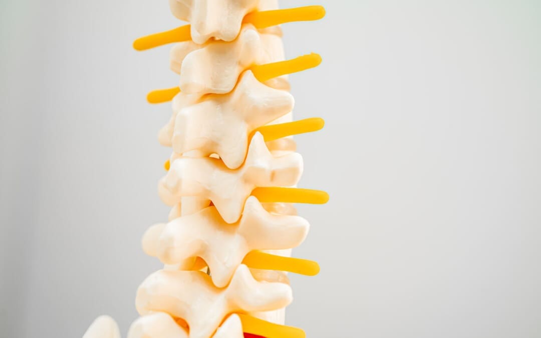
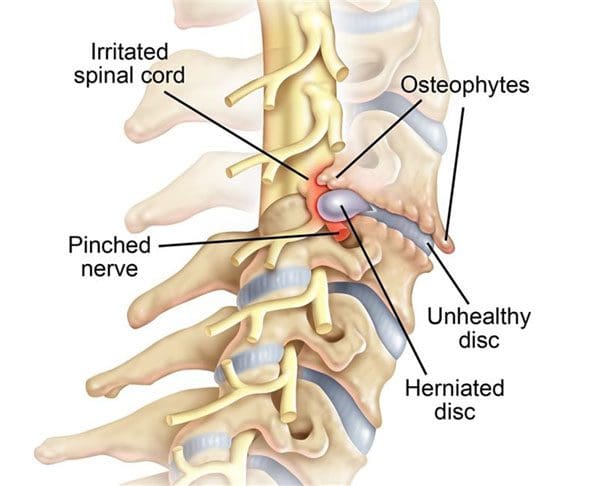
For individuals looking to improve their spinal health, can understanding the anatomy of the intervertebral foramen help in injury rehabilitation and prevention?
Intervertebral Foramen
The intervertebral foramen, aka neural foramen, is the opening between the vertebrae through which spinal nerve roots connect and exit to other body areas. If the foramina narrows, it can place added pressure on the nerve roots near and around them, causing pain symptoms and sensations. This is known as neuroforaminal stenosis. (Sumihisa Orita et al., 2016)
Anatomy
The vertebrae comprise the spinal column.
They protect and support the spinal cord and most of the weight placed on the spine.
Foramen is the singular form, and foramina is the plural form.
Structure
The body is the large, round part of the bone that makes up each vertebra.
The body of each vertebra is attached to a bony ring.
Stenosis can occur in the spinal canal, known as central canal stenosis, and the foramina.
Pain brought on by neuroforaminal spinal stenosis and arthritis-related bone growth/bone spurs/osteophytes that are present in one or more foramen rub against the nerve root that passes through the space, causing radicular pain.
Pain accompanied by other sensations, like tingling or numbness, is known as radiculopathy. (Young Kook Choi, 2019)
The main symptom is pain.
Numbness and/or tingling can present depending on the injury.
Neurogenic claudication occurs as a result of ischemia or a lack of blood circulation to the nerves and typically presents with a heaviness in the legs.
It is typically associated with central stenosis rather than foraminal stenosis.
Most individuals with spinal stenosis feel better when flexing or bending forward and worse when arching their backs.
Stenosis treatment aims to relieve pain and prevent nerve symptoms from occurring or worsening. Conservative treatments are recommended and can be highly effective.
These include:
Myelopathy in the neck and/or upper or mid-back (myelopathy symptoms are spinal cord related and occur in central canal stenosis) (Cleveland Clinic. 2021)
Intense incapacitating pain
Different surgical techniques include:
Decompression laminectomy – entails removing the buildup of bone in the spinal canal.
Spinal fusion – when there is instability of the spine or severe foraminal stenosis.
Orita, S., Inage, K., Eguchi, Y., Kubota, G., Aoki, Y., Nakamura, J., Matsuura, Y., Furuya, T., Koda, M., & Ohtori, S. (2016). Lumbar foraminal stenosis, the hidden stenosis including at L5/S1. European journal of orthopaedic surgery & traumatology : orthopedie traumatologie, 26(7), 685–693. https://doi.org/10.1007/s00590-016-1806-7
American Academy of Orthopaedic Surgeons. (2020). Spine Basics (OrthoInfo, Issue. https://orthoinfo.aaos.org/en/diseases–conditions/spine-basics/
American Academy of Orthopaedic Surgeons. (2021). Lumbar spinal stenosis (OrthoInfo, Issue. https://orthoinfo.aaos.org/en/diseases–conditions/lumbar-spinal-stenosis/
Choi Y. K. (2019). Lumbar foraminal neuropathy: an update on non-surgical management. The Korean journal of pain, 32(3), 147–159. https://doi.org/10.3344/kjp.2019.32.3.147
Lee, S. Y., Kim, T. H., Oh, J. K., Lee, S. J., & Park, M. S. (2015). Lumbar Stenosis: A Recent Update by Review of Literature. Asian spine journal, 9(5), 818–828. https://doi.org/10.4184/asj.2015.9.5.818
Lurie, J., & Tomkins-Lane, C. (2016). Management of lumbar spinal stenosis. BMJ (Clinical research ed.), 352, h6234. https://doi.org/10.1136/bmj.h6234
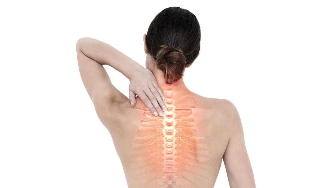
Can individuals with cervical spinal pain incorporate spinal decompression therapy to reduce neck pain and headaches?
Introduction
Many individuals deal with neck pain at some point, leading to many issues that can impact their daily lives. See, the neck is part of the cervical region of the musculoskeletal system. It is surrounded by muscles, soft tissues, and ligaments that protect the spinal cord while allowing the head to be mobile. Like back pain, neck pain is a common issue that causes pain and discomfort from associated environmental factors and traumatic injuries. When a person is dealing with neck pain, they are also coping with comorbidities that cause overlapping risk profiles like headaches and migraines. However, treatments like spinal decompression can help reduce cervical spinal pain affecting the neck and reduce the painful effects of headaches and migraines. Today’s article looks at the impact of cervical pain and headaches, how spinal decompression can reduce cervical spinal pain, and how it benefits from reducing headaches. We talk with certified medical providers who consolidate our patients’ information to assess how to mitigate cervical spinal pain from the neck. We also inform and guide patients on how spinal decompression can help reduce headaches caused by cervical spinal pain. We encourage our patients to ask their associated medical providers intricate and important questions about incorporating spinal decompression therapy as part of their routine to reduce headaches and migraines associated with the neck. Dr. Jimenez, D.C., includes this information as an academic service. Disclaimer.
The Effects Of Cervical Pain & Headaches
Do you feel stiffness on both sides of your neck that causes you limited mobility when you turn your neck? Have you experienced constant throbbing pain in your temples? Or do you feel muscle aches on your neck and shoulders from being hunched on the computer for an extended period? Many individuals dealing with these pain-like issues could be coping with cervical spinal pain. Various causes that can lead to the development of cervical spinal pain include herniated discs, pinched nerves, spinal stenosis, and muscle strain that originates from the neck region. This is because cervical spinal pain can be associated with environmental factors that can cause pain and discomfort, disability, and impaired quality of life as the surrounding neck muscles are overstretched and tight. (Ben Ayed et al., 2019) When people are dealing with cervical spinal pain, one of the symptoms it is associated with is headaches. This is because the intricated nerve pathways are connected to the neck and head. When cervical spinal pain is causing these issues, it can significantly impact a person’s daily body function as the pain is traveling upwards.
At the same time, neck pain is a multifactorial disease that can become a major issue worldwide. Like back pain, numerous risk factors can contribute to its development. (Kazeminasab et al., 2022) Some risk factors, like excessive phone usage, cause prolonged neck flexion to the neck and shoulders, causing static muscular loading with a lack of support to the upper extremities. (Al-Hadidi et al., 2019) To this point, environmental risk factors like excessive phone usage can make individuals develop a hunched position in their necks that can compress the spinal disc in the cervical region and aggravate the nerve roots to produce headaches and pain. However, many individuals have found ways to reduce cervical spinal pain and find pain relief from their headaches.
Home Exercises for Pain Relief-Video
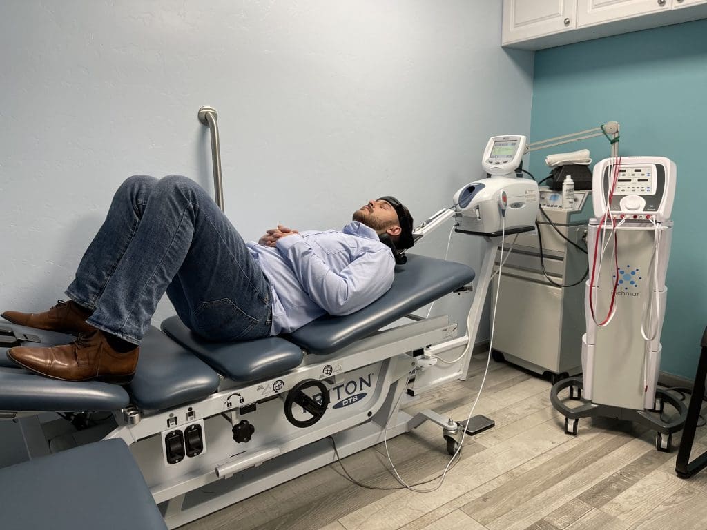
How Spinal Decompression Reduces Cervical Spinal Pain
When it comes to reducing cervical spinal pain, many individuals have experienced that spinal decompression can help mitigate the effects of cervical pain. Spinal decompression has increasingly been recognized as an effective non-surgical treatment when it comes to alleviating cervical spinal pain. What spinal decompression does is that it allows negative pressure on the cervical spine to relieve any herniated disc of the aggravated nerve roots and help improve neurological symptoms. (Kang et al., 2016) This is due to a person being strapped comfortably on a traction machine that gently stretches and decompresses the spinal vertebrae. Additionally, some of the benefits of spinal decompression for cervical spinal pain include:
Improved spinal alignment to reduce muscle strain on the neck muscles and joints.
Enhanced the body’s natural healing by increasing blood flow and nutrient exchange.
Increased neck mobility by decreasing muscle stiffness.
Reducing pain levels that are causing intense headaches.
The Benefits of Spinal Decompression For Headaches
Additionally, spinal decompression can help reduce headaches associated with cervical spinal pain as spinal decompression can be combined with other therapies like acupuncture and physical therapy to relieve the protruding spinal dice and stabilize within the annulus by spinal elongation. (Van Der Heijden et al., 1995) This is due to gentle traction on the neck that is causing the prolapsed disc to reposition itself while restoring disc height to minimize the pressure on the nerves. (Amjad et al., 2022) When a person is doing spinal decompression therapy consecutively, the pain-like effects of cervical spinal pain and the associated headaches begin to reduce over time, and many people will start to notice how their habits are in correlation with their pain. By incorporating spinal decompression therapy as part of their treatment, many people can make small changes in their routine and be more mindful of their bodies to prevent the progression of cervical spinal pain from returning.
References
Al-Hadidi, F., Bsisu, I., AlRyalat, S. A., Al-Zu’bi, B., Bsisu, R., Hamdan, M., Kanaan, T., Yasin, M., & Samarah, O. (2019). Association between mobile phone use and neck pain in university students: A cross-sectional study using numeric rating scale for evaluation of neck pain. PLOS ONE, 14(5), e0217231. https://doi.org/10.1371/journal.pone.0217231
Amjad, F., Mohseni-Bandpei, M. A., Gilani, S. A., Ahmad, A., & Hanif, A. (2022). Effects of non-surgical decompression therapy in addition to routine physical therapy on pain, range of motion, endurance, functional disability and quality of life versus routine physical therapy alone in patients with lumbar radiculopathy; a randomized controlled trial. BMC Musculoskelet Disord, 23(1), 255. https://doi.org/10.1186/s12891-022-05196-x
Ben Ayed, H., Yaich, S., Trigui, M., Ben Hmida, M., Ben Jemaa, M., Ammar, A., Jedidi, J., Karray, R., Feki, H., Mejdoub, Y., Kassis, M., & Damak, J. (2019). Prevalence, Risk Factors and Outcomes of Neck, Shoulders and Low-Back Pain in Secondary-School Children. J Res Health Sci, 19(1), e00440. https://www.ncbi.nlm.nih.gov/pubmed/31133629
Kang, J.-I., Jeong, D.-K., & Choi, H. (2016). Effect of spinal decompression on the lumbar muscle activity and disk height in patients with herniated intervertebral disk. Journal of Physical Therapy Science, 28(11), 3125-3130. https://doi.org/10.1589/jpts.28.3125
Kazeminasab, S., Nejadghaderi, S. A., Amiri, P., Pourfathi, H., Araj-Khodaei, M., Sullman, M. J. M., Kolahi, A. A., & Safiri, S. (2022). Neck pain: global epidemiology, trends and risk factors. BMC Musculoskelet Disord, 23(1), 26. https://doi.org/10.1186/s12891-021-04957-4
Van Der Heijden, G. J., Beurskens, A. J., Koes, B. W., Assendelft, W. J., De Vet, H. C., & Bouter, L. M. (1995). The Efficacy of Traction for Back and Neck Pain: A Systematic, Blinded Review of Randomized Clinical Trial Methods. Physical Therapy, 75(2), 93-104. https://doi.org/10.1093/ptj/75.2.93
For individuals dealing with musculoskeletal pain, can incorporating acupuncture and electroacupuncture therapy provide beneficial results?
Introduction
The upper and lower body quadrants are surrounded by muscles, soft tissues, and ligaments that allow the body to be mobile with feelings of pain or discomfort. Each muscle group has an important job providing sensory-motor functions like grasping objects, moving extremities, supporting the body in a correct posture, and stabilizing vertical axial weight. However, many people have adopted various habits from environmental factors or have been through traumatic injuries that can cause referred muscle pain in the upper and lower body quadrants. When this happens, it can lead to a life of disability, pain, and discomfort over time if it is not treated right away. To that point, musculoskeletal pain can also cause overlapping risk profiles with other comorbidities that can be pre-existing in the body. Fortunately, numerous treatments can help reduce musculoskeletal pain and benefit the body. Today’s article looks at two different non-surgical therapies, how each is beneficial to reducing musculoskeletal pain, and how effective they can help many people with musculoskeletal pain. We talk with certified medical providers who consolidate our patients’ information to assess how to reduce the pain-like effects of musculoskeletal pain with non-surgical treatments. We also guide patients on how these non-surgical treatments can help lessen the referred pain caused by various environmental factors affecting their musculoskeletal system. We encourage our patients to ask their associated medical providers intricate and important questions about incorporating non-surgical treatments into their health and wellness treatments. Dr. Jimenez, D.C., includes this information as an academic service. Disclaimer.
The Traditional Touch Of Acupuncture
After a long workday, do you feel soreness in your arms, legs, or feet? Have you experienced any symptoms of numbness or stiffness in the upper or lower portions of your body? Or do you feel muscle aches and pains after waking up in the morning? Around the world, many individuals have dealt with musculoskeletal pain at some point, which causes many people to miss out on numerous activities. Musculoskeletal pain is a multifactorial condition that any individual can develop over time. Some biological mechanisms contributing to the development of musculoskeletal pain can be heterogeneous, cardiometabolic, and systemic inflammation that can affect the body. (Dzakpasu et al., 2021) When many people are doing repetitive motions or have dealt with injuries, it can cause the various muscles to be overstretched, tightened, or weak, which can cause individuals to feel miserable and seek treatment. When people go to get treatment for their musculoskeletal pain, many people will tell their doctors about their pain experience and how it impacts their daily social well-being. By gaining information about how musculoskeletal pain negatively affects their lives, a multidisciplinary approach to pain management that emphasizes rehabilitation and non-surgical treatments can be the first step in effectively managing musculoskeletal pain. (Welsh et al., 2020)
Now, non-surgical treatments vary depending on the severity of musculoskeletal pain the person is experiencing. Since musculoskeletal pain is a multifactorial condition, many people could experience comorbidities that cause overlapping risk profiles that correlate with musculoskeletal pain, hence why many people incorporate non-surgical treatments since it is affordable and can be combined with other treatments. One of the oldest therapies that is still practiced today is acupuncture. Now, acupuncture involves the insertion of thin, solid needles into the body’s acupoints to restore the normal flow of energy through the body’s pathways. Highly trained professionals do acupuncture, and it is safe and effective for the person dealing with musculoskeletal pain. Additionally, acupuncture can positively affect the body as it can help change the pain perception of the affected muscle. (Kelly & Willis, 2019)
How Acupuncture Benefits Muscle Pain
Acupuncture can also provide beneficial results to individuals by emphasizing the mobilization of self-healing mechanisms to restore the body’s homeostasis to normal. (Wang et al., 2023) Some of the beneficial properties that people can experience with acupuncture include:
Provides natural pain relief by stimulating the release of endorphins in the affected muscle.
Reducing muscle inflammation in the affected muscle group area.
Improving blood flow circulation to decrease muscle stiffness and soreness.
Reducing stress and muscle tension in the affected area.
At the same time, acupuncture therapy for muscle pain can help reduce the inhibitory effects and modulate the feeling of pain, which then modifies central sensitization. (Zhu et al., 2021)
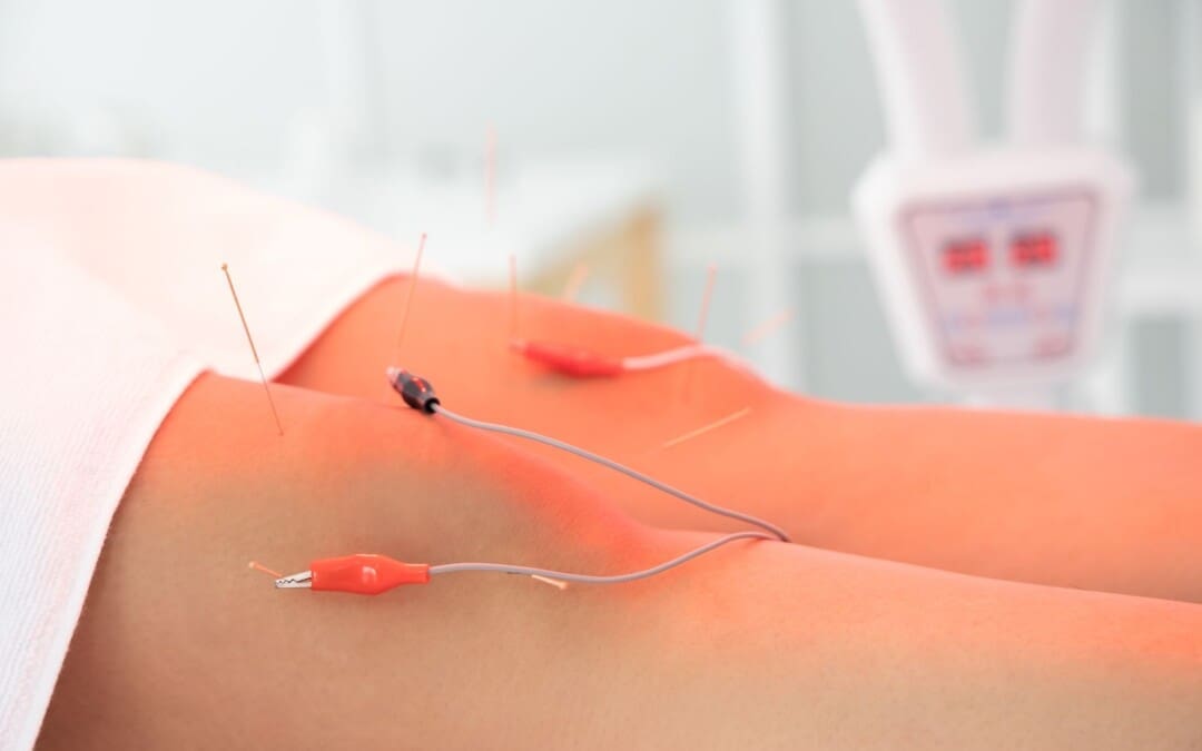
The Modern Twist Of Electroacupuncture
Now, electroacupuncture is a different form of acupuncture that uses the application of acupuncture needles and electric stimulation on the affected muscle. At the same time, when people are getting treated with electroacupuncture, their somatosensory afferent nerves provide pain relief. They are blocked to stop the pain signals from reaching the central nervous system. (Chen et al., 2021) This is because adding electric stimulation can enhance the therapeutic effects of the acupuncture points in the body.
How Electroacupuncture Benefits Muscle Pain
Regarding reducing muscle pain, electroacupuncture is more effective as acupuncturists can help adjust the intensity of the electric currents on the affected muscle to ensure comfort. Some of the benefits that electroacupuncture provides include:
Enhanced pain relief as the electric current can stimulate endorphin release.
Muscle relaxation from spasms in the affected muscle group.
Increased the healing rate by stimulating deeper muscles.
Help enhance muscle strength and flexibility to improve functionality.
Electroacupuncture can relieve pain and even adjust the biomechanical properties of the extensor-flexor muscles to improve abnormal joint loading caused by musculoskeletal pain. (Shi et al., 2020)
How These Two Treatments Help With Musculoskeletal Pain?
When it comes to acupuncture and electroacupuncture, it all depends on the severity of musculoskeletal pain affecting the body. Many people prefer traditional acupuncture for acute musculoskeletal pain in a more holistic approach. In comparison, others might prefer electroacupuncture to reduce the chronic pain effects of musculoskeletal pain. However, both of these treatments are non-surgical. They can be combined with other therapies like physical therapy or chiropractic care to help stimulate the body’s natural healing factor and relieve musculoskeletal pain. When these two treatments are combined with other therapies, the affected muscles are strengthened and provide mobility function back into the extremities. When people start thinking about their well-being, they can utilize these treatments to reduce the comorbidities associated with musculoskeletal pain that is affecting them. Thus allowing them to make small, healthy changes to their routine and live pain-free lives.
Beyond Adjustments: Chiropractic and Integrative Healthcare- Video
References
Chen, L., Wang, X., Zhang, X., Wan, H., Su, Y., He, W., Xie, Y., & Jing, X. (2021). Electroacupuncture and Moxibustion-Like Stimulation Relieves Inflammatory Muscle Pain by Activating Local Distinct Layer Somatosensory Afferent Fibers. Front Neurosci, 15, 695152. https://doi.org/10.3389/fnins.2021.695152
Dzakpasu, F. Q. S., Carver, A., Brakenridge, C. J., Cicuttini, F., Urquhart, D. M., Owen, N., & Dunstan, D. W. (2021). Musculoskeletal pain and sedentary behaviour in occupational and non-occupational settings: a systematic review with meta-analysis. Int J Behav Nutr Phys Act, 18(1), 159. https://doi.org/10.1186/s12966-021-01191-y
Shi, X., Yu, W., Wang, T., Battulga, O., Wang, C., Shu, Q., Yang, X., Liu, C., & Guo, C. (2020). Electroacupuncture alleviates cartilage degradation: Improvement in cartilage biomechanics via pain relief and potentiation of muscle function in a rabbit model of knee osteoarthritis. Biomed Pharmacother, 123, 109724. https://doi.org/10.1016/j.biopha.2019.109724
Wang, M., Liu, W., Ge, J., & Liu, S. (2023). The immunomodulatory mechanisms for acupuncture practice. Front Immunol, 14, 1147718. https://doi.org/10.3389/fimmu.2023.1147718
Welsh, T. P., Yang, A. E., & Makris, U. E. (2020). Musculoskeletal Pain in Older Adults: A Clinical Review. Med Clin North Am, 104(5), 855-872. https://doi.org/10.1016/j.mcna.2020.05.002
Zhu, J., Li, J., Yang, L., & Liu, S. (2021). Acupuncture, from the ancient to the current. Anat Rec (Hoboken), 304(11), 2365-2371. https://doi.org/10.1002/ar.24625
Can the effects of electroacupuncture reduce sciatica in individuals dealing with low back pain to restore their mobility?
Introduction
When many people start to overuse their muscles in the lower quadrants, it can lead to numerous issues that cause pain and discomfort. One of the most common pain issues in the lower quadrants of the musculoskeletal system is sciatica, which is associated with low back pain. This pain duo can affect a person’s daily routine and lead them to pain and discomfort. This musculoskeletal condition is common, and when it affects one of the legs and lower back, many people state that it’s a radiating shooting pain that doesn’t go away for a while. Luckily, there are treatments like electroacupuncture to reduce sciatica associated with low back pain. Today’s article looks at the sciatica-low-back connection, how electroacupuncture reduces this pain connection, and how electroacupuncture can restore mobility to the individual. We talk with certified medical providers who consolidate our patients’ information to assess how to reduce the sciatica-low-back connection with electroacupuncture. We also inform and guide patients on how electroacupuncture therapy can be combined with other therapies to restore mobility to the body. We encourage our patients to ask their associated medical providers intricate and important questions about incorporating electroacupuncture therapy as part of their routine to reduce sciatica associated with low back pain. Dr. Jimenez, D.C., includes this information as an academic service. Disclaimer.
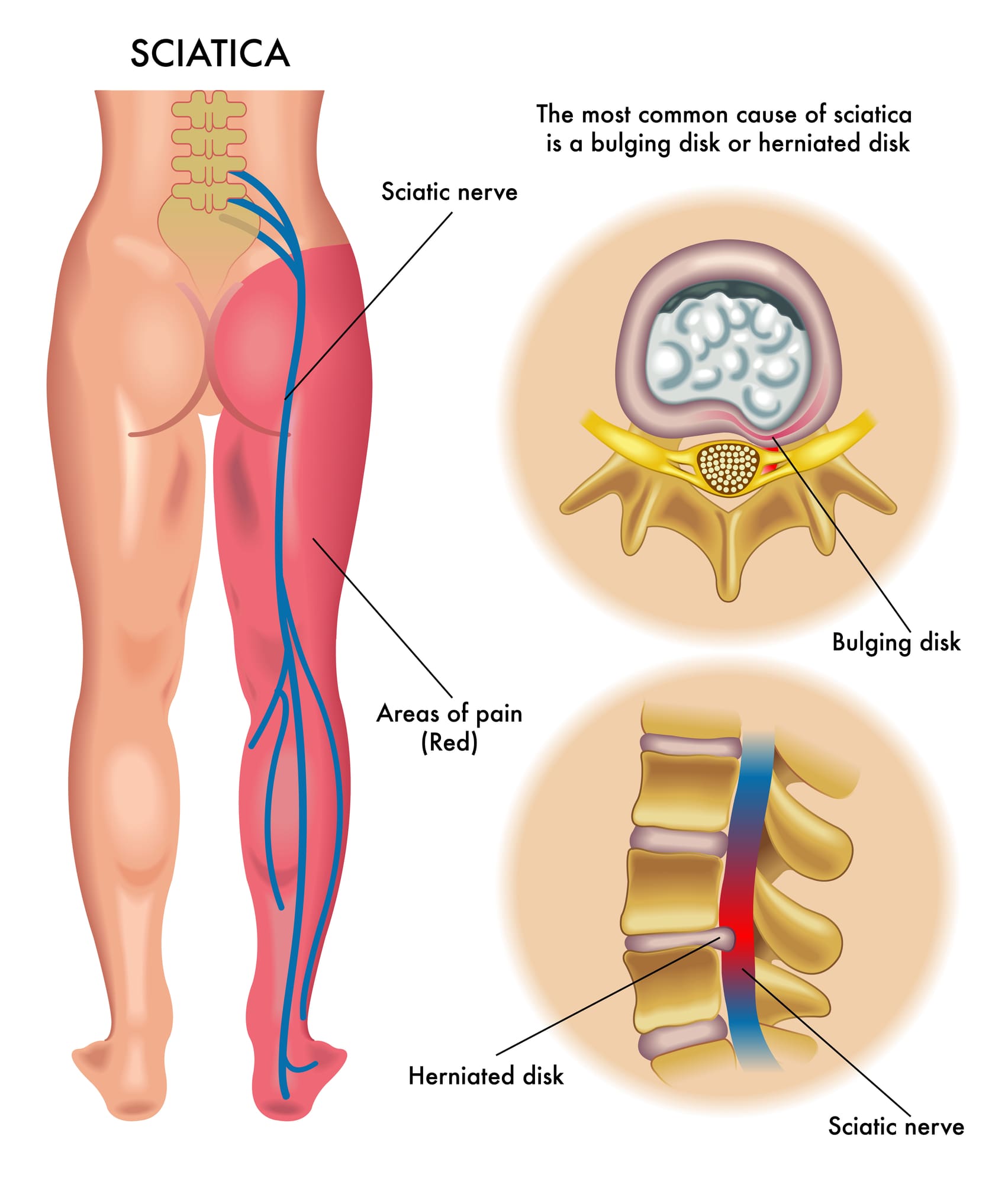
The Sciatica & Low Back Connection
Do you feel muscle aches or pain in your lower back or your legs? Do you experience radiating, throbbing pain in your legs that affects your walking ability? Or have you noticed that your legs and lower back ache more when carrying a heavy object? Many of these scenarios are associated with sciatica, which correlates with lower back pain. Now, sciatica is often characterized by aggravating pain traveling along the sciatic nerve from the lower back region, impairing a person’s quality of life. In the musculoskeletal system, the sciatic nerve plays an important role by providing motor function to the legs. (Davis et al., 2024) Now, when the sciatic nerve, the lumbar region also has a pivotal role. The lumbar region in the musculoskeletal region also has a crucial role in providing support, strength, and flexibility to the body. However, both the sciatic nerve and lumbar spinal region are more prone to stress and injuries from traumatic injuries and environmental factors that can impact the lumbar spinal discs and the sciatic nerve.
Repetitive motions, obesity, improper lifting, degenerative spinal issues, and musculoskeletal conditions are a few causes and risk factors contributing to the development of sciatica associated with the lower back. What eventually happens is that the water content and the progressive loss of the proteoglycans of the spinal discs break down between the vertebrae and protrude out to press on the sciatic nerve, which then can become irritated and cause referred radiating pain in the legs and lower back. (Zhou et al., 2021) The combination of sciatica and lower back pain can become a socio-economic issue depending on the severity of the pain that the sciatic nerve is causing and can make individuals miss out on any activities they are participating in. (Siddiq et al., 2020) While sciatica pain-like symptoms often correlate with the lumbar region, many individuals can find the relief they are looking for through various treatments.
Sciatica Causes- Video
Electroacupuncture Reducing The Sciatica-Low Back Connection
When it comes to reducing the sciatic-low-back connection, many individuals seek out treatment that is affordable and effective in decreasing pain-like issues. Non-surgical treatments like electroacupuncture can be beneficial to many individuals who are experiencing sciatica pain correlated with the lower back. Electroacupuncture is another form of traditional acupuncture therapy that originates in China. Highly trained acupuncturists follow the same acupuncture principles by placing solid thin needles at different acupoints in the body to restore qui or chi (energy flow). Electroacupuncture combines needles and electrostimulation to reduce the central pain-regulatory mechanisms causing low back pain and sciatica by blocking the pain signals and providing pain relief. (Kong, 2020) At the same time, electroacupuncture offers analgesic properties to stimulate endorphins and reduce pain medication for low back pain safely. (Sung et al., 2021)
Electroacupuncture Restoring Mobility
When the lower extremities are experiencing limited mobility due to sciatica associated with low back pain, electroacupuncture can help relax the muscles that are aggravating the sciatic nerve and even help improve blood flow to the lumbar muscles. That is because electroacupuncture can stimulate specific body regions to reduce the somato-vagal-adrenal reflexes to relieve and restore mobility to the lower extremities. (Liu et al., 2021) Additionally, electroacupuncture can be combined with other non-surgical therapies to help strengthen the core and lower back muscles, allowing people to be more mindful of what factors are causing sciatica and lower back pain. By doing this, many people struggling with sciatica associated with low back pain can incorporate electroacupuncture as part of their treatment program combined with holistic approaches to improving their quality of life and providing a pathway to improving their mobility.
Kong, J. T. (2020). Electroacupuncture for Treating Chronic Low-Back Pain: Preliminary Research Results. Med Acupunct, 32(6), 396-397. https://doi.org/10.1089/acu.2020.1495
Liu, S., Wang, Z., Su, Y., Qi, L., Yang, W., Fu, M., Jing, X., Wang, Y., & Ma, Q. (2021). A neuroanatomical basis for electroacupuncture to drive the vagal-adrenal axis. Nature, 598(7882), 641-645. https://doi.org/10.1038/s41586-021-04001-4
Siddiq, M. A. B., Clegg, D., Hasan, S. A., & Rasker, J. J. (2020). Extra-spinal sciatica and sciatica mimics: a scoping review. Korean J Pain, 33(4), 305-317. https://doi.org/10.3344/kjp.2020.33.4.305
Sung, W. S., Park, J. R., Park, K., Youn, I., Yeum, H. W., Kim, S., Choi, J., Cho, Y., Hong, Y., Park, Y., Kim, E. J., & Nam, D. (2021). The effectiveness and safety of electroacupuncture for nonspecific chronic low back pain: A protocol for systematic review and/or meta-analysis. Medicine (Baltimore), 100(4), e24281. https://doi.org/10.1097/MD.0000000000024281
Zhou, J., Mi, J., Peng, Y., Han, H., & Liu, Z. (2021). Causal Associations of Obesity With the Intervertebral Degeneration, Low Back Pain, and Sciatica: A Two-Sample Mendelian Randomization Study. Front Endocrinol (Lausanne), 12, 740200. https://doi.org/10.3389/fendo.2021.740200
IFM's Find A Practitioner tool is the largest referral network in Functional Medicine, created to help patients locate Functional Medicine practitioners anywhere in the world. IFM Certified Practitioners are listed first in the search results, given their extensive education in Functional Medicine