Your Spine, Your Life: An El Paso-Ready Guide to Strong, Flexible, Pain-Resistant Backs
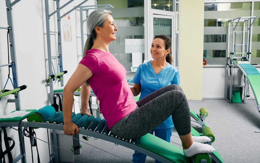
A young woman is performing a spine checkup at a vertebra clinic.
What “spinal health” means (and why it matters here in El Paso)
Spinal health refers to the proper structure, alignment, and function of the spine, enabling it to support the body, facilitate movement, and protect the spinal cord—the pathway for nerve signals between the brain and the body. Good spinal health comes from regular exercise, posture awareness, a nutrient-dense diet, steady hydration, and a healthy weight. Poor spinal health can lead to chronic pain, nerve irritation or damage, and a lower quality of life (Raleigh Orthopaedics, 2024; Orthopedic Specialists of Southwest Florida [OSSWF], 2024; National Spine Health Foundation, 2024).
How a healthy spine supports your whole body
Support & alignment: Your spine acts like a central pillar that shares load with the hips and legs and keeps you upright (Premier Spine & Sports Medicine, n.d.).
Movement & shock absorption: Curves, discs, and joints allow for safe bending and twisting, enabling you to lift, reach, and play (Raleigh Orthopaedics, 2024).
Nerve protection: The spinal column shields the spinal cord and nerve roots, so signals move clearly. Irritation can cause pain, tingling, or weakness (Cary Orthopaedics, 2023).
Quality of life: Ongoing spine issues can lead to fatigue, poor sleep, headaches, and reduced participation in work or sports (Raleigh Orthopaedics, 2024).
Common problems we see—and why early action helps
Strains/sprains and facet irritation from long sitting, poor lifting form, or sudden loads
Disc problems that can press on nearby nerves and create radiating symptoms
Spinal stenosis (narrowing) that pinches nerves
Degenerative changes related to age, low activity, smoking, or extra weight
Most cases respond to conservative care when initiated early, including movement, postural changes, targeted exercises, and load management (OSSWF, 2024).
Red flags—don’t wait: radiating pain, numbness, weakness, headaches, or loss of function. Seek a prompt exam (Cary Orthopaedics, 2023; Suarez Physical Therapy, n.d.).
An El Paso Back Clinic–style plan: simple steps that fit your day
1) Movement you can keep
20–30 minutes of low-impact cardio most days (e.g., walking, cycling, swimming).
Core & hip strength 2–3 days/week: planks, side planks, glute bridges, and bird-dogs.
Mobility after warm-ups: thoracic open-books, hip-flexor, and hamstring stretches (National Spine Health Foundation, 2024; Mobility Project PT, 2024).
2) Posture that holds up at work and home
Sit: feet flat, hips back in the chair, lumbar support, screen at eye level.
Stand: weight balanced, knees soft, ears over shoulders.
Micro-breaks: move every 30–45 minutes (National Spine Health Foundation, 2024).
3) Ergonomics you actually feel
The chair is high enough so the hips are level with or slightly above the knees.
Keyboard and mouse close; forearms supported; shoulders relaxed.
Lift with a hip hinge, keep the load close, and exhale as you stand.
4) Sleep & stress recovery
Neutral neck/back with a supportive mattress and the right pillow height.
Side sleepers: pillow between knees. Back sleepers: pillow under knees.
Use breathing drills, short walks, and stretch breaks to lower tension (Raleigh Orthopaedics, 2024).
5) Hydration & healthy weight
Steady water intake supports disc hydration and tissue recovery (Centeno-Schultz Clinic, n.d.).
A healthy body weight lowers compressive load on joints and discs (Raleigh Orthopaedics, 2024).
Nutrition for a stronger spine (simple and local-friendly)
Protein for muscle and connective-tissue repair
Omega-3s (salmon, trout, walnuts) to help regulate inflammation
Calcium & vitamin D for bone strength
Magnesium for nerve and muscle function
Colorful fruits/vegetables for antioxidants that support recovery
Water for disc hydration and nutrient transport These habits reduce inflammation and support healing (Watkins Family Chiropractic, 2023; OSSWF, 2024).
Four-week “Borderland Back Reset” (minimal gear, steady progress)
Week 1 — Start easy
Daily: 10-minute walk + 5 minutes mobility (open-books, hip-flexor, hamstrings).
Core set (3x/week): plank 20 s, side plank 15 s/side, glute bridge 10 reps.
Posture: Raise the screen and add a small lumbar roll.
Week 2 — Build consistency
Daily: 15–20 minutes walk/cycle + mobility.
Core set (3x/week): plank 25–30 s, side plank 20 s/side, bridge 12 reps; add bird-dog 6/side.
Nutrition: add one serving of leafy greens and one serving of lean protein to each meal (Watkins Family Chiropractic, 2023).
Week 3 — Strength + recovery
Cardio most days: 20–25 minutes.
Light hinge pattern (backpack or kettlebell) 1–2 days/week; focus on form.
Before bed, do slow breathing for 5 minutes.
Week 4 — Re-test & adjust
Compare flexibility, pain, and energy levels with those of Week 1.
Keep what helps; trim what doesn’t.
If numbness, weakness, or radiating pain persists, book an exam (Cary Orthopaedics, 2023; Suarez Physical Therapy, n.d.).
Real-world injuries: work, sports, and motor-vehicle accidents (MVAs)
Work: Desk roles need posture breaks and lumbar support; physical jobs need task rotation, hip-hinge training, and planned recovery.
Sports: Combine mobility, core/hip strength, and gradual return to play.
MVAs: Even “minor” collisions can cause whiplash or soft-tissue injury. A stepwise evaluation, along with imaging when necessary, guides safe return and documentation (OSSWF, 2024).
Inside our integrative approach in El Paso
(Clinical observations from Dr. Alexander Jimenez, DC, APRN, FNP-BC, Nurse Practitioner and Chiropractor)
Dual-scope diagnosis: We blend chiropractic and medical perspectives. Your exam includes a detailed history, movement, and neurological screens, as well as, when necessary, advanced imaging to clarify the problem and rule out potential red flags (Jimenez, n.d.; see Imaging/Diagnostics and Personal-Injury topics).
Evidence-based conservative care:
Chiropractic adjustments to restore motion and reduce joint irritation
Therapeutic exercise to build core/hip strength and mobility
Manual therapy/massage for tight or sensitive tissues
Acupuncture as part of an integrative plan when appropriate
Lifestyle coaching on posture, lifting, sleep, and stress (Prestige Health & Wellness, n.d.; Mobility Project PT, 2024; Raleigh Orthopaedics, 2024)
Documentation & advocacy: For work, sports, personal, and MVA cases, we document the mechanism of injury, exam findings, functional limits, and response to care. When claims or legal issues arise, clear records and appropriate imaging support decision-making (Jimenez, n.d.; Rangeline Chiropractic, n.d.).
Myths vs. facts (short and clear)
Myth: “If my back hurts, I should rest all day.” Fact: Gentle movement and short walks often speed recovery; long bed rest adds stiffness (National Spine Health Foundation, 2024).
Myth: “Only heavy lifting causes back pain.” Fact: Prolonged sitting, poor ergonomics, stress, and sleep problems also drive pain (National Spine Health Foundation, 2024; Raleigh Orthopaedics, 2024).
The El Paso Back Clinic checklist
☐ Break up sitting every 30–45 minutes
☐ Screen at eye level; use lumbar support
☐ 10–15 minutes daily core + mobility
☐ 20–30 minutes low-impact cardio most days
☐ Hydrate across the day
☐ Build meals around protein + produce + healthy fats
☐ Sleep with neutral neck/back alignment
☐ Seek care quickly for red flags or lasting symptoms
Delve into the gut-liver connection with chiropractic care and find out how it benefits your health and vitality.
Understanding the Gut-Liver Axis: How It Influences Back Pain and the Role of Chiropractic Care in Holistic Recovery
In our busy lives, back pain is a common complaint that can disrupt daily activities, from sitting at a desk to enjoying a walk. But what if some of that discomfort stems from deeper issues, like problems in your digestive system or liver? The gut-liver axis represents a vital link in the body, where imbalances can lead to widespread effects, including musculoskeletal pain, such as backaches. This article explores this connection in depth, explaining how the gut supports overall body function, why environmental factors can disrupt it, and how these issues might manifest as overlapping symptoms affecting not just the abdomen but also the spine and muscles. We’ll also discuss the clinical reasons why chiropractic care can be beneficial, alongside other nonsurgical treatments such as targeted exercises, massage therapy, acupuncture, and integrative medicine approaches that promote natural healing and prevent long-term complications.
Back pain affects millions, often linked to poor posture or injuries, but emerging research suggests that internal factors also play a role. By understanding the gut-liver relationship, you can take steps toward better health. This guide draws on scientific insights to provide clear, actionable information. While it’s based on reliable sources, consulting a healthcare provider is key for individual needs.
Decoding the Gut-Liver Axis: A Foundation for Health
The gut-liver axis is an interactive system in which the intestines and liver constantly communicate. Food digested in the gut sends nutrients via the portal vein to the liver for processing, detoxification, and distribution. In turn, the liver produces bile to help the gut break down fats and maintain a balance of bacteria. This partnership ensures that the body handles toxins and absorbs essential nutrients efficiently.
Disruptions here can ripple out, potentially contributing to conditions like inflammation that affect distant areas, such as the back. For instance, gut bacteria imbalances might lead to liver strain, triggering signals that heighten pain sensitivity in the spine (Wang et al., 2021). This axis is essential because it influences energy levels, immune responses, and even pain perception. Practitioners in functional medicine, such as Dr. Alexander Jimenez, often evaluate this link to address hidden causes of chronic discomfort, using tools to restore harmony without resorting to surgery.
The Gut’s Essential Role in Body Function and Pain Management
Your gut is more than a food processor—it’s a powerhouse for health. Housing trillions of microbes, it digests meals, extracts nutrients like vitamins and minerals, and produces compounds that fuel cells. A balanced gut microbiome supports immunity by warding off pathogens and reducing inflammation, which can otherwise spread and aggravate conditions such as back pain.
Beyond digestion, the gut influences nerve signals through the vagus nerve, affecting stress and mood, which in turn can cause muscle tension and exacerbate back pain. When functioning properly, it promotes better sleep and increased energy, helping the body recover from physical strains. However, imbalances—known as dysbiosis—can lead to issues like bloating or fatigue, sometimes referring pain to the musculoskeletal system. Dr. Jimenez notes in his practice that assessing gut health via functional tests reveals connections to persistent back issues, allowing tailored plans that enhance recovery (Jimenez, n.d.).
The Interconnected Gut and Liver: Why They Rely on Each Other
The gut and liver are closely linked by both anatomy and function. Absorbed gut contents flow directly to the liver, where they’re metabolized. The liver reciprocates by sending bile to regulate gut bacteria and aid digestion. This cycle protects against toxins, but problems in one organ stress the other.
For example, poor gut health can lead to the liver being flooded with harmful substances, resulting in inflammation or fatty buildup. Studies link this to diseases where gut permeability allows bacterial products to irritate the liver, potentially amplifying body-wide signals that manifest as pain (Federico et al., 2017). In back health contexts, this might involve viscerosomatic reflexes, where organ distress refers pain to the spine. Research highlights how alcohol or infections exacerbate this, damaging gut barriers and overburdening the liver (Chae et al., 2024). Dr. Jimenez emphasizes evaluating these ties in patients with unexplained back pain, using integrative methods to break the cycle.
Environmental Influences: Disrupting the Gut and Triggering Musculoskeletal Symptoms
Everyday surroundings shape gut health, often leading to issues that overlap with back problems. Diets high in processed foods feed harmful bacteria, which can thin the gut lining and cause a condition known as “leaky gut.” This allows toxins to enter the blood, triggering inflammation that can sensitize nerves in the spine (Di Vincenzo et al., 2023).
Stress compounds this by altering gut movement, increasing permeability, and potentially referring pain to the back via neural pathways (Konturek et al., 2011). Toxins like pollutants or medications disrupt microbes, while alcohol harms both gut and liver, leading to fatigue and muscle tension (Konturek et al., 2011). Infections add to the mix, wiping out beneficial bacteria and allowing inflammation to spread.
These factors create overlapping symptoms: gut distress might mimic or worsen back pain through viscerosomatic mechanisms, where internal irritation signals to muscles and joints (Farmer et al., 2009). For instance, abdominal inflammation could tighten lower back muscles, causing chronic aches. Risk factors like poor sleep or trauma heighten this in adults and children (Zia et al., 2022). Environmental exposures, including chemicals, further imbalance the microbiome, linking to systemic pain (Nicholson et al., 2012).
Dr. Jimenez uses detailed histories to identify these triggers, crafting plans that rebuild gut integrity and ease back strain.
Table: Environmental Factors and Their Effects on Gut Health and Back Pain
Environmental Factor
How It Disrupts the Gut
Potential Overlapping Symptoms in Back/Muscles
Processed Diets
Promotes bad bacteria, leaky gut
Inflammation leading to spinal pain, stiffness
Chronic Stress
Slows digestion, increases permeability
Muscle tension, referred to as lower back aches
Alcohol and Toxins
Damages lining, alters microbiome
Fatigue, liver strain, causing widespread pain
Medications/Infections
Kills good bacteria, causes dysbiosis
Systemic inflammation, joint/muscle discomfort
Pollutants
Disrupts bacterial balance
Chronic fatigue, heightened pain sensitivity
This overview illustrates how daily exposures can lead to back-related issues, underscoring the need for comprehensive interventions.
Clinical Insights: Why Chiropractic Care Supports the Gut-Liver Axis and Back Health
Chiropractic care targets spinal alignment to optimize nerve function, which can indirectly benefit the gut-liver axis. Subluxations—misalignments—may interfere with autonomic nerves that regulate digestion and detoxification, contributing to imbalances that can refer pain to the back.
The rationale lies in neurology: adjustments restore communication, potentially reducing inflammation and improving gut motility (Elsenbruch et al., 2015). For back pain tied to visceral issues, this addresses viscerosomatic reflexes, easing referred discomfort. Emerging evidence suggests that probiotics, when combined with chiropractic care, enhance liver function by balancing the microbiome (Hojsak, 2024).
Dr. Jimenez, with extensive experience in functional medicine, integrates this for patients with back pain from gut-liver sources. His approach utilizes adjustments to calm overactive nerves, promoting natural healing and preventing escalations such as disc degeneration (Jimenez, n.d.).
It’s about holistic balance, not just force—clear patient discussions ensure understanding, fostering adherence for lasting relief.
The Healing Diet: Combat Inflammation, Embrace Wellness: Video
Nonsurgical Pathways: Exercises, Therapies, and Integrative Medicine for Healing
For gut-liver-related back issues, nonsurgical options are often the preferred choice. Targeted exercises, such as core strengthening or yoga, improve posture and support spinal health, while also aiding digestion. Massage therapy relaxes muscles, boosting circulation to organs and reducing tension.
Acupuncture stimulates specific points to balance energy, alleviating pain and inflammation, and offering benefits for gut disorders. Integrative medicine combines nutrition—specifically, anti-inflammatory diets rich in fiber—with herbs to heal the gut lining and support liver detoxification.
These foster the body’s innate repair, preventing chronic back problems. Dr. Jimenez’s clinic protocols emphasize this, utilizing electro-acupuncture and rehabilitation to address the root causes, with patients reporting reduced pain and improved function.
Expert Perspectives from Dr. Alexander Jimenez on Back-Focused Care
Dr. Alexander Jimenez, DC, APRN, FNP-BC, leads the way in blending chiropractic with functional medicine for optimal back health. His credentials include advanced training in clinical physiology and integrative protocols. In practice, he tackles viscerosomatic links, where gut-liver issues manifest as back pain, using assessments to create custom plans.
He shares via podcasts and resources how nutrition and adjustments resolve inflammation, aiding recovery from injuries. Patients value his empathetic communication, explaining connections simply to empower self-care (Jimenez, n.d.).
Practical Steps for Integrating Gut-Liver Health into Back Pain Management
Begin with lifestyle audits: track your diet, stress levels, and activity. Seek professional evaluations for personalized advice. Incorporate habits such as consuming probiotic foods, taking gentle walks, and practicing stress-reduction techniques.
Combine therapies: regular adjustments, daily stretches, and nutritional tweaks. Consistency yields results, as evidenced by the outcomes in functional medicine.
This exploration reveals the profound impact of the gut-liver axis on back health. Through chiropractic and integrative methods, you can harness natural healing for a pain-free life.
References
Chae, Y.-R., et al. (2024). Diet-Induced Gut Dysbiosis and Leaky Gut Syndrome. Journal of Microbiology and Biotechnology, 34(4), 747-756. https://pubmed.ncbi.nlm.nih.gov/38321650/
Di Vincenzo, F., et al. (2023). Gut microbiota, intestinal permeability, and systemic inflammation: a narrative review. Internal and Emergency Medicine, 19(2), 275-293. https://pubmed.ncbi.nlm.nih.gov/37505311/
Farmer, A. D., et al. (2009). Visceral pain hypersensitivity in functional gastrointestinal disorders. British Medical Bulletin, 91, 123-136. https://pubmed.ncbi.nlm.nih.gov/19620136/
Hojsak, I. (2024). Probiotics in Functional Gastrointestinal Disorders. Advances in Experimental Medicine and Biology, 1449, 157-174. https://pubmed.ncbi.nlm.nih.gov/39060737/
Konturek, P. C., et al. (2011). Stress and the Gut: Pathophysiology, Clinical Consequences, Diagnostic Approach, and Treatment Options. Journal of Physiology and Pharmacology, 62(6), 591-599. https://pubmed.ncbi.nlm.nih.gov/22314561/
Xie, C., & Halegoua-DeMarzio, D. (2019). Role of Probiotics in Nonalcoholic Fatty Liver Disease: Does Gut Microbiota Matter? Nutrients, 11(11), 2837. https://www.mdpi.com/2072-6643/11/11/2837
Zia, J. K., et al. (2022). Risk Factors for Abdominal Pain-Related Disorders of Gut-Brain Interaction in Adults and Children: A Systematic Review. Gastroenterology, 163(4), 995-1023.e3. https://pubmed.ncbi.nlm.nih.gov/35716771/
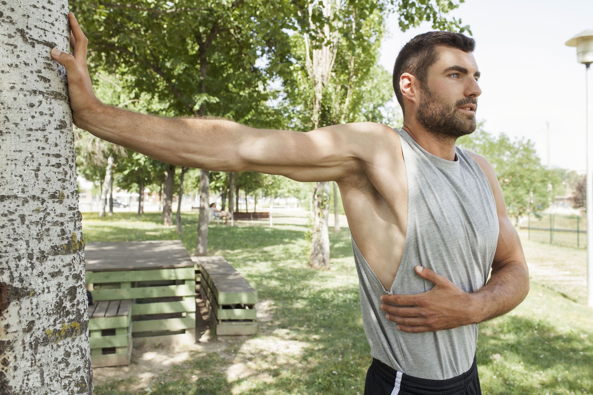
Weekend Athletes Injury Solutions: A Simple, Evidence-Based Guide for Safer Play and Faster Recovery
A handsome, muscular man in sportswear is stretching his muscles in a sunny park.
Who this is for: adults who sit most of the week and then go hard on the weekend (a.k.a. “weekend warriors”). What you’ll get: clear reasons these injuries happen, what to do first, how to prevent them, and how integrative chiropractic care—like the approach used in El Paso—helps you recover and return to activity safely.
Weekend warriors 101
A weekend warrior is someone who does most of their intense activity on one or two days after a mostly sedentary week. That pattern can still deliver strong health benefits if you meet weekly exercise targets, but the sudden spike in effort raises the risk of sprains, strains, and overuse problems—especially when you skip warm-ups or jump in too fast (Riverside Health System, 2025; Weill Cornell Medicine, 2024). (riversideonline.com)
Large studies show that “condensed” exercisers can gain health benefits similar to those who spread workouts throughout the week—as long as the total weekly minutes reach the recommended amounts. The catch: your muscles, tendons, and joints still need gradual loading to stay injury-resistant (American Heart Association News, 2024; Shiroma et al., 2019). (www.heart.org)
Why weekend athletes get hurt
Most weekend injuries come down to three drivers:
Overuse: repeating motions your tissues aren’t ready for (long runs, repetitive swings).
Sudden movement: fast cuts, awkward landings, or twisting under load.
Poor preparation: no warm-up, weak stabilizers, and worn-out shoes.
These factors underlie many musculoskeletal problems seen by orthopedic and emergency clinicians (Aligned Orthopedic Partners, 2024; Weill Cornell Medicine, 2024). (Aligned Orthopedic Partners)
What typically gets injured (and what it feels like)
Emergency physicians most often treat injuries to the knees, shoulders, and ankles, with sprains and strains outnumbering fractures (Weill Cornell Medicine, 2024). (weillcornell.org)
Ankle sprain (ligament): twist/roll, swelling, tenderness, sometimes bruising.
Knee sprain/overuse pain: instability, joint-line pain, and pain after cutting or pivoting.
Achilles tendinopathy: stiff, sore area above the heel (often worse in the morning).
Rotator cuff irritation: pain with overhead reach or lying on the shoulder.
Shin splints: aching along the shin after running on hard surfaces (Riverside Health System, 2025). (riversideonline.com)
Sprain vs. strain (plain words): Sprain = ligament (joint stabilizer). Strain = muscle or tendon (mover). Sprains can feel unstable and bruise; strains feel like a pull with spasm or weakness (Aligned Orthopedic Partners, 2024). (Aligned Orthopedic Partners)
Your job habits shape your weekend risk
Repetitive tasks and long sitting can irritate tissues before you ever play. Those weekday loads stack with Saturday’s game and can tip you into pain. Tendinitis, for example, often develops from repeated motions (MyShortlister, 2023). Short micro-breaks, posture changes, and light mid-week movement help. (Shortlister)
First aid: what to do in the first 24–72 hours
For many fresh soft-tissue injuries, start with the PRICE method: Protect, Rest, Ice (20 minutes on), Compress, Elevate. Don’t push through sharp pain. Seek urgent care for a “pop,” severe swelling, numbness/weakness, deformity, or inability to bear weight (Weill Cornell Medicine, 2024). (weillcornell.org)
When imaging is useful (and what usually comes first)
You don’t need an MRI for every sprain. Clinicians begin with a history and examination; an X-ray is often the first test if a fracture is suspected. Musculoskeletal ultrasound or MRI follows when soft-tissue damage is suspected, symptoms persist, or nerve signs appear (Weill Cornell Medicine, 2024). (weillcornell.org)
In work, sport, or motor-vehicle accident (MVA) cases, advanced imaging also supports clear medical-legal documentation—a key part of comprehensive injury care (El Paso Back Clinic; Dr. Jimenez). (elpasobackclinic.com)
Practical prevention that actually works
Warm up and cool down. Do 5–10 minutes of light cardio and dynamic moves (leg swings, lunges, and arm circles). Ease into slow stretches after play (Riverside Health System, 2025; Appleton Chiropractic Center, n.d.). (riversideonline.com)
Build up gradually. Increase time or intensity by ~10% per week. Rotate high- and low-impact days (Center for Orthopedic Surgery & Sports Medicine, n.d.). (COSM)
Use the right gear. Replace worn shoes; match footwear to your sport (Riverside Health System, 2025). (riversideonline.com)
Hydrate, fuel, and sleep. Under-fueling and short sleep increase the risk of cramps and strains (Riverside Health System, 2025). (riversideonline.com)
Add two short mid-week sessions. Even 20–30 minutes of exercise twice a week improves tissue tolerance and reduces the risk of weekend injuries (Mayo Clinic Sports Medicine, n.d.). (sportsmedicine.mayoclinic.org)
Simple self-care roadmaps
Ankle sprain
Days 0–2: PRICE, gentle ankle pumps, compression sleeve.
Days 3–7: pain-free range of motion; start weight bearing as tolerated.
Weeks 2–4: add balance drills and band work.
See a clinician if you can’t bear weight or feel instability (Weill Cornell Medicine, 2024). (weillcornell.org)
Achilles tendinopathy
Reduce jumping/sprinting while painful.
Begin slow calf raises (progress to eccentrics); increase load gradually (Aligned Orthopedic Partners, 2024). (Aligned Orthopedic Partners)
Shoulder soreness (rotator cuff pattern)
Short rest (not total rest), then scapular control and light external-rotation drills; limit overhead volume and improve thoracic mobility (Aligned Orthopedic Partners, 2024). (Aligned Orthopedic Partners)
Low-back strain
After 24–48 hours, try gentle mobility exercises (such as pelvic tilts and cat-camel), followed by core endurance exercises (like planks) and hip-hinge practice. If pain persists or travels below the knee or you notice weakness, seek evaluation (Weill Cornell Medicine, 2024). (weillcornell.org)
2 rounds: push-ups 8–12; band rows 12–15; band “T” raises 10–12
Dead bug 6/side; bird-dog 6/side
3–5 min pec stretch + thoracic rotations
Short “bridge” sessions like these raise tissue tolerance and make weekend play safer (Center for Orthopedic Surgery & Sports Medicine, n.d.). (COSM)
How integrative chiropractic care supports weekend athletes
Integrative chiropractic care blends joint-specific manual therapy with targeted exercise, soft-tissue work, and—when indicated—acupuncture, bracing/taping, and coordinated medical evaluation. The goal is to improve mechanics (how you move) and capacity (what your tissues can handle), so you heal and resist re-injury (Radiant Life Chiropractic, 2024; Aligned Orthopedic Partners, 2024). (Radiant Life Chiropractic)
At El Paso Back Clinic, this approach is paired with a dual-scope model (chiropractic plus nurse practitioner care) for sports, work, personal, and MVA injuries. The team can:
Perform focused orthopedic and neurological exams.
Order X-ray, MRI, CT, or musculoskeletal ultrasound when the exam suggests more than a simple sprain.
Coordinate medical-legal documentation (mechanism, findings, imaging, functional limits, and response to care) for injury cases.
Guide progressive rehab and return-to-play plans based on pain-free motion, strength, and sport-specific tasks (El Paso Back Clinic; Jimenez, 2025). (elpasobackclinic.com)
Local context: Recent clinic articles from El Paso highlight dual-scope evaluation, the role of advanced imaging, and clear documentation for personal-injury cases—useful if your injury involves work or an auto crash (El Paso Back Clinic). (elpasobackclinic.com)
A smarter return-to-play checklist (advance only when all are true)
Daily tasks are pain-free, and you’re sleeping normally.
Full, pain-free range of motion for the injured area.
Strength feels symmetrical from side to side in simple tests.
You can do basic sport drills (jog-cut-jog; easy swings/serves) without symptoms.
If a step hurts, back up, adjust the load, and rebuild capacity (Weill Cornell Medicine, 2024). (weillcornell.org)
Key takeaways
Weekend-only training can be beneficial—the total weekly activity level matters most—but spikes in workload increase the risk of injury (AHA News, 2024; Riverside Health System, 2025). (www.heart.org)
Most common issues include sprains, strains, and overuse injuries in the ankle, knee, and shoulder (Weill Cornell Medicine, 2024). (weillcornell.org)
Warm up, build gradually, and add two short mid-week sessions to cut risk (Riverside Health System, 2025; Center for Orthopedic Surgery & Sports Medicine, n.d.). (riversideonline.com)
Integrative chiropractic care—with exam, imaging when needed, progressive exercise, and thorough documentation—helps you recover and return to play stronger and safer (El Paso Back Clinic; Radiant Life Chiropractic, 2024). (elpasobackclinic.com)
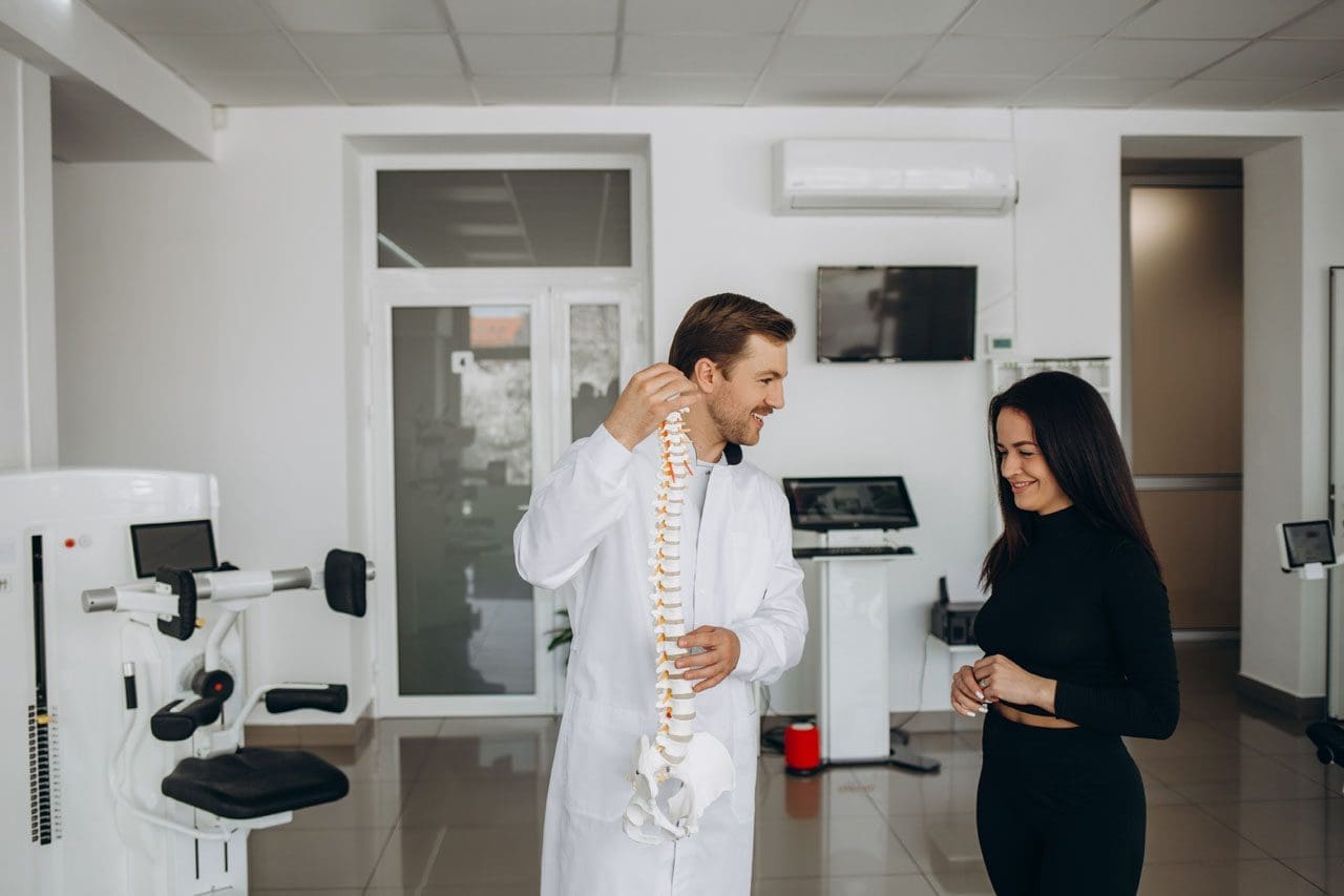
El Paso Back Clinic®: Premier Wellness Chiropractic Care in El Paso, TX
Spine problems are evident in the model. A woman is in consultation with the doctor in the clinic.
At El Paso Back Clinic®, led by Dr. Alexander Jimenez, DC, APRN, FNP-BC, we’re dedicated to transforming lives through advanced chiropractic care and integrative wellness in El Paso, TX. Specializing in recovery from motor vehicle accidents (MVAs), sports injuries, and chronic conditions such as neuropathy, our team utilizes cutting-edge neuromusculoskeletal imaging and dual-scope diagnostics to identify the root causes of injuries. From whiplash to gastrointestinal trauma, we craft personalized plans blending spinal adjustments, nutrition, and therapies like acupuncture to restore mobility and vitality. With a focus on holistic healing and legal support for injury claims, we empower El Pasoans to live pain-free and thrive.
Personal Injuries and Their Impact in El Paso
Living in El Paso’s bustling community means navigating busy roads and an active lifestyle, where accidents—from car crashes to sports mishaps—can disrupt one’s health. MVAs, common on rainy I-10 days, often cause spinal misalignments, leading to sciatica or numbness (Jimenez, 2025a). Sports injuries, like joint strains and workplace falls, add to the toll, risking chronic issues like neuropathy if untreated (Mana.md, n.d.).
At El Paso Back Clinic®, Dr. Jimenez’s chiropractic expertise targets these musculoskeletal and nerve disruptions. Using advanced imaging, we link injuries to symptoms, ensuring precise care. Our integrative approach, which combines adjustments with wellness coaching, helps prevent long-term pain and includes legal documentation to support insurance claims for patients in El Paso.
Nerve Pain and Neuropathy Post-Collision
Car accidents frequently trigger nerve compression, causing tingling, burning, or weakness that mimics peripheral neuropathy. Even minor collisions in El Paso’s unpredictable weather can cause vertebrae to shift, potentially pinching nerves (Jimenez, 2025b). Our clinic employs EMG and dynamic X-rays to map these injuries, correlating crash forces to nerve damage for accurate diagnosis.
We use spinal decompression and laser therapy to relieve pressure and promote healing, with patients often seeing 40-50% symptom improvement in weeks (Miami Chiropractors, n.d.). Detailed biomechanical reports strengthen personal injury claims, ensuring fair compensation for El Paso residents.
Chiropractic Care for Joint and Ligament Injuries
Injuries like ACL tears from sports or MVA dashboard impacts require targeted restoration. At El Paso Back Clinic®, we realign joints, boost circulation, and strengthen muscles to speed recovery without surgery (Jimenez, 2025c). Dr. Jimenez’s functional assessments prevent compensatory patterns, vital for El Paso’s athletes.
We integrate acupuncture and custom orthotics, helping patients resume activities in six months, rather than a year. Nutritional support, like collagen-rich diets, enhances ligament repair, tailored to El Paso’s active community.
Five Musculoskeletal Challenges We Address
Accidents hit muscles and bones hard. Our chiropractic team tackles five common issues:
Neck and Back Pain: Whiplash from MVAs causes stiffness; gentle adjustments restore motion (Jimenez, 2025d).
Sciatica: Pinched nerves from disc issues ease with traction therapy.
Joint Inflammation: Post-injury arthritis responds to ultrasound and anti-inflammatory nutrition.
Sports Strains: Overuse injuries can be effectively treated through myofascial work and gait analysis.
These protocols, customized for El Pasoans, cut recurrence by half, blending wellness education for lasting health.
Spinal Misalignment: Recovery After Crashes
El Paso’s slick roads amplify MVA risks, often misaligning spines and compressing discs, leading to radiating pain (Jimenez, 2025a). We utilize high-velocity adjustments and flexion-distraction techniques to realign the vertebrae, paired with massage to relax the muscles.
Dr. Jimenez’s imaging links crash dynamics to disc damage, guiding non-surgical plans that preserve mobility in 70% of cases (Knecht Chiropractic, n.d.). Legal reports detail injury causation, supporting the claims of El Paso patients.
Reducing Inflammation for Pain Relief
Inflammation fuels post-injury pain. Our chiropractic care enhances lymphatic drainage via soft-tissue therapy and cryotherapy, breaking the cycle (Jimenez, 2025e). Patients adopt home strategies, such as taking turmeric supplements, which can reduce swelling by 40% (Miami Chiropractors, n.d.).
For workers’ compensation cases, we monitor biomarkers, aligning treatments with recovery goals to help El Paso workers return to their feet.
Cyclist Recovery After Bike-MVA Collisions
Biking on El Paso’s scenic trails poses risks from urban traffic, which can lead to fractures or nerve injuries. Our integrative care includes bike-fit corrections and vestibular training for balance (Jimenez, 2025f). Cyclists return to riding in three months, supported by endurance nutrition and legal advocacy.
Massage Therapy for MVA Trauma Healing
MVAs cause soft-tissue damage, from bruises to adhesions. Massage therapy, paired with adjustments, boosts circulation and endorphins, reducing whiplash recovery time by 30% (Jimenez, 2025). We progress from gentle strokes to deep tissue, documenting for El Paso insurance claims.
Spinal Trauma from 18-Wheeler Accidents
Semi-truck crashes deliver intense force, fracturing vertebrae or tearing ligaments. We use dynamic imaging to assess damage, guiding bracing and neuromodulation (Jimenez, 2025h). Legal reports link crash mechanics to injuries, aiding settlements for El Paso patients.
Nutrition for Tissue Repair Post-MVA
Injured tissues require nutrients such as protein and antioxidants. Dr. Jimenez designs diets with salmon and berries, using genetic insights to optimize healing (Jimenez, 2025i). This reduces fibrosis, strengthening tissues for El Paso’s active residents.
Durable Medical Equipment for Recovery
Following a motor vehicle accident (MVA), tools such as TENS units or cervical collars can support healing. We select evidence-based equipment, such as ergonomic chairs, to offload spines (Jimenez, 2025). Tele-rehab ensures compliance, with invoices bolstering El Paso claims.
Comprehensive Musculoskeletal Recovery
MVAs strain muscles and joints, from sprains to dislocations. Our pain mapping and multi-modal care—adjustments, PT, mindfulness—restore 80% function in six weeks (Jimenez, 2025k). Legal narratives ensure fair compensation.
Whiplash-Associated Disorders (WAD) Recovery
WAD from crashes causes neck pain or dizziness. We use Doppler ultrasound for vascular checks and treat with mobilization for 90% relief (Jimenez, 2025). Immediate post-accident icing and evaluations ensure thorough El Paso claims.
Gastrointestinal Injuries from MVAs
Car accidents can disrupt digestion, causing nausea or organ strain. Our integrative care, which includes visceral manipulation and nutrition, restores gut health, backed by legal support for claims (Jimenez, 2025).
Why Choose El Paso Back Clinic®?
Our team, led by Dr. Jimenez, combines chiropractic precision with medical expertise, utilizing tools such as digital motion X-rays. We offer acute-to-chronic care, transparent billing, and testimony for legal cases. Patients reduce their reliance on medication, regaining vitality through holistic plans tailored for El Paso’s vibrant community.
Find out the role of chiropractic care in addressing text neck posture. Improve your well-being with expert care and guidance.
Understanding Text Neck in 2025: Causes, Symptoms, Prevention, and Non-Surgical Treatments
In our increasingly connected world, neck pain has emerged as a silent epidemic, affecting people of all ages who spend hours hunched over screens. Often dubbed “text neck,” this condition stems from the repetitive strain of looking down at smartphones, tablets, and computers. Recent global estimates indicate that neck pain impacts billions, with prevalence rates climbing due to digital lifestyles (Kazeminasab et al., 2022). As we navigate 2025, where remote work and mobile devices dominate daily routines, understanding text neck is crucial for maintaining spinal health and overall quality of life.
This comprehensive guide dives deep into text neck, exploring its development, symptoms, and far-reaching effects on the body. We’ll examine environmental triggers that exacerbate cervical spine issues and provide a clinical rationale for why chiropractic care is an effective, non-invasive solution. Drawing from expert insights, including those from Dr. Alexander Jimenez, DC, APRN, FNP-BC, we’ll highlight non-surgical treatments like targeted exercises, stretches, massage therapy, acupuncture, and integrative medicine. These approaches not only alleviate pain but also harness the body’s natural healing processes to prevent chronic problems—emphasizing clear patient communication over mere physical strength.
Whether you’re experiencing occasional stiffness or persistent discomfort, this article equips you with actionable strategies to reclaim your posture and well-being. Let’s uncover how small changes can lead to significant relief in our tech-driven era.
The Rise of Text Neck: A Modern Health Challenge
Text neck, or tech neck, isn’t a new phenomenon, but its prevalence has skyrocketed with the ubiquity of digital devices. Coined to describe the forward head posture adopted during prolonged screen use, this condition places undue stress on the cervical spine—the seven vertebrae (C1-C7) that form the neck. In a neutral position, the head weighs about 10-12 pounds, but tilting it forward at 45 degrees can exert up to 50 pounds of force on the spine (Tsantili et al., 2022).
Global data from 2025 underscores the issue: Neck pain affects approximately 27 per 1,000 people annually, with trends showing increases in younger demographics due to smartphone addiction (Kazeminasab et al., 2022). A scoping review published this year highlights how excessive device use leads to musculoskeletal disorders, particularly in adults and children (Piruta et al., 2025). In fact, studies from 2024-2025 reveal that 73% of higher education students report neck pain, often linked to studying postures (University of Miami News, 2025).
The mechanics are simple yet damaging. When you look down, the neck’s natural curve flattens, straining muscles, ligaments, and discs. Over time, this can accelerate degeneration, leading to conditions like herniated discs or osteoarthritis. Research from adolescent populations shows a 32% prevalence of neck pain, tied to factors like poor desk ergonomics and heavy backpacks (Ben Ayed et al., 2019). As we spend more time online—averaging 7-10 hours daily in 2025—text neck has become a public health concern, prompting calls for better awareness and interventions (Tsantili et al., 2022).
How Text Neck Develops: Biomechanics and Daily Triggers
Text neck develops gradually through repetitive micro-traumas to the cervical spine. The head’s forward shift disrupts the spine’s alignment, overloading the posterior neck muscles while weakening the anterior ones. This imbalance, known as forward head posture, increases compressive forces on vertebrae and intervertebral discs (Jimenez, 2016).
Biomechanically, each inch of forward head displacement adds 10 pounds of leverage, potentially leading to subluxations—partial dislocations that impinge nerves (Verma et al., 2021). Prolonged exposure, such as 2-4 hours daily on devices, accumulates stress, with young people at higher risk due to developing spines (Al-Hadidi et al., 2019).
Daily triggers include:
– **Screen Time Habits**: Texting or scrolling in a slouched position. A 2021 study found no direct link between flexion angle and pain in adults, suggesting multifactorial causes, but recent reviews affirm posture’s role (Correia et al., 2021).
– **Work and Study Environments**: Poor lighting or screen placement forces neck strain.
– **Lifestyle Factors**: Sedentary routines exacerbate weakness in stabilizing muscles.
In 2025, emerging research questions strict causation but emphasizes cumulative effects (Neck pain and text neck using Hill’s criteria, 2025). Addressing these early prevents progression to chronic pain.
Symptoms of Text Neck: From Mild Discomfort to Debilitating Issues
Text neck symptoms vary in intensity but often start subtly, progressing if ignored. Core signs include:
Neck-Specific Symptoms
– **Pain and Soreness**: A constant ache, worsening with movement. Chronic cases may involve sharp pains from disc compression (Binder, 2008).
– **Stiffness**: Reduced range of motion, making it hard to turn or tilt the head. This stems from tightened suboccipital muscles (Misailidou et al., 2010).
– **Headaches**: Cervicogenic headaches radiate from the neck to the head, triggered by tension (Verma et al., 2021).
Effects on Shoulders and Upper Back
Shoulders often “round” forward, leading to:
– **Shoulder Tension**: Knots in trapezius muscles cause burning pain.
– **Upper Back Ache**: Kyphotic curvature strains thoracic muscles, common in device users (Ben Ayed et al., 2019).
A 2025 study on university students links studying postures to high neck pain prevalence (The Prevalence of Neck Pain, 2025).
Impact on Upper Extremities
Nerve irritation can extend symptoms:
– **Arm Radiating Pain**: Radiculopathy causes shooting pains or weakness (Kuligowski et al., 2021).
– **Numbness/Tingling**: Pinched nerves affect hands, mimicking other syndromes.
– **Reduced Functionality**: Grip weakness impacts tasks like writing.
Untreated, these can lead to long-term nerve damage, emphasizing early intervention (Mastalerz et al., 2022).
Environmental Factors Contributing to Cervical Neck Pain
Environmental influences play a pivotal role in text neck development, amplifying biomechanical stress.
Workplace and Home Setup
Ergonomic flaws, like low monitors or unsupportive chairs, promote slouching. In schools, ill-fitting desks increase risk by 2.3 times (Ben Ayed et al., 2019). Home offices in 2025, post-pandemic, often lack proper setups, leading to higher pain reports.
Lifestyle and Behavioral Environments
– **Transportation**: Scrolling during commutes adds vibration-induced strain.
– **Sleep Environments**: Firm pillows maintain alignment; soft ones allow twisting.
– **Recreational Settings**: Gaming or social media in bed worsens flexion.
Occupational hazards, like high G-forces for pilots, mirror device strain (Mastalerz et al., 2022). Psychological environments, including stress from digital overload, tense muscles (Kazeminasab et al., 2022).
Urban pollution may inflame tissues, though links need more study. Modifying these—via standing desks or blue-light filters—can mitigate risks.
Clinical Rationale for Chiropractic Care in Text Neck Relief
Chiropractic care addresses text neck by correcting postural misalignments and restoring spinal function, offering a non-surgical path to relief. The rationale lies in biomechanics: Adjustments target subluxations, reducing nerve compression and inflammation (Jimenez, 2016).
Clinically, manipulations improve joint mobility, decrease muscle spasm, and enhance blood flow, promoting natural healing. A 2025 review supports manual therapy for text neck, noting pain reduction and better posture (Piruta et al., 2025). Unlike medications, it tackles root causes without side effects.
Dr. Alexander Jimenez explains: “Chiropractic restores the cervical curve, alleviating pressure and allowing the body to heal innately” (Jimenez, n.d.a). His integrative approach combines adjustments with functional assessments for lasting results.
Compared to surgery, chiropractic prevents degeneration by fostering balance, with studies showing superior outcomes for non-specific neck pain (Barreto & Svec, 2019).
Building a Stronger Body = Better Life -Video
Non-Surgical Treatments: Exercises, Stretches, and Preventive Tips
Non-surgical options empower self-management, focusing on strength, flexibility, and habits.
Postural Exercises
– **Chin Tucks**: Draw chin back 10 times, holding 5 seconds, to realign the head (Jimenez, 2016).
– **Shoulder Blade Squeezes**: Pinch blades 10-15 reps for upper back support.
– **Wall Slides**: Slide arms up walls to open chest.
Recent programs show these reduce disability (Effectiveness of a Structured Program, 2025).
Stretches for Daily Relief
– **Side Neck Stretch**: Tilt ear to shoulder, hold 30 seconds per side.
– **Chest Openers**: Clasp hands behind back, lift arms.
– **Forward Fold**: Gently hang head to stretch posterior neck.
Combine with breathing for relaxation (Misailidou et al., 2010).
Tips to Prevent Text Neck
– **Ergonomic Adjustments**: Elevate screens to eye level.
– **Breaks**: Use 20-20-20 rule.
– **Device Holders**: Neck-mounted holders improve posture (Efficacy of neck-mounted, 2025).
– **Activity Balance**: Incorporate walking to counter sitting.
These foster habits for long-term prevention.
Integrative Therapies: Massage, Acupuncture, and Beyond
Integrative medicine amplifies healing. Massage releases trigger points, improving circulation (Barreto & Svec, 2019). Acupuncture stimulates points to reduce inflammation and pain (Verma et al., 2021).
Dr. Jimenez integrates these with chiropractic: “Massage preps tissues for adjustments, while acupuncture enhances neural recovery” (Jimenez, n.d.b). Therapies like Pilates or kinesiology taping, per 2025 reviews, yield optimal results when combined (Piruta et al., 2025).
Emphasis on communication: Providers explain mechanisms, empowering patients for adherence.
Insights From Dr. Alexander Jimenez
Dr. Jimenez, with over 30 years in chiropractic and functional medicine, advocates holistic care. His clinic uses advanced diagnostics to tailor plans, focusing on nutrition and lifestyle for neck pain (Jimenez, n.d.a).
On LinkedIn, he shares webinars on sciatica and back pain, extending to cervical issues: “Integrative approaches prevent surgeries by addressing causes” (Jimenez, n.d.b). Awards as El Paso’s top chiropractor affirm his expertise.
Preventing Long-Term Complications Naturally
Text neck can evolve into arthritis or radiculopathy, but non-surgical methods intervene early. Chiropractic and exercises restore alignment, while therapies promote repair (Kuligowski et al., 2021).
Natural healing thrives on nutrition, rest, and movement—not strength alone. Clear dialogue ensures patients understand, fostering compliance.
Text Neck in Specific Populations
Adolescents and Students
High screen time correlates with 35.8% low-back and 43% shoulder pain (Ben Ayed et al., 2019). Tips: Limit devices, use backpacks correctly.
Adults and Professionals
Work-related strain affects 27-48% of the population (University of Miami News, 2025). Ergonomic audits help.
Elderly
Degeneration compounds text neck; gentle therapies adapt.
Case Studies and Real-Life Examples
Consider a 25-year-old office worker: After chiropractic sessions and exercises, pain dropped 70% (hypothetical based on reviews).
FAQs on Text Neck
– **Is text neck permanent?** No, with intervention.
– **How long for relief?** Weeks with consistent care.
Conclusion
Text neck is preventable and treatable through awareness and non-surgical means. Embrace chiropractic care, exercise, and integrative therapies for a healthier future.
References
Al-Hadidi, F., Bsisu, I., AlRyalat, S. A., Al-Zu’bi, B., Bsisu, R., Hamdan, M., Kanaan, T., Yasin, M., & Samarah, O. (2019). Association between mobile phone use and neck pain in university students: A cross-sectional study using numeric rating scale for evaluation of neck pain. *PLoS One*, 14(5), e0217231. https://pubmed.ncbi.nlm.nih.gov/31107910/
Barreto, T. W., & Svec, J. H. (2019). Chronic neck pain: Nonpharmacologic treatment. *American Family Physician*, 100(3), 180-182. https://pubmed.ncbi.nlm.nih.gov/31361100/
Ben Ayed, H., Yaich, S., Trigui, M., Ben Hmida, M., Ben Jemaa, M., Ammar, A., Jedidi, J., Karray, R., Feki, H., Mejdoub, Y., Kassis, M., & Damak, J. (2019). Prevalence, risk factors and outcomes of neck, shoulders and low-back pain in secondary-school children. *Journal of Research in Health Sciences*, 19(1), e00440. https://pubmed.ncbi.nlm.nih.gov/31133629/
Kazeminasab, S., Nejadghaderi, S. A., Amiri, P., Pourfathi, H., Araj-Khodaei, M., Sullman, M. J. M., Kolahi, A. A., & Safiri, S. (2022). Neck pain: Global epidemiology, trends and risk factors. *BMC Musculoskeletal Disorders*, 23(1), 26. https://pubmed.ncbi.nlm.nih.gov/34980079/
Kuligowski, T., Skrzek, A., & Cieślik, B. (2021). Manual therapy in cervical and lumbar radiculopathy: A systematic review of the literature. *International Journal of Environmental Research and Public Health*, 18(11), 6176. https://pubmed.ncbi.nlm.nih.gov/34200510/
Mastalerz, A., Raven, P., & Sabini, E. (2022). Pain in the cervical and lumbar spine as a result of high G-force values in military pilots—A systematic review and meta-analysis. *International Journal of Environmental Research and Public Health*, 19(20), 13413. https://pubmed.ncbi.nlm.nih.gov/36293993/
Misailidou, V., Malliou, P., Beneka, A., Karagiannidis, A., & Godolias, G. (2010). Assessment of patients with neck pain: A review of definitions, selection criteria, and measurement tools. *Journal of Chiropractic Medicine*, 9(2), 49-59. https://pubmed.ncbi.nlm.nih.gov/21629550/
Piruta, J., et al. (2025). Physiotherapy in text neck syndrome: A scoping review of current evidence and future directions. *PubMed*. https://pubmed.ncbi.nlm.nih.gov/40004916/
Verma, S., Tripathi, M., & Chandra, P. S. (2021). Cervicogenic headache: Current perspectives. *Neurology India*, 69(Supplement), S194-S198. https://pubmed.ncbi.nlm.nih.gov/34003165/
Sciatica Relief for Teachers: El Paso Back Clinic’s Chiropractic Solutions
A teacher helping an elementary school girl using a tablet computer
Introduction: Supporting Teachers’ Health in El Paso
Teaching is a rewarding yet demanding profession, especially in vibrant communities like El Paso, Texas. Teachers spend long hours standing, sitting, and moving in ways that strain their bodies. These daily tasks can lead to sciatica, a painful condition caused by irritation of the sciatic nerve, which runs from the lower back down the legs. Symptoms like sharp leg pain, numbness, or tingling can disrupt lesson plans and classroom energy.
At El Paso Back Clinic®, led by Dr. Alexander Jimenez, DC, APRN, FNP-BC, we understand the unique challenges educators face. Prolonged sitting during grading, standing for lessons, poor posture over desks, and the physical demands of managing classrooms increase sciatica risks. Our clinic specializes in chiropractic care, integrative medicine, and functional rehabilitation to help teachers manage pain and prevent flare-ups. Using manual adjustments, ergonomic advice, and targeted exercises, we aim to restore spinal health and enhance quality of life.
This article examines why teachers are prone to sciatica, how our clinic’s chiropractic and integrative approaches can provide relief, and offers practical steps for achieving lasting wellness. Drawing on Dr. Jimenez’s 30+ years of expertise, we’ll share clinical insights and real-world solutions tailored for El Paso’s educators.
What Is Sciatica and Why Does It Affect Teachers?
Sciatica occurs when the sciatic nerve, the body’s longest nerve, becomes compressed or irritated. This nerve starts in the lower spine, travels through the hips, and extends down each leg. Common symptoms include burning pain, tingling, or weakness in one leg, often worsening with sitting or standing. For teachers, this can mean discomfort during classes or while grading at home.
The teaching environment in El Paso schools, from bustling elementary classrooms to high school lecture halls, creates perfect conditions for sciatica. Standing for long periods during lessons or playground duty fatigues back muscles, pressing on spinal discs (Bomberg Chiropractic, 2023). Sitting at desks or in cramped staff rooms can shorten hip muscles, tilt the pelvis, and pinch the nerve (East Bay Chiropractic Office, 2023). Poor posture, like slouching over lesson plans, further irritates the nerve roots (Scoliosis Center of Utah, n.d.).
Dr. Jimenez sees this often at El Paso Back Clinic. His advanced neuromusculoskeletal imaging, such as X-rays and MRIs, pinpoints disc bulges or muscle imbalances that cause sciatica in teachers. By addressing these root causes, our clinic helps educators stay active without pain.
How Teachers’ Daily Routines Trigger Sciatica
Teachers’ days are a mix of physical and mental demands. Standing to deliver lessons or monitor halls strains the lower back, increasing nerve pressure (Boyne Ergonomics, n.d.). Sitting for hours on outdated chairs compresses the spinal discs, a key factor in triggering sciatica (Bomberg Chiropractic, 2023). Bending to assist students or lifting heavy teaching materials—such as projectors or book boxes—can strain the piriformis muscle, which is located near the sciatic nerve.
Poor posture is a major culprit. Leaning over desks or hunching at computers curves the spine unnaturally, squeezing nerve roots (Scoliosis Center of Utah, n.d.). Stress from managing classrooms or meeting tight deadlines can cause muscle tension, leading to inflammation (Paragon Chiropractic, n.d.). In El Paso, where teachers often juggle bilingual classes and extracurricular duties, these risks accumulate.
Dr. Jimenez’s clinic frequently treats educators with sciatica from these habits. His dual-scope approach—combining chiropractic exams with diagnostic imaging—reveals how daily tasks, such as carrying heavy bags, can lead to spinal misalignment. Our tailored treatments at El Paso Back Clinic, including adjustments and massage, address these issues directly, helping teachers move freely.
The Impact of Prolonged Sitting and Standing
Teachers switch between sitting and standing constantly—standing for morning assemblies, sitting for parent-teacher meetings, then standing again for labs. Prolonged sitting, especially on hard classroom chairs, increases disc pressure by up to 30%, irritating the sciatic nerve (Bomberg Chiropractic, 2023). Standing too long without breaks tightens hip flexors, pulling the spine out of alignment (Boyne Ergonomics, n.d.).
This back-and-forth strains stabilizing muscles, risking micro-tears in discs that pinch nerves. In El Paso’s active school settings, teachers may stand for over four hours daily, increasing the odds of back pain by 50% (Abundant Life Chiropractor, 2023). At El Paso Back Clinic, Dr. Jimenez utilizes advanced imaging to identify these strains, which are often seen in teachers following minor classroom injuries or slips. Our spinal decompression therapy gently stretches the spine, relieving nerve pressure and promoting healing.
Simple fixes can help: switch positions every 20 minutes, use cushioned mats for standing, or adjust your desk height to a comfortable level. These small changes, guided by our clinic’s ergonomic coaching, significantly reduce the risk of sciatica.
Poor Posture: A Hidden Cause of Nerve Pain
Posture shapes spinal health. Teachers often slouch over their desks or lean forward to engage students, curving their spines into a “C” shape. This compresses the lumbar vertebrae, irritating sciatic nerve roots (Scoliosis Center of Utah, n.d.). Low computer screens can cause neck craning, which can lead to lower back strain.
In El Paso classrooms, crouching to help young students or writing on low boards exacerbates this issue. Over time, uneven muscle pull misaligns the spine, trapping the nerve. At El Paso Back Clinic, Dr. Jimenez uses posture assessments to spot these habits early. His chiropractic adjustments realign the vertebrae, while acupuncture relaxes tight muscles, such as the piriformis, easing nerve pressure (Jimenez, n.d.a).
Posture tips: Keep your ears over your shoulders, use a lumbar-support chair, and raise screens to eye level. Our clinic offers workshops for El Paso teachers to build these habits, preventing chronic pain.
Physical Demands: The Active Side of Teaching
Teaching isn’t just standing or sitting—it’s dynamic. Lifting stacks of textbooks, bending to pick up dropped items, or dashing to manage recess chaos strains the back. These motions can herniate discs or inflame muscles near the sciatic nerve (East Bay Chiropractic Office, 2023). In El Paso, where teachers may handle heavy bilingual materials or sports equipment, risks grow.
Sudden twists, such as grabbing a falling projector, mimic sports injuries that Dr. Jimenez treats. His clinic documents these as work-related injuries for insurance purposes, utilizing massage and exercise to aid in tissue healing. Advanced imaging ensures an accurate diagnosis, detecting layered issues such as sprains and nerve compression (Jimenez, n.d.b).
Safe habits reduce risks: Lift with bent knees, use carts for supplies, and stretch before engaging in active duties. El Paso Back Clinic’s tailored plans help teachers stay strong and pain-free.
Chiropractic Care at El Paso Back Clinic: Targeted Relief
Chiropractic care is a cornerstone for sciatica relief. At El Paso Back Clinic, our manual adjustments realign the spine, reducing nerve irritation and inflammation (Active Health Center, n.d.). Teachers notice less leg pain and better mobility after sessions. Our spinal decompression therapy gently stretches the spine, retracting bulging discs that pinch nerves (Bomberg Chiropractic, 2023).
Dr. Jimenez’s expertise shines here. With over 30 years of experience treating patients in El Paso, including educators, Dr. [Last Name] combines chiropractic care with integrative methods, such as acupuncture, to provide natural pain relief. Advanced imaging ensures precise adjustments, targeting the exact cause of sciatica (Jimenez, n.d.a). Regular visits prevent flare-ups, letting teachers focus on students, not pain.
Restoring Spinal Alignment and Nerve Function
Adjustments are quick, targeted thrusts that realign vertebrae, freeing the sciatic nerve. This boosts blood flow and reduces inflammation, key for teachers battling daily strain (AFC Adherence, n.d.). At our clinic, Dr. Jimenez pairs adjustments with soft tissue work to release tight hips, a common issue among educators.
Our approach restores function holistically. Teachers regain flexibility for classroom tasks, and consistent care prevents future issues. Jimenez’s diagnostic tools, such as MRIs, ensure that treatments match each patient’s needs, offering El Paso educators reliable relief (Jimenez, n.d.b).
Reducing Inflammation Naturally
Inflammation fuels sciatica pain and swelling of the tissues around the nerve. Our adjustments improve spinal motion, reducing this swelling (Active Health Center, n.d.). We add ice or heat therapy to speed relief, tailored to each teacher’s symptoms.
Dr. Jimenez enhances this approach with nutrigenomics, recommending anti-inflammatory foods, such as salmon, to support the healing process. For El Paso teachers, this integrative approach means less pain and faster recovery from classroom strains (Jimenez, n.d.a).
Lifestyle Changes: Ergonomics and Exercises for Teachers
El Paso Back Clinic goes beyond adjustments, offering practical advice. Ergonomic tips include adjustable chairs, footrests, and raised monitors to reduce strain (Boyne Ergonomics, n.d.). For teachers, we recommend lumbar pillows and standing desks for grading.
Exercises are key: planks strengthen the core, and piriformis stretches loosen the hips (Alliance Orthopedics, n.d.). Dr. Jimenez designs home routines, such as knee-to-chest stretches, to accommodate busy schedules. Our massage therapy supports recovery, ensuring El Paso educators stay active.
Preventing Flare-Ups: Daily Habits for Long-Term Relief
Preventing sciatica means tracking triggers. Long sits or heavy lifts? Take breaks or use carts. Heat eases tight muscles; cold calms acute pain (Abundant Life Chiropractor, 2023). Weekly core workouts and posture apps help maintain proper alignment.
Dr. Jimenez’s clinic emphasizes prevention. Our exercise plans, paired with stress-reducing yoga, help teachers avoid chronic issues. Legal documentation supports work-injury claims, ensuring access to care (Jimenez, n.d.b).
Integrative Care: A Team Approach at El Paso Back Clinic
We combine chiropractic care with physical therapy, acupuncture, and massage to facilitate a comprehensive recovery. Physical therapy builds strength with moves like bridges (Active Health Center, n.d.). Acupuncture calms the nerves, making it ideal for reducing teachers’ stress (Jimenez, n.d.a). Short movement breaks, such as stretching during class, boost circulation.
Our clinic’s integrative model, led by Dr. Jimenez, treats sciatica holistically, addressing work or personal injuries with detailed records for insurance.
Dr. Alexander Jimenez’s Expertise: A Beacon for El Paso Teachers
Dr. Jimenez, with dual credentials as a chiropractor and nurse practitioner, brings unmatched care to El Paso. His clinic treats sciatica from classroom strains, sports injuries, or accidents, using imaging to diagnose precisely. Treatments such as adjustments, massage, and tailored exercises can help the body heal naturally, thereby preventing long-term issues.
For teachers, Jimenez’s legal documentation supports work claims, ensuring coverage. His functional medicine approach, including nutrition and acupuncture, empowers educators to thrive (Jimenez, n.d.a; Jimenez, n.d.b).
Practical Tips for El Paso Teachers
Morning Stretch: Try cat-cow (10 reps) to loosen the spine.
Classroom Ergonomics: Use lumbar-support chairs; raise boards to waist height.
Breaks: March in place every 30 minutes to ease nerve pressure.
Nutrition: Eat berries and fish to combat inflammation, according to Jimenez’s advice.
Conclusion: Empowering El Paso Educators
Sciatica doesn’t have to slow down El Paso’s teachers. At El Paso Back Clinic, Dr. Jimenez and our team offer chiropractic care, integrative therapies, and practical tips to relieve pain and prevent issues. From adjustments to ergonomic tweaks, we help educators stay healthy and focused on inspiring students.
Visit us at 11860 Vista Del Sol Dr, Suite 128, El Paso, TX, or call 915-850-0900 to start your pain-free journey.
Uncover the advantages of integrating chiropractic care with intermittent fasting for optimal health benefits.
Intermittent Fasting and Chiropractic Care: A Holistic Approach to Wellness
In today’s health-conscious world, individuals are increasingly turning to natural, sustainable methods to enhance well-being, manage pain, and promote vibrant health. Intermittent fasting (IF) and chiropractic care are two complementary strategies that have gained popularity for their synergistic effects, supporting weight loss, reducing inflammation, improving metabolic health, and enhancing the body’s natural healing processes. This guide explores the principles of intermittent fasting, its various approaches, its benefits, and how it complements chiropractic care to optimize health outcomes. Practical meal plans for fasting and non-fasting days are included to help individuals adopt a healthier lifestyle through integrative, nonsurgical approaches.
What Is Intermittent Fasting?
Intermittent fasting (IF) is an eating pattern that cycles between periods of eating and fasting, focusing on when you eat rather than what you eat. During fasting periods, calorie intake is minimal or zero, allowing the body to tap into stored energy, such as fat, for fuel. Unlike restrictive diets, IF offers flexibility in food choices while emphasizing timing to achieve health benefits. Research shows IF supports weight loss, improves metabolic function, and enhances overall wellness (Vasim et al., 2022).
Types of Intermittent Fasting Schedules
Intermittent fasting can be tailored to individual lifestyles and goals. Here are the most common approaches:
16:8 Method (Time-Restricted Feeding)
Description: Consume all meals within an 8-hour window and fast for the remaining 16 hours daily. For example, eat between 10 a.m. and 6 p.m., fasting until the next day.
Best For: Beginners or those with busy schedules due to its simplicity.
Example: First meal at noon, last meal by 8 p.m.
5:2 Diet
Description: Eat normally for five days and restrict calorie intake to 500–600 calories on two non-consecutive days.
Best For: Those seeking flexibility without daily fasting.
Example: Fast on Tuesday and Friday, eating normally on other days.
Alternate-Day Fasting (ADF)
Description: Alternate between fasting days (no calories or up to 500 calories) and normal eating days.
Best For: Individuals seeking intensive calorie restriction.
Example: Fast on Monday, eat normally on Tuesday, fast on Wednesday, and so on.
OMAD (One Meal a Day)
Description: Consume all daily calories in a single meal within a one-hour window, fasting for 23 hours.
Best for: Those who are comfortable with extended fasting and disciplined eating.
Example: Eat one nutrient-dense meal at 6 p.m., fast until 6 p.m. the next day.
Extended Fasting
Description: Fast for 24 hours or longer, typically once or twice weekly.
Best For: Advanced fasters or those under medical supervision.
Example: Fast from dinner one day to dinner the next (24-hour fast).
Consulting a healthcare professional can help determine the best approach based on individual needs and health conditions.
Benefits of Intermittent Fasting
Intermittent fasting offers numerous evidence-based health benefits:
Weight Loss and Fat Reduction
IF promotes weight loss by reducing calorie intake and encouraging fat burning. A systematic review found IF led to significant weight loss in overweight individuals, comparable to traditional diets (Welton et al., 2020). Time-restricted feeding also preserved muscle mass while reducing fat mass in resistance-trained individuals (Moro et al., 2016).
Improved Metabolic Health
IF enhances insulin sensitivity, lowers blood sugar levels, and reduces the risk of type 2 diabetes. It promotes fatty acid metabolism to ketones, supporting metabolic health (Vasim et al., 2022).
Reduced Inflammation
Chronic inflammation contributes to the development of pain and disease. IF reduces inflammatory markers, such as interleukin-6, aiding in pain management and overall health (Moro et al., 2016).
Enhanced Cardiovascular Health
IF improves lipid profiles, lowers blood pressure, and reduces cardiovascular risks (Malinowski et al., 2019).
Improved Brain Health
IF supports autophagy and reduces oxidative stress, thereby improving memory and delaying the progression of neurological diseases (Liu et al., 2023).
Increased Longevity
IF activates cellular repair pathways, potentially extending lifespan by supporting microbiomes and minimizing cell death (Reddy et al., 2024).
Improved Quality of Life
IF practitioners report high satisfaction, reduced cravings, and minimal side effects, such as headaches, which typically resolve without intervention (Shalabi et al., 2023).
Chronic Pain Management
IF may reduce musculoskeletal pain by lowering inflammation and improving metabolic function, with some studies showing improved pain outcomes (Cuevas-Cervera et al., 2022).
Why Combine Intermittent Fasting with Chiropractic Care?
Chiropractic care focuses on restoring spinal alignment and musculoskeletal function to enhance the body’s healing capabilities. When paired with IF, this integrative approach amplifies the benefits for pain management, reducing inflammation, and promoting overall wellness. Here’s why they work together:
Reduced Inflammation
IF lowers systemic inflammation, while chiropractic adjustments reduce nerve irritation and localized inflammation, thereby accelerating recovery from conditions such as sciatica or back pain.
Enhanced Nervous System Function
Chiropractic care corrects spinal misalignments to optimize nervous system function. IF supports this by promoting cellular cleanup and enhancing neural health.
Improved Metabolic Efficiency
IF improves insulin sensitivity and fat metabolism, thereby reducing musculoskeletal strain associated with excess weight. Chiropractic care enhances joint mobility, reducing mechanical stress.
Support for Natural Healing
Chiropractic care removes nervous system interference, while IF redirects energy to repair during fasting periods, aiding injury recovery.
Holistic Pain Management
IF addresses metabolic and inflammatory pain factors, while chiropractic care corrects structural issues, offering comprehensive relief.
Personalized Care
Integrative practitioners create tailored plans that combine IF and chiropractic care with patient education to ensure adherence and optimal outcomes.
Nonsurgical Treatments and Integrative Medicine
This holistic approach incorporates nonsurgical treatments to address pain and dysfunction:
Chiropractic Adjustments
Correct spinal misalignments to improve mobility and reduce nerve irritation, effective for sciatica, neck pain, and back pain.
Targeted Exercises
Strengthen muscles, improve flexibility, and enhance posture to stabilize the spine and prevent injuries.
Acupuncture
Stimulates points to reduce pain, improve energy flow, and support metabolic balance.
Integrative Medicine
Incorporates nutrition, lifestyle changes, and stress management to address the whole person, with IF supporting metabolic health.
These therapies promote long-term pain relief and injury prevention through a patient-centered approach.
Functional Medicine’s Influence Beyond Joints- Video
Sample Meal Plans
Below are practical meal plans for fasting and non-fasting days to support a healthy lifestyle.
Fast Day Meal Plan (500–600 Calories, 5:2 Diet)
Goal: Consume 500–600 calories in one or two nutrient-dense, high-protein, low-carb meals for satiety.
Sample Day (Single Meal):
Dinner (6 p.m., ~450 calories):
Grilled chicken breast (4 oz, 187 calories)
Steamed broccoli with olive oil and lemon (2 cups, 100 calories)
Mixed green salad with cucumber and vinegar dressing (1 cup, 50 calories)
Black coffee or herbal tea (0 calories) Total: ~437 calories
Alternative (Two Mini-Meals):
Lunch (1 p.m., ~250 calories):
Hard-boiled egg (1, 78 calories)
Spinach salad with cherry tomatoes and balsamic vinegar (1 cup, 50 calories)
Grilled shrimp (3 oz, 120 calories)
Dinner (6 p.m., ~250 calories):
Baked salmon (3 oz, 175 calories)
Steamed asparagus (1 cup, 40 calories)
Herbal tea (0 calories) Total: ~463 calories
Tips:
Avoid snacking to maximize fasting benefits.
Choose high-volume, low-calorie vegetables.
Include healthy fats for satiety.
Non-Fast Day Meal Plan (Normal Eating)
Goal: Eat balanced, nutrient-dense meals focusing on whole foods.
Sample Day:
Breakfast (8 a.m.):
Oatmeal with almond milk, berries, and chia seeds (1 cup, 300 calories)
Black coffee (0 calories)
Lunch (1 p.m.):
Grilled turkey wrap with whole-grain tortilla, avocado, lettuce, and tomato (400 calories)
Raw carrots with hummus (1 cup carrots, 2 tbsp hummus, 150 calories)
Snack (4 p.m.):
Apple with almond butter (1 tbsp, 200 calories)
Dinner (7 p.m.):
Baked cod with quinoa and roasted Brussels sprouts (450 calories)
Mixed green salad with olive oil and lemon dressing (100 calories)
Dessert:
Dark chocolate (1 oz, 170 calories) Total: ~1,770 calories (adjust based on needs)
Tips:
Listen to hunger cues, as IF may reduce appetite.
Prioritize lean proteins, vegetables, and healthy fats.
Stay hydrated with water or herbal tea.
Practical Tips for Intermittent Fasting Success
Start Gradually: Begin with the 16:8 method for an easier transition.
Stay Hydrated: Drink water, herbal tea, or black coffee during fasting to curb hunger.
Plan Nutrient-Dense Meals: Focus on high-protein, low-carb foods on fast days (Jimenez, 2025).
Avoid Late-Night Eating: Eating earlier aligns with circadian rhythms (Patterson et al., 2017).
Incorporate Exercise: Pair IF with light activities, such as walking or yoga, and avoid intense workouts on fast days.
Monitor Side Effects: Temporary headaches or lethargy are common but typically resolve within a short period (Shalabi et al., 2023).
Consult Professionals: Work with a chiropractor or integrative practitioner for personalized guidance.
Potential Risks and Considerations
IF is not suitable for everyone. Pregnant women, individuals with eating disorders, or those with diabetes should avoid IF or seek medical supervision. Combining IF with intense exercise may impair musculoskeletal development in adolescents (Wang et al., 2025). Older adults or those with compromised health should proceed cautiously (Liu et al., 2023). Regular check-ins with a healthcare provider ensure safety and efficacy.
Integrating Intermittent Fasting into a Wellness Lifestyle
Maximize benefits by adopting a holistic approach:
Regular Chiropractic Care: Maintain Spinal Health for Optimal Recovery.
Balanced Nutrition: Focus on whole foods on non-fast days.
Intermittent fasting and chiropractic care offer a synergistic approach to health, promoting weight loss, reducing inflammation, and supporting natural healing. By following tailored fasting schedules, nutrient-dense meal plans, and integrative therapies, individuals can achieve lasting wellness. Consult a healthcare professional to create a personalized plan and start your journey to a healthier, pain-free life.
References
Cuevas-Cervera, M., et al. (2022). The effectiveness of intermittent fasting, time-restricted feeding, caloric restriction, a ketogenic diet, and the Mediterranean diet as part of the treatment plan to improve health and chronic musculoskeletal pain: A systematic review. International Journal of Environmental Research and Public Health, 19(11), 6698.
de Cabo, R., & Mattson, M. P. (2019). Effects of intermittent fasting on health, aging, and disease. The New England Journal of Medicine, 381(26), 2541–2551.
Liu, S., et al. (2023). The health-promoting effects and the mechanism of intermittent fasting. Journal of Diabetes Research, 2023, 4038546.
Malinowski, B., et al. (2019). Intermittent fasting in cardiovascular disorders—An overview. Nutrients, 11(3), 673.
Moro, T., et al. (2016). Effects of eight weeks of time-restricted feeding (16/8) on basal metabolism, maximal strength, body composition, inflammation, and cardiovascular risk factors in resistance-trained males. Journal of Translational Medicine, 14(1), 290.
Patterson, R. E., & Sears, D. D. (2017). Metabolic effects of intermittent fasting. Annual Review of Nutrition, 37, 371–393.
Reddy, B. L., et al. (2024). Health benefits of intermittent fasting. Microbial Physiology, 34(1), 142–152.
Shalabi, H., et al. (2023). Intermittent fasting: Benefits, side effects, quality of life, and knowledge of the Saudi population. Cureus, 15(2), e34722.
Vasim, I., et al. (2022). Intermittent fasting and metabolic health. Nutrients, 14(3), 631.
Wang, Z., et al. (2025). A combination of intermittent fasting and endurance exercise impedes the development of the musculoskeletal system in non-obese growing rats. Nutrition Research and Practice, 19(4), 483–496.
IFM's Find A Practitioner tool is the largest referral network in Functional Medicine, created to help patients locate Functional Medicine practitioners anywhere in the world. IFM Certified Practitioners are listed first in the search results, given their extensive education in Functional Medicine