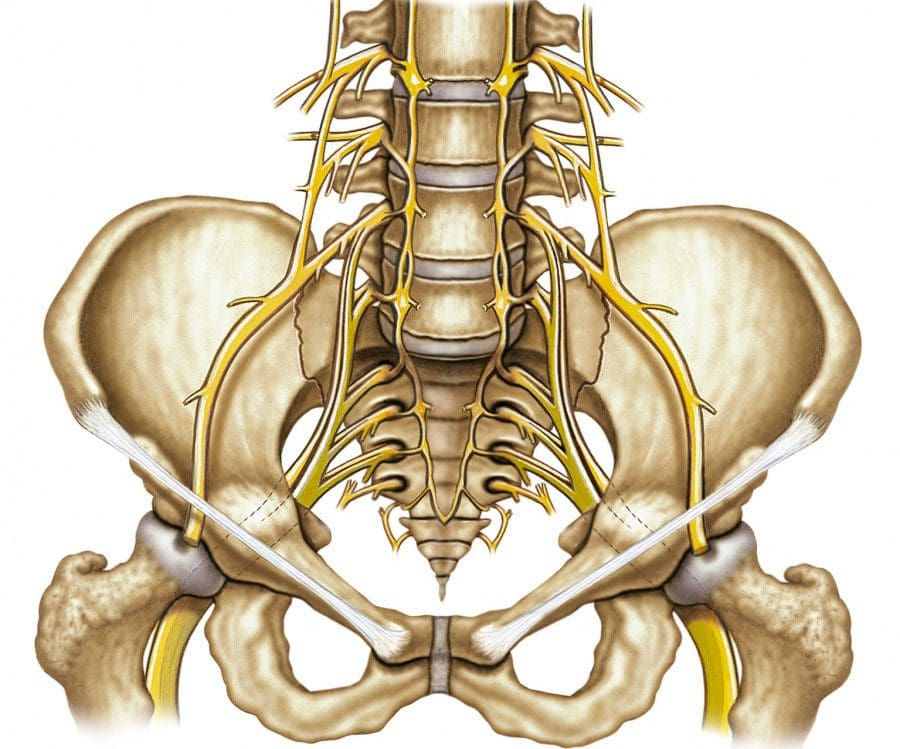
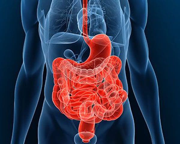
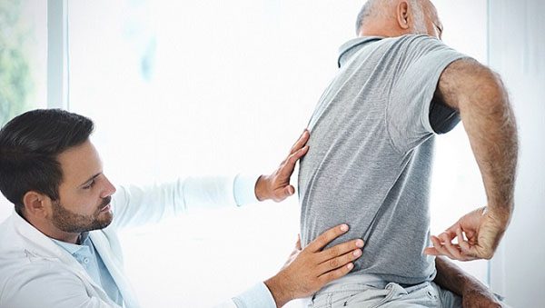
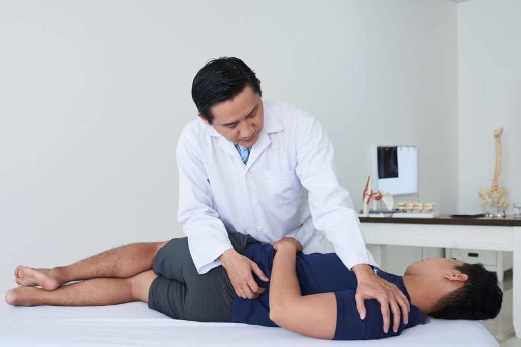
Stomach ache, acid reflux, gas, and other symptoms of gastric distress can be linked to spinal issues and misalignment. The spinal cord sends nerve signals to all parts of the body, specifically those affecting digestion functions. The lumbar spine/lower back includes the sacrum which is vital in terms of nerve function.
Various spinal cord issues could cause problems with the rest of the body. These include:
Disc compression
Herniated discs
Strained ligaments
Misalignments/problems in the lower back can result in gastric symptoms like:
Constipation
Diarrhea
Bloating
Gas
Bladder malfunction
This is because this area of the spine includes sympathetic and parasympathetic nerves that are connected to the digestive system. Any problem with these systems can result in miscommunicated signals to the rest of the body. The wide-range effects that compressed nerves can have on the body, as well as, how the spine is affected by the obstruction of these nerves, can be detrimental.
Chiropractic adjustments can help alleviate and release the gastric distress are able to correlate their spine�s role in gut health. This along with an education on the central nervous system. A chiropractic approach can help as a long-term solution to gastric distress.
The Nerves
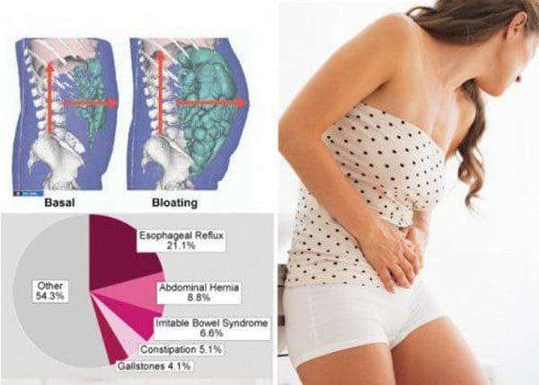
Every organ in the body functions by sending and receiving electrical impulses, transmitted through the nerves. These impulses direct the function of organs. If blocked or the signals are improperly/partially sent/received, various health issues can begin to present. For the gut, proper nerve signal transmission at full capacity is crucial. The stomach needs to be able to properly digest food while absorbing nutrients and preparing for waste removal. This is where gastric distress conditions begin like:
Irritable bowel syndrome – IBS
Gastroesophageal reflux disease – GERD
Abdominal pain syndrome – APS
Nerve conditions worsen with time if the health and function of the affected nerves are not restored. This could mean severe chronic symptoms and the possibility of permanent nerve damage.
Nerve Blockage
Messed up nerve signals are usually pinched, blocked, or displaced. Most nerve bundles exit through the spine and are usually where a chiropractic exam will start. Through palpitation of the spine along with diagnostic imaging, a chiropractor can track down exactly where the nerve blockage/s are taking place.
The lower back and upper back are common areas to examine. This is because a majority of abdominal organ nerves branch out from these spinal segments. If spinal subluxations are present, more than likely they are affecting the function of these organs. Chiropractic will adjust the spine and reset/realign the spine to its proper form, allowing for proper blood circulation. Compressed nerves can also cause inflammation that could require more complex treatment.
Listening to the Body
If the gut is presenting with aches, and bloating after every meal, it could be indicating that something is wrong or off. Individuals cannot feel blocked nerve signals, but the gut can. Listen to it when it is alerting an issue or problem. We want to educate our patients on gut and spinal health. Chronic gastric distress can be corrected with chiropractic.
Chiropractic Pain Relief
Dr. Alex Jimenez�s Blog Post Disclaimer
The scope of our information is limited to chiropractic, musculoskeletal, physical medicines, wellness, and sensitive health issues and/or functional medicine articles, topics, and discussions. We use functional health & wellness protocols to treat and support care for injuries or disorders of the musculoskeletal system. Our posts, topics, subjects, and insights cover clinical matters, issues, and topics that relate and support directly or indirectly our clinical scope of practice.*
Our office has made a reasonable attempt to provide supportive citations and has identified the relevant research study or studies supporting our posts. We also make copies of supporting research studies available to the board and or the public upon request. We understand that we cover matters that require an additional explanation as to how it may assist in a particular care plan or treatment protocol; therefore, to further discuss the subject matter above, please feel free to ask Dr. Alex Jimenez or contact us at 915-850-0900. The provider(s) Licensed in Texas& New Mexico*
References
Spiegel, Brennan M R et al. �Understanding gastrointestinal distress: a framework for clinical practice.��The American journal of gastroenterology�vol. 106,3 (2011): 380-5. doi:10.1038/ajg.2010.383
Kehl, Amy S et al. �Relationship between the gut and the spine: a pilot study of first-degree relatives of patients with ankylosing spondylitis.��RMD open�vol. 3,2 e000437. 16 Aug. 2017, doi:10.1136/rmdopen-2017-000437
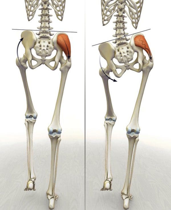
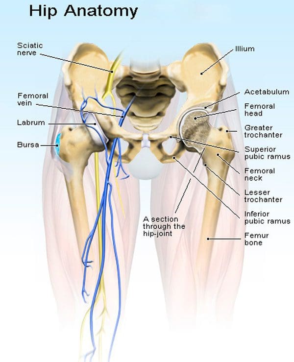
The number of individuals experiencing hip along with back pain is increasing and could benefit from chiropractic hip realignment. Chiropractic treatment is the recommended first-line option for hip pain and other issues related to misaligned hips. It is non-invasive and allows the body to heal naturally.
Shifted Hips
Hips that fall out of proper alignment have the potential to cause a variety of health issues in the body. The hips support a great deal of the body�s weight and facilitate substantial movement. The hips need to be properly balanced to allow for optimal mobility without compensation from the lower back and legs. A shift in the hips can occur from:
Shifted hips can cause pain in the low back, hips, and legs. This occurs from an altered gait and range of motion in this region. However, the hip joints, pelvic bones, muscles, and ligaments can all contribute to hip shifting and pain. Hip issues can also cause sciatica.
Hip Function
The hips do not receive as much attention until aches and pain start to present. Our hips are involved in nearly everything we do, providing numerous functions. It is no wonder they can cause tremendous pain when they’re out of alignment. The hips:
Keep the body upright
Bear the body’s weight
Allow for smooth:
Walking
Kicking
Running
Jumping
Sitting
Hip Adjustment
How to know if a hip adjustment is necessary? If there is any type of discomfort, soreness, and especially pain, achiropractor is the best medical professional to perform an examination and recommend if an adjustment is necessary or could just need ice/heat and rest.
However, if the source is being caused by another condition or injury the chiropractor will recommend the proper health care professional/specialist that can treat the issue. Hip pain can also be brought on from a different part of the body having its own issues. One of the most common types of hip pain is actually from a hernia or sciatica.
Chiropractic Hip Realignment
Chiropractic techniques focus on rebalancing the body, especially when realigning the hips. Manual manipulation and mobilization treatment techniques can improve flexibility, strength, and positioning to promote optimal balance and hip realignment.
A combination of adjustment techniques can be performed on the hip and spinal joints. Massage treatment can be incorporated into the treatment plan to loosen tight muscles, as the hips are realigned and are able to move freely. The holistic nature of chiropractic doesn’t just treat just the symptoms but identifies and treats the root cause. If the pain continues or worsens then the possibility for hip surgery could be recommended.
Complex Treatment
Working on imbalanced hips is not as straightforward as adjusting the low back. This is because there are a variety of arteries, nerves, joints, muscles, and other tissue structures that need to be considered during hip realignment. A chiropractor will use a careful combination of techniques when treating an imbalanced hip due to the complex nature of the area. In addition to chiropractic adjustments, chiropractors will suggest ways to improve hip mobility at home. This includes:
Stretches
Posture work
Exercises
These will help prevent the hips from shifting out of alignment. Treating pain at its source is what chiropractors do. Chiropractic hip realignment along with the realignment of the spine will allow the body to move freely, maintain balance and strength.
Hip Labral Tear Rehabilitation
Dr. Alex Jimenez�s Blog Post Disclaimer
The scope of our information is limited to chiropractic, musculoskeletal, physical medicines, wellness, and sensitive health issues and/or functional medicine articles, topics, and discussions. We use functional health & wellness protocols to treat and support care for injuries or disorders of the musculoskeletal system. Our posts, topics, subjects, and insights cover clinical matters, issues, and topics that relate and support directly or indirectly our clinical scope of practice.*
Our office has made a reasonable attempt to provide supportive citations and has identified the relevant research study or studies supporting our posts. We also make copies of supporting research studies available to the board and or the public upon request. We understand that we cover matters that require an additional explanation as to how it may assist in a particular care plan or treatment protocol; therefore, to further discuss the subject matter above, please feel free to ask Dr. Alex Jimenez or contact us at 915-850-0900. The provider(s) Licensed in Texas& New Mexico*
References
Okuzu, Yaichiro et al. �Hip-Spine Syndrome: Acetabular Anteversion Angle Is Associated with Anterior Pelvic Tilt and Lumbar Hyperlordosis in Patients with Acetabular Dysplasia: A Retrospective Study.��JB & JS open access�vol. 4,1 e0025. 29 Jan. 2019, doi:10.2106/JBJS.OA.18.00025
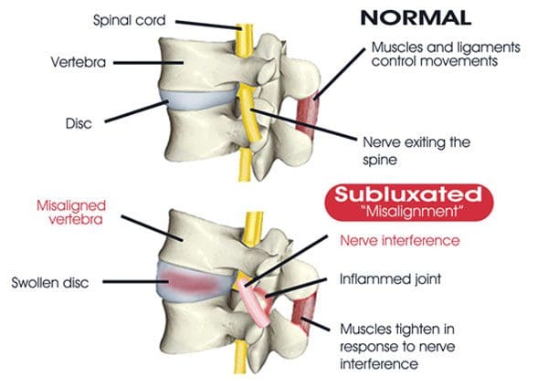
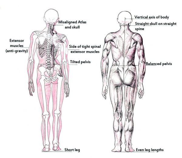
Most individuals go about their lives without thinking about poor posture and spinal misalignment, not to mention the spinal health problems/conditions that are associated with misaligned vertebrae. Most spinal misalignments come on as a result of:
Poor posture habits
Unhealthy diet
Destructive lifestyle choices
Sudden misalignments caused by accident/s or injury/s
Sudden misalignments can be associated with a direct cause that an individual can recognize increased symptoms and injury. It�s the long-term spinal misalignment that can become dangerous if left untreated and out of alignment. This is when it is time to see a chiropractor for an immediate spinal realignment.
Body Posture Mechanics
Poor posture along with poor body mechanics contributes to spinal misalignment. This creates unnecessary and consistent pressure on the:
Muscles
Ligaments
Joints
Discs
The pressure also causes a stretching of the nerves in the neck and low back. These nerves are responsible for transmitting, muscle expansion/contraction signals, vital information, and energy to the body’s organs. Any nerve interference will diminish the energy/blood flow to the limbs and organs. This can lead to disease and other major health complications like:
Chronic pain
Decreased mobility
Joint stiffness
Slouched posture
Reduced range of motion
Discomfort when sitting, standing, and lying down
Permanent joint/bone deformity/s
Broken bones, specifically in the spine
Most individuals do not recognize the gradual or long-term health problems linked to the spinal misalignment. Often the signs and symptoms are very subtle, so do not raise a cause for concern. The most direct symptoms of poor posture and spinal misalignment include:
Sore/stiff neck or back
Low energy
Tiredness/fatigue
Headaches
Back muscle spasms
Joint pain
Numbness
Tingling
Altered sensations
In addition to the symptoms, individuals should realize that underlying health conditions could also be linked to poor posture and spinal misalignment. If any of these symptoms are presenting see a chiropractor for an examination and proper diagnosis. These symptoms can be masked with pain killers, mattresses, or caffeinated drinks. However, these will not realign the spine to its proper form.
Chiropractic Lower Back Treatment
Dr. Alex Jimenez�s Blog Post Disclaimer
The scope of our information is limited to chiropractic, musculoskeletal, physical medicines, wellness, and sensitive health issues and/or functional medicine articles, topics, and discussions. We use functional health & wellness protocols to treat and support care for injuries or disorders of the musculoskeletal system. Our posts, topics, subjects, and insights cover clinical matters, issues, and topics that relate and support directly or indirectly our clinical scope of practice.*
Our office has made a reasonable attempt to provide supportive citations and has identified the relevant research study or studies supporting our posts. We also make copies of supporting research studies available to the board and or the public upon request. We understand that we cover matters that require an additional explanation as to how it may assist in a particular care plan or treatment protocol; therefore, to further discuss the subject matter above, please feel free to ask Dr. Alex Jimenez or contact us at 915-850-0900. The provider(s) Licensed in Texas& New Mexico*
References
Veintemillas Ar�iz, M T et al. �Changes in spinal alignment.� �Alteraciones de la alineaci�n vertebral.��Radiologia�vol. 58 Suppl 1 (2016): 115-27. doi:10.1016/j.rx.2016.01.007
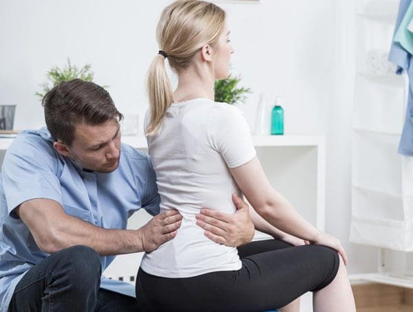
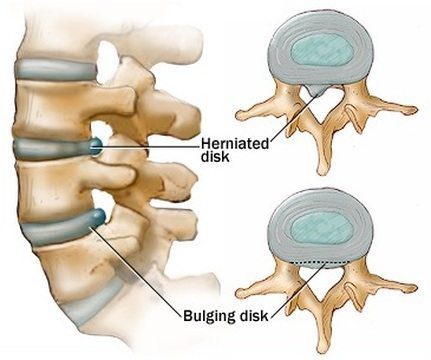
Back or disc pain is becoming increasingly common for individuals of all ages. The spinal discs are prone to injury because of the extreme amount of pressure/stress placed on the lower back and neck. Fortunately, most cases of back pain heal on their own, while others can cause long-term chronic pain that can be difficult to manage, without proper treatment.
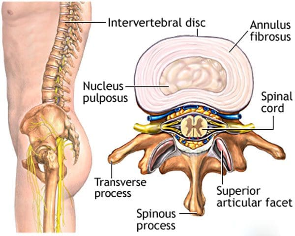
Different types of pain can be caused by problems with the spinal discs. The spinal discs are elastic sections of the spine that sit between the vertebrae. They are made up of materials called annulus fibrosus on the outside and a gel-like material called nucleus pulposus on the inside.
These discs are flexible, which allows for a range of motion to the spine and body shock absorption to increase comfort when in motion. When an individual visits a chiropractor, the practitioner will not know the exact cause of the pain, but only that it hurts around a certain area. Our spinal experts will help individuals understand the different types of ailments or conditions that could be affecting the spine. Then a customized treatment plan can be developed.
Disc problems
Spinal disc pain has two major sources of pain related to spinal discs: They are disc degeneration and nerve root pain. They are most common in the neck and the lower back. Individuals need to be informed of the difference to understand what is happening with the spine and potential treatment options to alleviate the pain and heal the problem.
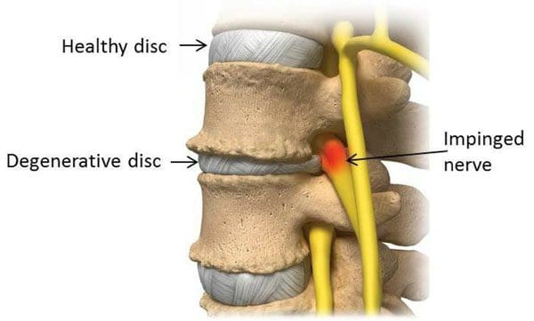
Degenerative disc disease
Degenerative disc disease is when the spinal discs begin to dry out and deteriorate. The discs start to shrink and can tear, which leaves that segment with little or no cushioning. This can cause pain, as well as inflammation of the surrounding muscles and joints. Disc degeneration usually presents with low levels of consistent pain and occasional severe flare-ups.
A chiropractor can utilize spinal manipulation to restore alignment, function, and mobility of the affected joint. Massages stretches and exercises will help alleviate the tension in the surrounding muscles along with strengthening them for better support.
Nerve root pain
Nerve root pain does not take place within the disc but is usually caused by a bulging or herniated disc. This condition can be called a slipped disc or pinched nerve, affect the nerves that are in close proximity to the neck and lower vertebrae.
The pain can lead to numbness, tingling, and weakness along the path of the nerve, and radiate out to the arms and legs. A bulging disc is when the spinal disc progressively protrudes through a narrow opening.
This can irritate any nearby nerve roots causing inflammation and pain. If the disc herniates or breaks through then the outer protective material tears, letting the cushion/gel leak out and come in contact with the nerve root/s, which could also cause pinching and inflammation. Treatment for a bulging or herniated disc includes:
Spinal manipulation
Corrective exercises
Physical therapy
Massage
Diet adjustments
These treatments can help realign the disc/s, moving them away from any nerves, and minimizing inflammation.
Proper Identification and Diagnosis
Spinal disc problems can be similar in their pain and symptoms. For example, degenerative disc disease can weaken the spinal discs to such a degree that nerve root pain follows creating a dual combination of pain. The conditions require various approaches and treatment methods often done in combination. However, they require a proper diagnosis to create a proper and custom treatment program for every individual. This will ensure the root cause of the disc pain is properly identified and handled. Call us to learn more.
Skateboarding Injury Treatment
Dr. Alex Jimenez�s Blog Post Disclaimer
The scope of our information is limited to chiropractic, musculoskeletal, physical medicines, wellness, and sensitive health issues and/or functional medicine articles, topics, and discussions. We use functional health & wellness protocols to treat and support care for injuries or disorders of the musculoskeletal system. Our posts, topics, subjects, and insights cover clinical matters, issues, and topics that relate and support directly or indirectly our clinical scope of practice.*
Our office has made a reasonable attempt to provide supportive citations and has identified the relevant research study or studies supporting our posts. We also make copies of supporting research studies available to the board and or the public upon request. We understand that we cover matters that require an additional explanation as to how it may assist in a particular care plan or treatment protocol; therefore, to further discuss the subject matter above, please feel free to ask Dr. Alex Jimenez or contact us at 915-850-0900. The provider(s) Licensed in Texas& New Mexico*
References
Browning, J E. �Chiropractic distractive decompression in the treatment of pelvic pain and organic dysfunction in patients with evidence of lower sacral nerve root compression.��Journal of manipulative and physiological therapeutics�vol. 11,5 (1988): 426-32.
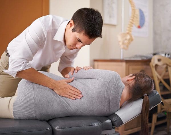
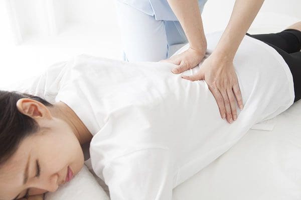
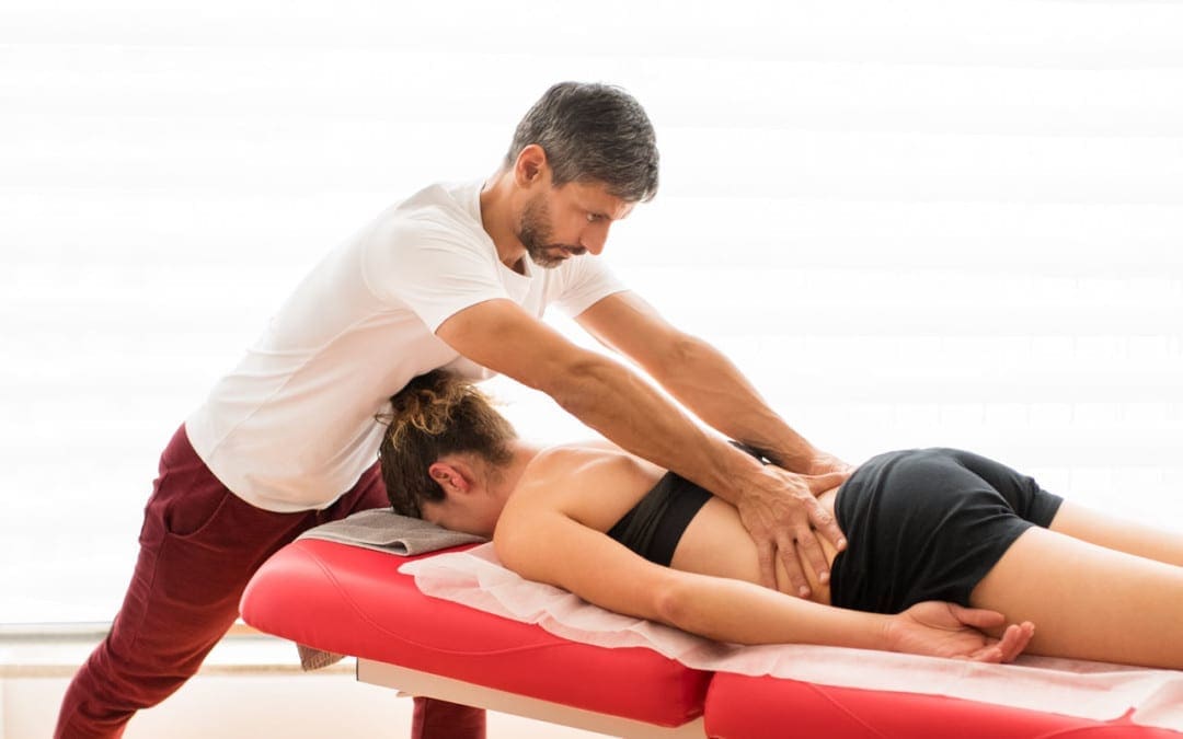
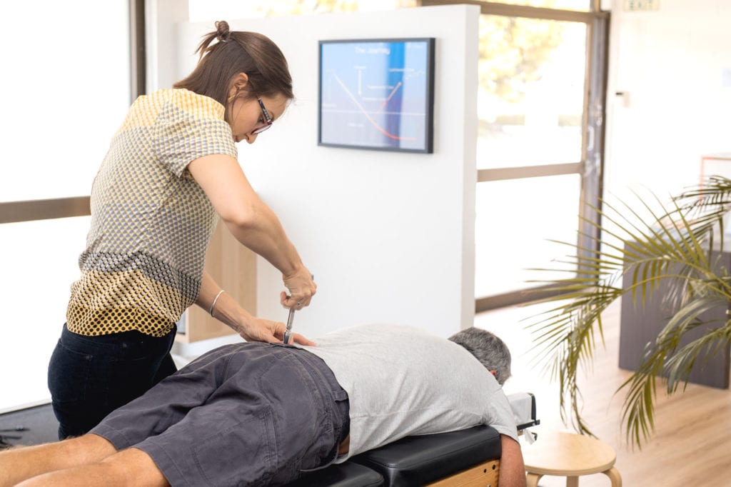
Chiropractic spinal mobilization techniques involve the slow and steady movements of the spine’s joints reestablishing their range of motion. Because it is a slower treatment style the techniques are done with the hands. However, a chiropractor can use various instruments/tools as well.
Spinal mobilization treatment has the same focus as spinal manipulation. To get the body back to optimal health and allow the body to heal itself naturally. However, there can be a variety of reasons for utilizing spinal stabilization, with treatment depending on the patient’s needs, if there are underlying conditions, or previous injury/s, and individual preference.
Some prefer mobilization because it is gentler and does not generate the pops or cracking sounds. And the chiropractor’s style/specialization comes into play. Some work in the firm manipulation high-velocity style, while others utilize the softer mobilization style and others work in combination.
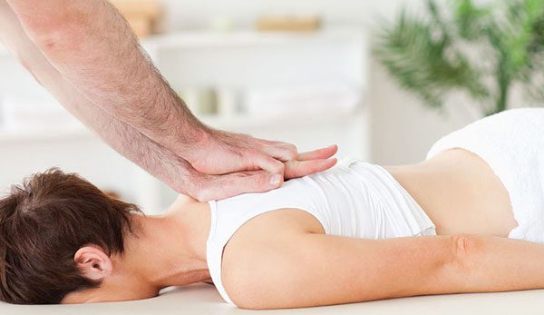
This adjustment re-alignment utilizes the necessary force to release the joint out of its restricted motion to improve mobility and reduce pain. There are various types of high-velocity low-amplitude manipulation approaches. These are the more common manipulation techniques:
Diversified Technique
This high-velocity low-amplitude technique is the one that is commonly associated with chiropractic manual adjustments. The chiropractor applies a short – low-amplitude, quick high-velocity thrust of the restricted joints. This is done one at a time with the objective to restore the normal range of motion. The patient is positioned in various positions to optimize the adjustment/alignment.
Gonstead Adjustment
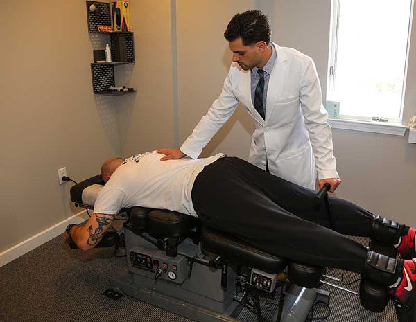
The Gonstead technique is another high-velocity low amplitude adjustment. It is similar to the diversified technique. The difference is the evaluation performed to specifically locate the painful joint and positioning of the body as the treatment is performed. Chiropractic or physical therapy chairs and tables can be used to position the patient for optimal treatment, like a cervical chair or a chest-knee table.
Thompson Terminal Point Drop Technique
Here specialized treatment tables with sections that drop down during a high-velocity low-amplitude thrust. The idea is that as the table drops the piece dropped allows for easier movement of the joint. A cracking sound can sometimes be heard. It depends on the patient and their condition. This type of manipulation can also be done in a gentle fashion making it a form of spinal mobilization.
Spinal mobilization
Slow steady motion/movements are performed to mobilize the joint. Spinal mobilization can be recommended for certain individuals for different reasons like:
Individual preference for spinal mobilization over spinal manipulation
Individuals with a sensitive nervous system can benefit from the gentle technique. This can keep the body from experiencing a negative reaction that can cause muscle spasms or other issues.
Individuals with certain conditions could be given a recommendation for spinal mobilization. This could be:
Individuals in the acute stage of their condition and experiencing severe pain
Obesity can be a factor as the positioning and the manipulation procedures can be a challenge for the provider and the patient requiring a low force approach.
Mobilization Approaches
The more common spinal mobilization approaches include:
Activator Technique
The Activator is a hand-held, spring-loaded tool that generates a low-force impulse. A patient lies face down on the adjustment table, while the chiropractor:
Examines leg length
Performs muscle testing
Adjusts the spine and/or extremity joints
Cox Flexion-Distraction Technique
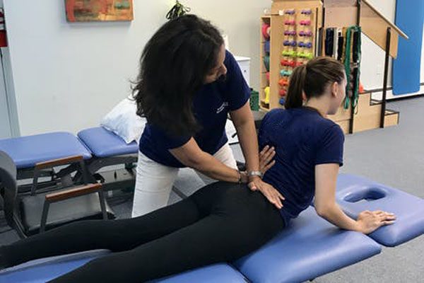
Here a gentle adjustment is designed to adjust the vertebrae by gently stretching the lower spine. This is usually performed in a series of repetitive slow movements like a steady rocking motion.
Toggle Drop
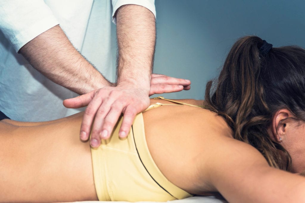
Here gravity is utilized to apply the adjustment. The chiropractors’ hands are crossed and on top of each other. Then the chiropractor presses down quickly and firmly on the area of the spine while a section of the table drops. The table sections can be raised and dropped according to the localization of the spinal adjustment.
McKenzie Technique
This technique incorporates active patient involvement, empowerment, and self-care as part of the treatment.
Spinal Release
The chiropractor separates the misaligned vertebrae by applying gentle pressure using the fingertips, with the objective to restore the spine back to a natural position.
Sacro-Occipital Technique – SOT
This technique utilizes wedges/blocks under the pelvis. This allows gravity with added low-force to assist the chiropractor to realign the pelvis.
Sciatica Alleviation
All of these techniques can be utilized by a chiropractor for sciatic nerve pain alleviation or can discover other conditions that could be mimicking sciatica.
Nerve mobilization techniques have been recently used as a method to adjust radiating pain related to disc disease, and in particular, mobilization techniques for the sciatic nerves improve mobility of the sciatic nerves, decrease mechanosensitivity of the nervous system, and heighten compliance of nerve tissues, relieving low back pain. Jeong, Ui-Cheol et al. �The effects of self-mobilization techniques for the sciatic nerves on physical functions and health of low back pain patients with lower limb radiating pain.��Journal of physical therapy science�vol. 28,1 (2016): 46-50. doi:10.1589/jpts.28.46
Sciatica Rehabilitation Causes and Symptoms
Dr. Alex Jimenez�s Blog Post Disclaimer
The scope of our information is limited to chiropractic, musculoskeletal, physical medicines, wellness, and sensitive health issues and/or functional medicine articles, topics, and discussions. We use functional health & wellness protocols to treat and support care for injuries or disorders of the musculoskeletal system. Our posts, topics, subjects, and insights cover clinical matters, issues, and topics that relate and support directly or indirectly our clinical scope of practice.*
Our office has made a reasonable attempt to provide supportive citations and has identified the relevant research study or studies supporting our posts. We also make copies of supporting research studies available to the board and or the public upon request. We understand that we cover matters that require an additional explanation as to how it may assist in a particular care plan or treatment protocol; therefore, to further discuss the subject matter above, please feel free to ask Dr. Alex Jimenez or contact us at 915-850-0900. The provider(s) Licensed in Texas& New Mexico*
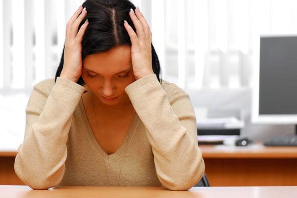
Seasonal Affective Disorder is a form of depression that has to do with the changing of the seasons, specifically when fall begins. It affects around 10 million Americans during the fall and winter seasons. Back pain can be a symptom of the disorder. For most individuals, the condition follows a pattern:
These are typical symptoms for many dealing with the condition. The individual can experience aches and pains throughout the body, but especially the back. Recent studies have shown how various forms of depression, like Seasonal Affective Disorder, can present with pain, specifically back/spinal pain. Many individuals play it off as the blues, but research has found, depression can have a much more significant impact on an individual’s quality of life. The exact cause is still unknown. But research is finding a connection related to the level of sunlight an individual receives throughout the day.
Research has shown there could be a biochemical imbalance in the brain when there is less daylight during fall and winter. As the seasons change from summer to fall individuals can experience a shift in circadian rhythms that can cause them to disrupt their regular daily routine. However, Seasonal Affective Disorder is more common for individuals living where the daylight lessens and has been shown to affect women more than men and young adults.
Depression and Back Pain Connection
Depression can present with pain symptoms and the pain can enhance the depression. Headaches, Body aches, and especially backaches are common symptoms of depression. Research has shown that individuals with severe depression can actually feel a more intense level of pain.
Physical symptomslike back pain or headaches can be the only or the beginning symptom/s of seasonal affective depression. As research continues to grow as to how the nervous system interacts with the body, pain symptoms have been found to be connected to biological mechanisms connected with stress, anxiety, and depression.
Living a sedentary lifestyle, with little or no physical activity and regular exercise can be another cause for Seasonal Affective Disorder. Depression itself can cause fatigue that restrains individuals from exercising and working the core muscles for optimal spinal strength and health.
This added stress on the spine’s discs, joints, and ligaments, makes the body more susceptible to low back pain, muscle strains, illness, and injuries. Pain can wear an individual down impacting mood and overall health.
Symptoms
The difference between Seasonal Affective Disorder and chronic depression is that Seasonal Affective Disorder is limited to the same time of the year during the time of less light, and winter months. Symptoms of Seasonal Affective Disorder can be the same as those associated with depression. They are:
Feeling Low/Depressed
Weight gain
Increased appetite
Craving sugar and carbohydrates
Sleeping all-day
Consistent drowsiness
Hopelessness
Loss of interest in enjoyed activities
Symptoms can also be related to low levels of Vitamin D, which is associated with anxiety and depression for individuals with fibromyalgia. Seasonal Affective Disorder can be related to chronic pain conditions the way depression can. Some individuals with chronic fatigue syndrome can also present with symptoms of Seasonal Affective Disorder.
Treatment
Diagnosis for Seasonal Affective Disorder requires an individual to experience at least two years of symptoms that become worse at a specific time of the year. And the depressive episodes have to significantly be worse than the non-seasonal episodes of depression. There are four types of treatments, that can be used individually or in combination. They are:
Cognitive-behavioral therapy/CBT is a form of psychotherapy that is effective for the disorder, as well as other conditions. It relies on techniques that identify negative thoughts and work on ways to not dwell on the negative and focus more on positive things and thoughts.
Lightbox Therapy
Diminished sunlight when fall and winter arrive can be replaced with regular exposure to a bright, artificial lightbox. Individuals sit or stand in front of the lightbox when they get up on a daily basis. This is done when fall begins and goes on until spring. The lightbox filters out ultraviolet rays and requires around 20 to 60 minutes of exposure to 10,000 lux of cool-white fluorescent light.
Vitamin D
Low levels of vitamin D were found in individuals with Seasonal Affective Disorder. Talk to a doctor about the proper level of vitamin D that should be taken through supplements.
The Pain and Backaches
Understanding how pain and depression are interconnected treating both conditions as part of an integrative treatment plan can include:
A light aerobic exercise program that stimulates serotonin levels and releases endorphins to relieve depression and pain.
Low-dose antidepressants can reduce depression symptoms and back pain. They work to inhibit the reuptake of neurotransmitters like serotonin and norepinephrine that is associated with a person�s mood and the way they perceive pain.
What Works
Everyone is unique, which means that different treatment plans and combinations of treatment plans may have to be tried out before finding the optimal one. A significant factor is not settling with the pain and just accepting it. Healing can be a unique and complex experience. The objective is for the individual and doctor to work collaboratively.
Depression and Chronic Pain
Dr. Alex Jimenez�s Blog Post Disclaimer
The scope of our information is limited to chiropractic, musculoskeletal, physical medicines, wellness, and sensitive health issues and/or functional medicine articles, topics, and discussions. We use functional health & wellness protocols to treat and support care for injuries or disorders of the musculoskeletal system. Our posts, topics, subjects, and insights cover clinical matters, issues, and topics that relate and support directly or indirectly our clinical scope of practice.*
Our office has made a reasonable attempt to provide supportive citations and has identified the relevant research study or studies supporting our posts. We also make copies of supporting research studies available to the board and or the public upon request. We understand that we cover matters that require an additional explanation as to how it may assist in a particular care plan or treatment protocol; therefore, to further discuss the subject matter above, please feel free to ask Dr. Alex Jimenez or contact us at 915-850-0900. The provider(s) Licensed in Texas& New Mexico*
References
Robertson, David et al. �Associations between low back pain and depression and somatization in a Canadian emerging adult population.��The Journal of the Canadian Chiropractic Association�vol. 61,2 (2017): 96-105.
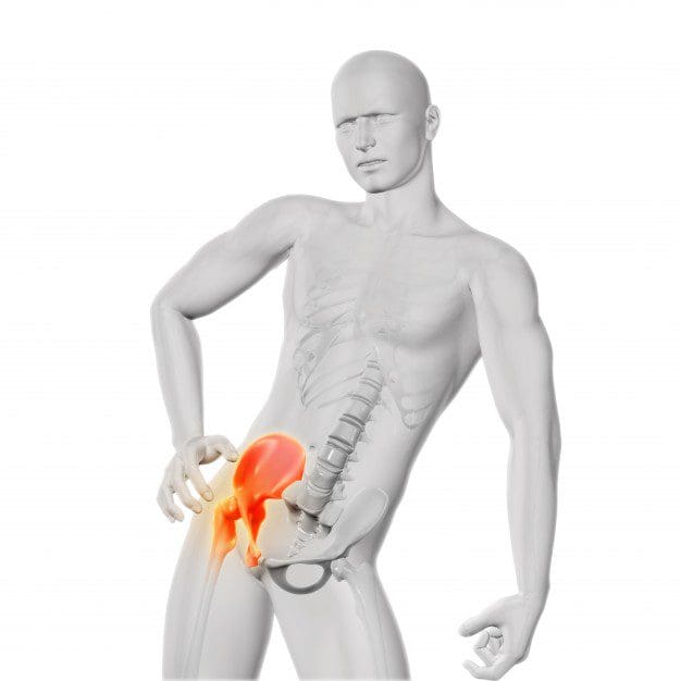
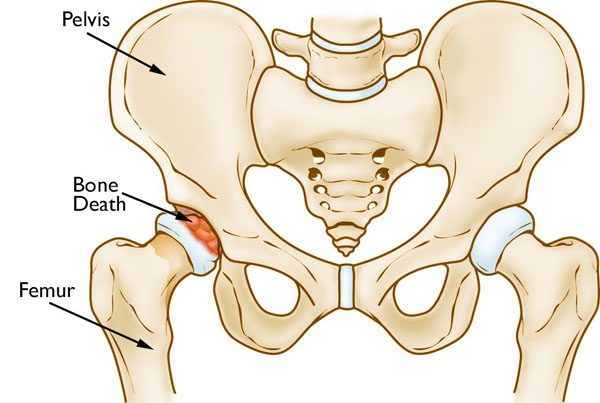
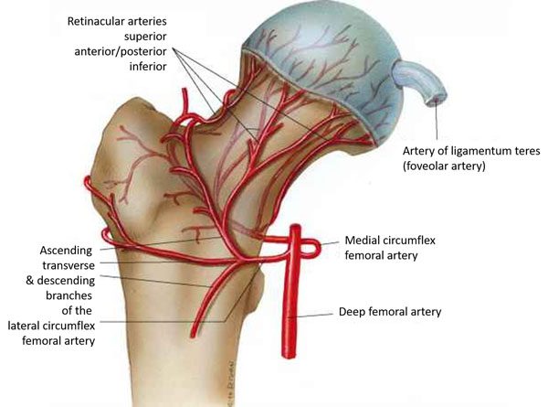
Osteonecrosis is a condition that causes the death of bone tissue from temporary or permanent loss of blood supply to the affected area. It is commonly known asAvascular necrosis and can lead to miniature/tiny breaks in the bone and the bone/s eventually collapsing. Specifically, it affects the upper part of the femur or femoral head and surrounding joints.
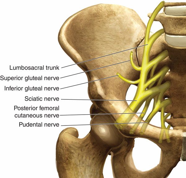
It can occur in any bone however, osteonecrosis typically affects the hip/s. Pain associated with osteonecrosis of the hip can be localized to the center of the groin, thigh, or buttock. Because of the hip joint’s close proximity to the sciatic nerve, misdiagnosis for sciatica is common.
Mimicking Sciatica Symptoms
Unfortunately, many health care providers can misdiagnose osteonecrosis hip pain as sciatica. Whatever the cause of the hip injury, most individuals with hip pathology report pain in the groin, upper thigh, and buttocks.
That is why a trained medical professional that knows the differences in the symptoms of each condition can make all the difference in making a proper diagnosis. And a proper diagnosis leads to proper and complete treatment of whichever condition it may be. With osteonecrosis, misdiagnosis often delays the proper treatment and continues to progress. Common symptoms of sciatica:
Leg pain is the primary symptom can be mild to severe
Low back pain is secondary can be mild to severe
Nerve-related symptoms
Numbness
Tingling
Shooting pain
Pins-and-needles sensation
Muscle weakness
Hip pain especially flexion and internal rotation of the hip.
Leg or foot weakness
Osteonecrosis Symptoms and Similarities
For many, there are no symptoms in the early stages of osteonecrosis. As the condition worsens, the affected joint could present pain symptoms only when weight is placed on it. Eventually, individuals begin to feel the pain even when lying down. Pain can be mild to severe with a gradual development. Other symptoms that mimick sciatica:
Walking Inability
Walking gait is complicated with both conditions which is a major cause behind the misdiagnosis.
Limping
Individuals often limp with osteonecrosis of the hip and spinal disc problems. This is another reason that the condition is misdiagnosed as a spinal disc problem or nerve root compression of the sciatic nerve.
Hip Pain
The tributaries/veins of the sciatic nerve also supply the hip area and often cause confusion between the two conditions.
Differences
Despite all of the similarities. There are differences in both conditions.
Nature of The Pain
With sciatica, the pain is related to the nervous system. Movement can complicate the pain. While rest helps to reduce the pain.
With Osteonecrosis the pain is geared toward the muscular. Rest does not help reduce the pain. In fact, the pain increases at night.
Location
Sciatica pain can radiate through the whole leg from the low back to the toe.
Osteonecrosis pain is confined to the hip joint, groin, and radiates to the knee joint only. Osteonecrosis pain does not radiate below the knee joint.
Restricted Movement
Osteonecrosis of the hip joint, means the movements involving the hip joint are restricted. Individuals cannot rotate the leg to the right and left. Individuals cannot bend or fold from the hip.
With sciatica, the rotation of the leg is not affected. Movements involving stretching the sciatic nerve can cause relief or pain.
Walking Gait Differences
Gait is the way an individual stands and walks.
Osteonecrosis of the hip joint causes individuals to not be able to open the hip joint properly or to step properly.
With sciatica, an individual tends to lean on their side to relax the compression on the nerve.
Risk Factors
More than 20,000 people enter hospitals for the treatment of osteonecrosis of the hip yearly. Other than the hip, areas of the body likely to be affected are the shoulder, knee, hand, and foot. The condition can occur for a variety of reasons. A few of these include:
Fracture – a broken bone can interrupt the blood flow to other sections of the bone.
Dislocation of bone or joint/s
Alcoholism
Trauma
Radiation damage
Steroid use
Some individuals can have more than one condition or injury that contributes to hip flexor pain. An example is that it is possible to have both hip osteoarthritis and hip impingement. Without proper treatment, the condition can worsen, causing joint or hip pain from the degradation of the bone.
Anyone can be affected, but osteonecrosis is most common in individuals aged 30 to 50. Treatment options include a total replacement of the hip known as arthroplasty. And if it is sciatica then chiropractic treatment is a first-line treatment protocol. However, a chiropractor can make the distinction between the two and treat sciatica or refer the patient to the proper specialist.
Lower Back Pain Relief
Dr. Alex Jimenez�s Blog Post Disclaimer
The scope of our information is limited to chiropractic, musculoskeletal, physical medicines, wellness, and sensitive health issues and/or functional medicine articles, topics, and discussions. We use functional health & wellness protocols to treat and support care for injuries or disorders of the musculoskeletal system. Our posts, topics, subjects, and insights cover clinical matters, issues, and topics that relate and support directly or indirectly our clinical scope of practice.*
Our office has made a reasonable attempt to provide supportive citations and has identified the relevant research study or studies supporting our posts. We also make copies of supporting research studies available to the board and or the public upon request. We understand that we cover matters that require an additional explanation as to how it may assist in a particular care plan or treatment protocol; therefore, to further discuss the subject matter above, please feel free to ask Dr. Alex Jimenez or contact us at 915-850-0900. The provider(s) Licensed in Texas& New Mexico*
References
Li, Wen-Long et al. �Exploring the Risk Factors for the Misdiagnosis of Osteonecrosis of Femoral Head: A Case-Control Study.��Orthopaedic surgery, 10.1111/os.12821. 16 Oct. 2020, doi:10.1111/os.12821
IFM's Find A Practitioner tool is the largest referral network in Functional Medicine, created to help patients locate Functional Medicine practitioners anywhere in the world. IFM Certified Practitioners are listed first in the search results, given their extensive education in Functional Medicine