BHRT, EvexiPEL, and Whole-Body Hormone Care at El Paso Back Clinic
Bioidentical Hormone Replacement Therapy, or BHRT, is often discussed as a way to help people feel more like themselves again when hormone levels drop or become unbalanced. It may help with symptoms such as low energy, poor sleep, mood changes, lower sex drive, mental fog, and body composition changes. But at El Paso Back Clinic, the message should be clear: hormone care should never be treated like a stand-alone shortcut. It works best when hormonal symptoms are reviewed alongside thyroid health, metabolic health, inflammation, gut function, stress load, and overall body mechanics. That type of full-picture care aligns with the clinic’s integrative model, which combines chiropractic care, functional medicine, and advanced nursing under the care of Dr. Alexander Jimenez, DC, APRN, FNP-BC. (Cleveland Clinic, 2022; EVEXIAS Health Solutions, n.d.; El Paso Back Clinic, 2026).
What BHRT Means
Bioidentical hormones are hormones designed to closely match those the human body naturally produces. Cleveland Clinic explains that BHRT is used to help manage symptoms related to menopause or other hormone imbalances, and that these hormones can come in several forms, including pills, creams, patches, gels, injections, and pellets. Cleveland Clinic also notes that some bioidentical options are FDA-approved, while custom-compounded versions are less studied and may carry more uncertainty. That matters because patients often hear the word “natural” and assume “risk-free,” but that is not always true. (Cleveland Clinic, 2022; Cleveland Clinic, 2024).
In simple terms, BHRT is not just about replacing hormones. It is about determining whether hormones are the primary issue, which hormones are low or imbalanced, and whether other systems are also involved. A person with fatigue, weight gain, poor focus, low motivation, or digestive problems may have a hormone imbalance, but they may also have thyroid dysfunction, insulin resistance, poor sleep, chronic stress, inflammation, or nutritional problems. That is why careful medical review matters before treatment begins. (Cleveland Clinic, 2024; EVEXIAS Health Solutions, n.d.).
Why This Topic Fits El Paso Back Clinic
El Paso Back Clinic is not just a back pain site. The published clinical model emphasizes integrative care that connects structural health, metabolic health, gut function, inflammation, and advanced nursing support. The clinic’s materials describe a team approach that combines chiropractic care, functional medicine, lab testing, and personalized plans. Dr. Alexander Jimenez’s published content also connects thyroid health, metabolism, inflammation, and gut function rather than treating each complaint as a separate issue. That makes BHRT a natural fit for the site when it is presented as one part of a broader healing strategy, not as a single magic answer. (El Paso Back Clinic, 2026; Jimenez, n.d.).
For a spine and wellness audience, this matters even more because hormone problems can affect the whole body, including:
energy and recovery
sleep quality
muscle tone and body composition
inflammation levels
mood and stress tolerance
motivation for exercise and rehab
digestive comfort and gut regularity
When those systems are off, recovery from back pain, mobility, and overall function can also suffer. That is why a whole-person clinic can add value to hormone care. (El Paso Back Clinic, 2026; EVEXIAS Health Solutions, n.d.).
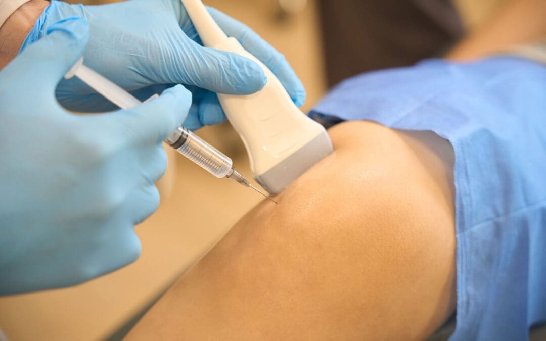
What EvexiPEL Pellet Therapy Is
EVEXIAS Health Solutions describes EvexiPEL as a clinically advanced BHRT method that uses tiny hormone pellets placed just under the skin during a simple in-office procedure. According to EVEXIAS, those pellets then release a steady, physiologic dose of hormones over about 3 to 6 months. The company presents this as a way to reduce the ups and downs that some people experience with daily creams, pills, patches, or more frequent injections. (EVEXIAS Health Solutions, n.d.).
That steady-release idea is one reason many patients are interested in pellet therapy. EVEXIAS states that pellets are designed to provide more consistent delivery and fewer “peaks and valleys” than some other delivery methods. For patients who do not want to remember daily or weekly dosing, that convenience can be appealing. At the same time, pellets are still a medical treatment, which means the patient needs the right workup, the right dosing plan, and the right follow-up. Convenience should never replace careful clinical judgment. (EVEXIAS Health Solutions, n.d.; Cleveland Clinic, 2024).
Why Thyroid and Metabolic Health Must Be Checked
One of the most important points for El Paso Back Clinic readers is that not every “hormone problem” starts with estrogen or testosterone. EVEXIAS says its testing protocols include sex hormone panels, advanced thyroid profiles with antibodies, adrenal stress and cortisol rhythm assessments, and metabolic markers such as insulin and A1C. That is a strong reminder that hormonal complaints often overlap with thyroid, adrenal, and metabolic health. (EVEXIAS Health Solutions, n.d.).
Dr. Jimenez’s metabolic thyroid content makes a similar point. His published thyroid articles explain that thyroid dysfunction can affect metabolism and can overlap with inflammation, chronic symptoms, and gut-related problems. In his educational materials, he also connects endocrine function with nutrition and whole-body recovery. This supports an important clinical idea: if someone has fatigue, poor exercise recovery, digestive symptoms, stubborn weight changes, or brain fog, the best next step is often a full workup rather than a guess. (Jimenez, n.d.).
This full workup may help answer questions like:
Is the problem mainly estrogen, progesterone, or testosterone related?
Is low thyroid function part of the picture?
Is stress chemistry affecting symptoms?
Is insulin resistance driving fatigue and weight gain?
Is chronic inflammation making everything worse?
Are gut issues interfering with absorption and recovery?
That kind of careful thinking aligns with how El Paso Back Clinic presents its broader care philosophy. (EVEXIAS Health Solutions, n.d.; El Paso Back Clinic, 2026).
Gut Health, Inflammation, and Hormone Balance
Many people who seek BHRT do not just complain about hormones. They also talk about bloating, constipation, poor digestion, mood swings, sleep trouble, and stubborn inflammation. The recent gut-health content from El Paso Back Clinic indicates a practical connection between the spine, gut, inflammation, and metabolism. The clinic’s published articles describe root-cause approaches that combine lab testing, nutrition support, and structural care. Dr. Jimenez’s thyroid and gut education also connects chronic inflammation with digestive imbalance and endocrine stress. (El Paso Back Clinic, 2026; Jimenez, n.d.).
This does not mean BHRT alone fixes gut health. It means hormone symptoms should be reviewed in a broader context. If a patient is exhausted, inflamed, constipated, bloated, gaining abdominal weight, and sleeping poorly, it makes sense to look at hormones, thyroid function, gut health, stress load, and nutrition together. That whole-body view is one of the strongest ways to position BHRT at El Paso Back Clinic. (EVEXIAS Health Solutions, n.d.; El Paso Back Clinic, 2026).
How an Integrative Clinic Can Improve BHRT Results
EVEXIAS says its broader model can include advanced lab testing, hormone therapy, targeted nutraceuticals, and peptide therapy as part of a personalized plan. Its functional and integrated health framework also includes support for the thyroid, adrenal, metabolic, and gut systems, as well as inflammation. That approach lines up well with the type of clinical ecosystem readers expect from El Paso Back Clinic. (EVEXIAS Health Solutions, n.d.).
At an integrative clinic, BHRT may be stronger when it is paired with:
full lab testing before treatment
thyroid and metabolic review
nutrition counseling
gut and inflammation support
peptide support when clinically appropriate
sleep, stress, and lifestyle coaching
chiropractic and rehab strategies that help the body move and recover better
El Paso Back Clinic’s own content states that the strongest results occur when chiropractic, functional medicine, and advanced nursing work together. The site describes this mix as a way to improve mobility, calm inflammation, support nerve function, and build long-term health. For a patient who is also struggling with low energy, hormone imbalance, or metabolic stress, that kind of coordinated care can be especially helpful. (El Paso Back Clinic, 2026; EVEXIAS Health Solutions, n.d.).
Clinical Observations From Dr. Alexander Jimenez
Dr. Alexander Jimenez’s published materials describe a multidisciplinary model built around chiropractic care, advanced nursing, functional medicine, imaging, lab review, and personalized recovery plans. El Paso Back Clinic’s recent clinical posts state that when structural treatment is paired with nutrition, hormone support, and metabolic care, patients often report increased energy, reduced inflammation, and improved overall function. The clinic also emphasizes that improved alignment, nerve function, and reduced inflammation can support recovery beyond just pain relief. (El Paso Back Clinic, 2026; Jimenez, n.d.; LinkedIn, n.d.).
For a BHRT article geared toward El Paso Back Clinic, the clinical takeaway is simple: the body functions as a single system. If hormones are off, the patient may also struggle with movement, sleep, inflammation, digestion, and stress resilience. If the spine and nervous system are stressed, that may also affect recovery, activity levels, and how well a patient responds to lifestyle changes. The strongest plan is one that respects both structure and chemistry. (El Paso Back Clinic, 2026).
Risks and Why Monitoring Matters
Cleveland Clinic is clear that all hormone replacement therapy comes with risks and that compounded bioidentical hormones may carry additional uncertainty because their long-term effects are not as well studied. Cleveland Clinic also says some people are not good candidates for hormone therapy and that treatment decisions should be based on symptoms, medical history, and an informed discussion with a healthcare provider. (Cleveland Clinic, 2022; Cleveland Clinic, 2024).
That is why a responsible BHRT program should include the following:
a full health history
lab work before treatment
a review of thyroid and metabolic markers
discussion of risks, benefits, and alternatives
regular follow-up for symptoms and side effects
dose adjustments when needed
For El Paso Back Clinic readers, this is an important message: smart hormone care is individualized, monitored, and tied to the patient’s bigger health picture. It is not just about giving more hormones. It is about finding the right level of support for the right patient at the right time. (Cleveland Clinic, 2024; EVEXIAS Health Solutions, n.d.).
Final Thoughts
BHRT can be a useful tool for the right patient, especially when symptoms are truly linked to hormone decline or imbalance. EvexiPEL pellet therapy offers a steady-delivery option that many patients find appealing, as it is designed to release hormones over 3 to 6 months. Still, the best hormone care does not stop at pellets or prescriptions. It looks at thyroid health, metabolism, inflammation, gut function, stress, nutrition, sleep, and physical recovery as a whole. That whole-body approach is exactly what makes this topic a strong fit for El Paso Back Clinic. (EVEXIAS Health Solutions, n.d.; El Paso Back Clinic, 2026; Cleveland Clinic, 2024).
Platelet-Rich Plasma (PRP) Therapy for Better Posture at El Paso Back Clinic: Natural Healing for Spine Strength and Daily Comfort
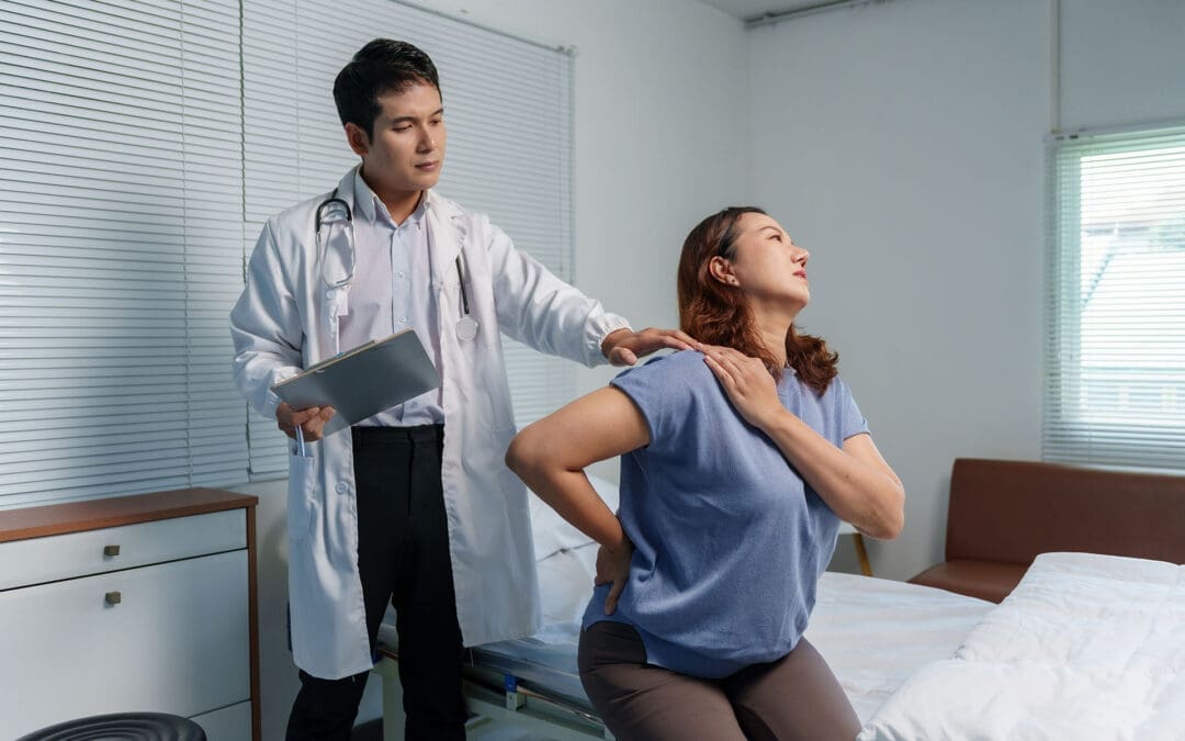
Many people in El Paso struggle with slouched shoulders or a rounded back that makes everyday tasks feel harder. These posture problems often hide more profound issues like pain, weak ligaments, or worn spinal discs. When it hurts to stand tall, the body chooses easier but unhealthy positions. Over time, this cycle worsens discomfort. At El Paso Back Clinic, platelet-rich plasma (PRP) therapy offers a natural way to break that cycle. PRP therapy can indirectly ease posture issues by calming the pain that forces bad habits, strengthening weak ligaments and tendons, and repairing degenerated spinal discs. When added to a full treatment plan at El Paso Back Clinic, PRP helps address the root musculoskeletal problems that cause poor posture. This leads to smoother movement and better body balance in the neck, back, and shoulders. Patients often turn to this path when exercises or pills stop working.
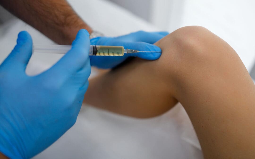
What Is Platelet-Rich Plasma Therapy at El Paso Back Clinic?
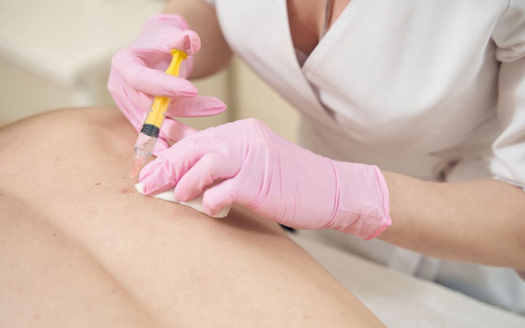
Platelet-rich plasma, or PRP, uses a small sample of your blood. Doctors at El Paso Back Clinic draw the blood, spin it in a centrifuge to concentrate the healing platelets, and inject it into sore areas with ultrasound guidance. These platelets release growth factors that kick-start the body’s repair process. The whole visit takes about 30 minutes, and no foreign drugs are used. This makes PRP a safe, natural choice for many El Paso residents dealing with back or neck pain.
Dr. Alexander Jimenez, DC, APRN, FNP-BC, CFMP, leads the multidisciplinary team at El Paso Back Clinic. His dual training as a chiropractor and family nurse practitioner lets him blend regenerative medicine with chiropractic care. In his clinical work, Dr. Jimenez notes that PRP supports the body’s natural healing processes, especially when combined with functional medicine and rehabilitation (Jimenez, n.d.). The clinic’s locations across El Paso, including the main site at 11860 Vista Del Sol, make this advanced care easy to reach.
PRP first helped athletes recover faster. Today, it is used to treat everyday wear and tear at locations such as El Paso Back Clinic. Johns Hopkins Medicine explains that PRP floods the area with growth factors to speed cell repair and reduce inflammation (Johns Hopkins Medicine, n.d.).
How PRP Injections Repair Damaged Tissues at the Clinic
Once injected, the concentrated platelets go right to work. They release growth factors that handle three key jobs:
Reduce swelling: Chronic inflammation keeps pain going and weakens tissues. PRP calms inflammation, so real healing can start.
Build stronger tissue: Growth factors boost collagen to toughen tendons and ligaments that support the spine.
Speed up repair: Platelets call in cells that fix tears and worn spots.
At El Paso Back Clinic, PRP is used to treat the spine for conditions like degenerative disc disease. Discs act like cushions between bones. When they wear down, pain spreads, and posture slumps. The clinic’s blog on PRP for spinal care reports that patients often experience improved disc health and reduced stiffness without surgery (El Paso Back Clinic, n.d.-a).
For shoulders, PRP helps rotator cuff tendons heal more quickly. Princeton Sports and Family Medicine reports that PRP boosts tendon growth and collagen, so people return to daily tasks faster (Princeton Sports and Family Medicine, n.d.).
Bullet points on the repair steps at El Paso Back Clinic:
Blood draw and spin create PRP with 2 to 8 times the platelet count of normal blood.
Ultrasound guides the needle to the exact spot for the best results.
Growth factors like PDGF, VEGF, and TGF-β promote the formation of new blood vessels and clear waste.
Benefits build over weeks to months, often after two or three sessions with rehab follow-up.
PRP Therapy and Spinal Disc Health in El Paso
Worn discs cause back pain that makes standing straight tough. PRP injections at El Paso Back Clinic go into the disc area or nearby joints. They cut inflammation and help discs hold more water for better cushioning. The Morrison Clinic’s review, used in the clinic’s protocols, notes improved flexibility after PRP for disc problems (The Morrison Clinic, n.d.). This added stability allows the spine to align naturally in daily life.
Dr. Jimenez’s clinical observations highlight that patients with disc wear regain mobility when PRP is combined with chiropractic adjustments. His team checks nutrition and inflammation levels to make results last longer (Jimenez, n.d.).
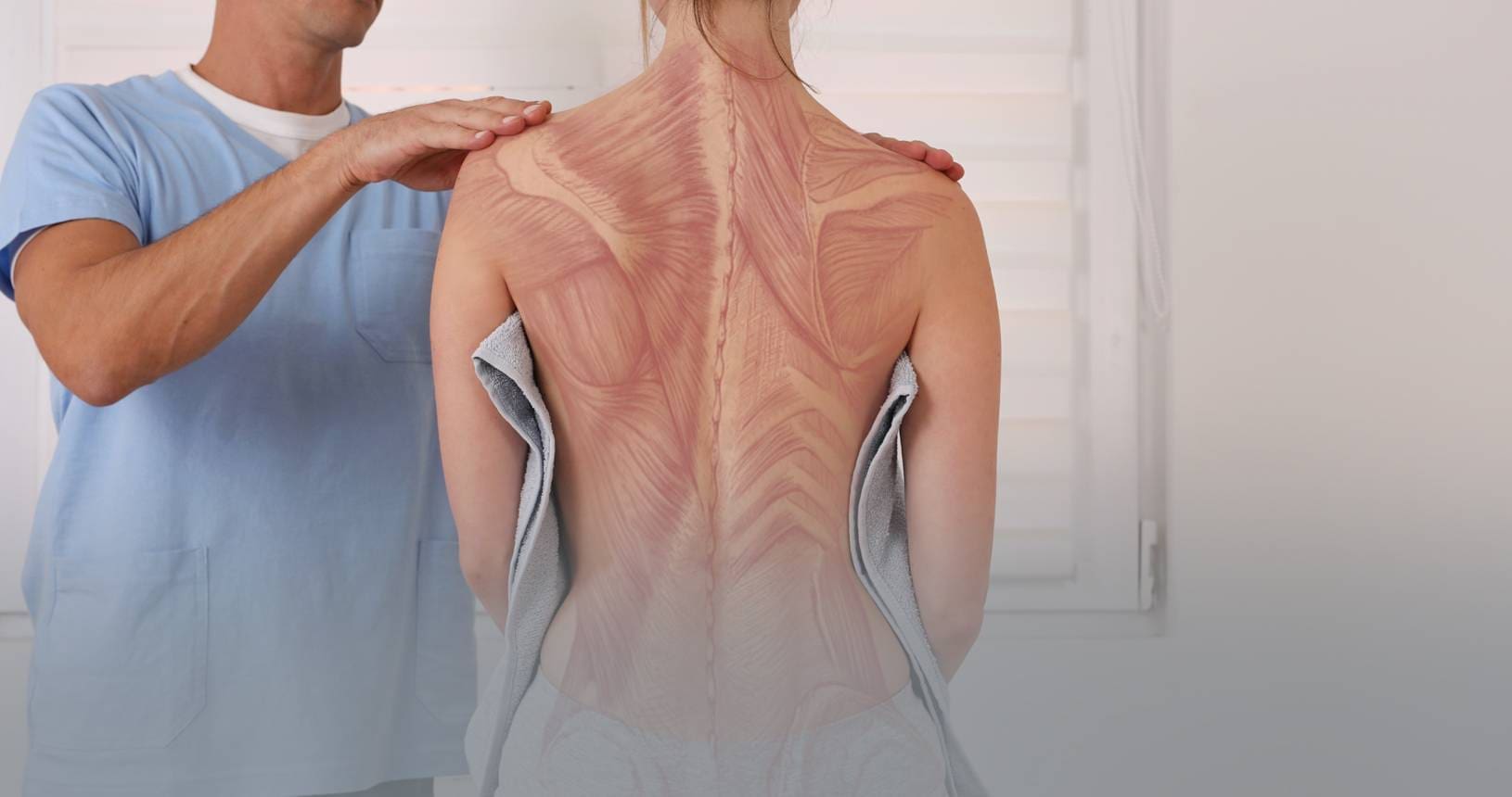
Strengthening Ligaments and Tendons for Posture Support
Ligaments and tendons hold the spine and shoulders upright like support wires. When they stretch or tear, posture suffers. PRP injections at El Paso Back Clinic strengthen these soft tissues by signaling cells to produce denser collagen. Princeton Medicine shows PRP reduces swelling in rotator cuff injuries and helps shoulders move with less effort (Princeton Sports and Family Medicine, n.d.).
In the neck and low back, stronger ligaments mean less forward head tilt or swayback. Patients at the clinic say they sit taller without constant reminders. Health Coach Clinic, aligned with the clinic’s functional medicine, notes PRP lowers the need for pain pills and keeps people active for natural posture training (Health Coach Clinic, n.d.-a).
How PRP Indirectly Boosts Mobility and Biomechanics
Pain blocks good posture the most. When your back or neck hurts, you hunch to guard it. PRP eases pain at the source at El Paso Back Clinic. With less discomfort, muscles relax and move freely. Better movement creates smoother walking, sitting, and lifting. Over time, the body adopts healthier patterns.
Bullet points on mobility gains from the clinic’s approach:
Less neck and shoulder pain allows the head to balance over the spine.
Stronger back ligaments reduce lower-back sway, which pulls the shoulders forward.
Healthier discs restore the spine’s natural curves.
Faster return to activities builds confidence and encourages movement.
A Journal of Pain Research review backs this, showing PRP gives longer relief for low-back pain by fixing the real damage (Akeda et al., 2019).
Limits of PRP: Not a Magic Fix for Habit-Based Posture
PRP works best for injury or instability. It does not retrain the brain if poor posture comes only from years of desk slouching. All Wells Scoliosis Centre reminds us that posture is a learned habit. Repetition of good movements retrains the brain, but pain must be removed first (All Wells Scoliosis Centre, n.d.).
That is why El Paso Back Clinic uses PRP as part of a bigger plan. Without exercises and habit changes, old ways may return once pain fades. Dr. Jimenez emphasizes that PRP repairs the structure, while chiropractic and rehabilitation address the habit.
The Integrative Chiropractic Approach at El Paso Back Clinic
When regular therapy or medicine falls short, patients choose El Paso Back Clinic’s team. Dr. Jimenez, as DC, APRN, FNP-BC, and CFMP, leads chiropractors, nurse practitioners, physical therapists, and nutritionists. They treat the whole person: spine alignment, nutrition, inflammation, and movement.
The clinic blends PRP with gentle adjustments, spinal decompression, and functional medicine testing. Dr. Jimenez’s writings show patients with sciatica or chronic pain heal faster when PRP repairs tissues and chiropractic keeps the spine moving right (Jimenez, n.d.). Nutrition coaches cut inflammatory foods, while rehab experts teach core strength. This team effort delivers results that single treatments cannot.
Saks Wellness Center ideas, echoed at the clinic, note that chiropractic finds muscle imbalances and fixes them with adjustments and exercises. When paired with PRP, the body receives support from both inside and out (Saks Wellness Center, n.d.).
Functional medicine lowers whole-body inflammation through diet and supplements.
APRNs and FNP-BCs safely oversee injections and track healing.
Regular check-ins catch small issues early.
Patients skip surgery and long-term medication use.
Is PRP Therapy Safe and Effective at the Clinic?
Most people handle PRP well since it uses their own blood. Mild soreness at the injection site fades quickly. Serious side effects are rare. MidJersey Orthopedics and the clinic’s own protocols report PRP eases or ends pain for many without steroid risks (MidJersey Orthopedics, n.d.).
Results vary, but many feel relief in four to six weeks. Riverside Online notes PRP shines with healthy lifestyle changes like better movement (Riverside Online, n.d.). At El Paso Back Clinic, patients see strong outcomes because PRP is integrated into full-body support plans, including recent guides on PRP for sciatica and spinal care (El Paso Back Clinic, n.d.-b).
Real-World Results from El Paso Back Clinic Patients
Picture a local office worker whose neck pain forces them to lean forward. After PRP injections into the cervical ligaments and discs, along with Dr. Jimenez’s chiropractic care, pain decreases and posture improves naturally. A construction worker with low-back disc issues regains lift strength safely. These stories happen often at the clinic because PRP addresses the “why” behind the slump.
Cedars-Sinai describes how platelets release growth factors that rebuild tissue and may avoid surgery (Cedars-Sinai, n.d.). Blue Ridge Ortho adds that PRP helps with back and shoulder problems, making daily life easier (Blue Ridge Ortho, n.d.). Dr. Jimenez’s patient stories on the clinic site echo this success with non-surgical recovery.
Moving Forward with PRP and Posture Care in El Paso
Platelet-rich plasma therapy does not replace good habits, but it clears the path so habits stick. By easing pain, mending discs, and strengthening ligaments and tendons, PRP gives the body a real chance at natural alignment. At El Paso Back Clinic, combining PRP with chiropractic care, functional medicine, and daily practice creates a comprehensive path to better posture and lasting comfort.
If chronic pain or instability keeps you from standing tall, reach out to El Paso Back Clinic. Their non-surgical, team-based approach using the body’s own tools can open the door to a straighter, stronger you. Call 915-850-0900 or visit their El Paso locations to learn more.
Akeda, K., Yamada, T., Takahashi, H., & Sudo, A. (2019). Platelet-rich plasma in the management of chronic low back pain: A critical review. Journal of Pain Research, 12, 753–767. https://pmc.ncbi.nlm.nih.gov/articles/PMC6394242/
Platelet-Rich Plasma (PRP) Therapy for Spinal Care: A Natural Path to Pain Relief and Healing
Platelet-rich plasma (PRP) therapy helps people with back pain find relief without surgery. Doctors take a small sample of the patient’s own blood and turn it into a powerful healing mixture. This mixture uses the body’s natural platelets to reduce swelling and repair damaged areas of the spine. Many patients with mild to moderate spine problems choose PRP after other treatments like physical therapy do not fully work.
What Is Platelet-Rich Plasma Therapy?
PRP therapy is a simple treatment that comes from the patient’s blood. A nurse or doctor draws a small amount of blood from the arm. Then the blood spins in a machine called a centrifuge. This step pulls out the platelets and makes them extra strong. The result is platelet-rich plasma, rich in growth factors. These growth factors act like signals that tell the body to start healing. PRP does not use drugs or chemicals from outside the body. It works with what the patient already has inside. This makes it a safe and natural choice for many people who want to avoid surgery.
How PRP Therapy Supports Spinal Healing
The spine has discs, facet joints, ligaments, and nerves that can wear down over time. PRP goes right to these spots and gets to work. The growth factors reduce inflammation and kick-start tissue repair. For example, they help degenerated discs hold more water and stay flexible. They also calm painful facet joints and strengthen loose ligaments. Because PRP comes from the patient’s own blood, the body accepts it and begins repairing the damage quickly. Studies show PRP can even help nerves heal and reduce chronic pain signals.
Releases growth factors that tell cells to grow and repair
Lowers swelling around discs and joints
Builds new blood vessels so nutrients can reach damaged areas
Helps ligaments and tendons get stronger
Supports natural disc repair without cutting into the body
Key Benefits of PRP for Back and Spine Issues
Patients often notice real changes after PRP. The treatment gives long-lasting pain relief instead of short-term fixes like steroid shots. Many people move better and feel more active in daily life. PRP also cuts the need for strong pain pills. Because it is minimally invasive, patients avoid hospital stays and big scars. Recovery is quick, and the risk of side effects stays low since the body uses its own material. Over time, PRP may slow down further spine wear.
Natural healing that lasts months or even years
Less pain without heavy medication
Better mobility and daily function
Quick return to normal activities
Lower chance of allergic reactions
Works well with other non-surgical care
Common Spinal Conditions PRP Can Help
Doctors use PRP for several spine problems that cause daily discomfort. It works best when the damage is mild to moderate. Conditions include degenerative disc disease, where discs lose height and cause stiffness. Spinal stenosis, which narrows the space around nerves, also responds well. Facet joint arthritis causes sharp pain that PRP can help ease. Herniated discs and ligament strains improve, too. Even chronic low back pain and sciatica often get better. Patients who tried rest, therapy, or meds without complete success often turn to PRP next.
The Step-by-Step PRP Procedure
The whole process feels straightforward and takes about an hour. First, the nurse draws blood from the arm. Next, the blood spins in the centrifuge to create the PRP. Then the doctor uses ultrasound or X-ray guidance to place the PRP exactly where it is needed. Patients stay awake and feel only mild pressure. No stitches or long cuts are involved. The clinic sends the patient home the same day with simple care instructions.
Blood draw (small amount from the arm)
Centrifuge step to concentrate platelets
Ultrasound-guided injection into the spine
Short rest period before going home
Follow-up visits to check progress
Who’s a Good Candidate for PRP Therapy?
PRP is suitable for people with mild to moderate spinal wear who have not found sufficient relief from physical therapy or medication. It is not usually the first choice for very severe damage. A doctor checks imaging and health history to decide. Patients who want to stay active and avoid surgery often like this option. Good health and realistic goals help the treatment work best.
Integrative Spinal Care: Combining PRP with Chiropractic and Functional Medicine
In clinics that blend different care styles, PRP becomes even more effective. An Advanced Practice Registered Nurse (APRN/FNP-BC) with functional medicine training (CFMP, IFMCP, ATN, CCST) can administer precise, ultrasound-guided PRP injections. At the same time, chiropractic adjustments keep the spine aligned. Nutritional support from functional medicine fixes any missing vitamins or inflammation triggers in the body. This team approach creates the perfect setting for repair. The body gets structural help, cellular healing, and inside support all at once.
Insights from Dr. Alexander Jimenez on PRP and Spine Health
Dr. Alexander Jimenez, DC, APRN, FNP-BC, sees PRP as part of whole-body healing in El Paso, Texas. As both a chiropractor and nurse practitioner, he combines spinal adjustments with regenerative shots and metabolic checks. His clinical work shows that patients with sciatica or disc problems heal faster when PRP teams up with chiropractic care and proper nutrition. Dr. Jimenez notes that this mix helps clear waste from injured tissues, builds stronger blood flow, and stops pain cycles. Many of his patients return to work and sports with less discomfort and more confidence.
What to Expect During Recovery
Most people feel mild soreness for a few days after the shot, like a deep bruise. Ice packs and gentle movement help. Light activities can start right away, but heavy lifting waits one to two weeks. Full benefits build over four to six weeks as the growth factors continue to work. Some patients need a second shot after a month or two for the best results. Follow-up visits track progress and adjust the plan.
Evidence and Safety of PRP Therapy
Research backs PRP for spine care. Clinical reviews show pain drops and better movement in patients with degenerative discs and facet problems. Nerve repair studies also point to positive results. Side effects are rare because the treatment uses the patient’s own blood. No major complications appear in most studies. Doctors continue to track long-term outcomes, but current data look promising for people who want natural options.
Conclusion
Platelet-rich plasma therapy offers a fresh way to handle spinal pain and damage. It uses the body’s own tools to reduce swelling, repair tissues, and restore movement. When paired with expert chiropractic and functional medicine, the results can feel even better. Patients who have struggled with ongoing back issues often discover new hope through PRP. Talking with a trained provider helps decide if this path fits personal needs. With steady advances in regenerative care, many more people may soon enjoy life with less spine pain and more freedom.
Neuropathy Relief Through Integrative Chiropractic Care in El Paso
Neuropathy can make daily life challenging. Many people experience burning pain, tingling, numbness, weakness, or a “pins and needles” sensation in the feet, legs, hands, or arms. These symptoms can affect sleep, walking, balance, exercise, and work. For many patients, the best long-term plan is not built around a single pill or a single procedure. It is built around a full recovery strategy that improves nerve function, supports the spine and joints, boosts circulation, and helps the body heal from the inside out.
At El Paso Back Clinic, the focus is naturally on integrative chiropractic care, physical therapy, rehabilitation, functional medicine, and lifestyle support. In that kind of setting, platelet-rich plasma, or PRP, may be used as a background regenerative option in selected cases, but it is not the center of the treatment plan. The main goal is to improve mobility, reduce pressure on irritated nerves, reduce inflammation, restore function, and address the root causes that sustain neuropathy (Mayo Clinic, 2023; NIDDK, 2018).
What Is Neuropathy?
Neuropathy means nerve damage. Peripheral neuropathy is one of the most common forms and often affects the hands and feet. Diabetic neuropathy is another major type and happens when high blood sugar damages nerves over time. Other causes may include spine problems, chronic inflammation, poor circulation, vitamin deficiencies, repetitive stress, injury, and metabolic imbalance (National Institute of Diabetes and Digestive and Kidney Diseases [NIDDK], 2018; Mayo Clinic, 2023).
Common symptoms include:
Burning pain
Tingling
Numbness
Sharp or shooting pain
Muscle weakness
Poor coordination
Trouble with balance
Reduced sensation in the feet or hands
These symptoms do not just affect comfort. They can also reduce quality of life and raise the risk of falls, poor posture, and less activity. Over time, that can create even more weakness and stiffness in the body (NIDDK, n.d.).
Why an Integrative Chiropractic Approach Matters
Neuropathy is often treated as if it were only a nerve problem. In reality, many cases involve much more than the nerve itself. The spine, muscles, joints, posture, circulation, inflammation levels, blood sugar control, and nutrition can all affect how nerves feel and function. That is why an integrative chiropractic and physical therapy setting can be such a good fit.
Instead of focusing only on symptom control, an integrative clinic may look at:
Spinal alignment and joint motion
Muscle tightness and weakness
Balance and gait problems
Mobility loss
Blood sugar and metabolic health
Inflammation
Nutritional deficiencies
Functional movement patterns
Daily habits that keep nerves irritated
This broader model is important because a nerve that is already stressed may be further affected by poor biomechanics, limited circulation, chronic inflammation, and weak supporting muscles (Mayo Clinic, 2023; Jimenez, n.d.-a).
How Chiropractic Care May Support Neuropathy Patients
Chiropractic care does not claim to “cure” every case of neuropathy. But it may help patients by addressing the mechanical and functional issues that often worsen nerve symptoms. When the spine and joints do not move well, the body may develop abnormal tension, poor posture, reduced mobility, and stress on surrounding tissues. These problems can increase pain and decrease function.
Chiropractic care may help by:
Improving spinal and joint motion
Reducing mechanical stress on irritated nerves
Helping posture and balance
Lowering muscle tension
Supporting better movement patterns
Improving comfort during walking and daily activity
Working together with rehabilitation and exercise care
For some patients, especially those with back-related leg symptoms, sciatica-like symptoms, or nerve irritation associated with spinal dysfunction, chiropractic treatment may be an important part of a broader care plan. When paired with rehab and lifestyle support, it can help patients move better and feel more stable (Lowery Chiropractic, n.d.; Mayo Clinic, 2023).
The Role of Physical Therapy and Rehabilitation
Search engines already recognize El Paso Back Clinic as a chiropractic and physical therapy clinic, and that makes sense because rehab is a major part of neuropathy support. A person with nerve pain often changes the way they walk, stand, bend, or exercise. Over time, those changes can create greater weakness, poorer balance, and increased strain on the body.
Physical therapy and rehab may focus on:
Balance training
Stretching tight muscles
Strengthening the legs, hips, core, and postural muscles
Improving gait
Restoring range of motion
Teaching safer movement patterns
Supporting better function in daily life
This matters because nerves do not work in isolation. They work inside a moving body. When muscles are weak and joints are stiff, the nervous system often performs worse. Better motion and stronger support can help reduce the overload on sensitive areas and improve quality of life (Mayo Clinic, 2023).
Functional Medicine and Nutritional Support for Root-Cause Care
A strong neuropathy program should also look at internal health. If the body is dealing with high blood sugar, insulin resistance, chronic inflammation, poor gut health, low B vitamins, oxidative stress, or other metabolic problems, nerve tissue may have a harder time recovering.
An integrative clinic may use functional and nutritional strategies to support:
Blood sugar control
Reduced inflammation
Better circulation
Healthier nerve metabolism
Improved energy production
Weight management
Better tissue healing
This root-cause approach fits neuropathy care very well. For example, diabetic neuropathy cannot be fully addressed without improving metabolic control. Even in non-diabetic cases, poor nutrition and chronic inflammation can make nerve symptoms worse. That is why APRNs, FNPs, CFMPs, and IFMCP-trained providers may add strong value to a chiropractic clinic model. They help connect musculoskeletal care with whole-body healing (NIDDK, 2018; Mayo Clinic, 2023).
Where PRP Fits In
PRP should be seen as a supportive regenerative option, not the main focus of a neuropathy article for a chiropractic and rehab-centered clinic. Platelet-rich plasma is made from the patient’s own blood and contains a higher concentration of platelets and growth factors. Research suggests these growth factors may help nerve healing, reduce inflammation, improve local blood flow, and support tissue repair (Shang et al., 2025; Wang et al., 2024).
As part of an integrative treatment plan, PRP may be considered in selected cases to support the recovery of damaged tissue and nerves. It may be especially useful when imaging-guided precision treatment is needed as part of a larger care strategy. Still, PRP is best understood as an added regenerative tool, not a replacement for chiropractic care, rehab, nutrition, exercise, and metabolic correction.
That is important because neuropathy usually does not improve with a single isolated treatment. Most patients need a layered approach to improve both nerve health and bodily function over time (Kennedy et al., 2025; Hassanien et al., 2020).
What the Research Says About PRP for Neuropathy
The research on PRP for neuropathy is promising but still developing. Reviews suggest PRP may support nerve regeneration, reduce neuropathic pain, and help with recovery in peripheral nerve conditions. Growth factors in PRP may stimulate Schwann cells, support axon repair, and improve the healing environment around injured nerves (Shang et al., 2025; Wang et al., 2022).
One randomized clinical study in patients with painful diabetic neuropathy found that ultrasound-guided perineural PRP, combined with medical treatment, improved pain and neuropathic symptoms more than medical treatment alone over several months (Hassanien et al., 2020). That is encouraging, but it does not mean PRP is a stand-alone answer for every patient.
The better message for a chiropractic and rehab audience is this: PRP may support healing in the background, but daily function improves most when patients also work on movement, stability, posture, circulation, metabolic health, and long-term lifestyle change.
Clinical Observations from Dr. Alexander Jimenez
Clinical materials from Dr. Alexander Jimenez, DC, APRN, FNP-BC, describe a multidisciplinary model that blends chiropractic care, physical medicine, rehabilitation, functional medicine, nutrition, and advanced clinical assessment. His observations support the idea that neuropathy care should not focus only on pain suppression. Instead, it should examine structure, movement, inflammation, metabolic health, and tissue healing together (Jimenez, n.d.-a; Jimenez, n.d.-b).
This type of model is a natural fit for El Paso Back Clinic because it keeps the main focus on the following:
Chiropractic treatment
Physical therapy and rehab
Functional movement
Metabolic and nutritional support
Whole-body recovery
Long-term improvement instead of short-term symptom masking
In this setting, regenerative treatments like PRP can stay in the background as one part of a broader plan rather than becoming the main message.
A Better Long-Term Message for Neuropathy Patients
Patients with neuropathy often want simple answers, but healing usually requires a combination of strategies. The best message is not “one injection fixes everything.” The stronger message is that an integrative chiropractic clinic can help patients improve function, reduce nerve stress, strengthen the body, and support healthier tissue over time.
A full neuropathy strategy may include:
Chiropractic adjustments when appropriate
Physical therapy and rehabilitation
Balance and gait training
Stretching and strengthening
Nutrition support
Functional medicine evaluation
Blood sugar and inflammation management
Imaging-guided regenerative support in select cases
This type of plan matches how real recovery works. Nerves need support, but so do muscles, joints, posture, circulation, and metabolism.
Final Thoughts
Neuropathy is complex, and many patients need more than symptom control. A chiropractic and physical therapy clinic like El Paso Back Clinic is well-positioned to help by focusing on biomechanics, movement, rehabilitation, and root-cause care. Integrative chiropractic treatment should remain front and center because it aligns with the clinic’s identity and offers patients a more comprehensive, natural path to relief.
PRP injections can be mentioned as a supportive regenerative option in the background, especially in selected cases where tissue repair and nerve support are part of the plan. But for search visibility and patient clarity, the stronger focus should stay on chiropractic care, rehabilitation, functional medicine, and long-term healing strategies that improve the body’s overall function.
When neuropathy care is built around structure, motion, metabolism, and recovery, patients get a more realistic and more complete path forward (Mayo Clinic, 2023; NIDDK, 2018; Jimenez, n.d.-a).
PRP Therapy for Sports Injuries: How It May Speed Healing Without Surgery
Sports injuries can slow life down fast. A sore tendon, a strained ligament, or a muscle tear can make it difficult to train, work, sleep, or even walk comfortably. That is one reason Platelet-Rich Plasma, or PRP, has gained attention in sports medicine. PRP is made from a patient’s own blood and then injected into an injured area to support healing. Medical centers such as Yale Medicine, Penn Medicine, Johns Hopkins Medicine, and Temple Health describe PRP as a biologic or regenerative treatment that may help repair tissue, lower pain, and improve function in certain musculoskeletal injuries. It is often used for tendon, ligament, muscle, cartilage, and joint problems, including some cases of osteoarthritis. (Johns Hopkins Medicine, n.d.; Penn Medicine, 2025; Yale Medicine, n.d.).
PRP is appealing because it is non-surgical and uses the body’s own healing tools. Still, it is not a miracle fix for every athlete or every injury. Research shows promising results in many cases, but outcomes can vary depending on the tissue involved, how long the injury has been present, how the PRP is prepared, and whether the person also follows a successful rehab plan. In other words, PRP works best as part of a comprehensive care strategy rather than a stand-alone shot. (Saini et al., 2021; Jimenez, n.d.).
What PRP Therapy Is
PRP stands for Platelet-Rich Plasma. Plasma is the liquid part of blood, and platelets are blood components best known for their role in clotting. However, platelets also carry growth factors and signaling molecules that help tissue repair. To make PRP, a clinician draws a small amount of blood, spins it in a centrifuge, and separates out a platelet-rich portion. That concentrated solution is then placed into the injured area. The goal is to increase healing signals directly at the site of tissue damage. (Johns Hopkins Medicine, n.d.; Yale Medicine, n.d.; HSS, n.d.; Penn Medicine, 2025).
A simple way to think about PRP is this: it does not just try to numb pain. It tries to support the body’s repair response. Hospital for Special Surgery describes PRP as a form of regenerative medicine that amplifies natural growth factors in blood cells to help damaged tissue heal. Johns Hopkins Medicine similarly explains that the concentrated growth factors in PRP may stimulate tissue regeneration and speed healing in the treated area. (HSS, n.d.; Johns Hopkins Medicine, n.d.).
What the procedure usually includes
A small blood draw from the patient
Processing the sample in a centrifuge
Preparing the platelet-rich portion
Injecting the PRP into the injured tissue
In some cases, using ultrasound to guide the injection
A visit that often takes less than an hour
This basic process is described by major medical centers, including Penn Medicine, Yale Medicine, and Johns Hopkins Medicine. (Johns Hopkins Medicine, n.d.; Penn Medicine, 2025; Yale Medicine, n.d.).
How PRP May Help Sports Injuries Heal
When tissue is injured, the body sends platelets to the area early in the healing process. Temple Health explains that platelets contain growth factors that help promote cell growth, repair tissue, and reduce inflammation. Yale Medicine notes that PRP contains concentrated platelets, cytokines, and growth factors with anti-inflammatory properties. This is why PRP is often used for injuries that have been slow to heal on their own. (Temple Health, 2021; Yale Medicine, n.d.).
PRP may be especially useful in tissues that do not receive a strong blood supply. The 2021 review in the Indian Journal of Orthopaedics notes that tendons heal more slowly than many other tissues because of their poor vascularity. That same review also explains that PRP has been studied in tendon disorders such as Achilles tendinopathy, rotator cuff tendinitis, and epicondylitis, as well as in muscle strains and osteoarthritis. (Saini et al., 2021).
For athletes, this matters because many sports injuries are overuse or repetitive-stress injuries. If a tendon stays irritated for months, or a ligament strain never fully calms down, the body may need extra support to restart a healthier repair process. Some research suggests earlier PRP use in select injuries may help guide inflammation toward recovery and restore tissue balance. Even so, researchers also note there is no universal PRP formula or perfect protocol yet, so treatment must be individualized. (Saini et al., 2021).
Common Sports Injuries PRP Is Used For
Medical centers and sports medicine sources commonly describe PRP for the following problems:
Chronic tendinitis or tendinopathy
Tennis elbow
Patellar tendinopathy or “jumper’s knee”
Achilles tendon problems
Ligament strains
Muscle strains and some muscle tears
Cartilage irritation
Osteoarthritis in active adults
These uses are repeatedly listed by Penn Medicine, Yale Medicine, Temple Health, and HSS. (Penn Medicine, 2025; Temple Health, 2021; Yale Medicine, n.d.; HSS, n.d.).
Temple Health highlights tennis elbow and jumper’s knee as common orthopedic conditions that may benefit from PRP. In its overview, Penn Medicine also lists structures such as the Achilles tendon, ACL, hamstring, patellar tendon, and cartilage as areas in sports medicine where PRP is used. Yale Medicine adds tendon, ligament, and muscle conditions, as well as degenerative joint conditions, to that list. (Penn Medicine, 2025; Temple Health, 2021; Yale Medicine, n.d.).
There is also supportive evidence for muscle injury care when injections are placed carefully. A 2014 study in Blood Transfusion reported that athletes with grade II muscle lesions who received ultrasound-guided PRP showed full healing on ultrasound, pain resolution, and return to sport, with only one relapse reported a year later. That does not prove PRP is right for every muscle injury, but it does show why sports clinicians remain interested in it. (Borrione et al., 2014).
What Recovery Feels Like After PRP
One important point for patients is that PRP can cause short-term soreness. Yale Medicine says the most common side effects are discomfort, pain, and stiffness at the injection site. Penn Medicine also notes that mild soreness, swelling, or stiffness is common for the first few days. Johns Hopkins Medicine adds that some people notice soreness and bruising after the procedure. In most cases, these effects are temporary. (Johns Hopkins Medicine, n.d.; Penn Medicine, 2025; Yale Medicine, n.d.).
Patients also need realistic expectations. PRP is not usually an instant pain reliever. Penn Medicine says improvement may take a few weeks to become noticeable, with fuller benefits developing over months. Yale Medicine reports that some people notice pain improvement in four to six weeks, with continued progress for up to a year. (Penn Medicine, 2025; Yale Medicine, n.d.).
Aftercare often includes
Resting the area for a short time
Avoiding hard exercise right away
Using a guided rehab plan
Following instructions about pain control
Avoiding some anti-inflammatory medicines when advised
Penn Medicine and HSS both note that anti-inflammatory medicines may interfere with the early healing response that PRP is meant to support, so patients should follow their treating clinician’s advice. (HSS, n.d.; Penn Medicine, 2025).
Why Ultrasound-Guided PRP Matters
Not every injection needs the same level of precision, but many sports injuries benefit from careful image guidance. Both Johns Hopkins Medicine and Yale Medicine acknowledge the use of ultrasound during PRP procedures. Research in athletes also supports this approach. The 2014 study on muscle injuries emphasized that ultrasound was important for both locating the lesion and guiding the needle accurately into it. The 2021 sports injury review similarly reported that ultrasound-guided injections improve accuracy, particularly for musculoskeletal conditions. (Johns Hopkins Medicine, n.d.; Yale Medicine, n.d.; Borrione et al., 2014; Saini et al., 2021).
On Dr. Alexander Jimenez’s public clinical website, one recent educational article describes ultrasound-guided intra-articular hip PRP as a precision-focused procedure in which ultrasound helps the clinician visualize anatomy, confirm correct placement, and improve safety. That same article stresses that biologic injections work best when they are combined with rehabilitation and movement-based recovery rather than used alone. (Jimenez, n.d.).
Dr. Alexander Jimenez’s Clinical Observations and the Value of Integrated Care
Dr. Alexander Jimenez, DC, APRN, FNP-BC, describes his El Paso practice as a multidisciplinary and integrative model that combines chiropractic care, functional medicine thinking, sports medicine principles, rehabilitation, and regenerative strategies. His website presents regenerative medicine as a natural, non-surgical option designed not only to reduce pain but also to improve structure, movement, and function. (Jimenez, n.d.).
That point matters in sports injury care. A tendon or muscle may not stay healthy if the athlete still has poor joint mechanics, weak stabilizers, incorrect loading patterns, or nutrition and recovery habits that slow healing. Dr. Jimenez’s site repeatedly frames recovery as a full process that includes a detailed history, physical evaluation, attention to biomechanics, regenerative options when appropriate, chiropractic care to improve motion, rehab planning, and follow-up focused on function. (Jimenez, n.d.).
In a comprehensive clinic model, that means PRP can be paired with structural care, progressive rehabilitation, and functional medicine support. The injection may help the tissue biologically, while rehab helps the athlete move better and reduce repeated stress on the injured area. This combined approach aligns with the broader message from both sports medicine research and Dr. Jimenez’s clinical content: better recovery usually comes from treating the tissue and the movement pattern together. (Borrione et al., 2014; Jimenez, n.d.; Saini et al., 2021).
Benefits and Limits of PRP
Possible benefits
Uses the patient’s own blood
Minimally invasive
May reduce pain and improve function
May help some chronic tendon, ligament, muscle, and joint problems
Can be part of a non-surgical recovery plan
Can be combined with rehab and other supportive care
These benefits are commonly described by Yale Medicine, Penn Medicine, Johns Hopkins Medicine, and HSS. (HSS, n.d.; Johns Hopkins Medicine, n.d.; Penn Medicine, 2025; Yale Medicine, n.d.).
Important limits
Results vary from person to person
Some injuries still need surgery or other procedures
Relief may take weeks or months, not days
PRP preparation methods are not fully standardized
Some tissues have stronger evidence than others
Those limits are important because proper medicine depends on the right treatment for the right injury at the right time. PRP may be a strong option, but it should be chosen carefully after a full exam and diagnosis. (Saini et al., 2021; Penn Medicine, 2025).
Final Thoughts
PRP therapy offers a promising non-surgical option for sports injuries because it delivers a concentrated dose of the patient’s own platelets to damaged tissue, where growth factors may support repair, reduce inflammation, and improve recovery. It is commonly used for chronic tendinopathy, ligament strain, muscle injury, and some joint conditions. Short-term soreness at the injection site can happen, but serious side effects are uncommon. The best results usually come when PRP is matched to the right injury and combined with smart rehabilitation, movement correction, and careful follow-up. (Johns Hopkins Medicine, n.d.; Penn Medicine, 2025; Yale Medicine, n.d.; Jimenez, n.d.).
Sciatica Relief in El Paso: How Integrative Chiropractic Care Supports Healing and Mobility
Sciatica can make daily life challenging. It often causes pain that starts in the lower back or buttocks and travels down the leg. Some people also feel tingling, numbness, burning, or weakness. In many cases, the problem begins when a lumbar disc, tight soft tissue, joint irritation, or spinal narrowing compresses a nerve root. Because sciatica can have multiple causes, treatment works best when it focuses on the whole person, not just the pain. That is why a chiropractic rehabilitation model aligns well with this topic for El Paso Back Clinic. The clinic publicly describes itself as a chiropractic rehabilitation and integrated medicine center focused on injury recovery, movement, function, and whole-person care. (Berry et al., 2019; El Paso Back Clinic, n.d.-a; El Paso Back Clinic, n.d.-b).
At El Paso Back Clinic, the public-facing message centers on chiropractic care, rehabilitation, mobility, flexibility, nutrition, and integrated support. The site describes Dr. Alexander Jimenez as both a chiropractor and a family nurse practitioner, leading a multidisciplinary team that blends evidence-based care with natural and functional approaches. That positioning is relevant for sciatica because many people improve with conservative care built around assessment, education, movement, and structured rehabilitation before more invasive options are considered. (El Paso Back Clinic, n.d.-a; El Paso Back Clinic, n.d.-c; Jimenez, n.d.).
What Sciatica Really Means
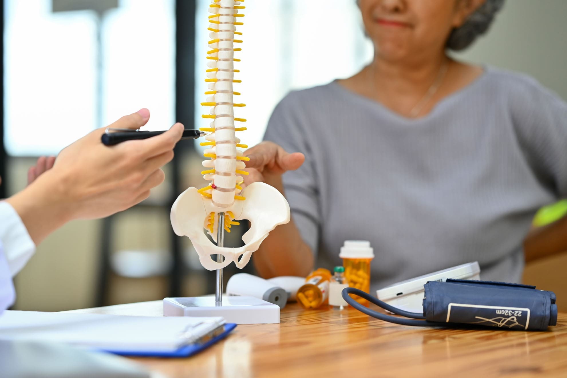
Sciatica is a symptom pattern, not a stand-alone diagnosis. It usually describes pain that follows the path of the sciatic nerve, often from the lower back into the buttocks, thigh, calf, or foot. A careful exam usually includes a history, strength testing, reflexes, sensation testing, and nerve tension testing. This matters because sciatica-like pain can arise from lumbar disc herniation, degenerative disc changes, facet joint irritation, spinal stenosis, piriformis-related irritation, or combined movement-related problems. When the source is correctly identified, treatment can be more specific and effective. (Berry et al., 2019).
Why a Chiropractic and Physical Rehabilitation Approach Fits So Well
Current guidance for lumbosacral radicular pain supports a stepped, conservative approach as first-line treatment. That usually means education, staying active, exercise therapy, and treatment matched to the patient’s symptoms and function. Recent guideline work also emphasizes clear communication, a gradual return to activity, and exercise therapy tailored to the person’s needs and tolerance. In other words, successful care is not just about lying down and waiting. It is about restoring motion, building support around the spine, and helping the nervous system calm down while the tissues recover. (Apeldoorn et al., 2024; Schmid & Tampin, 2023).
This conservative framework matches the public model of El Paso Back Clinic. The clinic’s website describes a whole-person plan that addresses posture, movement, daily habits, flexibility, strength, and nutrition. It also highlights chiropractic adjustments, rehabilitation-based care, and functional support rather than making injections the center of the message. That is a strong fit for a sciatica article aimed at a chiropractic and physical therapy audience. (El Paso Back Clinic, n.d.-d; El Paso Back Clinic, n.d.-e; El Paso Back Clinic, n.d.-f).
How Integrative Chiropractic Care May Help Sciatica
Chiropractic care for sciatica is not just one quick adjustment. In a more integrative setting, it can include a mix of spinal manipulation or mobilization, soft-tissue work, guided stretching, core-stability work, gait and posture correction, mobility drills, and progressive strengthening. The goal is to reduce mechanical stress, improve joint motion, improve movement patterns, and support the body’s own recovery. El Paso Back Clinic’s public materials describe a broader plan, including adjustments, exercises, and wellness strategies designed to restore mobility and reduce pressure on irritated structures. (El Paso Back Clinic, n.d.-b; El Paso Back Clinic, n.d.-d; El Paso Back Clinic, n.d.-e).
A 2024 narrative review on lumbar disc herniation with radiculopathy reported that spinal mobilization with leg movement, lumbar stabilization exercises, and manipulation can reduce symptoms and improve stability and mobility in selected patients. The same review emphasized that weak core muscles and poor spinal stability can delay healing, which is why structured rehabilitation matters so much. This supports a chiropractic rehabilitation strategy that focuses on both pain relief and rebuilding support around the lumbar spine. (El Melhat et al., 2024).
The Role of Exercise, Rehab, and Movement Training
For many people with sciatica, movement is medicine when it is used the right way. Recent physical therapy guidance recommends exercise therapy for patients who need help with daily activities, participation, or movement-related limits. The program should match irritability, tolerance, and function. In early stages, that may mean gentle pain-relieving movements, walking progressions, and avoiding positions that sharply increase symptoms. Later, it often expands into core work, hip strength, endurance, balance, and return-to-activity training. (Apeldoorn et al., 2024).
This is one of the biggest advantages of an integrative chiropractic clinic with a rehabilitation mindset. A patient is not just told where the pain is. They are shown how to move better, sit and lift with less strain, rebuild spinal support, and reduce the repeated stresses that may have contributed to the problem. El Paso Back Clinic’s site repeatedly highlights mobility, flexibility, sports medicine concepts, rehabilitation, and personalized exercise support as part of care. (El Paso Back Clinic, n.d.-d; El Paso Back Clinic, n.d.-f).
Common parts of a chiropractic rehabilitation plan for sciatica
Spinal adjustments or mobilization to improve motion
Soft tissue work for tight lumbar, hip, and gluteal tissues
Nerve-friendly movement progressions
Core stabilization exercises
Hip and pelvic strength work
Posture and ergonomic coaching
Walking programs and activity modification
Nutrition and inflammation support when needed
These tools do not all apply to every patient, but together they show why conservative care can be more than temporary pain relief. It can help correct the patterns that keep irritating the sciatic nerve. (Apeldoorn et al., 2024; El Melhat et al., 2024; El Paso Back Clinic, n.d.-e).
Clinical Observations from Dr. Alexander Jimenez
Dr. Alexander Jimenez’s public pages describe a dual-scope model that blends chiropractic care with nurse practitioner-level medical evaluation, functional medicine, and individualized rehabilitation planning. His clinic materials emphasize non-surgical recovery, movement restoration, advanced assessment, and whole-person healing. At El Paso Back Clinic, sciatica care is presented as a process of locating the source of the problem, improving alignment and mechanics, and guiding the patient back toward better function. That practical, layered approach is especially useful for chronic or recurring sciatica, where structural, inflammatory, stress-related, and lifestyle factors may overlap. (Jimenez, n.d.; El Paso Back Clinic, n.d.-a; El Paso Back Clinic, n.d.-b).
Where PRP Fits In
Platelet-Rich Plasma is made from a patient’s own blood and is used in regenerative medicine to deliver concentrated platelets and growth factors to a target area. In lumbar radiculopathy research, PRP injections have shown promising results in pain and function, and some studies suggest longer-lasting improvement than steroid injections in selected patients. Still, PRP is best presented as an adjunct option for carefully chosen cases, not as the foundation of care for every person with sciatica. (Gupta et al., 2024; Saraf et al., 2023).
That is also the most natural fit for a chiropractic and rehab-focused clinic. The main message should remain focused on conservative care, mechanical correction, mobility, strength, and function. PRP can be discussed as a secondary option for patients with persistent disc-related irritation who have not improved sufficiently with conservative care and who want a non-surgical option that goes beyond short-term symptom control. (Schmid & Tampin, 2023; Gupta et al., 2024; Saraf et al., 2023).
Why Whole-Person Care Matters
Sciatica is often worse when movement quality, stress load, inflammation, sleep, conditioning, and work demands are ignored. That is why integrative care can be valuable. A patient may need chiropractic treatment for joint motion, rehabilitation for core support and hip control, coaching on posture and lifting, and broader wellness strategies to reduce ongoing irritation. El Paso Back Clinic publicly describes this kind of combined approach, which includes chiropractic, rehabilitation, functional medicine, nutrition, and injury recovery planning. (El Paso Back Clinic, n.d.-c; El Paso Back Clinic, n.d.-f; Jimenez, n.d.).
Final Thoughts
For El Paso Back Clinic, the strongest sciatica message is clear: chiropractic rehabilitation should lead the conversation. People searching for help with sciatic pain often want answers that feel practical, natural, and functional. They want to know whether they can move again, work again, sleep better, and get back to life without jumping straight to drugs or procedures. A chiropractic and physical therapy-based strategy speaks directly to those goals. PRP can stay in the background as an advanced regenerative option for selected cases, but the heart of the article should stay on spinal mechanics, rehabilitation, movement, and whole-person recovery. That approach is consistent with both modern stepped-care guidance and the public identity of El Paso Back Clinic. (Apeldoorn et al., 2024; Schmid & Tampin, 2023; El Paso Back Clinic, n.d.-a).
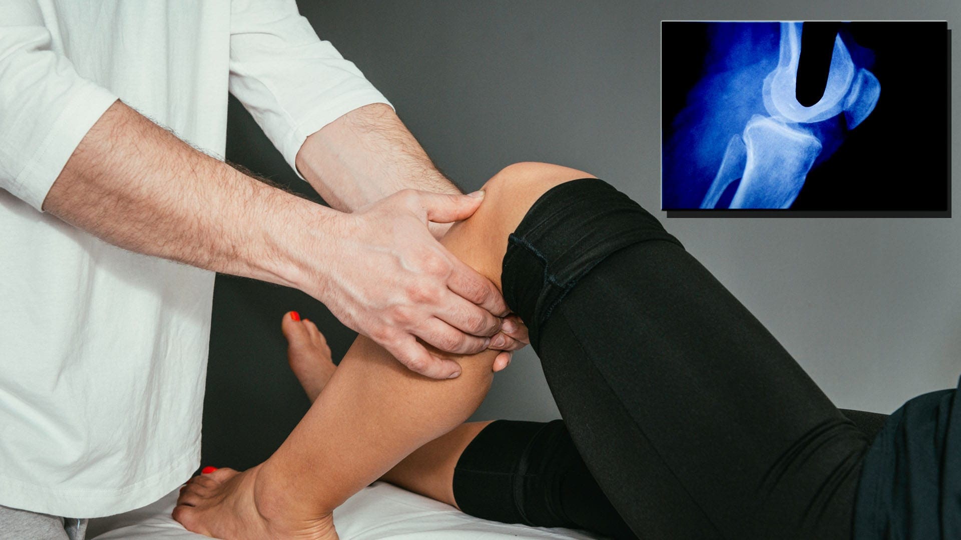
PRP and Integrative Chiropractic Care for Knee Meniscus Injuries
A knee meniscus tear can make simple movements feel difficult. Walking, bending, twisting, kneeling, or climbing stairs may cause pain, stiffness, swelling, or a feeling that the knee is not working right. Many people want to feel better without jumping straight to surgery. For that reason, conservative care has become a major focus for people dealing with knee injuries.
At El Paso Back Clinic, the focus is on improving how the knee moves, how the surrounding muscles support it, and how the whole body works together during healing. While regenerative options such as Platelet-Rich Plasma, or PRP, may be part of some care plans, the bigger picture is often about integrative chiropractic care, physical therapy-based rehabilitation, and functional recovery. This approach aims to reduce pain, improve joint mechanics, support natural healing, and help many patients return to daily activities with better comfort and confidence.
Understanding the Meniscus and Why It Matters
The meniscus is a strong piece of cartilage in the knee that acts like a shock absorber. Each knee has two menisci, and they help distribute weight, improve stability, protect the joint surfaces, and support smooth motion. When the meniscus is torn, the knee may become swollen, painful, weak, or unstable. Some people also notice catching, clicking, or a limited range of motion. (Andia & Maffulli, 2017; El Zouhbi et al., 2024)
A meniscus injury is important because the meniscus helps protect the knee over time. If the tear is not managed well, the joint can be placed under more stress, which may increase wear and tear later. That is why treatment should focus on both pain relief and long-term knee function.
Why Meniscus Tears Can Be Hard to Heal
Not every meniscus tear heals the same way. One major reason is blood flow. The outer part of the meniscus has a better blood supply, while the inner part has very little. This means that some tears have a better chance of healing than others. Tears in the outer zone often respond better to conservative treatment, whereas tears in the inner zone can be more challenging to treat. (Andia & Maffulli, 2017)
Other factors also affect healing, including:
The location of the tear
The size and pattern of the tear
The age of the patient
The condition of the knee joint
Strength and stability of the surrounding muscles
Activity level and movement habits
Because of this, a complete treatment plan should not focus only on the tear itself. It should also consider how the knee moves, how the hips and ankles support it, and how the body can be guided toward safer, stronger function.
The Role of Conservative, Integrative Care
At El Paso Back Clinic, a more chiropractic and rehabilitation-centered model makes sense for people who want a non-surgical path when appropriate. Conservative care often starts with reducing irritation in the knee, improving motion, correcting mechanical stress, and building strength around the joint. These steps can help lower pain and improve function while supporting the body’s natural healing process.
Integrative chiropractic care may include:
Careful assessment of gait and posture
Joint mobilization and chiropractic support for lower-body mechanics
Soft tissue work for muscles around the knee, hip, and lower leg
Stretching for tight structures that pull on the knee
Rehabilitation exercises to improve support and control
Movement retraining for walking, bending, and lifting
Physical therapy-based strengthening for the quadriceps, hamstrings, glutes, and core
This type of care is important because knee pain is often affected by more than the knee itself. Poor ankle motion, hip weakness, pelvic imbalance, altered posture, and abnormal walking patterns can all increase stress on the meniscus. Chiropractic and functional rehabilitation aim to improve those patterns so the knee is not constantly overloaded.
Why Joint Mechanics Matter So Much
Good joint mechanics are a major part of healing. If the knee continues to move poorly, the meniscus may remain irritated. If the hips are weak or the ankles are stiff, extra pressure may be placed on the knee with every step. Integrative chiropractic treatment works by looking at the whole movement chain, not just the painful spot.
For example, a patient with a meniscus injury may also have:
Poor hip stability
Tight hamstrings or calves
Weak glute muscles
Uneven weight shifting
Limited ankle mobility
Compensation in the low back or pelvis
When these problems are addressed, the knee often works more efficiently. This can reduce pain, improve balance, and help the person move with less strain. Chiropractic care in this setting is not just about an adjustment. It is about restoring better motion, reducing stress on injured tissues, and helping the body function as one connected system. (PCH Chiropractic, n.d.; LJ Chiropractic, n.d.)
Where PRP Fits Into the Bigger Picture
PRP is a regenerative treatment made from the patient’s own blood. After the blood is processed, a concentrated platelet layer is created. This contains growth factors that may support healing and help calm inflammation. In some cases, PRP may be considered as part of a broader plan for knee meniscus injuries, especially when a person wants to avoid surgery if possible. (Johns Hopkins Medicine, n.d.; El Zouhbi et al., 2024)
However, at a chiropractic and rehabilitation-centered clinic, PRP should be viewed as a background support tool rather than the main focus. The stronger message for patient care is that healing depends on function, stability, movement quality, and proper rehabilitation. Even with regenerative treatment, it works best when paired with mechanical support, strengthening, and guided recovery.
In other words, the knee does not heal well from an injection alone. It heals better when the whole joint environment improves.
What the Research Says About PRP for Meniscus Injuries
Research on PRP for meniscal injuries is promising but still developing. A 2024 narrative review reported that many studies showed short-term improvements in pain, function, and activity levels after PRP treatment, especially in follow-up periods of less than one year. At the same time, the review noted that long-term evidence remains mixed, and not every study showed clear differences over longer follow-up periods. (El Zouhbi et al., 2024)
This means PRP may help selected patients, but it is not a guaranteed answer for every tear. That is why it makes sense to keep the main focus on conservative, integrative care that improves knee function day after day.
Physical Therapy Principles in Meniscus Recovery
Physical therapy-based rehabilitation is a key part of non-surgical meniscus care. Strengthening the muscles around the knee helps reduce stress on the injured tissue. Improving balance and neuromuscular control helps the joint move more safely. Restoring range of motion helps reduce stiffness and improve confidence during activity. (Cognetti et al., 2024; Symmetry Physical Therapy, n.d.)
A typical conservative recovery plan may include:
Gentle mobility work early on
Swelling control and activity modification
Quadriceps activation exercises
Hamstring and glute strengthening
Core stabilization
Balance and coordination drills
Gradual return to walking, stairs, squatting, and sports tasks
This is one reason El Paso Back Clinic’s emphasis on chiropractic and rehab is so valuable. Patients often do best when they receive hands-on support plus guided therapeutic exercise rather than relying only on passive care.
Clinical Observations from Dr. Alexander Jimenez
Dr. Alexander Jimenez, DC, APRN, FNP-BC, has publicly described an integrative model that combines structural care, rehabilitation, functional medicine thinking, and movement-based recovery. His clinical observations support the idea that knee injuries often respond better when treatment focuses on reducing mechanical stress, improving movement quality, and promoting more complete healing. (Jimenez, 2026a, 2026b)
From that perspective, the most important message is not just that regenerative options exist. It is that the best outcomes often come from combining the following:
Better joint motion
Stronger muscular support
Improved gait and posture
Reduced inflammation
Progressive rehabilitation
Careful monitoring of function over time
That type of whole-body strategy fits well with a chiropractic and physical therapy-focused clinic identity.
Can This Approach Help People Avoid Surgery?
In some cases, yes. Not every meniscus tear needs surgery right away. Some patients improve with conservative care, especially when the tear is smaller, located in a better-healing zone, or does not cause severe locking or loss of function. When pain decreases, strength improves, swelling settles down, and movement becomes smoother, many people are able to return to normal activity without an operation. (El Zouhbi et al., 2024)
Still, it is important to be realistic. Some tears are too large, too unstable, or too mechanically disruptive to respond fully to conservative treatment. In those cases, an orthopedic referral may still be necessary. A patient-centered clinic should always support the treatment path that matches the injury.
Who May Benefit Most from Integrative Chiropractic and Rehab Care
A person may be a good candidate for a conservative, chiropractic-centered plan when they have the following:
Mild to moderate knee pain from a meniscus injury
Swelling or stiffness without major joint locking
Poor movement patterns that can be corrected
Muscle weakness around the knee and hips
A desire to avoid surgery if possible
A willingness to follow a rehabilitation plan
These patients often benefit from a program that restores motion, improves strength, and reduces stress on the injured knee over time.
The Value of a Whole-Body Recovery Plan
The knee is part of a larger movement system. If the hips, pelvis, low back, ankles, and feet are not working well, the knee may continue to struggle. That is why integrative chiropractic care can be so helpful. It goes beyond symptom relief to examine the full chain of motion.
A whole-body recovery plan may help:
Improve joint alignment and motion
Reduce strain on the meniscus
Build muscular support around the knee
Improve walking and standing mechanics
Lower the chance of repeated irritation
Support a safer return to work, exercise, and daily life
This type of care keeps the focus where it should be: on restoring function, improving resilience, and helping patients move better.
Conclusion
PRP may play a supportive role in the non-surgical management of some knee meniscus injuries, but the stronger long-term message for El Paso Back Clinic is the value of integrative chiropractic treatment and rehabilitation. Healing a meniscus injury is about more than one procedure. It is about improving how the knee moves, how the body supports it, and how the patient rebuilds strength and stability over time.
A conservative plan emphasizing chiropractic care, movement correction, soft-tissue support, and physical-therapy-based rehabilitation can help reduce pain and improve knee function in many patients. When appropriate, regenerative therapies may remain in the background as one part of a broader strategy. But the foundation of recovery is still mechanics, function, and whole-body care.
For many people with knee meniscus injuries, that kind of integrative approach offers a practical path toward healing without surgery while keeping the focus on strong movement, better stability, and long-term joint health.
IFM's Find A Practitioner tool is the largest referral network in Functional Medicine, created to help patients locate Functional Medicine practitioners anywhere in the world. IFM Certified Practitioners are listed first in the search results, given their extensive education in Functional Medicine