Back Clinic Mobility & Flexibility: The human body retains a natural level to ensure all its structures are functioning properly. The bones, muscles, ligaments, tendons, and other tissues work together to allow a range of movement and maintaining proper fitness and balanced nutrition can help keep the body functioning properly. Great mobility means executing functional movements with no restrictions in the range of motion (ROM).
Remember that flexibility is a mobility component, but extreme flexibility really is not required to perform functional movements. A flexible person can have core strength, balance, or coordination but cannot perform the same functional movements as a person with great mobility. According to Dr. Alex Jimenez’s compilation of articles on mobility and flexibility, individuals who don’t stretch their body often can experience shortened or stiffened muscles, decreasing their ability to move effectively.
Can various stretches be beneficial for individuals dealing with wrist and hand pain by reducing pain and discomfort to the extremities?
Introduction
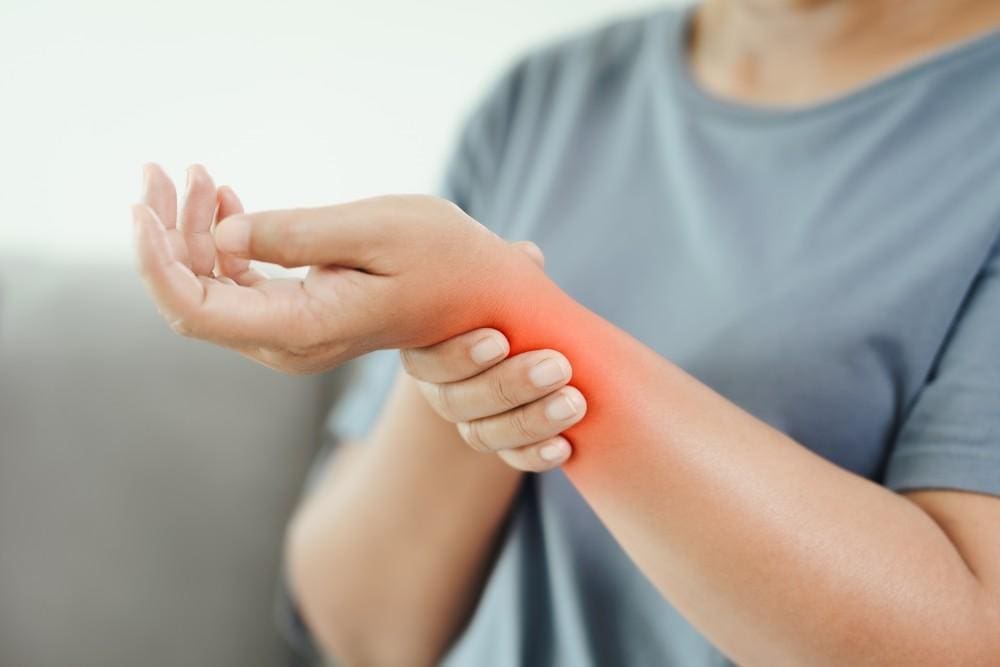
In a technological-driven world, it is common for people to experience wrist and hand pain at some point in their lives. The hands are part of the body’s upper extremities and are used for various tasks and chores throughout the entire day. The forearms provide a causal relationship with the hands and wrists for the upper extremities since they offer very important motor functions to the body. The hands support the body when carrying something; the various muscles, ligaments, tendons, and joints help the wrist with mobility and flexibility. However, when injuries or everyday movements begin to affect the forearms and cause issues with the hands and wrist, it can be difficult to do simple tasks and negatively impact a person’s way of life. Fortunately, numerous ways exist to reduce the pain and discomfort of the wrist and hands. Today’s article focuses on what causes wrist and hand pain, how to prevent wrist and hand pain from returning, and how incorporating various can help reduce the pain-like effects. We discuss with certified medical providers who consolidate our patients’ information to assess the multiple causes that lead to the development of wrist and hand pain. We also inform and guide patients on how various stretches and techniques can help reduce the chances of wrist and hand pain from returning. We also encourage our patients to ask their associated medical providers many intricate and important questions about incorporating these stretches and techniques into their daily routines to live healthier lives. Dr. Jimenez, D.C., includes this information as an academic service. Disclaimer.
What Causes Hand and Wrist Pain?
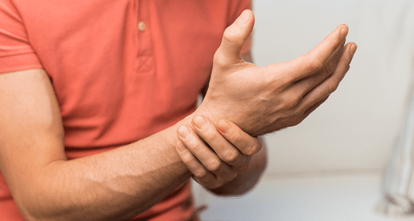
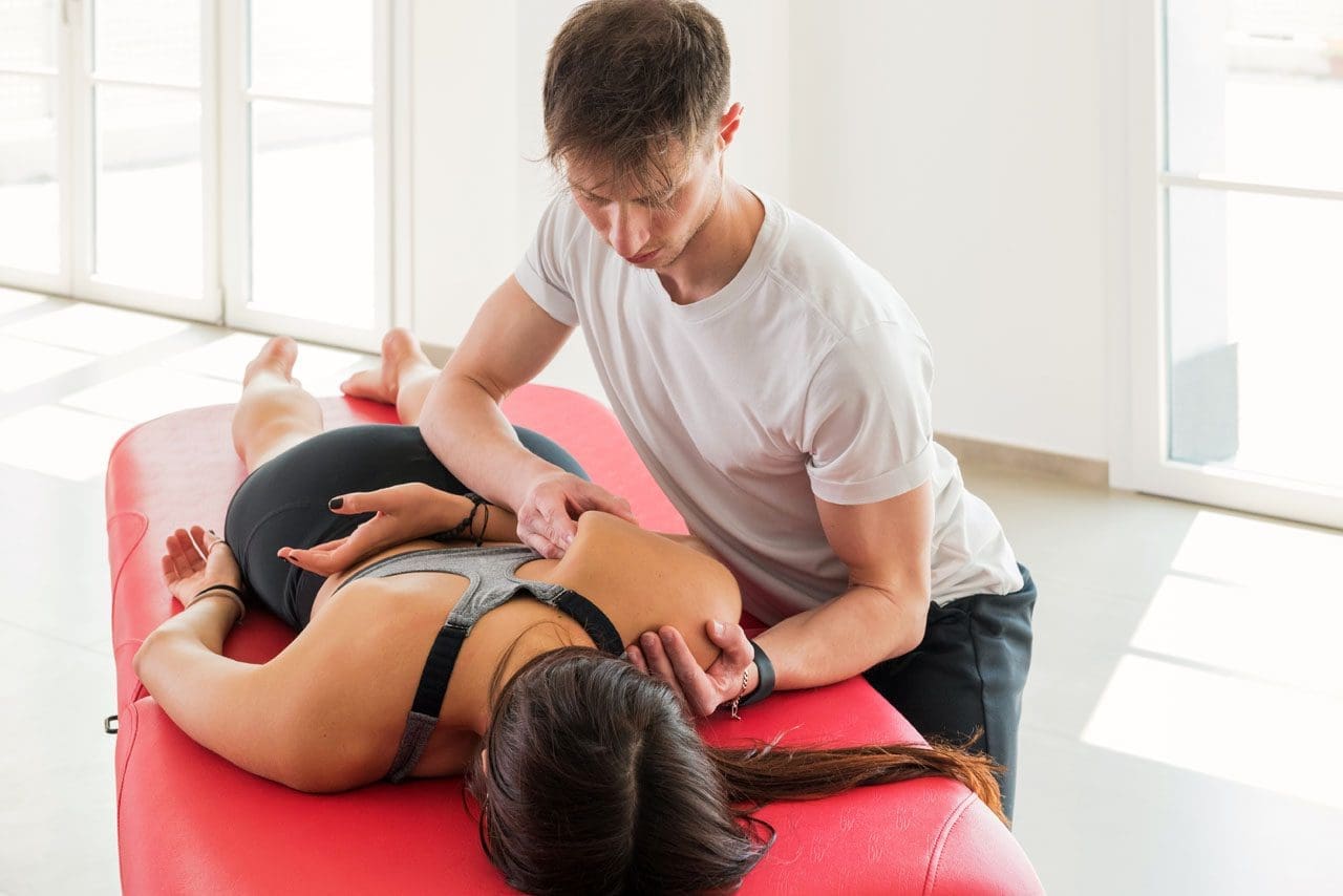
Do you often feel pain or stiffness in your wrist after typing all day on the computer or phone? Do you have trouble gripping items in your hands? Or how often do your hands ache that massaging them causes temporary relief? Many people, including older adults, have experienced pain at some point, and most of the time, it affects the hands and wrists. Since everyone uses their hands and wrists when performing various tasks, when injuries or repetitive movements start to affect the hands and wrists, it can have a huge impact on simple tasks. When dealing with wrist and hand pain, it can make life unbearable for the person. Since pain is a normal protective response to any injuries and potentially harmful stimuli in its acute form, when prolonged or dysfunctional neuromuscular issues start to affect the body, it may contribute to disability and pain. (Merkle et al., 2020) For wrist and hand pain, many occurrences that lead to its development result from micro-stress or repetitive tear usage.
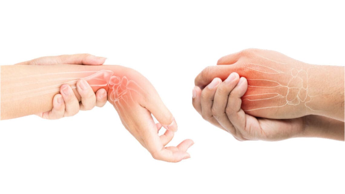
This is because since the world is technological-driven, many people are using computers or smartphones to communicate with each other, which can be one of the causes of the development of wrist and hand pain. When many people frequently use electronic devices, the frequent movements and uses of the thumbs will increase their load and become a higher prevalence of musculoskeletal disorders. (Baabdullah et al., 2020) Other studies stated that when many individuals begin to do repetitive movements constantly and have different positions of their wrist joints while using their electronic devices continually, it can cause pain to their wrist joints and affect the structure. (Amjad et al., 2020) Additionally, when repetitive vibration exposures or forceful angular motions affect the hands and wrists, it can lead to carpal tunnel syndrome and affect the hands. (Osiak et al., 2022) The various joints, tendons, and muscles also become affected in the hands and wrist as trigger points in the forearm. Fortunately, there are multiple ways that many people can reduce the pain-like effects of wrist and hand pain.
The Benefits of Stretching-Video
How To Prevent Wrist & Hand Pain From Returning
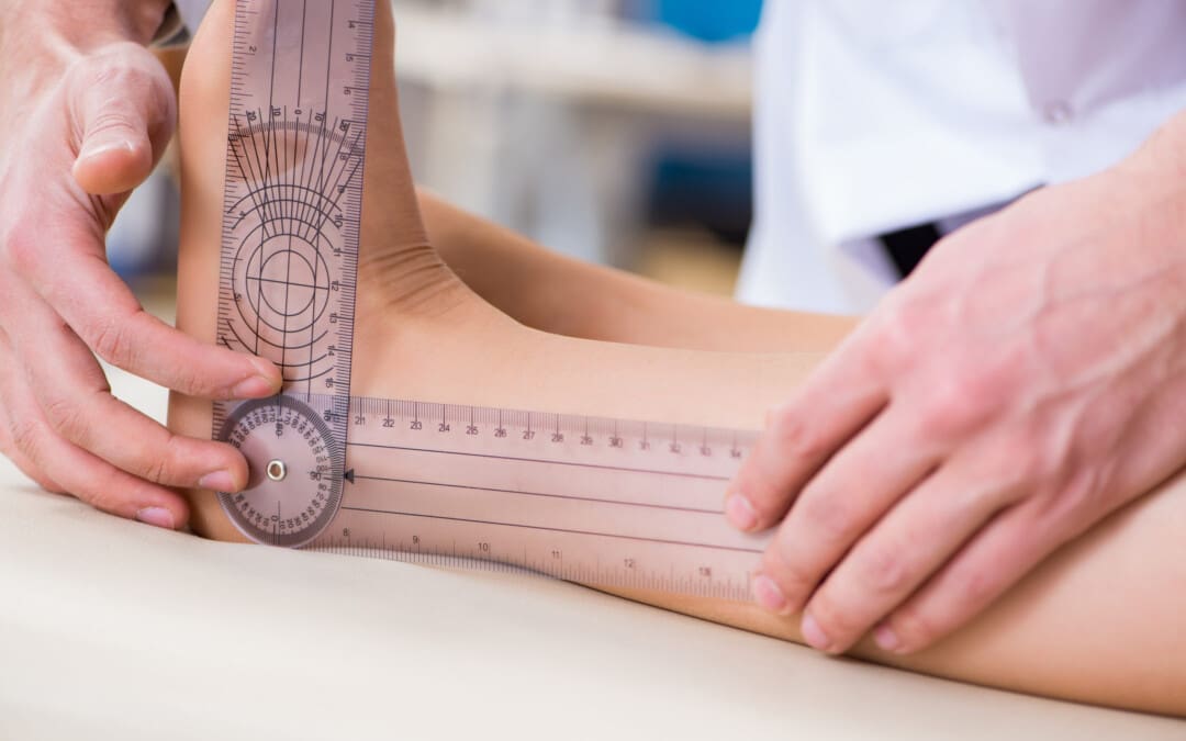
There are numerous ways to reduce wrist and hand pain, and many people try to find therapeutic solutions to mitigate the pain. Non-surgical treatments like manual therapy can help with wrist and hand pain by using mobilization forces to allow wrist flexion and extension to improve motor function. (Gutierrez-Espinoza et al., 2022) Another non-surgical treatment that can help with wrist and hand pain is acupuncture. Acupuncture utilizes small, solid, thin needles to be placed in various acupoints in the forearm to reduce the pain intensity and bring back the mobility function to the hands and wrist. (Trinh et al., 2022)
Various Stretches For Wrist & Hand Pain
Fortunately, there’s a simple and accessible way for many individuals to reduce the effects of wrist and hand pain-stretching and incorporating yoga into their routine. Yoga stretches for the hands and wrists can help decompress and reduce stiffness, and these stretches can be done for just a few minutes, providing beneficial results. (Gandolfi et al., 2023) Below are some of these stretches that can be easily incorporated into anyone’s routine, making it easier for you to take control of your wrist and hand health.
Wrist Flexor Stretch
How to Do It:
Extend your arm in front of you with your palm up.
Use your other hand to gently pull the fingers back toward the body until you feel a stretch in your forearm.
Hold this position for about 15 to 30 seconds.
Repeat 2-3 times with each wrist.
Wrist Extensor Stretch
How to Do It:
Extend your arm in front of your body with your palm facing down.
Gently pull the fingers towards your body with your other hand until you feel a stretch on the outside of your forearm.
Hold for 15 to 30 seconds.
Do this 2-3 times per wrist.
Prayer Stretch
How to Do It:
Put the palms together in a prayer position in front of the chest, below the chin.
Slowly lower the conjoined hands towards the waistline, keeping the hands close to your stomach and your palms together until you feel a stretch under your forearms.
Hold for at least 30 seconds and repeat a few times.
Tendon Glides
How to Do It:
Start with your fingers extended straight out.
Then, bend your fingers to form a hook fist; you should feel a stretch but no pain.
Return to the starting position and bend your fingers to touch the top of your palm, keeping your fingers straight.
Finally, bend your fingers into a full fist.
Repeat the sequence ten times.
Thumb Stretch
How to Do It:
Extend your hand with your fingers together.
Pull your thumb away from your fingers as far as comfortable.
Hold for 15 to 30 seconds.
Repeat 2-3 times with each thumb.
Shake It Out
How to Do It:
After stretching, shake your hands lightly as if trying to dry them off. This helps reduce tension and promote circulation.
References
Amjad, F., Farooq, M. N., Batool, R., & Irshad, A. (2020). Frequency of wrist pain and its associated risk factors in students using mobile phones. Pak J Med Sci, 36(4), 746-749. doi.org/10.12669/pjms.36.4.1797
Baabdullah, A., Bokhary, D., Kabli, Y., Saggaf, O., Daiwali, M., & Hamdi, A. (2020). The association between smartphone addiction and thumb/wrist pain: A cross-sectional study. Medicine (Baltimore), 99(10), e19124. doi.org/10.1097/MD.0000000000019124
Gandolfi, M. G., Zamparini, F., Spinelli, A., & Prati, C. (2023). Asana for Neck, Shoulders, and Wrists to Prevent Musculoskeletal Disorders among Dental Professionals: In-Office Yoga Protocol. J Funct Morphol Kinesiol, 8(1). doi.org/10.3390/jfmk8010026
Gutierrez-Espinoza, H., Araya-Quintanilla, F., Olguin-Huerta, C., Valenzuela-Fuenzalida, J., Gutierrez-Monclus, R., & Moncada-Ramirez, V. (2022). Effectiveness of manual therapy in patients with distal radius fracture: a systematic review and meta-analysis. J Man Manip Ther, 30(1), 33-45. doi.org/10.1080/10669817.2021.1992090
Merkle, S. L., Sluka, K. A., & Frey-Law, L. A. (2020). The interaction between pain and movement. J Hand Ther, 33(1), 60-66. doi.org/10.1016/j.jht.2018.05.001
Osiak, K., Elnazir, P., Walocha, J. A., & Pasternak, A. (2022). Carpal tunnel syndrome: state-of-the-art review. Folia Morphol (Warsz), 81(4), 851-862. doi.org/10.5603/FM.a2021.0121
Trinh, K., Zhou, F., Belski, N., Deng, J., & Wong, C. Y. (2022). The Effect of Acupuncture on Hand and Wrist Pain Intensity, Functional Status, and Quality of Life in Adults: A Systematic Review. Med Acupunct, 34(1), 34-48. doi.org/10.1089/acu.2021.0046
Can incorporating various yoga poses help reduce neck tension and provide pain relief for individuals dealing with neck pain?
Introduction
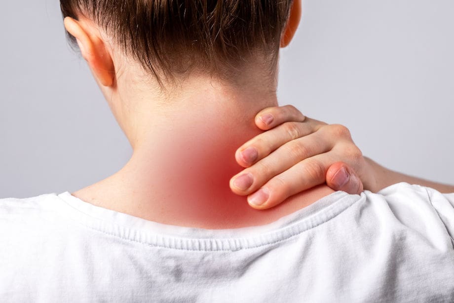
Within the hustling and bustling of modern life, it is common for many individuals to carry stress in their bodies. When the body deals with everyday stressors, tension, discomfort, and pain can often manifest in the upper and lower portions of the body. When the body’s upper and lower portions deal with these issues, they can cause overlapping risk profiles in the musculoskeletal system. One of the most common musculoskeletal issues is neck pain. It can cause many problems to the cervical portion of the spine and cause the surrounding muscles to become tense and in pain from the stress of everyday responsibilities. Luckily, there are numerous ways to reduce stress from the neck and help relax the affected muscles from discomfort, including yoga. In today’s article, we will look at how neck pain affects the upper body, the benefits of yoga for neck pain, and various yoga poses to reduce the overlapping effects of neck pain. We discuss with certified medical providers who consolidate our patients’ information to assess how neck pain is correlated with everyday stressors that affect the upper body. We also inform and guide patients on how yoga and the various poses can benefit the body and provide pain relief to the surrounding muscles. We also encourage our patients to ask their associated medical providers many intricate and important questions about incorporating yoga into their daily routine to reduce muscle tension and provide clarity to their bodies. Dr. Jimenez, D.C., includes this information as an academic service. Disclaimer.
How Does Neck Pain Affect The Upper Body?
Do you feel discomfort or pain in your neck and shoulders after a long, hard workday? Do you notice you hunched more than usual when doing your daily routine? Or do you see yourself developing a hunched posture from looking at the computer screen or phone for an extended period? Many of these normal motions are often correlated with the upper body, especially in the neck and shoulder regions, which causes neck pain. As one of the most common problems affecting many people worldwide, neck pain is a multifactorial disease with numerous risk factors contributing to its development. (Kazeminasab et al., 2022) Like back pain, neck pain can have acute and chronic stages depending on the severity and environmental factors leading to its development. The various muscles, ligaments, and tissues surrounding the neck and shoulders keep the neck stable and mobile. When many individuals overuse these muscles in the neck and shoulders repetitively, it can increase neck pain in the upper body in adulthood. (Ben Ayed et al., 2019)
When acute neck pain turns chronic, it can cause the individual to be in constant discomfort, pain, and misery, so they start to look for various solutions to reduce the correlating symptoms when speaking to their primary doctors. When many individuals begin to explain to their doctors what their daily routine looks like, many doctors will start to assess and formulate a plan that focuses on any specific description of any injuries, including potential mechanisms, inciting and relieving factors, and pain patterns they have encountered throughout the day to come up with a personalized treatment plan to not only reduce neck pain but also provide relief to tension and discomfort to the body. (Childress & Stuek, 2020)
The Science of Motion- Video
The Benefits Of Yoga For Neck Pain
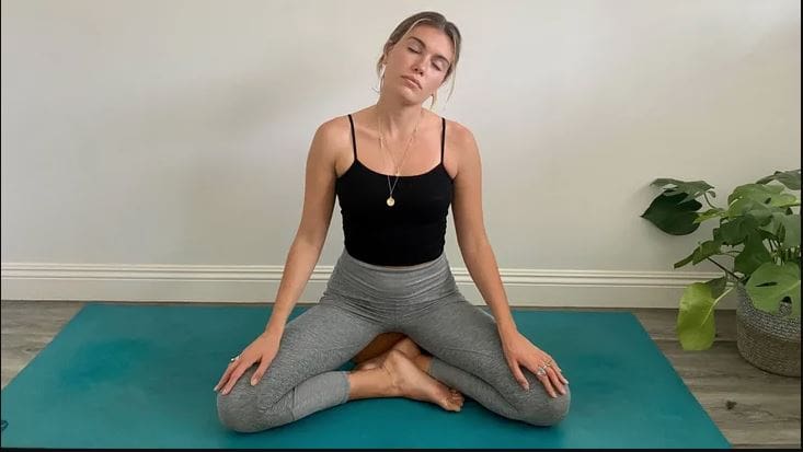
Many primary doctors will work with associated medical providers to develop a personalized plan to relieve neck pain and its associated symptoms in many individuals. Many of these customized treatment plans include spinal manipulation, acupuncture, massage, decompression therapy, and therapeutic exercises. One of the therapeutic exercises that many individuals have utilized is yoga. Yoga is a holistic practice encompassing breathing control, meditation, and various poses to stretch and strengthen the affected upper muscles. Yoga is excellent for reducing neck pain and helping with upper cervical spine mobility, stretching the neck musculature to help the individual improve mobility and flexibility. (Raja et al., 2021) Additionally, the effects of yoga and its many poses can reduce tension, give clarity to the mind, and allow the nutrients and oxygen to the musculo-articular system to naturally heal the body itself. (Gandolfi et al., 2023)
Yoga Poses For Neck Pain
At the same time, many individuals with sedentary jobs that correlate to neck pain have implemented yoga as part of their routine. Yoga improves their range of joint motion and cognitive function and helps relieve musculoskeletal discomfort in the neck and shoulder regions. (Thanasilungkoon et al., 2023) Below are some of the various yoga poses that can help reduce the pain-like symptoms of neck pain and ease the surrounding muscles.
Seated Neck Stretches
For seated neck stretches, this yoga pose helps stretch and release the neck muscles that carry tension and stress in the cervical region of the body.
In a seated upright position, turn the head to the right and gently lift the chin.
You should feel a stretch along the left side of the neck and shoulders.
Hold the position for three to five breaths and repeat on the left side.
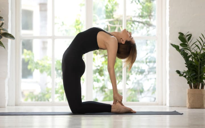
Camel Pose
For the camel pose, this yoga pose helps strengthen the front neck muscles while easing tension on the shoulders and back of the neck.
You can kneel on a yoga mat by keeping your knees and feet hip-distance apart while keeping the pelvis neutral.
Lift the chest while arching your back and pressing the pelvis slightly forward.
Bring the fingertips to the heels or yoga blocks beside the ankles.
Focus on drawing the chin close to the neck while pressing the feet to the mat.
Hold the position for three to five breaths before releasing and lifting the sternum to rise back up.
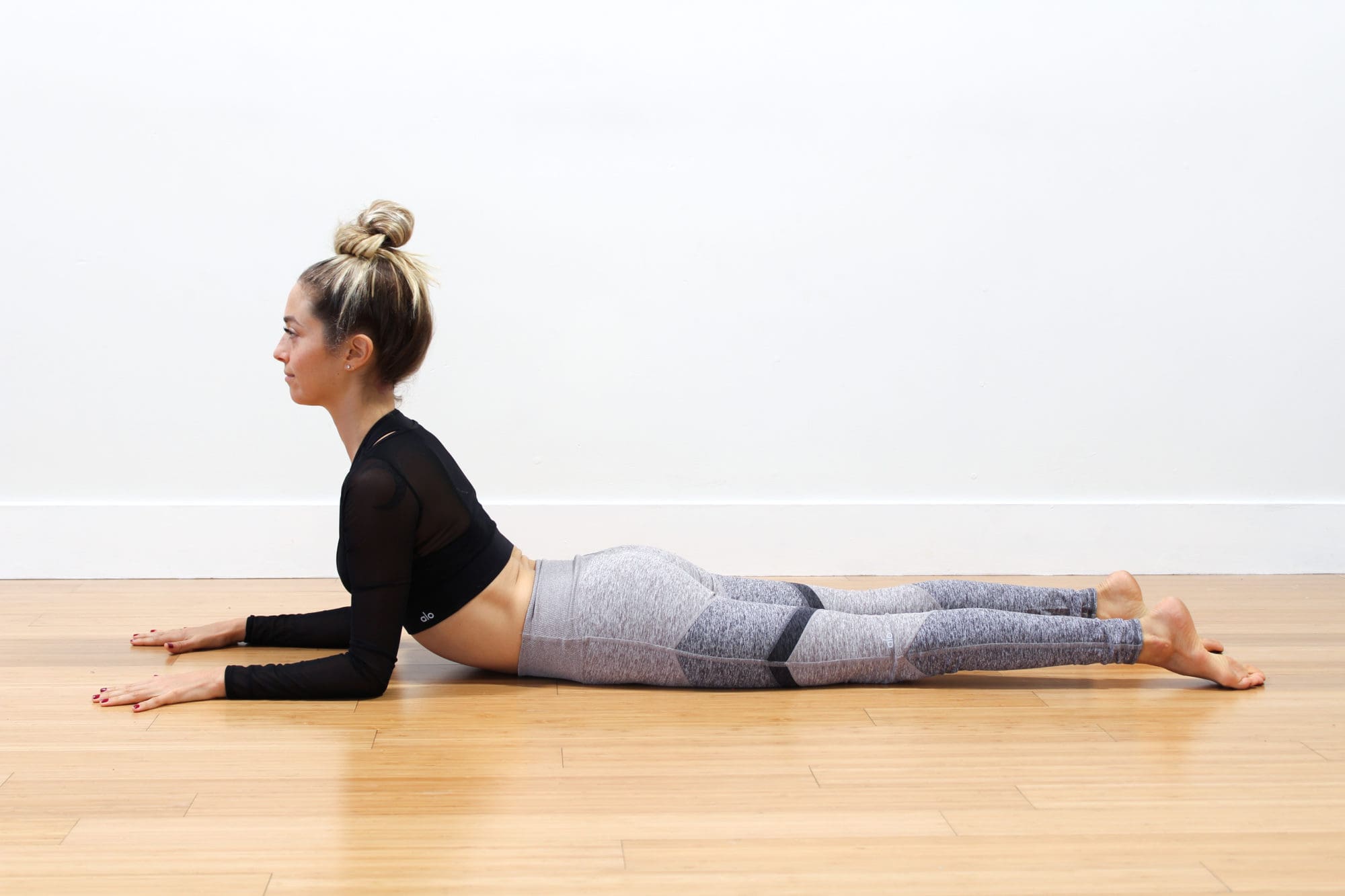
Sphinx Pose
The sphinx pose allows you to lengthen and strengthen the spine while stretching the shoulders and releasing tension.
On a yoga mat, lie on your stomach with the elbows under the shoulders.
Press your palms and forearms on the mat and tighten the lower half to support you as you lift your upper torso and head.
Keep looking straight ahead as you are being mindful of lengthening the spine.
Hold this position for three to five breaths.
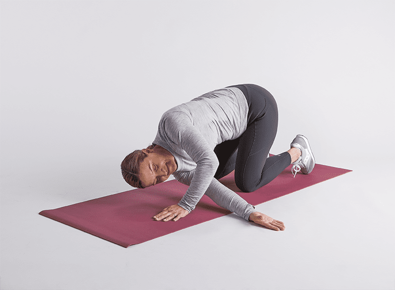
Thread The Needle Pose
The thread-the-needle pose helps release tension stored in the neck, shoulders, and back.
On a yoga mat, start in an all-fours position with the wrist under the shoulders and the knees under the hips.
Lift the right hand and move it to the left along the floor with the palm facing up.
Hold the position for three to five breaths for thirty seconds and release.
Return to the all-fours position and repeat to the left side.
Conclusion
Overall, incorporating yoga as part of a daily routine can provide beneficial results in reducing neck pain and its associated comorbidities. Yoga does not require hours of practice or even contorting into various poses, as just a few minutes of gentle stretching and mindful breathing each day can provide positive results. When people start to utilize yoga as part of their daily activities, they will notice their posture improving, their minds clearer than ever, and live a happier, healthier life without dealing with neck pain.
References
Ben Ayed, H., Yaich, S., Trigui, M., Ben Hmida, M., Ben Jemaa, M., Ammar, A., Jedidi, J., Karray, R., Feki, H., Mejdoub, Y., Kassis, M., & Damak, J. (2019). Prevalence, Risk Factors and Outcomes of Neck, Shoulders and Low-Back Pain in Secondary-School Children. J Res Health Sci, 19(1), e00440. www.ncbi.nlm.nih.gov/pubmed/31133629
Childress, M. A., & Stuek, S. J. (2020). Neck Pain: Initial Evaluation and Management. American Family Physician, 102(3), 150-156. www.ncbi.nlm.nih.gov/pubmed/32735440
Gandolfi, M. G., Zamparini, F., Spinelli, A., & Prati, C. (2023). Asana for Neck, Shoulders, and Wrists to Prevent Musculoskeletal Disorders among Dental Professionals: In-Office Yoga Protocol. J Funct Morphol Kinesiol, 8(1). doi.org/10.3390/jfmk8010026
Kazeminasab, S., Nejadghaderi, S. A., Amiri, P., Pourfathi, H., Araj-Khodaei, M., Sullman, M. J. M., Kolahi, A. A., & Safiri, S. (2022). Neck pain: global epidemiology, trends and risk factors. BMC Musculoskelet Disord, 23(1), 26. doi.org/10.1186/s12891-021-04957-4
Raja, G. P., Bhat, N. S., Fernandez-de-Las-Penas, C., Gangavelli, R., Davis, F., Shankar, R., & Prabhu, A. (2021). Effectiveness of deep cervical fascial manipulation and yoga postures on pain, function, and oculomotor control in patients with mechanical neck pain: study protocol of a pragmatic, parallel-group, randomized, controlled trial. Trials, 22(1), 574. doi.org/10.1186/s13063-021-05533-w
Thanasilungkoon, B., Niempoog, S., Sriyakul, K., Tungsukruthai, P., Kamalashiran, C., & Kietinun, S. (2023). The Efficacy of Ruesi Dadton and Yoga on Reducing Neck and Shoulder Pain in Office Workers. Int J Exerc Sci, 16(7), 1113-1130. www.ncbi.nlm.nih.gov/pubmed/38287934
Can individuals with Ehlers-Danlos syndrome find relief through various non-surgical treatments to reduce joint instability?
Introduction
The joints and ligaments surrounding the musculoskeletal system allow the upper and lower extremities to stabilize the body and be mobile. The various muscles and soft connective tissues that surround the joints help protect them from injuries. When environmental factors or disorders start to affect the body, many people develop issues that cause overlapping risk profiles, which then affect the stability of the joints. One of the disorders that affect the joints and connective tissue is EDS or Ehlers-Danlos syndrome. This connective tissue disorder can cause the joints in the body to be hypermobile. It can cause joint instability in the upper and lower extremities, thus leaving the individual to be in constant pain. Today’s article focuses on Ehlers-Danlos syndrome and its symptoms and how there are non-surgical ways to manage this connective tissue disorder. We discuss with certified medical providers who consolidate our patients’ information to assess how Ehlers-Danlos syndrome can correlate with other musculoskeletal disorders. We also inform and guide patients on how various non-surgical treatments can help reduce pain-like symptoms and manage Ehlers-Danlos syndrome. We also encourage our patients to ask their associated medical providers many intricate and important questions about incorporating various non-surgical therapies as part of their daily routine to manage the effects of Ehlers-Danlos syndrome. Dr. Jimenez, D.C., includes this information as an academic service. Disclaimer.
What Is Ehlers-Danlos Syndrome?
Do you often feel extremely tired throughout the day, even after a full night of sleep? Do you bruise easily and wonder where these bruises are coming from? Or have you noticed that you have an increased range in your joints? Many of these issues are often correlated with a disorder known as Ehlers-Danlos syndrome or EDS that affects their joints and connective tissue. EDS affects the connective tissues in the body. The connective tissues in the body help provide strength and elasticity to the skin, joints, as well as blood vessel walls, so when a person is dealing with EDS, it can cause a significant disruption to the musculoskeletal system. EDS is largely diagnosed clinically, and many doctors have identified that the gene coding of the collagen and proteins that interact in the body can help determine what type of EDS affects the individual. (Miklovic & Sieg, 2024)
The Symptoms
When understanding EDS, it is essential to know the complexities of this connective tissue disorder. EDS is classified into numerous types with distinct features and challenges that vary depending on the severity. One of the most common types of EDS is hypermobile Ehlers-Danlos syndrome. This type of EDS is characterized by general joint hypermobility, joint instability, and pain. Some of the symptoms that are associated with hypermobile EDS include subluxation, dislocations, and soft tissue injuries that are common and may occur spontaneously or with minimal trauma. (Hakim, 1993) This can often cause acute pain to the joints in the upper and lower extremities. With its broad range of symptoms and the personal nature of the condition itself, many often don’t realize that joint hypermobility is common in the general population and may present no complications that indicate that it is a connective tissue disorder. (Gensemer et al., 2021) Additionally, hypermobile EDS can lead to spinal deformity due to the hyperextensibility of the skin, joints, and various tissue fragility. The pathophysiology of spinal deformity associated with hypermobile EDS is primarily due to muscle hypotonia and ligament laxity. (Uehara et al., 2023) This causes many people to reduce their quality of life and daily living activities significantly. However, there are ways to manage EDS and its correlating symptoms to reduce joint instability.
Movement Medicine: Chiropractic Care-Video
Ways To Manage EDS
When it comes to looking for ways to manage EDS to reduce pain and joint instability, non-surgical treatments can help address the physical and emotional aspects of the condition. Non-surgical treatments for individuals with EDS commonly focus on optimizing the body’s physical function while improving muscular strength and joint stabilization. (Buryk-Iggers et al., 2022) Many individuals with EDS will try to incorporate pain management techniques and physical therapy anduse braces and assistive devices to reduce the effects of EDS and improve their quality of life.
Non-surgical Treatments For EDS
Various non-surgical treatments like MET (muscle energy technique), electrotherapy, light physical therapy, chiropractic care, and massages can help strengthen while toning the surrounding muscles around the joints, provide sufficient pain relief, and limit long-term dependence on medications. (Broida et al., 2021) Additionally, individuals dealing with EDS aim to strengthen the affected muscles, stabilize the joints, and improve proprioception. Non-surgical treatments allow the individual to have a customized treatment plan for the severity of EDS symptoms and help reduce the pain associated with the condition. Many individuals, when going through their treatment plan consecutively to manage their EDS and reduce the pain-like symptoms, will notice improvement in symptomatic discomfort. (Khokhar et al., 2023) This means that non-surgical treatments allow individuals to be more mindful of their bodies and reduce the pain-like effects of EDS, thus allowing many individuals with EDS to lead fuller, more comfortable lives without feeling pain and discomfort.
References
Broida, S. E., Sweeney, A. P., Gottschalk, M. B., & Wagner, E. R. (2021). Management of shoulder instability in hypermobility-type Ehlers-Danlos syndrome. JSES Rev Rep Tech, 1(3), 155-164. doi.org/10.1016/j.xrrt.2021.03.002
Buryk-Iggers, S., Mittal, N., Santa Mina, D., Adams, S. C., Englesakis, M., Rachinsky, M., Lopez-Hernandez, L., Hussey, L., McGillis, L., McLean, L., Laflamme, C., Rozenberg, D., & Clarke, H. (2022). Exercise and Rehabilitation in People With Ehlers-Danlos Syndrome: A Systematic Review. Arch Rehabil Res Clin Transl, 4(2), 100189. doi.org/10.1016/j.arrct.2022.100189
Gensemer, C., Burks, R., Kautz, S., Judge, D. P., Lavallee, M., & Norris, R. A. (2021). Hypermobile Ehlers-Danlos syndromes: Complex phenotypes, challenging diagnoses, and poorly understood causes. Dev Dyn, 250(3), 318-344. doi.org/10.1002/dvdy.220
Hakim, A. (1993). Hypermobile Ehlers-Danlos Syndrome. In M. P. Adam, J. Feldman, G. M. Mirzaa, R. A. Pagon, S. E. Wallace, L. J. H. Bean, K. W. Gripp, & A. Amemiya (Eds.), GeneReviews((R)). www.ncbi.nlm.nih.gov/pubmed/20301456
Khokhar, D., Powers, B., Yamani, M., & Edwards, M. A. (2023). The Benefits of Osteopathic Manipulative Treatment on a Patient With Ehlers-Danlos Syndrome. Cureus, 15(5), e38698. doi.org/10.7759/cureus.38698
Can understanding the body’s hinge joints and how they operate help with mobility and flexibility problems and manage conditions for individuals with difficulty fully bending or extending their fingers, toes, elbows, ankles, or knees?
Hinge Joints
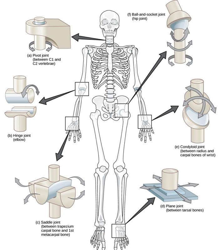
A joint forms where one bone connects to another, allowing motion. Different types of joints differ in structure and movement depending on their location. These include hinge, ball and socket, planar, pivot, saddle, and ellipsoid joints. (Boundless. General Biology, N.D.) Hinge joints are synovial joints that move through one plane of motion: flexion and extension. Hinge joints are found in the fingers, elbows, knees, ankles, and toes and control movement for various functions. Injuries, osteoarthritis, and autoimmune conditions can affect hinge joints. Rest, medication, ice, and physical therapy can help alleviate pain, improve strength and range of motion, and help manage conditions.
Anatomy
A joint is formed by the joining of two or more bones. The human body has three main classifications of joints, categorized by the degree to which they can move. These include: (Boundless. General Biology, N.D.)
Synarthroses
These are fixed, immovable joints.
Formed by two or more bones.
Amphiarthroses
Also known as cartilaginous joints.
A fibrocartilage disc separates the bones that form the joints.
These movable joints allow for a slight degree of movement.
Diarthroses
Also known as synovial joints.
These are the most common freely mobile joints that allow movement in multiple directions.
The bones that form the joints are lined with articular cartilage and enclosed in a joint capsule filled with synovial fluid that allows for smooth motion.
Synovial joints are classified into different types depending on differences in structure and the number of motion planes they allow. A hinge joint is a synovial joint that allows movement in one plane of motion, similar to a door hinge that moves forward and backward. Within the joint, the end of one bone is typically convex/pointed outward, with the other concave/rounded inward to allow the ends to fit smoothly. Because hinge joints only move through one plane of movement, they tend to be more stable than other synovial joints. (Boundless. General Biology, N.D.) Hinge joints include:
The finger and toe joints – allow the fingers and toes to bend and extend.
The elbow joint – allows the elbow to bend and extend.
The knee joint – allows the knee to bend and extend.
The talocrural joint of the ankle – allows the ankle to move up/dorsiflexion and down/plantarflexion.
Hinge joints allow the limbs, fingers, and toes to extend away and bend toward the body. This movement is essential for activities of daily living, such as showering, getting dressed, eating, walking, standing up, and sitting down.
Conditions
Osteoarthritis and inflammatory forms of arthritis can affect any joint (Arthritis Foundation. N.D.) Autoimmune inflammatory forms of arthritis, including rheumatoid and psoriatic arthritis, can cause the body to attack its own joints. These commonly affect the knees and fingers, resulting in swelling, stiffness, and pain. (Kamata, M., Tada, Y. 2020) Gout is an inflammatory form of arthritis that develops from elevated levels of uric acid in the blood and most commonly affects the hinge joint of the big toe. Other conditions that affect hinge joints include:
Injuries to the cartilage within the joints or ligaments that stabilize the outside of the joints.
Ligament sprains or tears can result from jammed fingers or toes, rolled ankles, twisting injuries, and direct impact on the knee.
These injuries can also affect the meniscus, the tough cartilage within the knee joint that helps cushion and absorb shock.
Rehabilitation
Conditions that affect hinge joints often cause inflammation and swelling, resulting in pain and limited mobility.
After an injury or during an inflammatory condition flare-up, limiting active movement and resting the affected joint can reduce increased stress and pain.
Applying ice can decrease inflammation and swelling.
Once the pain and swelling start to subside, physical and/or occupational therapy can help rehabilitate the affected areas.
A therapist will provide stretches and exercises to help improve the joint range of motion and strengthen the supporting muscles.
For individuals experiencing hinge joint pain from an autoimmune condition, biologic medications to decrease the body’s autoimmune activity are administered through infusions delivered every several weeks or months. (Kamata, M., Tada, Y. 2020)
Cortisone injections may also be used to decrease inflammation.
At Injury Medical Chiropractic and Functional Medicine Clinic, we passionately focus on treating patients’ injuries and chronic pain syndromes and improving ability through flexibility, mobility, and agility programs tailored to the individual. Our providers use an integrated approach to create personalized care plans that include Functional Medicine, Acupuncture, Electro-Acupuncture, and Sports Medicine protocols. Our goal is to relieve pain naturally by restoring health and function to the body. If the individual needs other treatment, they will be referred to a clinic or physician best suited for them. Dr. Jimenez has teamed up with the top surgeons, clinical specialists, medical researchers, and premier rehabilitation providers to provide the most effective clinical treatments.
Kamata, M., & Tada, Y. (2020). Efficacy and Safety of Biologics for Psoriasis and Psoriatic Arthritis and Their Impact on Comorbidities: A Literature Review. International journal of molecular sciences, 21(5), 1690. doi.org/10.3390/ijms21051690
For individuals experiencing shoulder and upper back pain, could periscapular bursitis be a possible cause?
Periscapular Bursitis
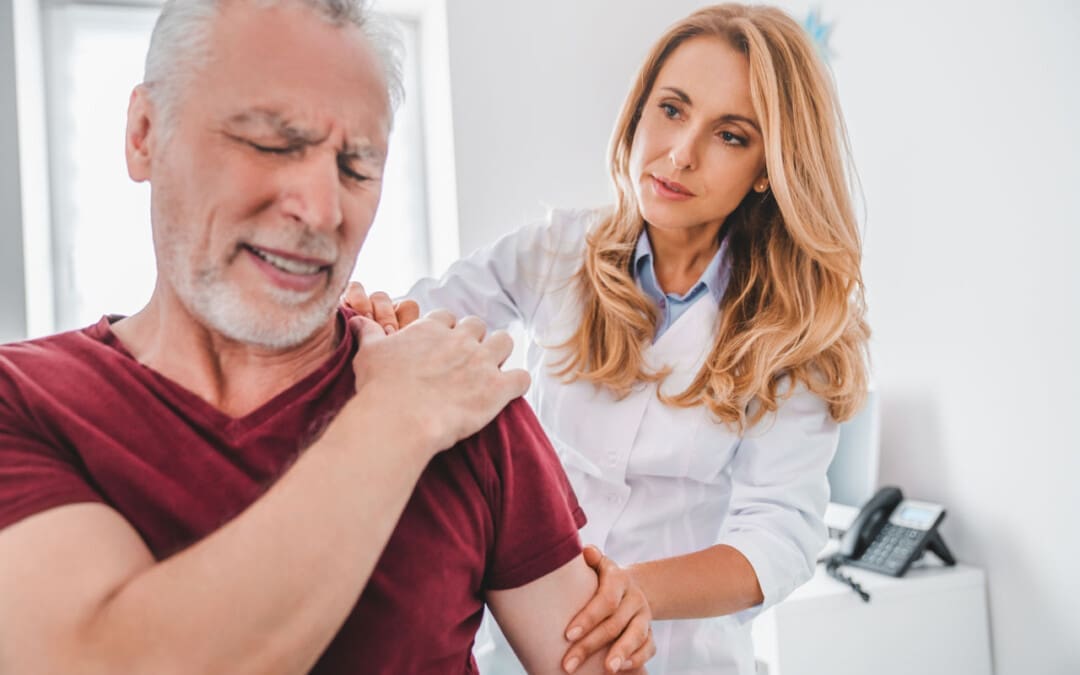
The scapula/shoulder blade is a bone that shifts position with upper body and shoulder movement. The scapula motion is critical to the normal function of the shoulder and the spine. When abnormal or sudden shoulder movements occur, inflammation and pain symptoms can develop. (Augustine H. Conduah et al., 2010)
Normal Scapula Function
The scapula is a triangular bone on the upper back outside the rib cage. Its outer or lateral side contains the shoulder joint socket /glenoid, while the rest of the bone serves as attachment points for the different shoulder and back muscles. The scapula shifts on the rib cage when moving the arm forward and back. This movement is called scapulothoracic motion and is critical to the normal function of the upper extremity and the shoulder joint. When the scapula does not glide in a coordinated motion, the function of the torso and shoulder joints can become stiff and painful. (J. E. Kuhn et al., 1998)
Scapular Bursa
A bursa is a fluid-filled sac that allows smooth, gliding motion between structures, body tissues, bones, and tendons. Bursae are found throughout the body, including those in front of the kneecap, outside the hip, and at the shoulder joint. When a bursa becomes inflamed and irritated, normal movements can become painful. There are bursae around the scapula in the upper back. Two of these bursa sacs are between the bones and the serratus anterior muscle that controls scapular movement on the chest wall. One bursa sac is located on the upper corner of the scapula, close to the spine at the base of the neck, and the other is at the bottom corner of the scapula, close to the mid-back. Either or both bursa sacs can be affected by periscapular bursitis. There are other bursae around the scapula and the surrounding tendons, but the two corner sacs tend to be the primary bursae that develop periscapular bursitis.
Inflammation
When these bursae become inflamed and irritated, swollen, and thickened, the condition known as bursitis results. When bursitis occurs near the scapula, muscle, and shoulder blade movements can lead to discomfort and pain. The most common symptoms of periscapular bursitis include:
An examination of the scapula may display abnormal movements of the shoulder blade. This can lead to winging, where the shoulder blade is not held correctly to the rib cage and protrudes abnormally. Individuals with winging of the scapula typically have abnormal shoulder joint mechanics because the shoulder’s positioning is altered.
Causes
The causes of periscapular bursitis can be varied. The most common is overuse syndrome, where a specific activity is causing irritation to the bursa. These can include:
Sports-related activities that result from repetitive use.
Work-related activities that result from repetitive use.
Traumatic injuries that cause inflammation or irritation to the bursa.
Some conditions can cause abnormal anatomy or bone protuberances, irritating the bursa. One condition is a benign bone growth known as an osteochondroma. (Antônio Marcelo Gonçalves de Souza and Rosalvo Zósimo Bispo Júnior 2014) These growths can project off the scapula, leading to irritation and inflammation.
Treatment
Treatment of periscapular bursitis begins with conservative therapies. Invasive treatments are rarely needed to correct the problem. Treatment can include:
Rest
The first step is to rest the irritated bursa and settle the inflammation.
This can take a few weeks and can be accomplished by modifying physical, sports, or work-related activities.
Ice
Ice is useful for reducing inflammation and controlling pain.
Knowing how to ice an injury properly can help manage the pain and swelling.
Physical Therapy
Physical therapy can alleviate the symptoms of inflammation through various exercises and stretches.
The therapy can improve scapular mechanics so the injury does not become ongoing and recurrent.
Abnormal movement of the scapula on the rib cage can not only lead to the development of bursitis, but if these abnormal mechanics are not addressed, the problem may recur.
Anti-Inflammatory Medications
Non-steroidal anti-inflammatory medications are used to control the inflammation in the short term. (Augustine H. Conduah et al., 2010)
The medications can help block the inflammatory response.
Before taking any medication, individuals should confirm with their healthcare provider that it is safe.
Cortisone Injections
Successful treatment with a cortisone shot is a sign that surgery will be more effective for individuals who may need surgery.
Cortisone injections can be very helpful in delivering a powerful anti-inflammatory dose directly to the site of inflammation. (Augustine H. Conduah et al., 2010)
Cortisone injections should be limited in terms of how many injections are offered to an individual, but in limited doses can be very helpful.
However, cortisone shots should only be performed once the diagnosis is confirmed.
Surgery
Surgery is seldom necessary but can be effective in individuals who are unable to find relief with conservative treatments.
Surgery is often used for individuals with abnormal scapular anatomy, like bone growths or tumors.
At Injury Medical Chiropractic and Functional Medicine Clinic, we treat injuries and chronic pain syndromes by improving an individual’s ability through flexibility, mobility, and agility programs tailored for all age groups and disabilities. Our chiropractor care plans and clinical services are specialized and focused on injuries and the complete recovery process. If other treatment is needed, individuals will be referred to a clinic or physician best suited to their injury, condition, and/or ailment.
Scapular Winging in Depth
References
Conduah, A. H., Baker, C. L., 3rd, & Baker, C. L., Jr (2010). Clinical management of scapulothoracic bursitis and the snapping scapula. Sports health, 2(2), 147–155. doi.org/10.1177/1941738109338359
Kuhn, J. E., Plancher, K. D., & Hawkins, R. J. (1998). Symptomatic scapulothoracic crepitus and bursitis. The Journal of the American Academy of Orthopaedic Surgeons, 6(5), 267–273. doi.org/10.5435/00124635-199809000-00001
de Souza, A. M., & Bispo Júnior, R. Z. (2014). Osteochondroma: ignore or investigate?. Revista brasileira de ortopedia, 49(6), 555–564. doi.org/10.1016/j.rboe.2013.10.002
Can individuals with joint hypermobility find relief through nonsurgical treatments in reducing pain and restoring body mobility?
Introduction
When a person moves their body, the surrounding muscles, joints, and ligaments are incorporated into various tasks that allow them to stretch and be flexible without pain or discomfort. Many repetitive motions enable the individual to continue their routine. However, when the joints, muscles, and ligaments are stretched farther than normal in the upper and lower extremities without pain, it is known as joint hypermobility. This connective tissue disorder can correlate with other symptoms that affect the body and cause many people to seek treatment to manage joint hypermobility symptoms. In today’s article, we will look at joint hypermobility and how various non-surgical treatments can help reduce pain caused by joint hypermobility and restore body mobility. We talk with certified medical providers who consolidate our patients’ information to assess how their pain may be associated with joint hypermobility. We also inform and guide patients on how integrating various non-surgical treatments can help improve joint function while managing the associated symptoms. We encourage our patients to ask their associated medical providers intricate and insightful questions about incorporating non-surgical therapies as part of their routine to reduce pain and discomfort from joint hypermobility. Dr. Jimenez, D.C., includes this information as an academic service. Disclaimer.
What Is Joint Hypermobility?
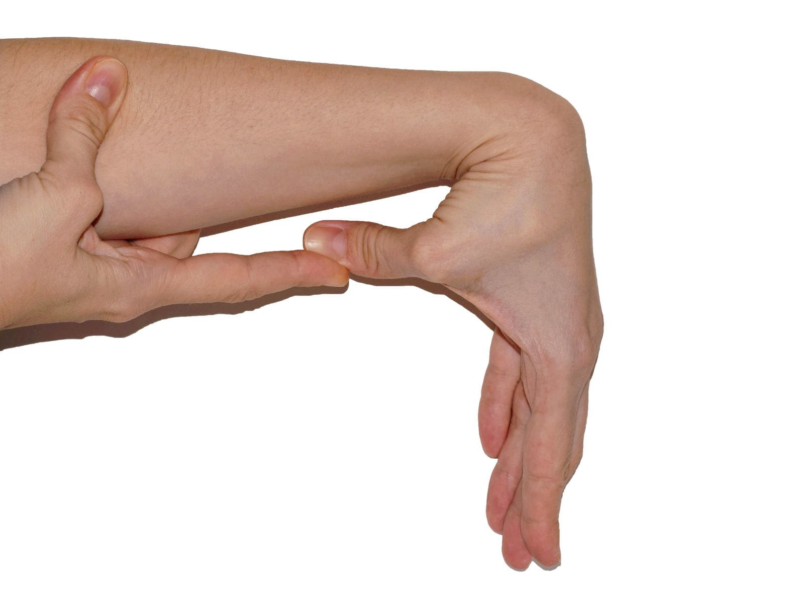
Do you often feel your joints locked up in your hands, wrists, knees, and elbows? Do you experience pain and fatigue in your joints when your body feels constantly tired? Or when you stretch your extremities, do they extend farther than usual to feel the relief? Many of these various scenarios are often correlated with individuals experiencing joint hypermobility. Joint hypermobility is an inherited disorder with autosomal dominant patterns that characterize joint hyperlaxity and musculoskeletal pain within the body extremities. (Carbonell-Bobadilla et al., 2020) This connective tissue condition is often related to the flexibility of the connected tissues like ligaments and tendons in the body. An example would be if a person’s thumb is touching their inner forearm without feeling pain or discomfort, they have joint hypermobility. Additionally, many individuals dealing with joint hypermobility will often have a difficult diagnosis as they will develop skin and tissue fragility over time, causing musculoskeletal complications. (Tofts et al., 2023)
When individuals deal with joint hypermobility over time, many often have symptomatic joint hypermobility. They will present with musculoskeletal and systemic symptoms that lead to displaying skeletal deformities, tissue and skin fragility, and structural differences in the body’s system. (Nicholson et al., 2022) Some of the symptoms that joint hypermobility are shown in a diagnosis include:
Muscle pain and joint stiffness
Clicking joints
Fatigue
Digestive issues
Balance issues
Luckily, there are various treatments that many people can use to help restrengthen the surrounding muscles around the joints and reduce the correlating symptoms caused by joint hypermobility.
Movement As Medicine-Video
Nonsurgical Treatments For Joint Hypermobility
When dealing with joint hypermobility, many individuals need to seek treatments to reduce the correlating pain-like symptoms of joint hypermobility and help relieve the body’s extremities while restoring mobility. Some excellent treatments for joint hypermobility are non-surgical therapies that are non-invasive, gentle on the joints and muscles, and cost-effective. Various non-surgical treatments can be customized for the individual depending on how severe their joint hypermobility and comorbidities affect the person’s body. Non-surgical treatments can relieve the body from joint hypermobility by treating the causes of the pain through reduction and maximizing functional capacity and restoring a person’s quality of life. (Atwell et al., 2021) The three non-surgical treatments that are excellent for reducing pain from joint hypermobility and helping strengthen the surrounding muscles are below.
Chiropractic Care
Chiropractic care utilizes spinal manipulation and helps restore joint mobility in the body to reduce the effects of joint hypermobility by stabilizing the affected joints from the hypermobile extremities. (Boudreau et al., 2020) Chiropractors incorporate mechanical and manual manipulation and various techniques to help many individuals improve their posture by being more mindful of their bodies and work with multiple other therapies to emphasize controlled movements. With other comorbidities associated with joint hypermobility, like back and neck pain, chiropractic care can reduce these comorbidity symptoms and allow the individual to regain their quality of life.
Acupuncture
Another non-surgical treatment that many individuals can incorporate to reduce joint hypermobility and its comorbidities is acupuncture. Acupuncture utilizes small, thin, solid needles that acupuncturists use to block pain receptors and restore the body’s energy flow. When many individuals are dealing with joint hypermobility, their extremities in the legs, hands, and feet are in pain over time, which can cause the body to be unstable. What acupuncture does is help reduce the pain caused by joint hypermobility associated with the extremities and restore balance and functionality to the body (Luan et al., 2023). This means that if a person is dealing with stiffness and muscle pain from joint hypermobility, acupuncture can help rewire the pain by placing the needles in the body’s acupoints to provide relief.
Physical Therapy
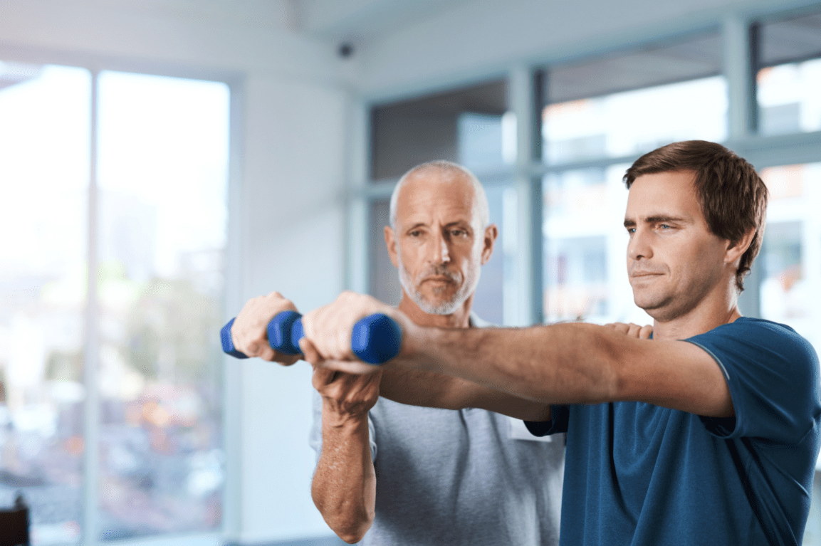
Physical therapy is the last non-surgical treatment many people can incorporate into their daily routine. Physical therapy can help manage joint hypermobility that are tailored to help strengthen weak muscles that are surrounding the affected joints, improving a person’s stability and helping reduce the risk of dislocation. Additionally, many individuals can use low-impact exercise to ensure optimal motor control when doing regular exercises without putting excessive strain on the joints. (Russek et al., 2022)
By incorporating these three non-surgical treatments as part of a customized treatment for joint hypermobility, many individuals will begin to feel a difference in their balance. They will not experience joint pain by being more mindful of the body and incorporating small changes in their routine. Even though living with joint hypermobility can be a challenge for many individuals, by integrating and utilizing the right combination of non-surgical treatments, many can begin to lead active and fulfilling lives.
References
Atwell, K., Michael, W., Dubey, J., James, S., Martonffy, A., Anderson, S., Rudin, N., & Schrager, S. (2021). Diagnosis and Management of Hypermobility Spectrum Disorders in Primary Care. J Am Board Fam Med, 34(4), 838-848. doi.org/10.3122/jabfm.2021.04.200374
Boudreau, P. A., Steiman, I., & Mior, S. (2020). Clinical management of benign joint hypermobility syndrome: a case series. J Can Chiropr Assoc, 64(1), 43-54. www.ncbi.nlm.nih.gov/pubmed/32476667
Carbonell-Bobadilla, N., Rodriguez-Alvarez, A. A., Rojas-Garcia, G., Barragan-Garfias, J. A., Orrantia-Vertiz, M., & Rodriguez-Romo, R. (2020). [Joint hypermobility syndrome]. Acta Ortop Mex, 34(6), 441-449. www.ncbi.nlm.nih.gov/pubmed/34020527 (Sindrome de hipermovilidad articular.)
Luan, L., Zhu, M., Adams, R., Witchalls, J., Pranata, A., & Han, J. (2023). Effects of acupuncture or similar needling therapy on pain, proprioception, balance, and self-reported function in individuals with chronic ankle instability: A systematic review and meta-analysis. Complement Ther Med, 77, 102983. doi.org/10.1016/j.ctim.2023.102983
Nicholson, L. L., Simmonds, J., Pacey, V., De Wandele, I., Rombaut, L., Williams, C. M., & Chan, C. (2022). International Perspectives on Joint Hypermobility: A Synthesis of Current Science to Guide Clinical and Research Directions. J Clin Rheumatol, 28(6), 314-320. doi.org/10.1097/RHU.0000000000001864
Russek, L. N., Block, N. P., Byrne, E., Chalela, S., Chan, C., Comerford, M., Frost, N., Hennessey, S., McCarthy, A., Nicholson, L. L., Parry, J., Simmonds, J., Stott, P. J., Thomas, L., Treleaven, J., Wagner, W., & Hakim, A. (2022). Presentation and physical therapy management of upper cervical instability in patients with symptomatic generalized joint hypermobility: International expert consensus recommendations. Front Med (Lausanne), 9, 1072764. doi.org/10.3389/fmed.2022.1072764
Tofts, L. J., Simmonds, J., Schwartz, S. B., Richheimer, R. M., O’Connor, C., Elias, E., Engelbert, R., Cleary, K., Tinkle, B. T., Kline, A. D., Hakim, A. J., van Rossum, M. A. J., & Pacey, V. (2023). Pediatric joint hypermobility: a diagnostic framework and narrative review. Orphanet J Rare Dis, 18(1), 104. doi.org/10.1186/s13023-023-02717-2
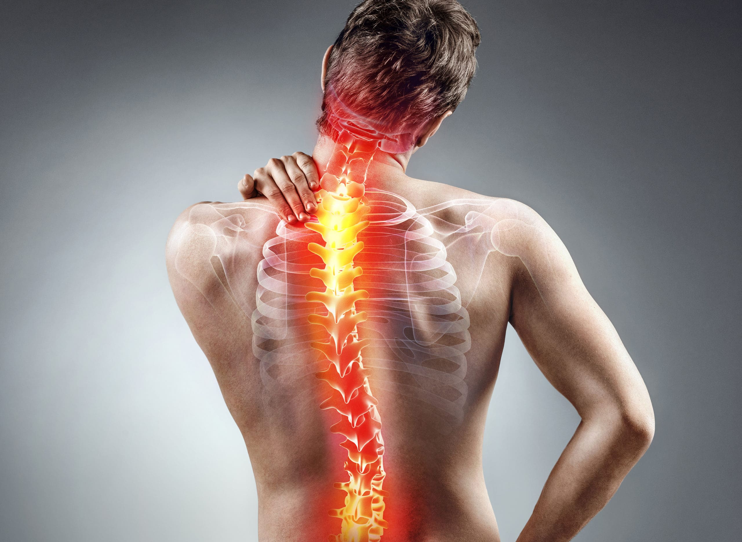
Can individuals with spinal pain in their necks and back utilize decompression therapy to restore spinal disc height and find relief?
Introduction
Many people don’t realize that as the body gets older, so does the spine. The spine is part of the musculoskeletal system that provides structural support to the body by keeping it upright. The surrounding muscles, ligaments, and tissues surrounding the spine help with stability and mobility, while the spinal disc and joints provide shock absorption from the sheer vertical weight. When a person is on the move with their daily activities, the spine can allow the individual to be mobile without pain or discomfort. However, as time passes, the spine goes through degenerative changes that can cause pain and discomfort to the body, thus leaving the individual to deal with overlapping risk profiles that can affect their neck and back. To that point, many people seek out treatments to reduce the pain affecting their spine and restore the disc height in their bodies. Today’s article looks at how spinal pain affects a person’s neck and back and how treatments like spinal decompression can reduce spinal pain and restore disc height. We talk with certified medical providers who consolidate our patients’ information to assess how spinal pain can significantly impact a person’s well-being and quality of life in their bodies. We also inform and guide patients on how integrating spinal decompression can help reduce spinal pain and restore spinal disc height. We encourage our patients to ask their associated medical providers intricate and important questions about incorporating non-surgical treatments into a health and wellness routine to relieve spinal pain and regain their quality of life. Dr. Jimenez, D.C., includes this information as an academic service. Disclaimer.
How Spinal Pain Affects A Person’s Neck & Back
Do you feel constant muscle aches and pains in your neck and back? Have you experienced stiffness and limited mobility when you are twisting and turning? Or do heavy objects cause muscle strain when moving from one location to another? Many individuals will be on the move and be in weird positions without feeling pain and discomfort when it comes to the spine. This is due to the surrounding muscles and tissues being stretched and the spinal discs taking on the vertical pressure on the spine. However, when environmental factors, traumatic injuries, or natural aging start to affect the spine, it can lead to the development of spinal pain. This is because the outer portion of the spinal disc is intact, and the inner portion of the disc is being affected. When abnormal stresses start to reduce the water intake within the disc, it can internally stimulate the pain receptors without nerve root symptoms inside the disc. (Zhang et al., 2009) This causes many individuals to deal with neck and back pain in their bodies and reduces their quality of life.
Spinal pain can lead to overlapping risk profiles that cause many individuals to deal with severe low back pain and neck pain, which then causes the surrounding muscles to become weak, tight, and overstretched. At the same time, the surrounding nerve roots are also affected as the nerve fibers surround the outer and inner parts of the spinal disc, which causes nociceptive pain properties to the neck and back region and leads to discogenic pain. (Coppes et al., 1997) When many individuals are dealing with muscle pain correlated with the spinal discs, it causes a pain-spasm-pain cycle that can affect their bodies due to not moving enough and causing painful muscular activities when trying to be mobile. (Roland, 1986) When a person has limited mobility cause they are experiencing spinal pain, their natural disc height slowly degenerates, causing more issues to their bodies and socioeconomic burdens. Fortunately, when many individuals are dealing with spinal pain, numerous treatments can reduce spinal pain and restore their disc height.
Movement Medicine- Video
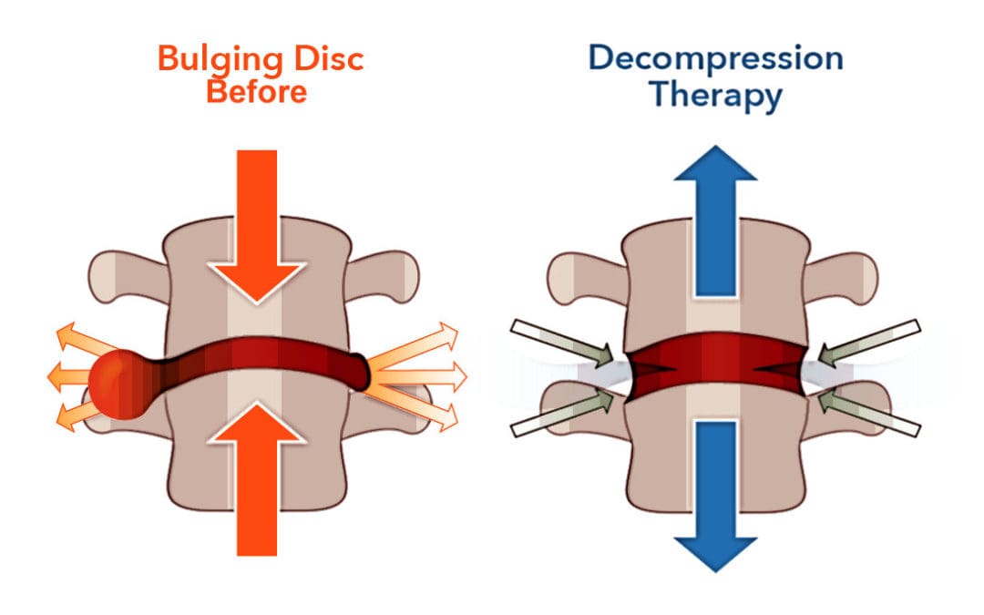
How Spinal Decompression Reduces Spinal Pain
When people are seeking treatments for their spinal pain, many will seek surgical treatments to reduce their pain, but it will be a bit pricey. However, many individuals will opt for non-surgical treatments due to their affordability. Non-surgical treatments are cost-effective and customizable to a person’s pain and discomfort. From chiropractic care to acupuncture, depending on the severity of the person’s pain, many will find the relief they seek. One of the most innovative treatments for reducing spinal pain is spinal decompression. Spinal decompression allows the individual to be strapped into a traction table. This is because it gently pulls on the spine to realign the spinal disc by reducing the pressure on the spine to invoke the body’s natural healing process to relieve pain. (Ramos & Martin, 1994) Additionally, when many individuals are using spinal decompression, the gentle traction provides a motorized distraction to the spine that may induce physical changes to the spinal disc and help restore a person’s range of motion, flexibility, and mobility. (Amjad et al., 2022)
Spinal Decompression Restoring Spinal Disc Height
When a person is being strapped into the spinal decompression machine, the gentle traction helps the spinal disc return to the spine, allowing the fluids and nutrients to rehydrate the spine, increasing the spine’s disc height. This is because spinal decompression creates negative pressure on the spine, allowing the spinal disc to return to its original height and providing relief. Plus, the amazing thing that spinal decompression does is that it can be combined with physical therapy to help stretch and strengthen the surrounding muscles near the spine to provide more stability and flexibility. (Vanti et al., 2023) This allows the individual to be more mindful of their bodies and start incorporating small habit changes to reduce the pain from returning. When many people begin to think about their health and wellness by going to treatment, they will regain their quality of life and get back to their daily routine without the issues affecting their spine.
References
Amjad, F., Mohseni-Bandpei, M. A., Gilani, S. A., Ahmad, A., & Hanif, A. (2022). Effects of non-surgical decompression therapy in addition to routine physical therapy on pain, range of motion, endurance, functional disability and quality of life versus routine physical therapy alone in patients with lumbar radiculopathy; a randomized controlled trial. BMC Musculoskelet Disord, 23(1), 255. doi.org/10.1186/s12891-022-05196-x
Coppes, M. H., Marani, E., Thomeer, R. T., & Groen, G. J. (1997). Innervation of “painful” lumbar discs. Spine (Phila Pa 1976), 22(20), 2342-2349; discussion 2349-2350. doi.org/10.1097/00007632-199710150-00005
Ramos, G., & Martin, W. (1994). Effects of vertebral axial decompression on intradiscal pressure. J Neurosurg, 81(3), 350-353. doi.org/10.3171/jns.1994.81.3.0350
Roland, M. O. (1986). A critical review of the evidence for a pain-spasm-pain cycle in spinal disorders. Clin Biomech (Bristol, Avon), 1(2), 102-109. doi.org/10.1016/0268-0033(86)90085-9
Vanti, C., Saccardo, K., Panizzolo, A., Turone, L., Guccione, A. A., & Pillastrini, P. (2023). The effects of the addition of mechanical traction to physical therapy on low back pain? A systematic review with meta-analysis. Acta Orthop Traumatol Turc, 57(1), 3-16. doi.org/10.5152/j.aott.2023.21323
Zhang, Y. G., Guo, T. M., Guo, X., & Wu, S. X. (2009). Clinical diagnosis for discogenic low back pain. Int J Biol Sci, 5(7), 647-658. doi.org/10.7150/ijbs.5.647
IFM's Find A Practitioner tool is the largest referral network in Functional Medicine, created to help patients locate Functional Medicine practitioners anywhere in the world. IFM Certified Practitioners are listed first in the search results, given their extensive education in Functional Medicine