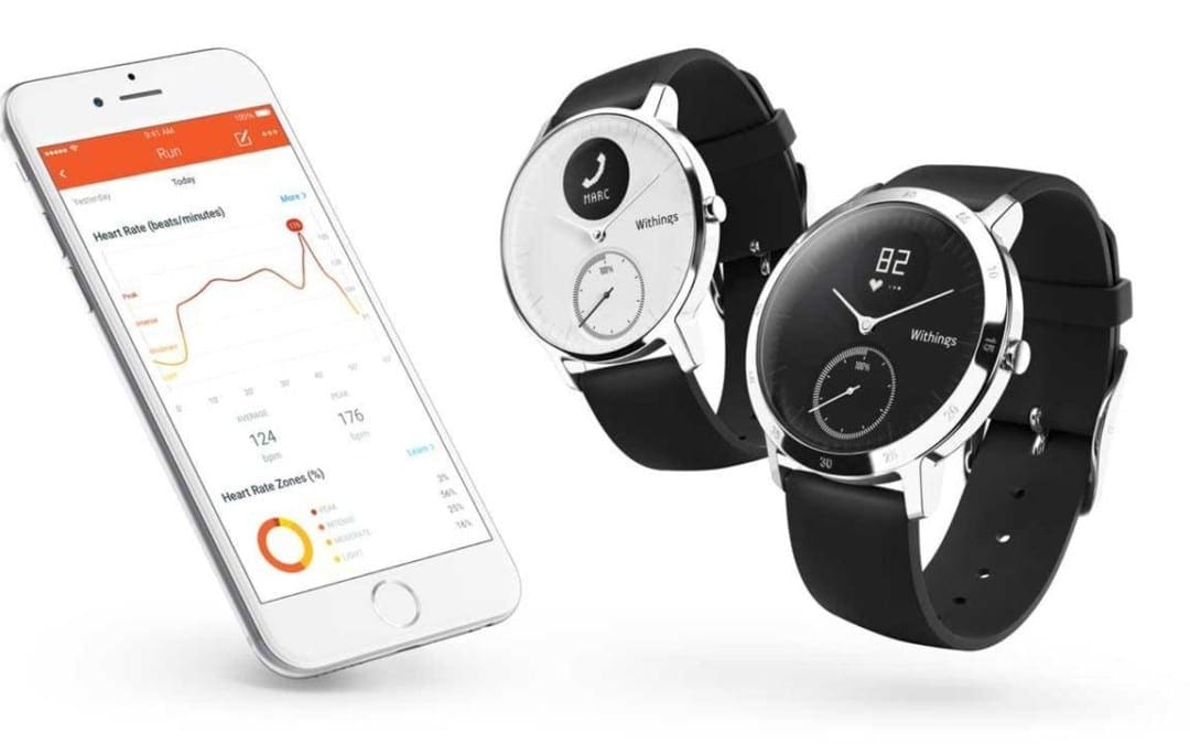
The first thing you notice when you see the Withings Steel HR is how little it resembles what most of us have come to expect from most fitness trackers in terms of shape and design. At first glance, it looks and feels like a classic analog watch: the silicone strap (or leather, if you choose that option) connects to a slim, round display with minute markers dotting the edges and hands to show you the time. The one giveaway that this is not your typical watch is the small circular OLED display at the top, and little would you know that housed behind that round face is enough hardware to power a very capable fitness tracker.
Contents
What is the Withings Steel HR?
The Withings Steel HR (from $179.95, amazon.com, bestbuy.com) has all the standard features one would expect from a fitness tracker: it lets you know how many calories you’ve burned, offers heart-rate monitoring, automatically tracks your sleep, counts your steps, pulls notifications from your phone and has an alarm to gently wake you up.
Aside from its design, one thing that really helps to set the Steel HR apart from a lot of other fitness trackers is its incredible battery life. Most fitness trackers can last you a few days before it needs to be charged again. Withings boasts that it can last up to 25 days on a single charge and that it has the longest battery life of any heart-rate tracker on the market. After spending nearly three straight weeks with the Steel HR on my wrist and over a dozen workouts tracked in that time, there was still enough juice left in the tank to leave me with little reason to doubt them on that claim.
Using the Steel HR is simple enough. There is a single button on the right side of the watch that you press to turn on the display and to cycle through the different stats. It was a minor nuisance having to press the button every time to turn the OLED display on, as the display doesn�t automatically light up if you pull the watch up toward your face. However, the goal meter below the OLED display is a nice touch on the watch face. You can quickly see how close you are to hitting your daily activity goal by looking at the meter as it climbs towards 100%.
Withings likely had to make some compromises for the sake of battery life and overall look and I found the OLED display to be one of te biggest weaknesses of the watch. While it�s relatively easy to read in regular usage if you�re just casually checking to see how many steps you walked, all of that changes if you�re engaged in a workout or on a run. If you�re running, trying to read the display or cycling through the different screens was a bit of a challenge. And you won�t get a lot of useful real-time feedback during a workout. With such a small screen, you can�t just casually glance at the display to get a quick readout. Notifications from your phone also don�t give you any real useful information.
It appears that GPS functionality was another compromise that Withings likely made. Not every fitness tracker comes equipped with GPS built into the device. Instead, they typically rely on your phone�s GPS to track you. However, the Steel HR isn�t even able to use your phone�s GPS, so it�s hard to put much faith into the accuracy of the distance measurements. It instead uses sensors and data about you to generate the distance, which is far from accurate. If you�re a runner, this is a pretty glaring omission.
Workouts are automatically detected, which seemed to work fine. Like most fitness trackers that have this feature, it will occasionally falter, but you can typically rely on it to record your workout in case you forgot to do so. You also have the option of tracking a workout manually by holding the button on the side, and workouts can be logged in the app after you sync your watch.
The heart-rate monitor on the Steel HR has two modes: workout and smart mode. Smart mode is constantly running and takes measurements about every 10 minutes, which helps to preserve battery life. However, if you switch to workout mode, it continuously runs throughout your workout. Every fitness tracker that I�ve used has had odd, random spikes that occur every now and then�this was no different for the Steel HR. I frequently wore a Fitbit Charge 2 along with the Steel HR to compare and noticed that the Steel HR always tracked higher than the Fitbit. It wasn�t enough to give me any real cause for concern, but it�s something that should be noted nonetheless. The Steel HR also seemed slower to normalize, frequently spiking at the start of a workout before eventually coming back down to a more reasonable level.
Sleep tracking worked as well as the Fitbit Charge 2, and the fact that you barely notice the watch while you�re wearing it makes it easier to wear at night compared to some of the bulkier fitness trackers out there.
If you�re the type of person that wants the features of a fitness tracker but don�t like how most of them look, the Withings Steel HR may be the tracker for you. It has a sleek and stylish design that looks good whether you�re in the gym or out on the town. Most people that see it won�t even be able to tell that you�re wearing a fitness tracker. For those that may be runners or just a little more serious about fitness, you may find the Steel HR lacks in some areas. Although, even with the compromises that Withings made with the Steel HR, this is the best hybrid option available.
For more information, Please feel free to ask Dr. Jimenez or contact us at 915-850-0900 .
Whole Body Wellness
Overall health and wellness can be achieved by following a proper nutrition and engaging in regular exercise and/or physical activities. While these are some of the most common ways to ensure whole body health and wellness, visiting a qualified and experienced healthcare professional can also grant your body additional benefits. Chiropractic care, for instance, is a safe and effective alternative treatment option utilized by people to maintain well-being.
No matter what size you are, you may have some fat between your back and arms that spills over your bra�also known as “bra bulge.” Some of that’s due to genetics, but an unbalanced workout routine can play a role as well. Many women neglect their arms, chest, and back due to a misguided fear of getting bulky. And while you may not love your bra bulge, a weak upper body can also wreck your posture and bring on back pain.
Barry’s Bootcamp instructor and celeb personal trainer Astrid Swan wants you to get over your fear of upper body workouts, so she created this exclusive routine for Health. This seven-move sequence revs your heart rate to torch calories and melt away fat from your whole body (including your back). Plus, these sculpting exercises will perk up your posture, which may minimize the appearance bra bulge. Use a pair of heavy dumbbells; Swan suggests 12 lb. or higher, depending on your strength level.
Contents
Pushups to superman
Lower all the way to the floor slowly as you do a pushup. Lay flat and extend arms forward to a superman position, lifting chest and thighs off the floor. Pull elbows down to goal-post position and lower your body down to press back up into the top of the pushup. Do 10 repetitions.
Plank renegade rows
Start in plank position, using the dumbbells as handles. Keep feet slightly wider than hip width, be sure to keep hips parallel to the floor and abs engaged. Alternate renegade rows, 10 repetitions per side, for 20 total reps.
Start with dumbbells on shoulders, feet hip-width apart with feet slightly turned out. Lower down into a squat position, keeping chest tall and abs engaged. Power from the core and glutes to press the weights above head in a press. Be sure to avoid locking out your knees as you press the weights up to the top. Do 10 reps.
Combine all three moves minus the superman. Using the dumbbells as handles, do one pushup, at the top of the pushup complete renegade rows on the right side then left side. Next, jump your feet forward sand land in the bottom of your squat. Be sure to keep your core engaged as you thrust the weights above your head. Do 10 repetitions.
Snatch passes
Using one dumbbell, bend knees slightly to hoist up the weight and snatch it to the top. Be sure to keep hips tucked and keep a small bend in the knees as you extend the arm. Return to starting position and pass the weight to the other side. Do 10 reps per side.
Triceps extensions
Depending on your strength, you can continue using both weights or drop down to one. Do a triceps extension slowly; think three counts to lower and one count to press up. Keep your elbows tight, framing your face. Do 10 reps, slowly and with intention.
Hold one dumbbell on each end. Start with the dumbbell right under your chin, and pass it around your head clockwise for 10 reps, then counterclockwise for another 10 reps. Be sure to keep your elbows tight, framing your face, and bring the weight around your head (like a “halo”) with elbows bent.
For more information, please feel free to ask Dr. Jimenez or contact us at 915-850-0900 .
Chiropractic and Athletic Performance
Many athletes who are injured performing their specific sport or physical activity, frequently seek treatment from chiropractors. Chiropractic care focuses on the prevention, diagnosis and treatment of injuries and conditions affecting the musculoskeletal and nervous system. While chiropractic is a safe and effective form of conservative care for a variety of ailments, chiropractic can also be utilized to enhance athletic performance.
In celebrity yoga instructor Hilaria Baldwin�s new book The Living Clearly Method: 5 Principles for a Fit Body, Healthy Mind & Joyful Life, she outlines her method for combining movement and mindfulness to lead a more balanced life. Her strategy includes five simple principles: perspective, breathing, grounding, balance, and letting go. But how exactly does the celeb and mother of three stay so centered with such a crazy schedule? One of her go-to ways to bring all her principles into practice is through yoga. Watch this video to learn one of Baldwin�s go-to yoga sequences that incorporates all elements of her method into a movement format.
Here, she guides you through tree pose, high lunge, warrior II, side angle A, plank, chaturanga, upward facing dog, and finally downward facing dog. After completing the sequence on one side, you roll up slowly and repeat it on the other. Within this sequence, you get a touch of balance and grounding, thanks to tree pose. And as you conquer the challenging transitions from high lunge to side angle A, consistent breathing plays a role, helping you get calm and centered.
Baldwin describes this practice as an �all-purpose flow,� meaning you can do it at any time of day�whether you want to wake your body up in the morning, get your heart rate up in the afternoon, or close out your day the right way with a moving meditation before you go to sleep. Whenever you do this sequence, it�s an efficient workout that can easily become a part of your daily routine. The goal is to simply set aside some time for yourself to help unwind and connect with your body and mind. Watch the video to learn more about how to master this flow.
For more information, please feel free to ask Dr. Jimenez or contact us at 915-850-0900 .�
If just thinking about a HIIT workout seems tiring, let the music play. A Journal of Sports Sciences study found that when people performed four 30-second all-out sprint intervals on a bike while listening to music, they had a more positive workout experience than when they pedaled without tunes�possibly because music helps distract you from the, uh, discomfort of a tough sprint. Try biking (or running or rowing) it out to one of these songs recommended by Steph Dietz, lead instructor at Cyc Fitness, an indoor-cycling studio chain.
“They�re perfect for intervals because they slowly build to the chorus, where the beat drops, picking up speed and intensity,” says Dietz. “Each song has about two or three HIIT interval builds.”
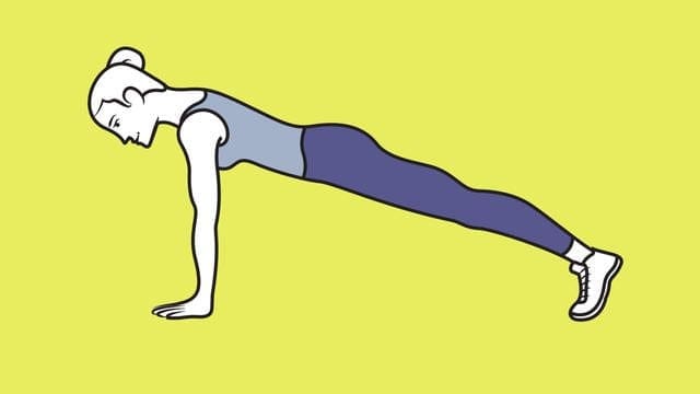
Trying to be a better runner? It’s not just about logging miles (although that certainly helps). The key to running strong and long also has a lot to do with shoring up your muscles, activating your core and back in addition to your lower body,�and keeping your movements fluid. To help do that, start incorporating these full-body strengthening�moves from Nike+ Run Club coach Julia Lucas to your routine three days a week, before or after a run. You’ll start noticing a difference in your strength in no time.
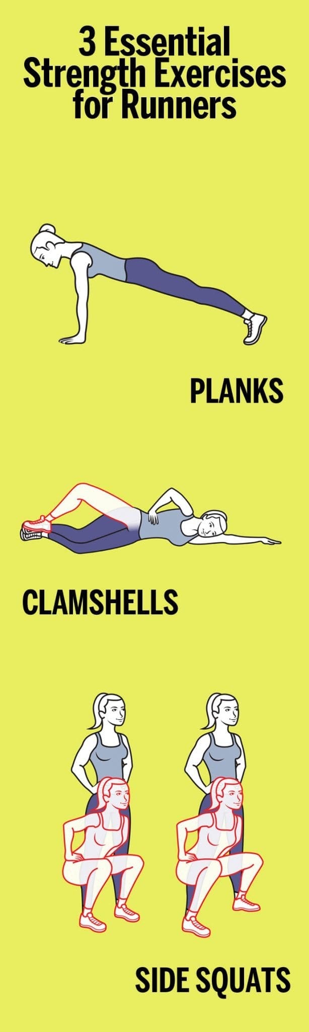
1. Planks
�
Planks have long been considered one of the best exercises for your core. In addition to your abs, this move engages your back, quads, and hamstrings, making it a great full-body exercise for runners. To do it, get into the �up� part of a push-up, with palms�on the floor directly under shoulders and legs extended behind you, forming a straight�line from head to heels. Hold for 10 to 30 seconds, keeping abs tight. Do�2 or 3 sets.
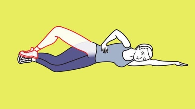
Clamshells work your hips and glutes, parts of the body that runners regularly need to activate. To do them, start out by lying�on your side with legs stacked and knees bent at 45 degrees. Rest head on arm; place top hand on hip. With inside edges of feet touching, lift top knee as high as you can without shifting hips or pelvis. Pause; lower knee. Do 2 or 3 sets of 10 reps per side.
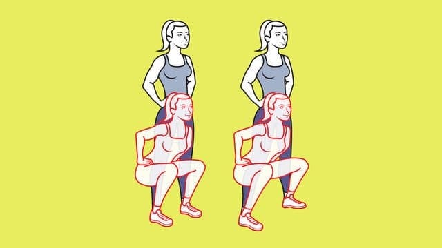
3. Side squats
Side squats are a great way to strengthen your outer highs, hips, and glutes. To do, stand with feet hip-width apart, hands on hips; squat. Stand; move left foot a step out. Squat again; step left foot in as you rise. Continue, alternating sides. Do 2 or 3 sets of 10 to 12 reps per side.
Men and women of a certain age realize quickly that their muscles diminish faster than they used to, and that muscles do not respond to exercise the way they used to. A new study shows that it’s all in the mitochondria — the part of the cell responsible for energy and vitality — and there is an exercise plan that can benefit the older crowd, even surpassing — surprisingly — benefits for the younger crowd.
Among various regimens of exercise, researchers found that interval training for a group older than 64 altered the working mechanisms in an amazing 400 genes — compared to only 274 for a group 30 or younger. The vitality in the older crowd’s cells responded more robustly than the younger crowd — adding another layer to the need for older folks to hit the gym.
Here’s how the experiment was conducted: 72 healthy but sedentary men and women were divided into two groups — 30 or younger, or older than 64 by researchers at the Mayo Clinic in Rochester, Minn. Their vitals were measured, including blood-sugar levels, gene activity, and mitochondrial health in their muscle cells. Then the volunteers were randomly assigned to 1 of 3 exercise regimens.
Some did vigorous weight training several times a week; some did brief interval training three times a week on stationary bicycles (pedaling hard for four minutes, resting for three minutes, then repeating for three times); some rode stationary bikes at a moderate pace for 30 minutes a few times a week and lifted weights lightly on other days. For control purposes, a fourth group did not exercise.
After 12 weeks, vitals were again checked for all involved. All exercise groups experienced improvements in fitness and blood sugar regulation.
Strength and endurance were affected differently, but predictively: The gains in muscle mass and strength were greater for those who exercised only with weights, while interval training had the strongest influence on endurance.
But biopsied muscle cell activity proved to be surprisingly different. Among the 30 and younger who went through interval training, the activity levels had changed in 274 genes, compared with 170 genes for those who exercised more moderately and 74 for the weightlifters. In the older crowd, almost 400 genes were working differently — more activley — for interval training, compared with 33 for the weightlifters and only 19 for the moderate exercisers.
Those who did the interval workouts showed increases in the number and health of their mitochondria. The takeaway: Interval training seems to be the best way to achieve vital cell health for muscle mass —particularly for those who are age 64 and older. Better muscle mass means a healthier, stronger body.
The decline in the cellular health of muscles associated with aging seemed corrected with interval exercise, especially if intense, Dr. Sreekumaran Nair, a professor of medicine and an endocrinologist at the Mayo Clinic and the study’s senior author, told The New York Times. Moreover, as his results show, older people’s cells responded in some ways more intensely than the cells of the younger group — suggesting, he says, it is never too late to benefit from exercise. Nair and his research team’s results were published in the journal Cell Metabolism.
New research has found more evidence to suggest a positive link between exercise and depression, this time finding that children who exercise could benefit from a reduce risk of developing depression in the future.
Carried out by a team from The Hospital for Sick Children (SickKids) and University of Calgary researchers at the Alberta Children’s Hospital, the study is the first meta-analysis to examine the potential protective effect of childhood physical activity on depression later in life.
According to the Canadian Mental Health Association 3.2 million children in Canada between the ages of 12 and 19 are at risk for developing depression.
A number of exercise intervention programs for children have been launched in recent years to support treatment for mental health issues, however current research shows large discrepancies on the effectiveness of exercise. Although some studies show strong support for physical activity’s effect on reducing depression, other studies show no relationship at all.
To look further into the validity of exercise interventions based on the existing evidence the team conducted a meta-analysis of 40 studies involving a total of 90,000 participants between the ages of eight and 19 years old. Study participants were healthy and had not been diagnosed with depression.
The team found a statistically significant association between increased physical activity and a lower risk of future depressive symptoms; however, the link was not as strong as they expected.
Explaining the results principal investigator, Dr. Daphne Korczak, said, “This suggests that physical activity is one factor, but that there are other factors that are important in determining a child’s risk for developing depression,” adding that factors such as having a family history of depression, particularly in a parent, or struggling at school academically or socially can all play a role.
Korczak added that further research looking at children with depression or examining the frequency, type or intensity of exercise would be useful in developing a better understanding of how physical activity affects the brain and the body to impact someone’s mood.
The Canadian Psychological Association recommends children and adolescents get 60 minutes of physical activity a day, but statistics published by the Canadian Society for Exercise Physiology suggest that only 15 percent of children (5 to 11 years) and five percent of adolescents (12 to 17 years) meet this recommended amount.
The study can be found online published in the journal Pediatrics.
Historically athletes were barefoot in the sporting arena and it is only a relatively recent phenomenon for shoes to be worn in competition. In Roman times wrestlers competed barefoot, whilst runners wore little more than thin leather sandals to compete over long distances.
More recently several athletes have achieved significant success competing barefoot: Abebe Bikila from Ethiopia won the Rome Olympic marathon in 1960, and Zola Budd became the world record holder over 5000 meters. Since the 1970�s athletic shoe manufacture has boomed and with it so too has the incidence of running-related lower limb injuries. This prompted the question of whether these new designs were to blame for the injuries or simply reflected the growing interest in distance running as a sport. That notwithstanding, the interest around barefoot running to reduce such injuries has grown exponentially. This account aims to appraise some of the literature on this contentious subject.
Contents
Gait Cycle & Running Biomechanics
The normal gait cycle consists of both stance and swing phases. The stance phase occupies 60% and swing 40% of the time taken to complete one cycle of consecutive heel striking by the same foot. The stance phase itself is divided into contact, midstance and propulsive phases. It begins and ends with both feet in contact with the ground known as the �double support phase�. The swing phase is divided into follow-through, forward swing and foot descent phases. The phases of running are very similar except for the fact that there is a flight phase when neither foot is in contact with the ground between stance phases. Evidently, with slower jogging the stance phase is longer than the flight phase, however, during sprinting this relationship reverses and the stance phase becomes the shorter of the two phases.
There are several key biomechanical considerations that must be borne in mind before a comparison can be made between barefoot and shod running. During running there is an increase in rotation at the pelvis, hip, and knee which must be absorbed by increasing the muscle forces acting over these joints. Moreover, as running speed increases the point of foot impact changes from predominantly heelstriking to that of forefoot weight-bearing when sprinting. The normal angle of gait is approximately 100 abducted from the line of progression. As speed increases this angle decreases approaching zero as the foot strike nears the line of progression. Runners who have developed stride patterns that incorporate low levels of impact force and rapid pronation are at a reduced risk for over-use running injuries such as stress fractures, plantar fasciitis, and ligamentous sprains. It is important to note that many shod runners never develop injuries, however, the available data indicates that 19-79% will develop an injury over their years spent running.
Biomechanical Abnormalities and Injury
Excessive Pronation � Pronation of the foot occurs at the sub-talar joint and when it occurs in excess is associated with many running-related injuries. Examples include, first metatarsophalangeal joint abnormalities, medial arch and plantar fascia strain, Achilles and tibialisposterior tendinopathy, patellofemoral joint dysfunction, and stress fractures. One study illustrated that shod running decreased torsion and increased pronation significantly, the paper concluded that the reduction in torsion produced by stiff soled shoes may well be a factor in running injuries caused by excessive pronation.
Excessive Supination
This movement also occurs at the subtalar joint and may compensate for a weakness of the antagonist pronating musculature (e.g. peroneal) or as a result of spasm or tightness of the supinating musculature (e.g. tibialis posterior, and the gastrocnemius- soleus complex). The supinated foot is less mobile and provides inferior shock-absorption which may well predispose to the development of stress fractures of the tibia, fibula, calcaneus and metatarsals. Lateral instability of the foot and ankle may be associated with excessive supination resulting in an increase incidence of ligamentous sprains of the foot and ankle. Such a lateral stress on the lower limb could result in tightening of the ileo-tibial band with associated bursitis of the femoral epicondyle.
Abnormal Pelvic Mechanics
During normal running the pelvis assumes a rotated position with anterior-posterior and lateral tilt. Weakness in the muscles needed for stabilisation of this position will result in excessive movement in any one of the three planes. A less efficient transfer of force will subsequently occur. The most common pelvic abnormalities are excessive anterior tilt, excessive lateral tilt and asymetrical pelvic movement. The complex inter-play of musculature to compensate for each of these abnormalities may well result in muscle tightness, strains and tendinopathy. Adaptation and biomechanics of running barefoot A leading study on the subject of barefoot running was conducted by Lieberman et al. who compared foot striking patterns and collision forces in habitually barefoot with shod runners. They found that habitually barefoot endurance runners often land on the fore-foot (fore-foot strike) before bringing down the heel. Less frequently they may also land with a flat foot (mid-foot strike), or even less often, on the heel (rear-foot strike). In contrast, shod runners mostly rear-foot strike which is facilitated by the elevated and cushioned heel of the modern running shoe.
The same study conducted kinematic and kinetic analyses on the two populations and discovered that even on hard surfaces, barefoot runners who fore-foot strike generate smaller collision forces than shod rear-foot strikers. This is brought about by the manner in which the barefoot runner�s foot is more plantarflexed at landing with a greater degree of ankle compliance at impact. These features combine to decrease the effective mass of the body that collides with the ground and so potentially reducing injury through repeated heavy loading. In addition, the stride length of barefoot runners is shorter and the strides have a greater vertical leg compliance which acts to lower the body�s centre of mass relative to the force of impact. Again, these features work to reduce jarring and result in a smoother running motion experienced by the individual.
Footwear and Injury
It has been surmised that modern footwear produces a lower level of perceived foot impact than that actually experienced and thus increases injury risk. There is good evidence to show that the more cushioning runners believe to be under the foot, the harder they strike. Furthermore, modern shoe designs are far more forgiving on poor running technique and since the athlete suffers less pain bad habits become re-inforced. In contrast, barefoot runners have been found to have a reduction in impact peak with reduced mechanical stress and enhanced ankle extensor function. In one particular study peak load in the hip and knee joints of participants with osteoarthritis was decreased significantly in barefoot walkers. These findings appear to point to the supposition that shoes may increase loads in poor physiological patterns and thus perpetuate injury.
Bipedalism has been around for millions of years and it is only relatively very recently that humans have been shod. The running technique of early distance running bipeds almost certainly differed enormously from the style that is seen today with modern foot-wear. The pre-historic �hunter-gatherer� would be more likely to have had a fore-foot or mid-foot-strike gait which studies have shown to be protective from many of the running injuries seen today.
Modern running shoes allow a greater degree of �laziness� in running style and in so doing ingrain bad habits which ultimately predispose to injury. Information on how barefoot running can be integrated into one�s training and how to overcome the obvious hazards of penetrating and friction injuries are growing at a great rate through internet forums and sites. There are already products available such as the Vibram FiveFingers� which are gaining in
popularity as the option of running barefoot or �nearly barefoot� grows. Furthermore, interest in the subject has been helped enormously by popular literature such as the bestselling book �Born to Run� by Christopher McDougall which follows the Tarahumara Indians of Northern Mexico who run ultra-marathons in simple leather strapped sandals.
In short, the evidence indicates two clear points. Firstly, from observations of populations who run barefoot or are habitually barefoot there appears to be lower injury rates versus the shod population. Secondly, the wearing of modern running shoes promotes a heavy impacting heelstrike gait which predisposes to injury. For the subject to gain wide-spread acceptance there will need to be an increase in the number of well designed prospective and randomised controlled trials on the subject.
References:
1. Clinical Sports Medicine by Peter Brukner and Karim Khan. Third Edition, Chapter 3; pp.45-55
2. Hreljac A. Impact and overuse injuries in runners.
Med Sci Sports Exerc 2004; 36:845-9 3. van Gent RN, Siem D, van Middelkoop M, van Os AG, Bierma-Zeinstra SM, Koes BW. Incidence and
determinants of lower extremity running injuries in long distance runners: a systematic review. Br J Sports Med 2007; 41(8):469-80
4. Buschbacher R, Prahlow N, Dave SJ (eds). Sports Medicine and Rehabilitation: A Sports Specific
Approac, 2nd ed. Baltimore (MD): Lippincott Williams and Wilkins; 2008, p. 200-1
5. Stacoff A, Kaelin X, Stuessi, Segesser B. The torsion of the foot in running. Int J Biomech 1989; 5:375-89
6. Lieberman DE, Venkadesan M, Werbel WA, Daoud AI, D-Andrea S, Davis IS, Mang-Eni RO, Pitsiladis Y. Foot strike patterns and collision forces in the
habitually barefoot versus shod runners. Nature 2010; 463:531-535 7. Jungers WL. Barefoot running strikes back. Nature
2010; 463:433-434 8. Robbins S, Waked E. Hazard of deceptive advertising of athletic footwear. Br J Sports Med
1997; 31(4):299-303. 9. Divert C, Mornieux G, BaurH, et al. Mechanical comparison of barefoot and shod running. Int J
Sports Med 2005; 26:593-8 10. Shakoor N, Block JA. Walking barefoot decreases loading on the lower extremity joints in knee
osteoarthritis. Arthritis Rheum 2006; 54:2923-7 11. Christopher McDougall. Born to run: the hidden
tribe, the ultra-runners and the greatest race the world has never seen. Profile books, published 2009.
12. Robbins SE, Hanna AM. Running-related injury prevention through barefoot adaptations. Med Sci Sports Exerc 1987.;19:148-56
For more information, please feel free to ask Dr. Jimenez or contact us at 915-850-0900 .
Additional Topics: What is Chiropractic?
Chiropractic care is an well-known, alternative treatment option utilized to prevent, diagnose and treat a variety of injuries and conditions associated with the spine, primarily subluxations or spinal misalignments. Chiropractic focuses on restoring and maintaining the overall health and wellness of the musculoskeletal and nervous systems. Through the use of spinal adjustments and manual manipulations, a chiropractor, or doctor of chiropractic, can carefully re-align the spine, improving a patient�s strength, mobility and flexibility.
Plantar fasciitis is a common affliction affecting many athletes, in particular runners. Adam Smith has written a great piece in the September issue of Sports Injury Bulletin outlining the relevant anatomy, how the injury occurs, how to differentiate from other similar pathologies, such as neural irritation in the tarsal tunnel, and finally how to manage it.
Speaking from experience as a former sufferer of plantar fasciitis, it can be a frustratingly recalcitrant condition and I have heard of some extreme measures to manage it. Read on for a story on the drastic measures an AFL player took to overcome the problem, and to understand more about the condition.
Many years ago an elite level AFL player had suffered a 2 year history of plantar fasciitis with no relief from any form of treatment. In the end the sports doctor at the club involved injected the plantar fascia origin with a corticosteroid injection the day before a game.
The hope was that as the plantar fascia weakened due to the steroid injection, the player would rupture it, go through the standard week rehab protocol, and then be pain free for ever more.
And yes, the player did rupture the plantar fascia during during the game and was consequently placed in a boot for about 10 days. He soon was walking, then running, and was playing again within four weeks with no more problems. The podiatrist made an orthotic to control the dropped arch and all the problems went away.
What has happened to that player now is anyone’s guess. He may now suffer from long term issues due to a poorly controlled arch that have caused other issues such as achilles tendon, knee pain and/or hip pain.
So do we really need the plantar fascia and why is it such a problem when it is injured?
Being bipedal (walking on two leg) animals, the plantar fascia gives the natural plantar arch support in weight bearing positions. It is a passive structure that acts like a high tension wire to keep the arch bones supinated as we push off.
Without a plantar fascia in place, we would need a better active system to create the arch support, such as the intrinsic plantar arch muscles, and also the extrinsic long arch support muscles such as the tibialis posterior, flexor hallucis longus (FHL) and the flexor digitorum longus (FDL). These muscles would need extra work to improve their arch control abilities. Alternatively, we could use a passive support mechanism in the form of an orthotic to control the arch position.
The majority of plantar fascia problems stem from a build up of tensile and compressive forces that degenerate the plantar fascia origin against the heel bone. The combination of tensile (stretch) force due to overpronation and the added compressive force as the plantar fascia is pushed against the heel bone leads to a pathological state whereby the plantar fascia degenerates and creates dysfunction and pain.
Therefore like other degenerative tendon issues (such as Achilles tendons) once the patient starts to feel pain often the injury has been building for months to years. Which explains why it then becomes so problematic to deal with.
Proper management takes time to not only correct the muscle imbalances that cause it � such as tight calves, poor hip control, poor pronation control � but due to its degenerative nature it requires a huge amount of time to even slightly change the existing pathology.
For more information, please feel free to ask Dr. Jimenez or contact us at 915-850-0900 .
Additional Topics: What is Chiropractic?
Chiropractic care is an well-known, alternative treatment option utilized to prevent, diagnose and treat a variety of injuries and conditions associated with the spine, primarily subluxations or spinal misalignments. Chiropractic focuses on restoring and maintaining the overall health and wellness of the musculoskeletal and nervous systems. Through the use of spinal adjustments and manual manipulations, a chiropractor, or doctor of chiropractic, can carefully re-align the spine, improving a patient�s strength, mobility and flexibility.
Stress fractures to the ribs occur in rowers, golfer, canoeists, lacrosse players and baseball pitchers. They are more common in sports involving an element of trunk rotation with scapula movement across the rib cage.
A stress fracture is described as an overload to the bone caused by repetitive loading due to a particular movement. Any load on the bone will create a stress in the bone. However, given enough recovery time the bone heals and ends up stronger. This is known as Wolfe�s law. But, if the bone load is too high or too frequent, then the bone does not repair quickly enough, a stress response occurs and a fracture follows.
In rowing, the repetitive loading is created by a number of factors. Muscles such as the serratus anterior and abdominals that directly attach to the ribs can lead to loading on the ribcage due to contraction. Bad rowing technique, perhaps caused by poor hip flexibility, which then requires an excessive compensatory thoracic rotation, may then lead to rib breakdown.
Other causes include equipment issues such as the oar type (lighter carbon oars increase rib loading), bigger boats with more drag and position in the boat (bow rowers have less incidence due to lower stroke rate and force). Rib cross section and density also influence the chance of stress fractures, and women have a higher chance due to greater likelihood of bone density issues. Finally, training variables such as volume, intensity, type of loading and off water training can also be factors in stress fracture development.
The signs and symptoms are usually straight forward. These include generalised rib pain with a focused spot of tenderness, pain rolling onto the ribs whilst sleeping and pain with deep breathing. They can be confirmed with bone scan (black spot) and/or MRI (white spot).
Unfortunately for the rower, the immediate management of the injury involves rest. Usually 4-6 weeks away from rowing will be enough to allow some bone healing and this is followed by a progressive increase in rowing load over another 4 weeks before the athlete is back to full training.
The scope of our information is limited to chiropractic and spinal injuries and conditions. To discuss options on the subject matter, please feel free to ask Dr. Jimenez or contact us at 915-850-0900 .
Additional Topics: Chiropractic and Athletic Performance
Chiropractic care is a popular, alternative treatment option which focuses on the diagnosis, treatment and prevention of injuries and/or conditions associated to the musculoskeletal and nervous system, primarily the spine. Many athletes, and civilians alike, seek chiropractic care to restore their natural health and wellness, however, chiropractic has been demonstrated to benefit athletes by increasing their athletic performance.
IFM's Find A Practitioner tool is the largest referral network in Functional Medicine, created to help patients locate Functional Medicine practitioners anywhere in the world. IFM Certified Practitioners are listed first in the search results, given their extensive education in Functional Medicine