Although oral devices, such as splints and bite guards, are the most prevalent treatments for facial pain associated with temporomandibular disorders, or TMD, patients have found that these remedies are frequently less effective than self-care techniques, such as jaw exercises or warm compresses, according to a new research study published by researchers at the New York University (NYU) College of Dentistry in New York City.
The research study, published in the journal Clinical Oral Investigations, demonstrates that self-care techniques should primarily be utilized to help treat muscle-related temporomandibular disorders or TMD.
TMD, occasionally known as TMJ after the temporomandibular joint, is a collection of prevalent painful conditions which develop in the jaw joint and its surrounding muscles. Myofascial temporomandibular disorder, or mTMD, is a muscular condition which affects over 10 percent of women. Individuals with TMD often suffer from other chronic pain conditions. Research studies found that 7 to 18 percent of people with TMD also experience fibromyalgia, a condition characterized by widespread pain.
Treatments for TMD and Fibromyalgia
Dentists and patients utilize an assortment of treatments to help manage facial pain, such as oral devices like splints and bite guards, pain medicines, including nonsteroidal anti-inflammatory drugs, and self-care methods like jaw exercises and hot compresses.
Oral devices are a prevalent first-line treatment for TMD, regardless of research study outcome measures regarding their advantages, stated Vivian Santiago, Ph.D., MPH, research study scientist at the Department of Oral and Maxillofacial Pathology, Radiology, and Medicine at NYU College of Dentistry, and the research study’s leading author.
“While oral splints have been discovered to have some benefits, they have yet to be found to be as successful for patients who have widespread pain when treating mTMD,” she explained.
In this research study, the researchers evaluated what non-medication remedies women with mTMD utilized to handle their pain as well as how successful patients perceived these remedies. The researchers interviewed a total of 125 women including 26 women who had fibromyalgia and mTMD, so as to find out whether treatment differed for patients.
The most frequent treatments reported were oral devices (utilized by 59 percent of participants), physical therapy (utilized by 54 percent of participants), and at-home jaw exercises (utilized by 34 percent of participants). The least frequent treatments reported were acupuncture (utilized by 20 percent), chiropractic care (utilized by 18 percent), trigger point injections (utilized by 14 percent), yoga (utilized by 7 percent), and meditation (utilized by 6 percent). Participants frequently used more than one treatment.
Participants reported the most improvement in their pain from well-known self-care techniques, such as jaw exercises, yoga, meditation, massage, and warm compresses, with over 84 percent reporting that these techniques helped reduce painful symptoms. Only 64 percent of participants who used the oral devices reported that they helped improve their pain. About 11 percent of women who used oral devices stated that these made their pain worse, an area which warrants further research studies.
Oral devices failed to outperform self-care techniques in improving facial pain, according to Karen Raphael, Ph.D., professor at the Department of Oral and Maxillofacial Pathology, Radiology, and Medicine at NYU College of Dentistry, and the research study’s co-author.
“Our outcome measures encourage utilizing self-care techniques as the first line of treatment for mTMD before contemplating more costly interventions,” stated Raphael.
The researchers didn’t find substantial differences between the amount of remedies reported by women with and without fibromyalgia. While the use of alternative treatment options for mTMD was reported among women with fibromyalgia, further research studies are still required. Pain relief tended to be greater through the use of self-care techniques in women with and without fibromyalgia.
“While fibromyalgia is diagnosed by a healthcare professional, such as a rheumatologist, TMD is typically diagnosed and treated by a dentist,” said Santiago. “Our research study demonstrates that dentists must ask patients with facial pain if they also have widespread chronic pain because this might provide more information to help plan their treatment.”
Fibromyalgia is a health issue characterized by widespread chronic pain accompanied by fatigue, sleep, memory and mood problems. Fibromyalgia has been associated with a variety of other health issues, such as TMD and/or TMJ. Individuals with this painful disorder may often struggle to engage in their everyday physical activities. As a qualified and experienced chiropractor, I’ve helped treat numerous patients with fibromyalgia. It’s important for patients to know that they are not alone when it comes to treating their painful symptoms. Chiropractic care is an alternative treatment option which can help treat a variety of health issues, including fibromyalgia.
Dr. Alex Jimenez D.C., C.C.S.T. Insight
The scope of our information is limited to chiropractic, spinal health issues, and functional medicine articles, topics, and discussions. To further discuss the subject matter above, please feel free to ask Dr. Alex Jimenez or contact us at 915-850-0900 .
Curated by Dr. Alex Jimenez
Additional Topic Discussion: Acute Back Pain
Back pain is one of the most prevalent causes of disability and missed days at work worldwide. Back pain attributes to the second most common reason for doctor office visits, outnumbered only by upper-respiratory infections. Approximately 80 percent of the population will experience back pain at least once throughout their life. Your spine is a complex structure made up of bones, joints, ligaments, and muscles, among other soft tissues. Injuries and/or aggravated conditions, such as herniated discs, can eventually lead to symptoms of back pain. Sports injuries or automobile accident injuries are often the most frequent cause of back pain, however, sometimes the simplest of movements can have painful results. Fortunately, alternative treatment options, such as chiropractic care, can help ease back pain through the use of spinal adjustments and manual manipulations, ultimately improving pain relief.
XYMOGEN�s Exclusive Professional Formulas are available through select licensed health care professionals. The internet sale and discounting of XYMOGEN formulas are strictly prohibited.
Proudly, Dr. Alexander Jimenez makes XYMOGEN formulas available only to patients under our care.
Please call our office in order for us to assign a doctor consultation for immediate access.
If you are a patient of Injury Medical & Chiropractic Clinic, you may inquire about XYMOGEN by calling 915-850-0900.
For your convenience and review of the XYMOGEN products please review the following link.*XYMOGEN-Catalog-Download
* All the above XYMOGEN policies remain strictly in force.
A heart-healthy diet rich in fruits, vegetables, fish, and nuts which is also low in meat, and full-fat dairy is related to improved middle-aged cognitive function, such as memory and thinking skills, according to a research study published in the journal “Neurology”.
The research study, directed by Claire McEvoy, Ph.D., of Queen’s University Belfast in Northern Ireland, analyzed which dietary patterns, including the Mediterranean diet plan, Dietary Approaches to Stop Hypertension (DASH), and the A Priori Diet Quality Score (APDQS), throughout early adulthood have been correlated with middle age brain function and cognitive performance.
Nutrition and Better Brain Function
Researchers evaluated 2,621 Coronary Artery Risk Development in Young Adults, or CARDIA, study participants with a mean age of 25 years old throughout a 30-year period. The study participants were asked about their diet at the start of the research study, after seven years, and after 20 years. The study participants’ brain function was analyzed when they were 50 and 55 years old.
For every diet, the participants of the research study were separated into one of 3 categories, low, moderate, or higher adherence, dependent on how closely they followed their diet plan. The evaluation demonstrated that the DASH diet wasn’t associated with any alteration in cognitive function. A Mediterranean diet or a APDQS diet also resulted in a lesser decline in cognitive functioning.
Individuals with higher adherence to the Mediterranean diet were 46 percent less likely to experience poor memory and thinking skills than individuals with reduced adherence to this diet plan, according to the research study. Of those individuals in the adherence group, 9 percent had poor memory and thinking skills, compared to 29 percent of those 798 men and women in the adherence group.
Individuals with higher adherence to the APDQS diet were 52 percent less likely to have poor memory and thinking skills than individuals with reduced adherence to the diet plan. Of the 938 men and women in the adherence class, 6 percent had poor memory and thinking skills, compared to 32 percent of those 805 men and women in the adherence group, according to researchers.
The outcome measures of the research study were adjusted for other variables that may affect cognitive functioning, including the degree of education, as well as smoking, physical activity or exercise, and health issues like diabetes, according to the research study.
Numerous research studies have attempted to evaluate the effects of nutrition on brain health. Recent research studies, however, have demonstrated that certain diet plans, such as the Mediterranean diet, the DASH diet, and the APDQS diet, can help decrease the decline of cognitive function during middle age. The research study also demonstrated the effects these diets have on overall brain health. A balanced nutrition is essential towards optimal well-being. Before following any of the diet plans described in the following article, make sure to talk to a healthcare professional about your options.
Dr. Alex Jimenez D.C., C.C.S.T. Insight
Types of Diets for Improved Brain Function
The Mediterranean diet emphasizes the consumption of whole grains, fruits, vegetables, legumes, nuts, healthy unsaturated fats, and fish while restricting the consumption of red meat, poultry, and full-fat dairy. In contrast, the DASH diet emphasizes the consumption of grains, fruits, vegetables, legumes, nuts, and low-fat dairy while restricting the consumption of meat, fish, poultry, total fat, saturated fat, sweets, and sodium. The APDQS diet emphasizes the consumption of fruits, vegetables, legumes, low-fat fish, poultry, and alcohol while restricting the consumption of fried foods, salty snacks, sweets, high-fat dairy, and sugar-sweetened soft drinks.
The Mediterranean diet and the DASH diet regularly rank as some of the top diets for humans, according to the U.S. News and World Report’s yearly evaluation. According to researchers, it’s uncertain why the DASH diet didn’t result in improved cognitive functioning.
Further research studies are required to specify how nutrition, diet, and food contributes towards optimal brain health throughout our life span. According to a statement published by the American Academy of Neurology, “While we do not yet understand the perfect nutritional supplement for brain health, shifting towards a heart-healthy diet might be a relatively safe and effective approach to decrease the danger of developing health issues with memory and thinking skills throughout our middle age.”
The scope of our information is limited to chiropractic, spinal health issues, and functional medicine articles, topics, and discussions. To further discuss the subject matter above, please feel free to ask Dr. Alex Jimenez or contact us at 915-850-0900 .
Curated by Dr. Alex Jimenez
Additional Topic Discussion: Acute Back Pain
Back pain is one of the most prevalent causes of disability and missed days at work worldwide. Back pain attributes to the second most common reason for doctor office visits, outnumbered only by upper-respiratory infections. Approximately 80 percent of the population will experience back pain at least once throughout their life. Your spine is a complex structure made up of bones, joints, ligaments, and muscles, among other soft tissues. Injuries and/or aggravated conditions, such as herniated discs, can eventually lead to symptoms of back pain. Sports injuries or automobile accident injuries are often the most frequent cause of back pain, however, sometimes the simplest of movements can have painful results. Fortunately, alternative treatment options, such as chiropractic care, can help ease back pain through the use of spinal adjustments and manual manipulations, ultimately improving pain relief.
XYMOGEN�s Exclusive Professional Formulas are available through select licensed health care professionals. The internet sale and discounting of XYMOGEN formulas are strictly prohibited.
Proudly, Dr. Alexander Jimenez makes XYMOGEN formulas available only to patients under our care.
Please call our office in order for us to assign a doctor consultation for immediate access.
If you are a patient of Injury Medical & Chiropractic Clinic, you may inquire about XYMOGEN by calling 915-850-0900.
For your convenience and review of the XYMOGEN products please review the following link.*XYMOGEN-Catalog-Download
* All the above XYMOGEN policies remain strictly in force.
Hormones can also play a significant part in the pain. One of the many changes that occur in a pregnant woman�s body is an increase in the production of a hormone called Relaxin. The job of this hormone is to �relax� the ligaments or soften them. This allows the pelvis to spread so that the baby can be born. However, while the body is making these positive and necessary preparations, it can also allow bones to shift or move in directions that they shouldn�t. The result is an impingement or compression on the nerves exiting the spinal canal, causing the tissues in the area to become irritated and inflamed.
Another effect of the increased production of relaxin is that it can lead to intrauterine constraint. This condition causes the pelvic bones to become misaligned which obstructs the baby�s natural movement in utero while it develops. This can keep the baby from moving into the birthing position with the head down. As the bones move out of their natural place, it can stress, pull, and twist the attached ligaments. The baby doesn�t have as much space as it should so it can�t move like a normal fetus can. This can also cause breech births. Stress, poor posture, and overexertion can also cause pregnancy back pain.
Why is pregnancy low back pain so difficult to treat and what treatments are available?
Medications given to the mother will cross over the placenta delivering a dose of the medication to the baby. Because of this, pain medications are generally off limits for pregnant women. In order to protect the baby, natural pain control is preferred. This makes chiropractic a preferred treatment for pregnancy low back pain. The chiropractor may perform a spinal subluxation to bring the spine back into alignment and the body back onto balance.
He or she may also make some recommendations to help the woman manage her pain on her own, including improving her posture, stretching, and special exercises. Other recommendations may include:
Not wearing high heels. They put strain on the back when a woman isn�t pregnant. On a pregnant body, the strain is even worse.
Avoid bending over to pick up things, but instead, squat � or ask for help.
Sleeping on the left side and use pillows under the belly to support it as well as a pillow between her legs.
Rest, lots of rest and elevating her feet.
Regular chiropractic care and following the doctor�s instructions can help greatly decrease low back pain for the mom to be so that she can better enjoy the excitement and joy of her pregnancy.
Lower Back Pain Pregnancy Chiropractic Treatment El Paso, TX
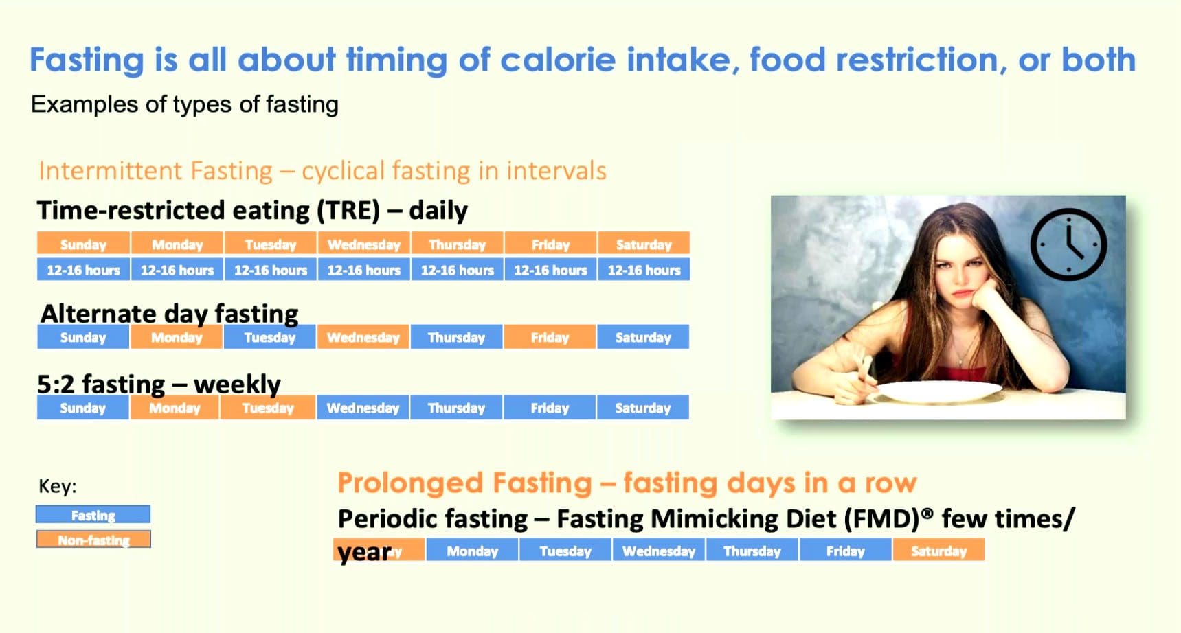
Recent science has unleashed the power of fasting to optimize body aging and dismantle chronic diseases, e.g., obesity, diabetes, cancer, autoimmune disorders, Alzheimer�s and cardiovascular disease. At El Paso Back Clinic, Dr. Jimenez’s mission is to educate the public about the health benefits of fasting. The idea is to introduce it to them in a safe, effective way through the Fasting Mimicking Diet (FMD).
Pre-clinical and clinical studies have proven that periodic fasting, done for several consecutive days, is a very powerful intervention that our bodies learned to naturally cope with by protecting and rejuvenating itself. These two factors are both anti-aging measures that offer additional health benefits. The 5-Day ProLon Fasting Mimicking Diet has been clinically tested and found to promote beneficial effects in a wide variety of conditions ranging from excess weight and fasting blood glucose, to growth factors associated with DNA damage and aging.
Min Wei; Sebastian Brandhorst et al. Fasting?Mimicking Diet and Risk Factors for Aging, Diabetes, Cancer and Cardiovascular Disease.
5-Day Program
The ProLon meal plan is followed 5 days per month. Once an individual has finished the five-day plan, they go back to a normal healthy dietthe last twenty-five days. Fasting with the Prolon� plan follows a low carbohydrate/protein meal and contains the good kind of fatty acids. The FMD� recipe keeps your body on a fasting-type mode, that triggers protection measures that the body has developed. This causes the body to optimize its performance, rejuvenate cells, and thrive.
Unboxing the ProLon Fasting Mimicking Diet
Age-related Disease: A Revolution Is Coming
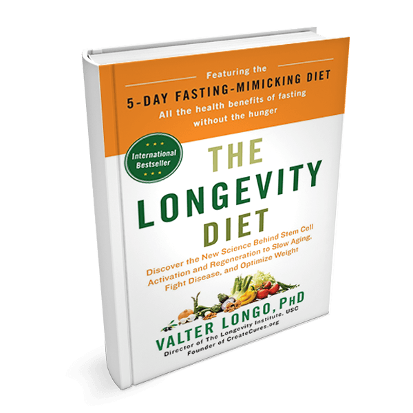
I just finished reading a groundbreaking book called The Longevity Diet: Discover the New Science Behind Stem Cell Activation and Regeneration to Slow Aging, Fight Disease, and Optimize Weight, which was written by Valter Longo, PhD,1 who is director of the Longevity Institute at the University of Southern California and a principal scientist in the development and study of the fasting mimicking diet (FMD). I have followed Dr. Longo�s career for many years with great admiration. He has been published in top-tier journals. In his book, Dr. Longo writes about some exciting findings regarding the FMD research group consisting of 16-month-old mice, which are described as being the equivalent of a 45-year-old human: �A stem cell-dependent process rejuvenated the immune system. Regeneration also occurred in the liver, muscle, and brain. Levels of several types of stem cells increased.�1 He went on to explain: �The fasting itself destroys many damaged cells and damaged components inside the cells but it also activates stem cells.�1
Jeffrey S. Bland, PhD, FACN, FACB, Associate Editor
Get Your Free Copy At El Paso Back Clinic!
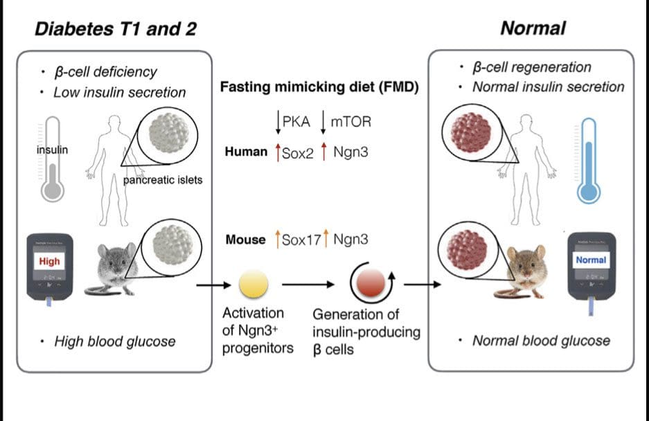
Fasting-Mimicking Diet Promotes b-Cell Regeneration to Reverse Diabetes
Fasting mimicking diet induces prenatal-development gene expression in adult pancreas
FMD promotes Ngn3 expression to generate insulin-producing b cells
Cycles of FMD reverse b-cell failure and rescue mice fromT1D and T2Dd
Inhibition of PKA or mTOR promotes Ngn3-driven b-cell regeneration in human T1D islets
Stem-cell-based therapies can potentially reverse organ dysfunction and diseases, but the removal of impaired tissue and activation of a program leading to organ regeneration pose major challenges. In mice, 4-day fasting mimicking diet (FMD) induces a stepwise expression of Sox17 and Pdx-1, followed by Ngn3-driven generation of insulin-producing b cells, resembling that observed during pancreatic development. FMD cycles restore insulin secretion and glucose homeostasis in both type 2 and type 1diabetes mouse models.
In human type 1 diabetes pancreatic islets, fasting conditions reduce PKAand mTOR activity and induce Sox2 and Ngn3expression and insulin production. The effects of the FMD are reversed by IGF-1 treatment and recapitulated by PKA and mTOR inhibition. These results indicate that an FMD promotes the reprogramming of pancreatic cells to restore insulin generation in islets from T1D patients and reverse both T1D andT2D phenotypes in mouse models.
Chia-Wei Cheng,1,6,7Valentina Villani,2,7Roberta Buono,1,5,7Min Wei,1Sanjeev Kumar,4Omer H. Yilmaz,6Pinchas Cohen,1Julie B. Sneddon,3Laura Perin,2and Valter D. Longo1,4,5,8,*
Individuals getting a very low percentage of their daily calories from carbohydrates, such as fruits, grains, and starchy vegetables, are more likely to develop atrial fibrillation, or AFib. This health issue is one of the most prevalent heart rhythm disorders, according to a new research study being presented at the American College of Cardiology’s 68th Annual Scientific Session.
The research study examined the health records of almost 14,000 people spanning two or more decades. Researchers brought data from Atherosclerosis Risk in Communities, or ARIC, a research study controlled by the National Institutes of Health which was conducted from 1985 to 2016. Of almost 1,900 participants that were diagnosed through a mean of 22 years of follow-up, a majority of them were identified with AFib by researchers. The details of the research study are described below.
AFib and Carbohydrates
Research study participants were requested to report the everyday consumption of 66 distinct food items in a poll. The researchers utilized this information to gauge the percentage of calories which came from carbohydrates from each participant’s calorie intake. Carbohydrates were contained in roughly half of the daily calories consumed by the participants.
Researchers subsequently separated the participants into three separate groups categorized by low, moderate, and high carbohydrate intake, representing diets where carbohydrates consisted less than 44.8 percent of their daily calories, followed by 44.8 to 52.4 percent, and finally where carbohydrates consisted more than 52.4 percent of their daily calories, respectively.
Participants who reporting reduced carbohydrate consumption were the ones who had the highest probability of developing AFib, according to researchers. As the statistics of the research study later demonstrated, these participants were also 18 percent more likely to come up with AFib compared to those with moderate carbohydrate intake and 16 percent more likely to come up with AFib compared to those with high carbohydrate ingestion. Some diets can also help decrease the risk of heart rhythm disorders.
The type of carbohydrates you eat can make a huge difference in your overall health and wellness. Complex carbohydrates are digested more slowly than simple carbohydrates and these release a steady release of sugar, or glucose, into the blood stream. Complex carbohydrates, often referred to as “starchy” foods, include legumes, starchy vegetables, whole grain, and fiber. According to the research study in the following article, consuming low amounts of carbohydrates, which often includes fruits, vegetables, and whole grains, can contribute to cardiovascular diseases, such as atrial fibrillation. When it comes to carbohydrates, it’s important to consume this essential macronutrient for overall health and wellness.
Dr. Alex Jimenez D.C., C.C.S.T. Insight
Nutrition for AFib
Restricting carbohydrates has become a popular weight loss plan. Many diets, such as the Paleo and the ketogenic diet, highlight the consumption of proteins. According to Xiaodong Zhuang, MD, PhD, cardiologist and the research study’s lead author, “The long-term impact of carbohydrate restriction remains controversial, particularly with respect to its own influence on cardiovascular disease.” “Considering the possible effects on arrhythmia, our research study indicates that this popular weight control system ought to be recommended carefully,” he stated in a statement published by the ACC.
The findings complement previous research studies, a number of which have correlated both polyunsaturated and high-carbohydrate diets with a greater probability of death. While previous research studies indicated that this part of the diet affected the outcome measures found, the research study itself didn’t determine these findings. “Low carbohydrate diets have been associated with greater risk of developing AFib irrespective of the type of fat or protein utilized to substitute the carbohydrate,” Zhuang said.
“Several possible mechanisms could explain why limiting carbohydrates may contribute to AFib,” Zhuang said. One is that individuals eating a low-carbohydrate diet often consume fewer fruits, vegetables, and whole grains. Without these foods, individuals may experience more widespread inflammation, which has been connected with AFib. According to the research study, another potential explanation is that eating more fat and protein instead of carbohydrate-rich foods can result in oxidative stress, which has also been connected to AFib. The effect may be associated with an increased risk of other types of cardiovascular disease.
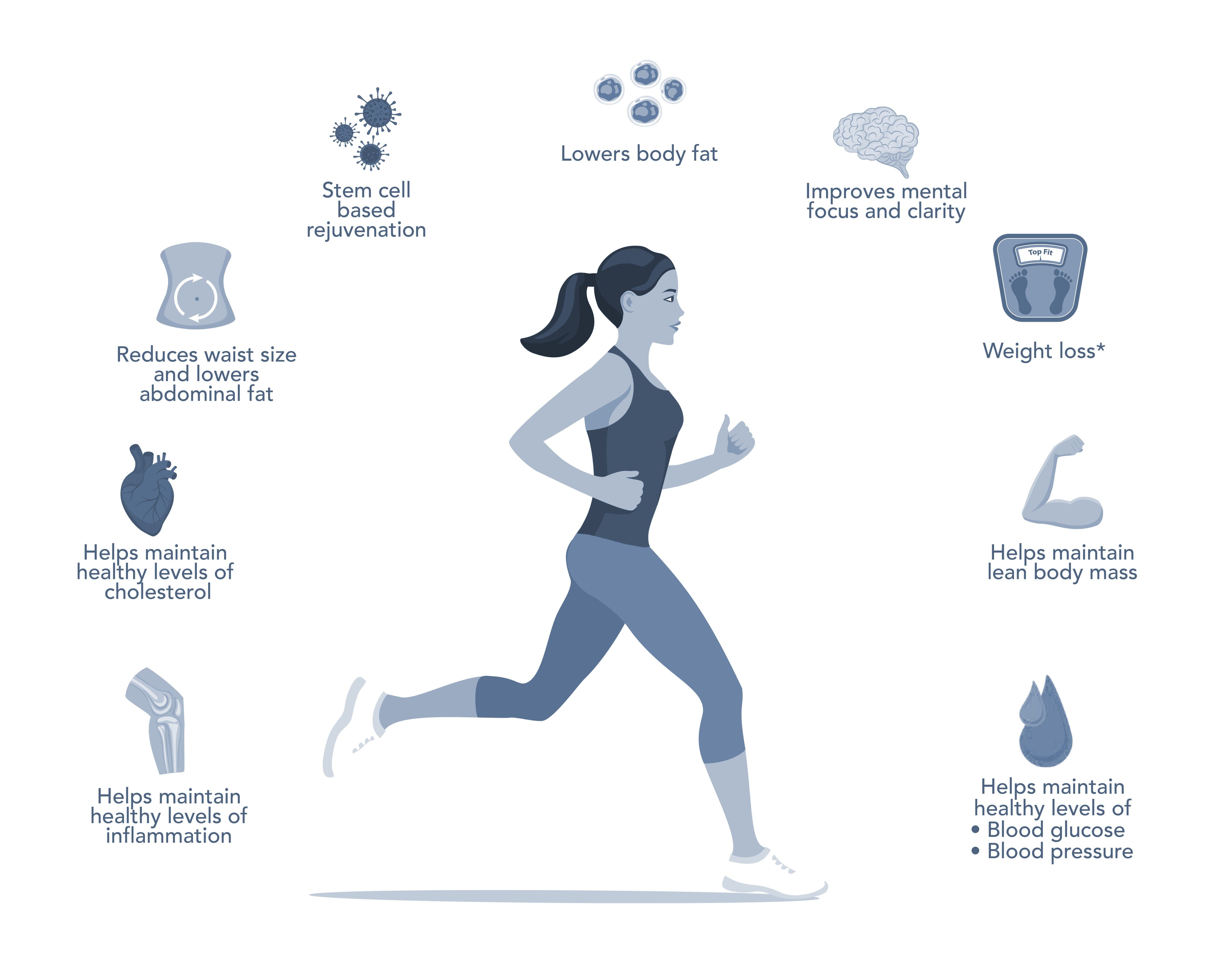
The Longevity Diet Plan, presented in the book by Dr. Valter Longo, eliminates the consumption of processed foods which can cause inflammation, promoting well-being and longevity. While this diet program doesn’t focus on weight loss, the emphasis of the longevity diet plan is on eating healthier. The Longevity Diet Plan has been demonstrated to help activate stem cell-based renewal, reduce abdominal fat, and prevent age-related bone and muscle loss, as well as build resistance to developing cardiovascular disease.
The fasting mimicking diet, or FMD, allows you to experience the benefits of traditional fasting without depriving your body of food. The main difference of the FMD is that instead of completely eliminating all food for several days or even weeks, you only restrict your calorie intake for five days out of the month. The FMD can be practiced once a month to help promote overall health and wellness.
While anyone can follow the FMD on their own, the ProLon� fasting mimicking diet offers a 5-day meal program which has been individually packed and labeled for each day, which serves the foods you need for the FMD in precise quantities and combinations. The meal program is made up of ready-to-eat and easy-to-prepare, plant-based foods, including bars, soups, snacks, supplements, a drink concentrate, and teas. Before starting the ProLon� fasting mimicking diet, 5-day meal program, or any of the lifestyle modifications described above, please make sure to talk to a healthcare professional to find out if this dietary program is right for you.
Furthermore, the research study didn’t monitor participants with asymptomatic AFib, or people who had AFib but were never admitted to a hospital. It didn’t investigate subtypes of AFib, therefore it’s unknown if patients were far more likely to have episodes of persistent or arrhythmia AFib. Zhuang reported that the research study didn’t show cause and effect. A randomized trial could be required to validate the connection between AFib and carbohydrate intake to evaluate the result in a more diverse population.
The scope of our information is limited to chiropractic, spinal health issues, and functional medicine articles, topics, and discussions. To further discuss the subject matter above, please feel free to ask Dr. Alex Jimenez or contact us at 915-850-0900 .
Curated by Dr. Alex Jimenez
Additional Topic Discussion: Acute Back Pain
Back pain is one of the most prevalent causes of disability and missed days at work worldwide. Back pain attributes to the second most common reason for doctor office visits, outnumbered only by upper-respiratory infections. Approximately 80 percent of the population will experience back pain at least once throughout their life. Your spine is a complex structure made up of bones, joints, ligaments, and muscles, among other soft tissues. Injuries and/or aggravated conditions, such as herniated discs, can eventually lead to symptoms of back pain. Sports injuries or automobile accident injuries are often the most frequent cause of back pain, however, sometimes the simplest of movements can have painful results. Fortunately, alternative treatment options, such as chiropractic care, can help ease back pain through the use of spinal adjustments and manual manipulations, ultimately improving pain relief.
XYMOGEN�s Exclusive Professional Formulas are available through select licensed health care professionals. The internet sale and discounting of XYMOGEN formulas are strictly prohibited.
Proudly, Dr. Alexander Jimenez makes XYMOGEN formulas available only to patients under our care.
Please call our office in order for us to assign a doctor consultation for immediate access.
If you are a patient of Injury Medical & Chiropractic Clinic, you may inquire about XYMOGEN by calling 915-850-0900.
For your convenience and review of the XYMOGEN products please review the following link.*XYMOGEN-Catalog-Download
* All the above XYMOGEN policies remain strictly in force.
Cycling is a great cardiovascular workout, building strength, stamina, and balance. However, it can be tough on the body, especially if you put in a lot of miles. This is true of any activity that is repetitive, though. Certain muscles can become tense and others that aren�t used become weaker. This can cause your spine to become misaligned. Bending over the handlebars can also cause back pain as well as wrist, neck and shoulder pain and affect your posture if you remain in that position for long stretches or are not using proper form. Chiropractic for cyclists can help combat these effects and keep your body in alignment.
Hip and Knee Pain
Cycling puts a lot of strain on your lower body, particularly the hips and legs. The more developed these muscles get, the tighter they can become if you aren�t stretching before and after your ride (and getting regular chiropractic care).
Inflammation and stiffness of the sacroiliac joint are very common with cyclists. It can cause a significant decrease in range of motion and flexibility, not to mention cause pain. This issue can really affect your ride. The stress that occurs in this area can affect nearby large joints, such as the knees. Chiropractic can release those joints and address any issues associated with it.
Lower Back Out of Alignment
Spending a lot of time hunched over your handlebars can cause your back muscles to become stretched out. When you combine that with hunching over a desk or computer all day, it can leave you with a misaligned spine.
The tight muscles of the lower body, including hip flexors and hamstrings, work against the stretched out back muscles to weaken the spinal support. Once your spine is misaligned, it can lead to back pain, hip pain, and make it difficult for you to ride effectively or comfortably.
Getting good, regular chiropractic adjustments, along with some core strength exercises, stretching and exercises to increase the range of motion, you can get your spine aligned � and keep it that way. It is also worth mentioning that proper bike fit is absolutely integral to keeping your spine and body healthy and aligned.
Numb, Tingling Hands
Numb, tingling hands are very common among cyclists. There are several reasons that this can occur, but the most common culprits are stress on the upper back from riding in a hunched position, or wrist issues that come from the stress of supporting your upper body as you are hunched over, and from holding the handlebars.
Proper bike fit can go a long way in preventing this, as does maintaining proper form and technique while riding. This will help you avoid compression, undue stress, and overreaching so that your arms are in a more natural, comfortable position. Your chiropractor can help alleviate the pain and correct the condition with spinal manipulation and other techniques applied to the joints.
Chiropractic is exceptional for cyclists because it not only addresses structural issues in the body, it also provides whole body care. Your chiropractor may recommend certain exercises and stretches, supplements, and lifestyle changes to help improve your ride and help you live a healthier life. You will combat the strain that the sport puts on your body and learn powerful techniques for avoiding injury.
If you are a regular cyclist, whether you ride competitively, ride with a group, or bike to work, you will find that your body will respond very well to chiropractic treatments. When performed regularly, you will find that your body will get stronger and your posture will be better. You will feel better too.
Labrum Tear Hip Treatment El Paso, TX Chiropractor
Do you sometimes feel like your chronic pain becomes worse after eating certain foods? As a matter of fact, research studies have demonstrated that eating several types of foods can trigger an inflammatory response in the human body. And we all know that inflammation can be one of the primary causes for your chronic pain flare-ups. Before we discuss the foods that can cause inflammation and the foods that can fight against inflammation, let’s discuss what is inflammation and how you can measure inflammation.
What is Inflammation?
Inflammation is the immune system’s natural defense mechanism. It functions by protecting the human body from injury, illness, and infection. Inflammation helps to maintain overall health and wellness. Allergic reactions can also result in inflammation. When you’re injured or you have an infection, you can see symptoms of inflammation: or swollen, red, and hot spots. However, inflammation may occur seemingly without a cause. The ideal way to diagnose inflammation is to measure specific biomarkers through blood tests.
The C-reactive protein, or CRP, a substance produced by the liver, is one of the best biomarkers of inflammation. CRP levels increase as inflammation increases, therefore, you can know a lot about what’s happening inside your own body by looking at your CRP levels. According to the American Heart Association and the Centers for Disease Control and Prevention, a CRP concentration of under 1.0 mg/L suggests a low risk for heart issues; between 1.0 to 3.0 mg/L suggests an average risk for heart issues; and over 3.0 mg/L suggests a high risk for heart issues. Substantial levels of CRP (greater than 10 mg/L) may also suggest a risk of developing other health issues.
Other biomarkers like activated monocytes, cytokines, chemokines, various adhesion molecules, adiponectin, fibrinogen, and serum amyloid alpha, are other biomarkers which can be measured through blood tests to diagnose inflammation. Inflammatory responses consist of sympathetic activity, oxidative stress, nuclear factor kappaB (NF-kB) activation, and proinflammatory cytokine production.
White blood cells play an important part in the human body’s immune system. Every time a bacteria or virus enters the bloodstream, the white blood cells, or leukocytes, recognize and destroy the foreign invaders. You might believe that an increased white blood cell count may be beneficial since white blood cells fight infection, however, this may not necessarily be the case. An increased white blood cell count may indicate the presence of another health issue, although a large white blood cell count is not a problem itself.
Foods that Cause Inflammation
Not surprisingly, the same types of foods which can cause inflammation are also generally considered to be bad for our health, such as refined carbohydrates, and sodas as well as red meat, and processed meats. Inflammation is an important underlying mechanism which has been associated with an increased risk for chronic diseases like type 2 diabetes and heart disease, among other health issues.
Unhealthy foods also contribute to weight gain, which is itself a risk factor for inflammation. In several research studies, even after researchers took obesity into account, the connection between inflammation and these foods remained, which suggests that weight gain is not a cause of inflammation. Some foods have an increased effect on inflammation and increased caloric consumption.
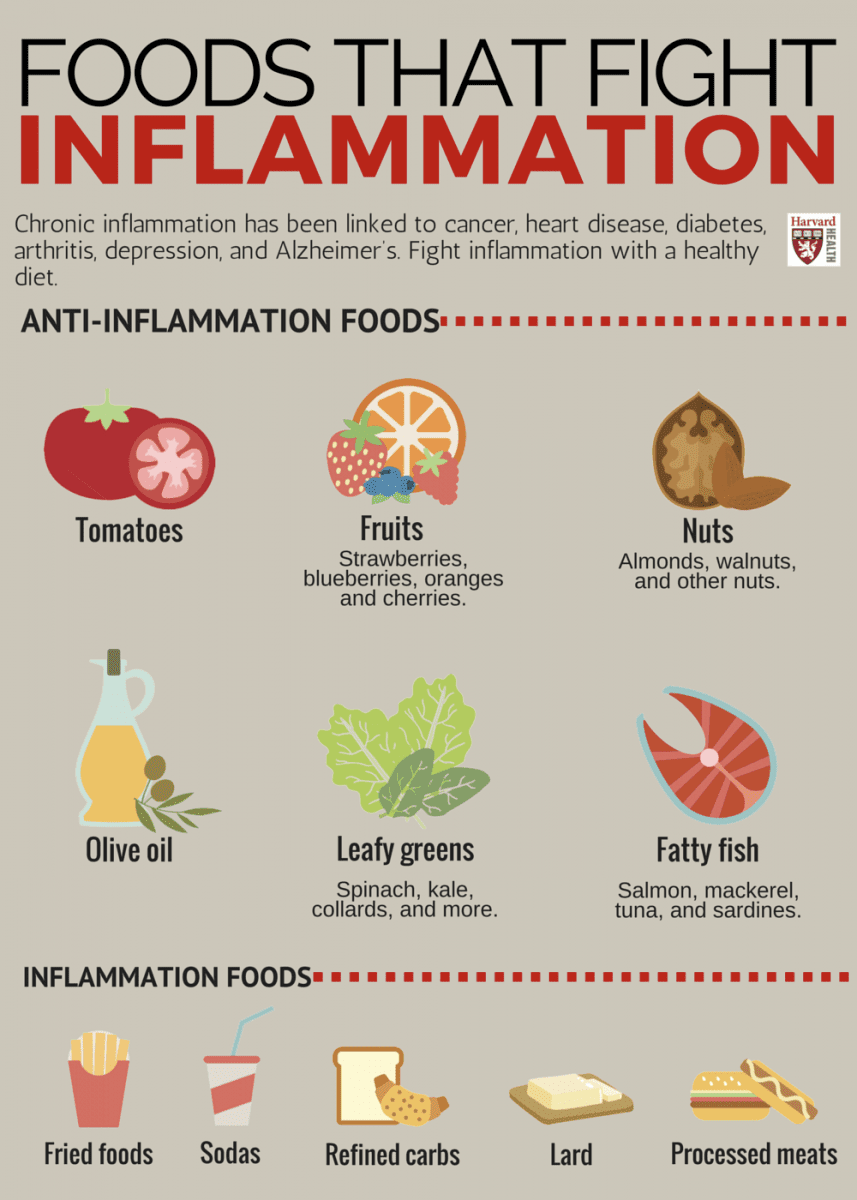
Foods that can cause inflammation include:
Refined carbohydrates, such as white bread and pastries
French fries and other fried foods
Sodas and other sugar-sweetened drinks
Red meat like burgers and steaks as well as processed meat like hot dogs and sausage
Margarine, shortening, and lard
Foods that Fight Against Inflammation
Alternatively, there are foods that fight against inflammation, and with it, chronic disease. Certain fruits and vegetables, such as blueberries, apples, and leafy greens, are high in polyphenols and antioxidants, which are components that may have anti-inflammatory effects. Research studies also have associated nuts with reduced biomarkers of inflammation and a decreased risk of diabetes and cardiovascular disease. Coffee may protect against inflammation, as well. Choose anti-inflammatory foods and you could improve your overall health and wellness. Choose inflammatory foods and you might increase the risk of inflammation and chronic pain.
Foods that can fight against inflammation include:
Tomatoes
Olive oil
Green leafy vegetables, such as spinach, kale, and collards
Nuts like almonds and walnuts
Fatty fish, such as salmon, tuna, mackerel, and sardines
Fruits like strawberries, blueberries, cherries, and oranges
Healthcare professionals are learning that one of the greatest ways to reduce inflammation is found. not in the medicine cabinet, but in the refrigerator. An anti-inflammatory diet can ultimately help reduce the human body’s inflammatory response. The immune system triggers inflammation to protect the human body from injury, illness, and infection. But if inflammation continues, it can cause a variety of health issues, including chronic pain symptoms. Research studies have demonstrated that certain food can influence the effects of inflammation in the human body.
Dr. Alex Jimenez D.C., C.C.S.T. Insight
Anti-Inflammatory Diets
To reduce inflammation, focus on following an overall healthier diet. If you’re looking for an anti-inflammatory diet, consider following the Mediterranean diet, which is high in fruits, vegetables, nuts, whole grains, fish, and oils. The Longevity Diet Plan, presented in the book by Dr. Valter Longo, also eliminates foods which can cause inflammation, promoting well-being and longevity. Fasting, or caloric restriction, has long been known to decrease oxidative stress and slow down the mechanisms of aging in various organisms.
And if fasting is not for you, Dr. Valter Longo’s longevity diet plan also includes the fasting mimicking diet, or FMD, which allows you to experience the benefits of traditional fasting without depriving your body of food. The main difference of the FMD is that instead of eliminating all food for several days or even weeks, you only restrict your calorie intake for five days out of the month. The FMD can be practiced once a month to help promote overall health and wellness as well as to help reduce inflammation and chronic pain.
While anyone can follow the FMD on their own, Dr. Valter Longo offers the ProLon� fasting mimicking diet, a 5-day meal program which has been individually packed and labeled to serves the foods you need for the FMD in precise quantities and combinations. The meal program consists of ready-to-eat and easy-to-prepare, plant-based foods, including bars, soups, snacks, supplements, a drink concentrate, and teas. However, before starting the ProLon� fasting mimicking diet, 5-day meal program, or any of the lifestyle modifications described above, please make sure to talk to a doctor to find out which chronic pain treatment is right for you.
In addition to reducing inflammation, a more natural, less processed diet can have noticeable effects on your physical and emotional health. The scope of our information is limited to chiropractic, spinal health issues, and functional medicine articles, topics, and discussions. To further discuss the subject matter above, please feel free to ask Dr. Alex Jimenez or contact us at 915-850-0900 .
Curated by Dr. Alex Jimenez
Additional Topic Discussion: Acute Back Pain
Back pain is one of the most prevalent causes of disability and missed days at work worldwide. Back pain attributes to the second most common reason for doctor office visits, outnumbered only by upper-respiratory infections. Approximately 80 percent of the population will experience back pain at least once throughout their life. Your spine is a complex structure made up of bones, joints, ligaments, and muscles, among other soft tissues. Injuries and/or aggravated conditions, such as herniated discs, can eventually lead to symptoms of back pain. Sports injuries or automobile accident injuries are often the most frequent cause of back pain, however, sometimes the simplest of movements can have painful results. Fortunately, alternative treatment options, such as chiropractic care, can help ease back pain through the use of spinal adjustments and manual manipulations, ultimately improving pain relief.
XYMOGEN�s Exclusive Professional Formulas are available through select licensed health care professionals. The internet sale and discounting of XYMOGEN formulas are strictly prohibited.
Proudly, Dr. Alexander Jimenez makes XYMOGEN formulas available only to patients under our care.
Please call our office in order for us to assign a doctor consultation for immediate access.
If you are a patient of Injury Medical & Chiropractic Clinic, you may inquire about XYMOGEN by calling 915-850-0900.
For your convenience and review of the XYMOGEN products please review the following link.*XYMOGEN-Catalog-Download
* All the above XYMOGEN policies remain strictly in force.
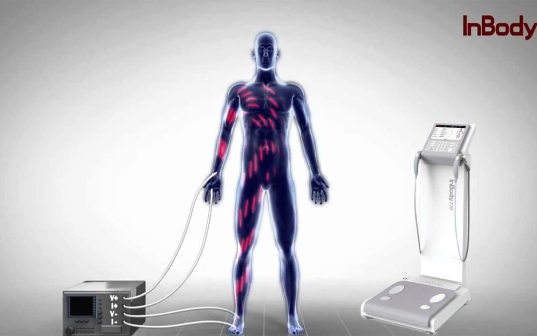
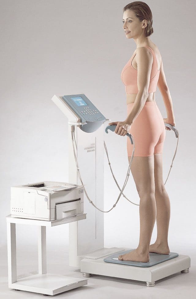
InBody devices use a method called Bio-electrical Impedance Analysis (BIA) to measure body composition. This divides your weight into different components, e.g., lean body mass and fat mass are utilized in the assessment of health and nutrition.
InBody Technology
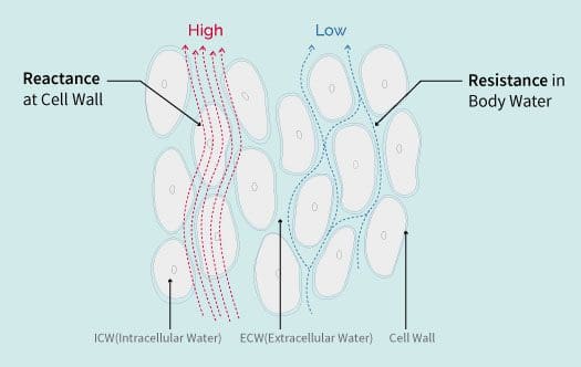
Resistance Concept
An illustration of how this works:
Imagine cars in traffic
Your car is the voltage-current
The highway is body water
If no other cars were around, you could roll right through
If the human body was only water and nothing else, there would be no resistance.
But water is not the only element
You are not the only car on the freeway
The more traffic gets onto the freeway, the longer it takes for you. This is resistance.
Other elements:
Fat
Muscle
Bone
Minerals
Create resistance to the current going through the body
In BIA testing, the more water in the body equals less resistance
The muscle in the body contains water
The more muscle you have, the more body water
The more body water, the less the resistance on the current
Bringing It Together
Impedance is the vector sum of the resistance
Reactance is the measurement BIA devices use to determine body composition
BIA applies cylinder model for the relationship between impedance and a body
Impedance is calculated by using two formulas:
The volume of a cylinder (Volume = Length x Area)
Impedance is inversely proportional to cross-sectional area and directly proportional to length.
Knowing the impedance and length of the cylinder, a measurement can be made of the volume of total body water.
In the body, the same formula applies, where the length is the height.
Calculation of the volume of the total body water can be made by knowing the impedance and height.
This is why it’s imperative to have correct height.
BIA Technology Has Been Revolutionized With InBody
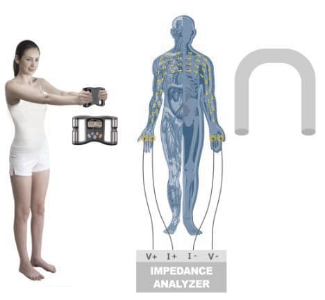
Measuring impedance with electrodes creates contact resistance.
InBody accounts for this by strategically placing electrodes to accurately measure.
InBody provides independent measurements for the body�s 5 cylinders:
Left Arm
Right Arm
Left Leg
Right Leg
The Torso
InBody uses multiple currents and varying frequencies.
No empirical estimations are used to calculate body composition.
InBody measures impedance independently, so results are not affected by age, ethnicity, or gender.
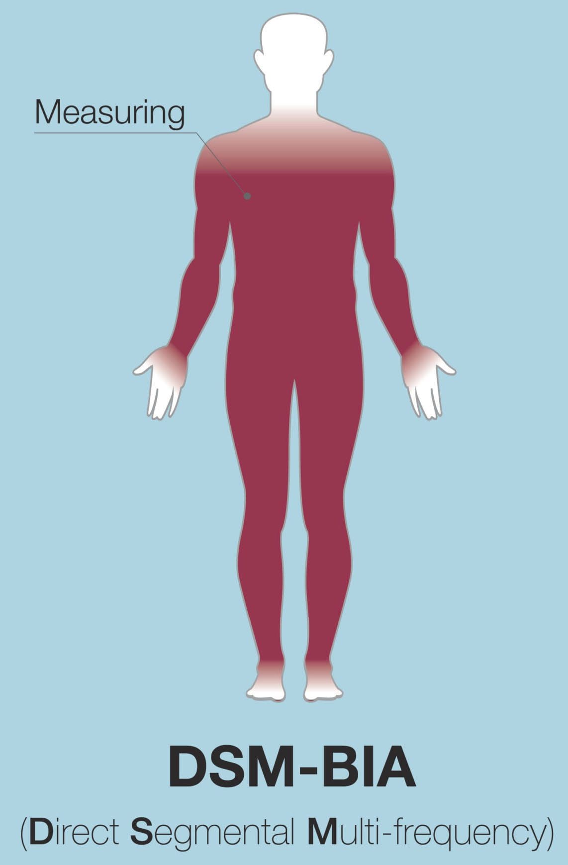
Direct Segmental Multi-frequency Bioelectrical Impedance Analysis
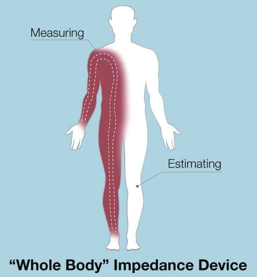
Traditional BIA systems view the body as a single cylinder and use whole-body impedance to determine total body water.
This method has a number of flaws:
It assumes the distribution of lean body mass and body fat are constant.
The shape & length of the arms, legs, and torso differ so the body cannot be seen as just one, but five separate parts.
Impedance is based on length and cross-sectional area, the calculation of TBW is inaccurate because each segment has different length and cross-section.
One major problem with the one-cylinder method is the lack of a torso measurement.
The torso has the lowest length and highest cross-section area.
This results in a very low impedance (10-40 ohms).
However, the trunk comprises about 50% of an individual�s lean body mass (LBM).
In the whole-body impedance measurement, the torso impedance is ignored and so changes the body torso impedance.
If the body torso is not measured separately then the impedance of the torso could be overlooked.
Because the body torso contains more water and muscles than the limbs, 1 ohm of torso impedance and 1 ohm of limb impedance can be completely different.
A difference of even 1-2 ohms can lead to a significant error in the determination of TBW.
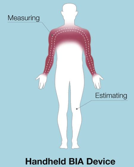
Some BIA devices only measure the impedance values of two cylinders and estimate the rest.
Some BIA scales only measure the legs.
For BIA handheld devices, only the arms are measured.
Some devices say they measure the whole body when they only measure one arm, one leg, and estimate the rest.
When using a BIA device, find one that actually measures the torso and measures it separately.
Otherwise, the estimations can lead to large errors.
InBody devices do not estimate through Direct Segmental Multi-frequency BIA, which in simpler terms, which means that each segment of the body right arm, left arm, torso, right leg, left leg are measured separately.
History of Bioimpedance Technology
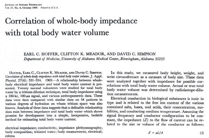
In 1969 came The Hoffer and Impedance Index
In 1969, Hoffer experimented to prove that total body water and biological impedance were highly interconnected. This meant that impedance measurement could be used to determine total body water.
He showed the squared value of height divided by impedance was highly correlated with total body water.
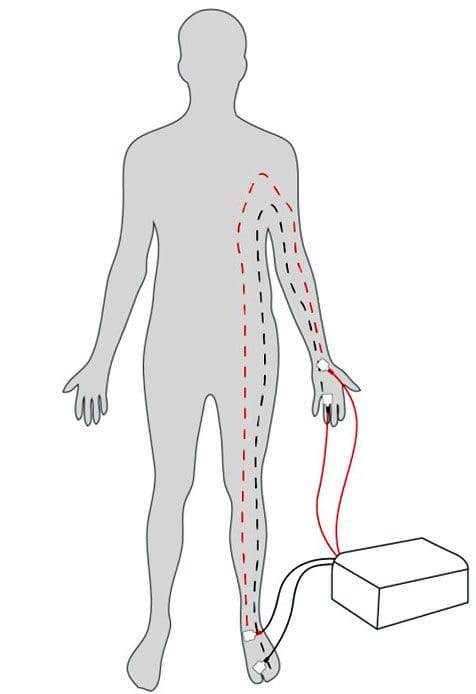
He took impedance measurements of the right half of the body. These included the right arm, torso, and right leg.
The equation he proved is the impedance index used in Bioelectrical analysis today.
In 1979 came the RJL System and First Impedance Meter
In 1979, RJL Systems brought the impedance meter and the BIA method began.
The device measured impedance through attachment of electrodes on the back of the right hand and on top of the right foot. A current of 50kHz was run through the right half of the body.
Before this, body composition could only be measured with calipers or underwater weighing.
These techniques had to be carried out by skilled people and was not easy.
Only specific types of patients were able to benefit from them, as well.
However, this was fast, less expensive and less intrusive. Thus, body composition analysts, nutritionists, and medical experts started using BIA technology.
In the 1980’s BIA limitations Emerged along with…
Lukaski, Segal and other scholars are the ones that accelerated the evolution of BIA.
Their studies proved that BIA had a high correlation with top standard methods, e.g., Underwater Weighing and DEXA.
However, there were technical limitations with BIA which surfaced towards the end of 1980s.
A common limitation was that BIA would assumed the human body was a cylinder shape and so only used a single frequency of 50 kHz.
Through research, various equations evolved (along with the impedance index). This complemented the technical limitation of BIA and was able to achieve greater accuracy for patients of different age, gender etc.
Lukaski & Kushner Develop Empirical Data Equation
This increased the accuracy of the results.
These equations utilized empirical data:
Gender and
Age to calculate a person�s body composition.
Empirical data is defined, as knowledge acquired by means of observation or experimentation.
By collecting data on a sample population that (hopefully) represents the variance of the entire population, researchers attempt to derive trends that may be used to predict outcomes.
In body composition, researchers identify these trends in muscle and fat mass; they use this data to predict body composition based on specific variables (age, gender, ethnicity, etc.)
Although empirical estimations could give you an accurate estimate of a general user�s body composition, there are significant problems when they are used for medical purposes.
Suppose there is a device that uses empirical equations to calculate TBW.
And there are two individuals who have same amount of lean body mass, however, one is 30 years old and the other is 40 years old.
Even though they have the same amount of LBM, the empirical equation will calculate that both will have 0.8 L difference in their TBW. This only because of age, which is not fair or accurate.
Home BIA Devices Start Showing Up
Because of technological limitations, BIA devices turned into home devices instead of hospital devices.
Then Japanese manufacturers released a variety of BIA body composition devices that the general public could easily use.
Some measured the impedance between two feet while standing on a scale. Others would hold the device and then measure the impedance between the hands.
Then In 1992 Kushner Proposed Multi-Frequencies & Segmental Analysis
Kushner claimed the human body to be made from five cylinders
Right Arm
Left Arm
Torso
Right Leg
Left Leg
Since the torso makes up 50% of lean body mass, Kushner emphasized measuring the impedance of the torso separately would be very significant.
Measuring total impedance alone is not sufficient. However, when all five parts are measured separately at different frequencies, then a distinction between extracellular water and intracellular water can be made.
In 1996 Dr. Cha Creates InBody Composition Analyzer
In 1996, Dr. Kichul Cha, a bioengineer from Harvard Medical School, develops the first 8-point electrode system with direct segmental analysis, which measures impedance for the five cylinders of the body at multiple frequencies.
This allows separate checking of torso impedance.
With this technology highly accurate results, without using empirical data, were able to be yielded.
InBody body composition analyzers became precision based medical devices. Impedance values for all of the body’s cylinders can be found on the InBody Result Sheet.
Many BIA products today provide muscle mass for each section of the body.
However, you can see the impedance values of all five parts of the body with the use of both high and low frequencies.
InBody Spotlight – Rachel Cosgrove of Results Fitness
History Body Model
Body composition analysis lends itself to a number of techniques depending upon the specific needs of your patients.
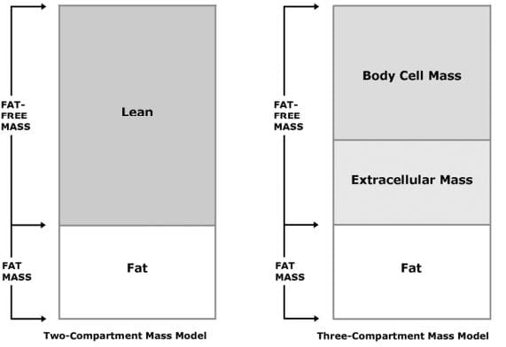
Two-Compartment Mass Model: The two-compartment mass model divides the body into Fat-Free Mass and Fat Mass.
This simple model is useful when evaluating basic nutritional, fitness, and weight management needs of patients.
Three-Compartment Mass Model: The three-compartment model divides the body into:
Body Cell Mass
Extracellular Mass
Fat Mass
This model is often used in support of nutritional counseling and monitoring changes associated with aging.
Appropriate for full range of patients.
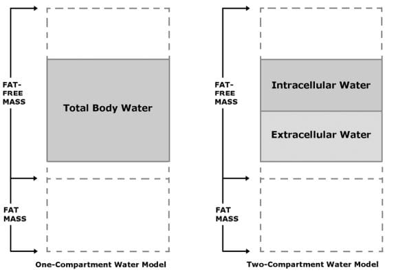
One-Compartment Water Model The one-compartment water model accounts for Total Body Water (TBW).
Total Body Water is the sum of Intracellular Water plus Extracellular Water and is wholly contained within Fat-Free Mass.
Normally, about 73% of Fat-Free Mass is water.
This model is handy for evaluating basic hydration status of patients.
Two-Compartment Water Model The two-compartment water model divides:
Total Body Water into
Intracellular Water and
Extracellular Water
This model is often used for the assessment of fluid balance associated with the treatment of conditions in a clinical setting.
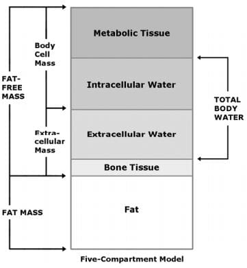
Five-Compartment Model divides the body into:
Metabolic Tissue
Intracellular Water
Extracellular Water
Bone Tissue
Fat Mass
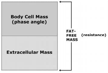
Impedance Model Application
When monitoring:
Compartment
Fat-Free Mass Resistance
Body Cell Mass
Total Body Water
Intracellular Water
Chart
Resistance
Phase Angle Resistance
Phase Angle
Application Guide
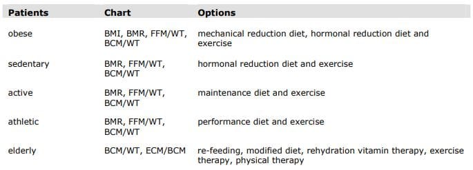
Obesity
The United States has long recognized obesity as a serious health condition.
In February 1985, the National Institutes of Health (NIH) in its Consensus Development Conference Statement (1) declared “The evidence is now overwhelming that obesity, defined as excessive storage of fat, has adverse effects on health and longevity.”
Obesity has long been associated with health risks.
While the specific mechanisms linking obesity to health risks are not fully understood, recent research focused on genes that express only in fat tissue has shown promise.
These genes code for hormones associated with insulin resistance and cardiovascular plaques.
The obesity epidemic continues to grow unabated in the United States (2,3).
Now, it has become a serious health problem in “both developed and developing countries in the Western Pacific Region of the World Health Organization” (4).
A useful definition of obesity is “excess fat mass resulting in mechanical or hormonal stress on the cardiovascular system, organs, and muscular-skeletal system.”
Diagnostic Criteria Body Mass Index (BMI) of 30 or greater. or
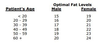
Fat Mass that is greater than 25 percent for males or greater than 30 percent for females.
Mechanical Reduction We recommend a two-step approach to normal function.
The first step is weight reduction to relieve mechanical stress on body systems.
Estimate total daily caloric expenditure = BMR * 1.2.
Set dietary intake = caloric expenditure – 700 calories per day.
Continue diet until BMI = 30.
NOTE: The initial dietary intake for obese patients will appear to be very high.
For example, a patient weighing 300 lb with 40 percent body fat will have a basal metabolic rate of 2550 calories, a total caloric expenditure of 3060 calories, and dietary intake of 2360 calories per day.
Hormonal Reduction
The second step is to decrease the ratio of fat mass to fat-free mass to reduce the incidence of fat-related hormones.
Measure the basal metabolic rate and fat-free mass.
Estimate caloric expenditure = BMR * 1.2.
Set dietary intake = caloric expenditure – 500 calories per day.
Continue until percentage fat mass reaches optimal level.
NOTE: Exercise is important in BIA because weight loss from dieting alone is comprised of 45 percent fat-free mass and 55 percent fat mass per pound. Exercise can alter this ratio to 25 percent fat-free mass and 75 percent fat mass.
References:
1. NIH Consensus Conference Statement, Health Implications of Obesity. Annals of Internal Medicine, 1985; 103 (6 pt 2):1073-1077.
2. Mokdad AH, et al. The spread of the obesity epidemic in the United States, 1991 – 1998. Journal of American Medical Association, 1999; 282:1519-1522.
3. Blackburn GL. Managing obesity in America: An overview. Advanced Studies in Medicine 2002;2(2):40-49.
4. Regional Office for the Western Pacific of the World Health Organization, the International Association for the Study of Obesity and the International Obesity Task Force. The Asia-Pacific perspective: Redefining obesity and its treatment. Health Communications Australia Pty. Limited, February 2000.
Chronic pain is a common health issue which affects many people in the United States. While several medical conditions, such as fibromyalgia and myofascial pain syndrome, can cause chronic pain, it may also develop due to a variety of other health issues. Research studies have found that widespread inflammation is the leading cause of chronic pain. Inflammation is a natural defense mechanism to injury, illness, or infection. But, if the inflammatory process continues for too long, it can become problematic.
Inflammation signals the immune system to heal and repair damaged tissue as well as to protect itself against bacteria and viruses. As mentioned above, however, chronic inflammation can cause a variety of health issues, including chronic pain symptoms. Healthy lifestyle modifications can help manage chronic pain, but first, let’s understand the common causes of chronic pain.
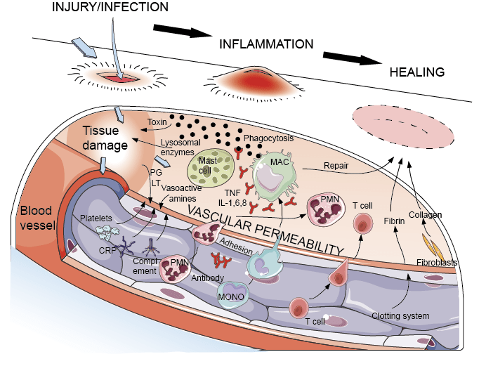
What is Acute Inflammation?
Acute inflammation, by way of instance, occurs following an injury or something as simple as a sore throat. It is a natural response with adverse effects, meaning it works locally in the region where the health issue is found. The common signs of acute inflammation include swelling, redness, warmth, pain and loss of function, as stated by the National Library of Medicine. When acute inflammation develops, the blood vessels dilate causing blood flow to increase, and white blood cells in the injured region promote recovery.
During severe inflammation, compounds called cytokines are released by the damaged tissue. The cytokines act as “emergency signals” which bring on the human body’s own immune cells, as well as hormones and numerous nutrients to repair the health issue. Additionally, hormone-like substances, known as prostaglandins, cause blood clots to heal damaged tissue, and these may also trigger fever and pain as part of the inflammatory procedure. As the damage or injury recovers, the inflammation subsides.
What is Chronic Inflammation?
Unlike acute inflammation, chronic inflammation has long-term effects. Chronic inflammation, also known as persistent inflammation, produces low-levels of inflammation throughout the human body, as demonstrated by an increase in immune system markers located in blood and cell tissues. Chronic inflammation may also cause the progression of various diseases and conditions. Elevated levels of inflammation may sometimes trigger even if there is no injury, illness, or infection, which may also cause the immune system to react.
As a result, the human body’s immune system could begin attacking healthy cells, tissues, or organs. Researchers are still trying to understand the consequences of chronic inflammation in the human body and the mechanisms involved in this natural defense process. By way of instance, chronic inflammation has been associated with a variety of health issues, such as heart disease, and stroke.
One theory suggests that when inflammation remains in the blood vessels, it can encourage the accumulation of plaque. According to the American Heart Association, or the AHA, if the immune system identifies plaque as a foreign invader, the white blood cells can attempt to wall off the plaque found in the blood flowing through the arteries. This can create a blood clot which may block the blood flow to the heart or brain, causing it to become unstable and rupture. Cancer is another health issue associated with chronic inflammation. Furthermore, according to the National Cancer Institute, DNA damage can also be caused by chronic inflammation.
Persistent, low-grade inflammation frequently doesn’t have any symptoms, but healthcare professionals can check for a C-reactive protein, or CRP, known as lipoic acid, a marker for inflammation found in the blood. Elevated levels of CRP are associated with an increased risk of cardiovascular disease. Elevated CRP levels may be found in chronic disorders like lupus or rheumatoid arthritis.
In the case of other chronic conditions, such as fibromyalgia, the nervous system over-reacts to specific stimulation, however, it’s inflammation which causes chronic pain symptoms. Subjectively, it’s almost impossible to tell the difference between the chronic pain caused by an oversensitive nervous system and the chronic pain caused by widespread inflammation. Apart from searching for clues in the bloodstream, a person’s nutrition, lifestyle habits, and environmental exposures, can also promote chronic inflammation.
Inflammation is the immune system’s natural defense mechanism against injury, illness, or infection. While this inflammatory response can help heal and repair tissues, chronic, widespread inflammation can cause a variety of health issues, including chronic pain symptoms. A balanced nutrition, including a variety of diets and fasting, can help reduce inflammation. Fasting, also known as caloric restriction, promotes cell apoptosis and mitochondrial recovery. The fasting mimicking diet, which is a part of the longevity diet plan, is a dietary program which “tricks” the human body into a fasting state to experience the benefits of traditional fasting. Before following any of the diets described in this article, make sure to consult a doctor.
Dr. Alex Jimenez D.C., C.C.S.T. Insight
Nutrition, Diets, Fasting and Chronic Pain
Anti-inflammatory diets mainly consist of eating fresh fruits and vegetables, fish, and fats. The Mediterranean diet plan, by way of instance, is an anti-inflammatory diet which promotes eating moderate amounts of nuts, ingesting very little meat, and drinking wine. Anti-inflammatory food parts, such as omega-3 fatty acids, protect the human body against the damage brought on by inflammation.
An anti-inflammatory diet also involves staying away from foods which could promote inflammation. It is ideal to decrease the amount of foods you eat which are high in trans and saturated fats, such as meats. Additionally, an anti-inflammatory diet limits the consumption of refined carbohydrates and foods, such as bread and rice. These also promote cutting back on the utilization of margarine and oils that are packed with omega-6 fatty acids, such as sunflower, safflower and corn oils.
Fasting, or caloric restriction, has long been known to decrease oxidative stress and slow down the mechanisms of aging in various organisms. The effects of fasting involve programmed cell death, or apoptosis, transcription, mobile energy efficiency, mitochondrial biogenesis, antioxidant mechanisms, and circadian rhythm. Fasting also contributes to mitochondrial autophagy, known as mitophagy, where genes in the mitochondria are stimulated to undergo apoptosis, which promotes mitochondrial recovery.
Intermittent fasting can help you fight inflammation, improve digestion, and boost your longevity. The human body is designed to be able to survive for extended periods of time without food. Research studies have demonstrated that intermittent fasting can have positive changes in the overall composition of your gut microbiota. Moreover, intermittent fasting can reduce insulin resistance while increasing the immune system response. Finally, intermittent fasting can promote the production of a substance, known as ?-hydroxybutyrate, that blocks a portion of the immune system involved in inflammatory ailments as well as substantially reducing the production of inflammatory markers, such as cytokines and the C-reactive protein, or CRP, previously mentioned above.
The Longevity Diet Plan, presented in the book by Dr. Valter Longo, eliminates the consumption of processed foods which can cause inflammation, promoting well-being and longevity. This unique dietary program, unlike most traditional diets, doesn’t promote weight loss. Although you may experience weight reduction, the emphasis of this unique dietary program is on eating healthier. The Longevity Diet Plan has been demonstrated to help activate stem cell-based renewal, reduce abdominal fat, and prevent age-related bone and muscle loss, as well as build resistance to developing cardiovascular disease, Alzheimer’s disease, diabetes, and cancer.
The fasting mimicking diet, or FMD, allows you to experience the benefits of traditional fasting without depriving your body of food. The main difference of the FMD is that instead of completely eliminating all food for several days or even weeks, you only restrict your calorie intake for five days out of the month. The FMD can be practiced once a month to help promote overall health and wellness.
While anyone can follow the FMD on their own, the ProLon� fasting mimicking diet offers a 5-day meal program which has been individually packed and labeled for each day, that serves the foods you need for the FMD in precise quantities and combinations. The meal program is made up of ready-to-eat or easy-to-prepare, plant-based foods, including bars, soups, snacks, supplements, a drink concentrate, and teas. Before starting the ProLon� fasting mimicking diet, 5-day meal program, or any of the lifestyle modifications described above, please make sure to talk to a healthcare professional to find out which chronic pain treatment is right for you.
The scope of our information is limited to chiropractic, spinal health issues, and functional medicine articles, topics, and discussions. To further discuss the subject matter above, please feel free to ask Dr. Alex Jimenez or contact us at 915-850-0900 .
Curated by Dr. Alex Jimenez
Additional Topic Discussion: Acute Back Pain
Back pain is one of the most prevalent causes of disability and missed days at work worldwide. Back pain attributes to the second most common reason for doctor office visits, outnumbered only by upper-respiratory infections. Approximately 80 percent of the population will experience back pain at least once throughout their life. Your spine is a complex structure made up of bones, joints, ligaments, and muscles, among other soft tissues. Injuries and/or aggravated conditions, such as herniated discs, can eventually lead to symptoms of back pain. Sports injuries or automobile accident injuries are often the most frequent cause of back pain, however, sometimes the simplest of movements can have painful results. Fortunately, alternative treatment options, such as chiropractic care, can help ease back pain through the use of spinal adjustments and manual manipulations, ultimately improving pain relief.
XYMOGEN�s Exclusive Professional Formulas are available through select licensed health care professionals. The internet sale and discounting of XYMOGEN formulas are strictly prohibited.
Proudly, Dr. Alexander Jimenez makes XYMOGEN formulas available only to patients under our care.
Please call our office in order for us to assign a doctor consultation for immediate access.
If you are a patient of Injury Medical & Chiropractic Clinic, you may inquire about XYMOGEN by calling 915-850-0900.
For your convenience and review of the XYMOGEN products please review the following link.*XYMOGEN-Catalog-Download
* All the above XYMOGEN policies remain strictly in force.
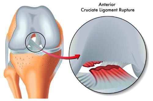
ACL injuries are some of the most common sports-related injuries that doctors and chiropractors see. It occurs when one of the ligaments in the knee, the ACL, is torn. If left untreated, it can cause the person to have difficulty moving their knee or controlling its movements because of the breakdown of knee support. When this happens, the bones of the joint often rub against each other. Over time this can cause a condition called chronic ACL deficiency. Osteoarthritis can also occur due to the bones rubbing against each other, eroding the cartilage and meniscus. A chiropractor can help reduce the pain of an ACL injury and help prevent further damage.
What is an ACL Injury?
The anterior cruciate ligament, or ACL, is a major ligament in the knee that connects the femur (thighbone) to the tibia (shinbone) in the knee joint and provides stability for the knee. An ACL injury does not occur gradually, but instead usually happens suddenly. Most often they happen when a person is playing sports and jumps, stops suddenly, or makes a sudden change in direction. Soccer, basketball, tennis, volleyball, downhill skiing, gymnastics, and football.
What causes an ACL injury?
Sports and fitness activities, typically ones that place stress on the knee, are the most common causes of ACL injury. Specific actions that can lead to the tears include:
Pivoting when the foot is planted firmly
Sudden stops
Cutting or slowing and changing direction suddenly
Landing incorrectly from a jump
Being involved in a collision where the knee is hit or it causes any of the other listed actions
Receiving a direct, sudden, hard hit to the knee
The resulting tear can be minor and small or it can be severe, including a complete tear. In very mild cases, the ligament may be overextended but still intact.
What are the symptoms of an ACL injury?
People who experience an ACL injury will often hear a loud POP when it occurs. Other symptoms of an ACL injury include:
Severe or intense pain
Swelling (begins in the first few hours after the injury)
A feeling that the knee is unstable
A popping sensation in the knee
The knee feels like it �gives away� when bearing weight
Unable to continue the activity they were doing when the injury occurred
Loss of or decreased range of motion
Who is most at risk of having an ACL injury?
Statistically, when both men and women are participating in the same sports, women are more likely to sustain an ACL injury. Research shows that, generally speaking, women have a tendency to have an imbalance of strength in their thighs. Specifically, the quadriceps, the muscles in the front are typically stronger that the hamstrings, the muscles in the back. It�s the hamstrings that work to keep the shinbone from extending too far forward � the type of movement that overextends the ACL. Additionally, men and women athlete jump differently with women more likely to land in a manner that places extra stress on the knees.
How is an ACL injury treated?
There are several treatments for an ACL injury. Immediate treatment can help reduce swelling and pain that the injury causes. The R.I.C.E. model is the recommended self-care that the patient can do at home:
Rice
Ice
Compression
Elevation
In some cases, surgery may be recommended, but usually the best course of treatment includes physical therapy and chiropractic care. The knee may be braced and the patient may have to rest for a while before beginning physical therapy. A chiropractor can help not only treat the ACL injury, but also help correct any muscular imbalances that the patient may have.
IFM's Find A Practitioner tool is the largest referral network in Functional Medicine, created to help patients locate Functional Medicine practitioners anywhere in the world. IFM Certified Practitioners are listed first in the search results, given their extensive education in Functional Medicine