The human body can absorb toxins in a variety of ways, from the type of foods we eat to the external and environmental factors we’re exposed to on a regular basis. Fortunately, the human body can also eliminate toxins in a variety of ways. Healthcare professionals have recognized that the accumulation of toxins in the human body can cause numerous health issues, including inflammation which may lead to back pain and sciatica. A good detox plan can help improve your overall health and wellness from the inside. Detoxing is also a fundamental process which can help relieve sciatica and back pain.
Contents
Back Pain and Sciatica
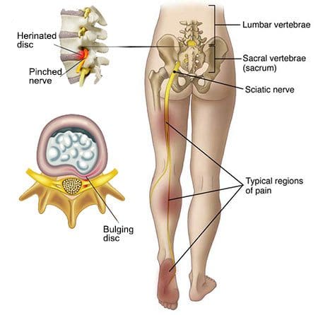
Back pain is one of the most common health issues reported among the general population. Although back pain affects approximately 80 percent of people at least once throughout their lifetime, sciatica continues to be one of the most frequently misunderstood health issues across the world. Also known as sciatic nerve pain, sciatica is characterized by irritation or inflammation due to the compression or impingement of the sciatic nerve, the longest and largest nerve in the human body. The sciatic nerve travels from the lower back, down the hips and buttocks, into the legs and feet. �
A variety of alternative treatment options, such as chiropractic care, can help safely and effectively treat sciatica symptoms without the side-effects of drugs and/or medications. Many healthcare professionals may recommend the use of drugs and/or medications to help offer sciatic nerve pain relief, however, these are only managing the symptoms rather than treating the health issue at the source. Research studies demonstrated that 76 percent of patients with sciatica reported complete relief of their symptoms without any toxic side-effects after following a detox plan for 45 days. �
Detox for Sciatica and Back Pain
For those people who may not be sure how or where to start with a detox plan, it’s fundamental for you to know that you can detox the human body on a variety of levels. First, you can start off with a simple foot detox. Healthcare professionals have demonstrated that a detox foot bath is a good way to cleanse the human body. Best known as the BioElectric Field Enhancement Unit, this helps generate positive and negative ions which creates the pH balance in the human body. As a result, the human body’s negative ions decrease while the human body’s positive ions increased to provide pain relief. �
To perform a detox foot bath, soak your feet for 30 minutes in warm salt water together with the foot coil. This process can help give muscles the strength and endurance they need to deal with back pain and sciatica symptoms. Moreover, a detox foot bath can also help eliminate free radicals which enhance range of motion. A detox foot bath also supports circulation. A good detox plan to help improve sciatica and back pain can also include following proper nutrition. Healthcare professionals can recommend a diet food plan which can help detox the human body from the inside with other treatment approaches. �
As previously mentioned, nutrition and lifestyle modifications below can help cleanse and detox the human body, including: �
Drinking more water, at least a minimum of two liters a day.
Drinking freshly squeezed juices without adding sugar, several times a day.
Replacing one meal with fresh smoothies, preferably breakfast.
Eating a healthy diet without processed foods, red meat, sugar, and dairy products.
According to healthcare professionals, it is a healthy practice to follow a detox plan once every 3 to 4 months. Do you remember when was the last time you had a detox? Or do you at least remember ever having a moment where you considered detoxification? Whether you followed a detox plan a few months ago, a year ago, several years ago, many years ago or simply never, it doesn�t matter. There is no better time then to start now. A detox plan cleanses the bloodstream, improves gut health, channels energy levels and strengthens muscles. Detox helps improve sciatica from its source. �
Sciatica is a collection of symptoms which can manifest due to a variety of underlying health issues. However, numerous research studies have demonstrated that following a detox plan can help tremendously reduce irritation and inflammation associated with sciatic nerve pain. Following a proper nutrition and lifestyle modifications together with a detox plan can ultimately help improve overall health and wellness, including sciatica and back pain. – Dr. Alex Jimenez D.C., C.C.S.T. Insight
Non-Invasive Treatments for Chronic Low Back Pain
Following a detox plan may help pain and inflammation associated with sciatica. The scope of our information is limited to chiropractic, musculoskeletal and nervous health issues as well as functional medicine articles, topics, and discussions. To further discuss the subject matter above, please feel free to ask Dr. Alex Jimenez or contact us at 915-850-0900�. �
Curated by Dr. Alex Jimenez �
Additional Topic Discussion: Severe Sciatica
Back pain�is one of the most prevalent causes of disability and missed days at work worldwide. Back pain attributes to the second most common reason for doctor office visits, outnumbered only by upper-respiratory infections. Approximately 80 percent of the population will experience back pain at least once throughout their life. Your spine is a complex structure made up of bones, joints, ligaments, and muscles, among other soft tissues. Injuries and/or aggravated conditions, such as�herniated discs, can eventually lead to symptoms of sciatica, or sciatic nerve pain. Sports injuries or automobile accident injuries are often the most frequent cause of painful symptoms, however, sometimes the simplest of movements can have these results. Fortunately, alternative treatment options, such as chiropractic care, can help ease sciatic nerve pain, or sciatica, through the utilization of spinal adjustments and manual manipulations, ultimately improving pain relief. �
�
Formulas for Methylation Support
�
XYMOGEN�s Exclusive Professional Formulas are available through select licensed health care professionals. The internet sale and discounting of XYMOGEN formulas are strictly prohibited.
Proudly,�Dr. Alexander Jimenez makes XYMOGEN formulas available only to patients under our care.
Please call our office in order for us to assign a doctor consultation for immediate access.
If you are a patient of Injury Medical & Chiropractic�Clinic, you may inquire about XYMOGEN by calling 915-850-0900.
�
� For your convenience and review of the XYMOGEN products please review the following link.*XYMOGEN-Catalog-Download �
* All of the above XYMOGEN policies remain strictly in force. �
As you move into your senior years, you may expect some discomfort and loss of mobility. Aging is hard on the body. But it is important to understand that there are things you can do to feel better. Regular exercise, a healthy diet, and chiropractic care can all make your senior years active and thriving.
Contents
How Chiropractic Can Make Your Senior Years Fantastic
The benefits of chiropractic for seniors can be considerable, including:
1. Takes away the pain.
If you are experiencing pain due to spinal or joint issues, chiropractic can help. Chiropractic care focuses on providing pain relief without the need for prescription medications or surgery.
Research has demonstrated the effectiveness of chiropractic for pain relief. In fact, one study showed that chiropractic was considerably more effective than prescribed medications. Unfortunately, many seniors assume that their pain is here to stay when it may be reduced or eliminated through regular chiropractic care.
2. Improves balance.
Seniors who are suffering from balance and coordination issues often have problems with the cervical spine, the technical term for the neck. When degenerative disc disease or other injuries disrupt the function of the mechanoreceptors located in the neck responsible for sending to the brain important information related to balance, then problems with balance can result.
Chiropractic adjustments and other related therapies are quite effective in treating neck injuries and degenerative disc disease. Regular adjustments from your chiropractor may be enough to help you get your balance back.
3. Improves mobility.
Injuries and aging can lead to a loss of range of motion, both in the spine and in the extremities. Fortunately, chiropractic care is designed to help you regain range of motion.
Your chiropractor has a variety of treatment methods to heal existing injuries while also improving range of motion. It may take several treatments to achieve the results you want, but a few visits to the chiropractor are well worth the increased range of motion you will enjoy.
4. Wear and tear on the joints are reduced.
The spine and joints need to be aligned to minimize wear on discs, bones, and other tissues. When you go for an extended period of time with a misaligned spine, it will increase the rate that your joints wear down.
One of the major benefits of chiropractic care is the way it keeps your whole body in alignment. As long as alignment is maintained, wear is minimized. Of course, alignment is lost in day-to-day life, but you can get it back by revisiting the chiropractor.
5. Remain healthy.
Chiropractors are known for adjusting spines. An aligned spine helps the body function at its peak. But chiropractors do more than adjustments. They focus on improving your overall health.
When you visit your chiropractor, he or she will ask you several questions and listen to your story to better understand the challenges you are dealing with. Once the chiropractor understands your situation, he or she will design an overall treatment plan designed to help you be as healthy as possible. Seniors that get regular chiropractic care often say they feel much better than they did before, which allows them to do more of the things they love to do.
General Disclaimer *
The information herein is not intended to replace a one-on-one relationship with a qualified healthcare professional or licensed physician and is not medical advice. We encourage you to make your own health care decisions based on your research and partnership with a qualified health care professional. Our information scope is limited to chiropractic, musculoskeletal, physical medicines, wellness, sensitive health issues, functional medicine articles, topics, and discussions. We provide and present clinical collaboration with specialists from a wide array of disciplines. Each specialist is governed by their professional scope of practice and their jurisdiction of licensure. We use functional health & wellness protocols to treat and support care for the injuries or disorders of the musculoskeletal system. Our videos, posts, topics, subjects, and insights cover clinical matters, issues, and topics that relate to and support, directly or indirectly, our clinical scope of practice.* Our office has made a reasonable attempt to provide supportive citations and has identified the relevant research study or studies supporting our posts. We provide copies of supporting research studies available to regulatory boards and the public upon request.
We understand that we cover matters that require an additional explanation of how it may assist in a particular care plan or treatment protocol; therefore, to further discuss the subject matter above, please feel free to ask Dr. Alex Jimenez or contact us at 915-850-0900.
Dr. Alex JimenezDC,MSACP, CCST, IFMCP*, CIFM*, ATN*
email: [email protected]
Licensed in: Texas & New Mexico*
Sciatica is a health issue caused by the compression or impingement of the sciatic nerve, or the longest and largest nerve in the human body. Patients with sciatica can experience painful symptoms anywhere along the length of the sciatic nerve. Common symptoms associated with sciatica include pain and discomfort, tingling sensations, numbness, and weakness. �
Sciatic nerve pain can be caused by a variety of health issues,� such as herniated discs, spinal stenosis, pregnancy, scar tissue, tight muscles, sacroiliac joint dysfunction, degenerative disc disease, tumors, and infection. Sciatic nerve pain can occur along one or both sides of the human body. According to numerous research studies, inflammation caused by an improper diet can cause sciatic nerve pain. Below, we will discuss how certain types of foods, both good and bad, can affect sciatica. �
Contents
Good Foods for Sciatica
Patients with sciatica caused by muscle spasms, such as piriformis syndrome, can benefit from consuming a variety of magnesium-rich foods. The human body uses magnesium to help release muscle contractions. Several magnesium-rich foods include dairy products, fish, meat, seafood, apples, apricots, brown rice, dulse, and lima beans. Foods with considerable amounts of vitamin B-12, such as liver, clams, oysters, lamb, and cheese, might also be beneficial for sciatic nerve pain. �
Halibut is a magnesium-rich food which may help treat sciatica associated with tight muscles or muscle spasms. Halibut contains numerous nutrients, including tryptophan, selenium, phosphorus, magnesium, protein, omega-3 fatty acids and vitamins B3, B6 and B12. The significantly increased levels of vitamin B12 in halibut can also help reduce painful symptoms. Halibut is also used to help treat cardiovascular problems, such as heart arrhythmia and elevated blood pressure. �
Because sciatica is a collection of symptoms rather than a single condition, patients with sciatic nerve pain should seek help from a healthcare professional to determine if an underlying health issue is causing the painful symptoms. In some cases, sciatic nerve pain may be caused by a serious problem, such as a tumor. Although further research studies may be needed to confirm the benefits of foods for sciatica, more research studies still have demonstrated how foods can also affect sciatica. �
Bad Foods for Sciatica
According to numerous research studies, eating a low-nutrient diet or a diet which may cause weight gain can ultimately cause a variety of health issues, including sciatic nerve pain and inflammation. Consuming foods rich in B-vitamins are essential for healthy nerve tissue. When we eat refined grain products, valuable nutrients are lost through the process, including B-vitamins. To make sure we eat nutrient dense foods, choose whole grains over refined grain products, such as white bread, instant rice, enriched pasta, low-fiber cereals and baked goods prepared with white, baking or cake flour. �
Added sugars are ingredients which add sweet flavor and calories to foods, however, they offer very little nutrients. These are also high-glycemic and they may have a considerable impact on blood sugar levels. A high-glycemic diet can increase inflammation and it also leaves less room for beneficial, anti-inflammatory foods, including fresh fruits and vegetables. Foods and beverages particularly high in added sugars include regular soft drinks, candy, pancake syrup, frosting, sweetened cereals, frozen desserts and commercially-prepared cakes, cookies, pies, and brownies, among other foods and beverages. �
Saturated fats can also increase inflammation. The American Heart Association recommends restricting the consumption of saturated fats to less than 7 percent of total daily calories. Common sources of saturated fats include red and processed meats, dark-meat poultry, poultry skin, high-fat dairy products, fried foods, and egg yolks. Healthcare professionals recommend replacing saturated fats in your diet with omega-3 fatty acids, or healthy fats with anti-inflammatory properties, to reduce sciatica. Sources of omega-3s include cold-water fish like salmon and mackerel, flaxseeds, canola oil, and walnuts. �
Trans-fats, also well-known as trans-fatty acids, are chemically-produced fats which can increase LDL, or “bad,” cholesterol and decrease HDL, or “good,” cholesterol. According to research studies, trans-fats are also pro-inflammatory substances and they can ultimately account for less than 1 percent of the calories in a heart-healthy diet. Common sources of trans-fats include stick margarine, shortening, and commercial foods which list hydrogenated vegetable oil as an ingredient. �
Sciatica, or sciatic nerve pain, can be caused by a variety of underlying health issues, including herniated discs and spinal stenosis, among other spine problems. However, numerous research studies have demonstrated that the foods we eat can affect painful symptoms, such as those associated with sciatic nerve pain or sciatica. “Good” foods can help reduce sciatica symptoms while “bad” foods can increase sciatica symptoms, affecting overall health and wellness. – Dr. Alex Jimenez D.C., C.C.S.T. Insight
Non-Invasive Treatments for Chronic Low Back Pain
�
�
A healthy diet may help manage pain and inflammation associated with sciatica. The scope of our information is limited to chiropractic, musculoskeletal and nervous health issues as well as functional medicine articles, topics, and discussions. To further discuss the subject matter above, please feel free to ask Dr. Alex Jimenez or contact us at 915-850-0900�. �
Curated by Dr. Alex Jimenez �
Additional Topic Discussion: Severe Sciatica
Back pain�is one of the most prevalent causes of disability and missed days at work worldwide. Back pain attributes to the second most common reason for doctor office visits, outnumbered only by upper-respiratory infections. Approximately 80 percent of the population will experience back pain at least once throughout their life. Your spine is a complex structure made up of bones, joints, ligaments, and muscles, among other soft tissues. Injuries and/or aggravated conditions, such as�herniated discs, can eventually lead to symptoms of sciatica, or sciatic nerve pain. Sports injuries or automobile accident injuries are often the most frequent cause of painful symptoms, however, sometimes the simplest of movements can have these results. Fortunately, alternative treatment options, such as chiropractic care, can help ease sciatic nerve pain, or sciatica, through the utilization of spinal adjustments and manual manipulations, ultimately improving pain relief.
�
�
Formulas for Methylation Support
XYMOGEN�s Exclusive Professional Formulas are available through select licensed health care professionals. The internet sale and discounting of XYMOGEN formulas are strictly prohibited.
Proudly,�Dr. Alexander Jimenez makes XYMOGEN formulas available only to patients under our care.
Please call our office in order for us to assign a doctor consultation for immediate access.
If you are a patient of Injury Medical & Chiropractic�Clinic, you may inquire about XYMOGEN by calling 915-850-0900.
For your convenience and review of the XYMOGEN products please review the following link.*XYMOGEN-Catalog-Download � * All of the above XYMOGEN policies remain strictly in force. �
College can be one of the most exciting times in a person�s life. With everything that comes with college also comes massive amounts of physical stress. Sitting all day, hunched over textbooks and laptops, even watching videos on your phone or texting, put constant strain on the spine that you may not be aware of. Chiropractic�can help alleviate the stress, relax the muscles and align the spine for optimal health, making it an ideal health solution for your college lifestyle.
Contents
How Chiropractic Benefits College Lifestyle
There are a lot of ways that chiropractic can help you feel better and even perform better at college. A healthy body makes everything easier, and chiropractic is designed to aid in a healthy lifestyle.
Some of the ways chiropractic can help include:
1. Heals the damage caused by extended sitting.
Sitting for long periods every day has been shown to increase the risk of death, and has been connected with a number of different health issues. The most important thing you can do each day is get up and move around every half hour or so, if possible.
But there is another thing you can do to address the tightness in soft tissues, compression of spinal discs and other effects sitting has on your body�get chiropractic adjustments. Regular adjustments realign your spine, reduce pressure on discs and help to relax the tightness in muscles and other soft tissues.
2. Helps correct poor posture.
Studying and working on the computer for long periods of time can be exhausting, there is no denying. Unfortunately, as you get tired you tend to hunch and slouch in ways that are not good for your back and neck. While paying attention to your posture is important and can help somewhat, the reality is that no one can maintain the perfect posture for hours and hours.
By visiting your chiropractor, you can get much-needed adjustments to your back and neck that will help to correct damage caused by poor posture. Your chiropractor can also give you tips on how to maintain the best possible posture and ergonomics on a daily basis to minimize further damage.
Meeting Talking Discussion Brainstorming Communication Concept
3. Heals neck injuries like text neck.
According to recent research, looking down at your phone for an extended period of time is the equivalent of putting a 60-pound weight on your neck. As you go about your day, notice how often you are staring down at your phone.
Whether it is texting with friends, watching a video or looking something up, you spend a considerable amount of time with that 60-pound weight on your neck. If your neck hurts, it is easy to understand why.
Chiropractic care is an excellent way to heal neck injuries including those caused by text neck. Regular adjustments can bring back the space between your vertebrae and help your neck heal.
4. Maintains spinal health.
Whether you hit the gym every day, play sports, or try to never move from the couch, you are always putting pressure on your spine. Your chiropractor is trained to help you maintain spinal health in a variety of ways, including chiropractic adjustments and lifestyle advice concerning healthy movement. By developing a relationship with your chiropractor that includes regular visits for adjustments and consultations on spinal issues you are experiencing, you can enjoy much better spinal health.
Contact Us & Schedule An Appointment
The best way to learn the benefits of chiropractic care is to meet with an experienced chiropractor. Please contact us to schedule an appointment. Let us help you enjoy a healthier spine and all the benefits that come with it.
Sciatica is commonly characterized by a collection of symptoms, such as pain, discomfort, tingling sensations and numbness, frequently caused by the compression of the sciatic nerve. In a variety of circumstances, the cause of a patient’s sciatic nerve pain may be due to inflammation, even without any underlying health issues. � Many healthcare professionals will prescribe anti-inflammatory drugs and/or medications to help treat sciatica. However, these can only do so much when it comes to treating the source of sciatica. The type of foods you eat also plays a fundamental role in reducing overall inflammation in the human body. Below, we will describe what a comprehensive diet food plan should include to help improve sciatic nerve pain symptoms. �
Contents
Anti-Inflammatory Foods for Sciatica
� There are many types of anti-inflammatory foods which can help ease pain, discomfort, and swelling, which may ultimately be beneficial for sciatica, or sciatic nerve pain. The best anti-inflammatory foods include: �
Spices: Several varieties of spices contain antioxidants which can help reduce the human body’s inflammatory response. Rosemary, cloves, ginger, and turmeric can help reduce inflammation.
Dark leafy greens: Vegetables such as collard greens, Swiss chard, kale, and spinach also contain antioxidants which can help fight against cellular damage, among other health issues like sciatica.
Green tea: Japanese matcha tea contains 17 times more antioxidants than wild blueberries, another food which is also rich in antioxidants. These anti-inflammatory foods can help reduce sciatic nerve pain.
Fermented vegetables: Gut health is essential to help boost your immune system and reduce inflammation. Anti-inflammatory foods, such as natto, kimchi, miso, pickles, and sauerkraut, are excellent choices. You may also eat fermented dairy products, such as yogurt and kefir, to help reduce inflammation.
� When purchasing any of the anti-inflammatory foods listed above, make sure they come from organic providers to help minimize your risk of consuming pesticides and other harmful toxins. As for the dairy foods listed above, make sure they come from healthy, grass-fed cows to make sure that you’re consuming a nutritious, high-quality product. The milk harvested from cows raised in concentrated animal feeding operations, or CAFOs, often contain antibiotics and other additives which can affect your overall well-being. �
Eat Foods Rich in B Vitamins
� The vitamin B family is fundamental for the structure and function of the human body because it helps regulate the nervous system. Vitamin B deficiencies can cause transmitters to start working inefficiently which may slow down the sciatica recovery process. Below are some of the key B vitamins you should consume, including: �
Vitamin B6: Also known as pyridoxine, vitamin B6 can help support the metabolism of the human body’s neurotransmitters. Foods rich in this nutrient include grass-fed meat, fish, bananas, and legumes.
Vitamin B12: Vitamin B12 plays an essential role in maintaining the health of myelin, or the fatty substance which wraps around nerve fibers to speed up neurotransmitter communications. Foods rich in this nutrient include pasture-raised chicken and eggs, wild-caught fatty fish and grass-fed dairy products.
Folate: This vital vitamin is crucial for the production of neurotransmitters. Vegetables are good sources of folate and the best choices include asparagus, broccoli, bok choy, romaine lettuce, and cauliflower.
Eat Foods to Promote Muscle Repair
� In a variety of circumstances, muscle health issues can cause sciatica, such as piriformis syndrome. Add foods into your sciatica diet food plan treatment which can help nourish and repair your muscles properly in order to help prevent injuries and/or aggravated conditions. Some of the best foods for muscle repair include: �
Nuts and seeds: These two popular snack options contain plant-based omega-3 fatty acids which can tremendously help reduce inflammation as well as provide protein for muscle synthesis and growth.
Wild-caught salmon: Wild-caught salmon can greatly increase your omega-3 intake. It also contains a good source of lean protein to promote muscle-building for overall muscle repair, health, and wellness.
Tart cherries: These small fruits should be a part of your diet food plan as they may help decrease pain, discomfort, and inflammation, according to the Journal of the International Society of Sports Nutrition.
Pasture-raised eggs: Eggs contain generous amounts of protein which can ultimately help repair and strengthen your muscles to reduce sciatica symptoms, as well as increase energy production.
� Make sure to eat only moderate amounts of protein on a daily basis, preferably 1 gram for every kilogram of your lean body mass, according to healthcare professionals. This will lower the risk of stimulating mTOR, or mammalian target of rapamycin, which is associated with cancer when protein is consumed excessively. �
Sciatica is a collection of symptoms characterized by pain, discomfort, tingling sensations, and/or numbness, among other common symptoms. Sciatica, also known as sciatic nerve pain, is generally caused by a variety of underlying health issues, however, research studies have demonstrated that inflammation caused by an improper nutrition can also cause sciatic nerve pain. Following a sciatica diet food plan treatment can help improve sciatic nerve pain. – Dr. Alex Jimenez D.C., C.C.S.T. Insight
Mix and Match Diet Food Plan
� With the numerous food choices listed in this article, you can surely put together a sciatica diet food plan treatment which can help reduce overall inflammation while simultaneously building your muscles and supporting your nervous system health. If you�re unsure of what to eat due to allergies, consult a healthcare professional. Afterward, you can come up with healthy meals which can improve your overall well-being. � Sciatica is a collection of symptoms caused by the compression or impingement of the spinal cord and/or nerve roots. Understanding the symptoms of sciatic nerve pain is essential towards obtaining a diagnosis in order to follow up with the best treatment. The scope of our information is limited to chiropractic, musculoskeletal and nervous health issues as well as functional medicine articles, topics, and discussions. To further discuss the subject matter above, please feel free to ask Dr. Alex Jimenez or contact us at 915-850-0900�. �
Curated by Dr. Alex Jimenez �
Additional Topic Discussion: Severe Sciatica
Back pain�is one of the most prevalent causes of disability and missed days at work worldwide. Back pain attributes to the second most common reason for doctor office visits, outnumbered only by upper-respiratory infections. Approximately 80 percent of the population will experience back pain at least once throughout their life. Your spine is a complex structure made up of bones, joints, ligaments, and muscles, among other soft tissues. Injuries and/or aggravated conditions, such as�herniated discs, can eventually lead to symptoms of sciatica, or sciatic nerve pain. Sports injuries or automobile accident injuries are often the most frequent cause of painful symptoms, however, sometimes the simplest of movements can have these results. Fortunately, alternative treatment options, such as chiropractic care, can help ease sciatic nerve pain, or sciatica, through the utilization of spinal adjustments and manual manipulations, ultimately improving pain relief. �
�
Formulas for Methylation Support
�
XYMOGEN�s Exclusive Professional Formulas are available through select licensed health care professionals. The internet sale and discounting of XYMOGEN formulas are strictly prohibited.
Proudly,�Dr. Alexander Jimenez makes XYMOGEN formulas available only to patients under our care.
Please call our office in order for us to assign a doctor consultation for immediate access.
If you are a patient of Injury Medical & Chiropractic�Clinic, you may inquire about XYMOGEN by calling 915-850-0900.
�
For your convenience and review of the XYMOGEN products please review the following link.*XYMOGEN-Catalog-Download �
* All of the above XYMOGEN policies remain strictly in force. �
Manuel Lozano’s ability to participate in his daily responsibilities was lost after he was involved in an auto accident. Mr. Lozano’s symptoms worsened and took hold of his life. He couldn’t take it anymore and sought treatment. Local El Paso chiropractor Dr. Alex Jimenez treated and restored Mr. Lozano’s health to optimal performance, and he recommends El Paso Back Clinic & Dr. Jimenez for auto accident injuries.
El Paso Back Clinic
We are blessed to present El Paso’s Premier Wellness & Injury Care Clinic to you.
At El Paso’s Chiropractic Rehabilitation Clinic & Integrated Medicine Center, we are passionately focused on treating patients after frustrating injuries and chronic pain syndromes. We focus on improving your ability through flexibility, mobility, and agility programs tailored for all age groups and disabilities.
We were hoping you could live a life filled with more energy, a positive attitude, better sleep, less pain, proper body weight, and education on maintaining this way of life.
Helping You Heal To Optimal Performance!
Please feel free to subscribe and recommend us if you have enjoyed this video and we have helped you.
Sciatica can cause pain, discomfort, tingling sensations and numbness along the entire length of the sciatic nerve. Also known as sciatic nerve pain, is a collection of symptoms caused by a variety of health issues. Many people will turn to the use of drugs and/or medications as well as surgery to treat sciatica, however, research studies have demonstrated that functional medicine can help improve sciatic nerve pain. � As a matter of fact, a 2010 research study published in the Journal of Manipulative Physiological Therapies demonstrated that approximately 60 percent of patients with sciatica benefited from alternative treatment options, such as chiropractic care. Moreover, functional medicine approaches, including acupuncture, yoga, and massage therapy, among other alternative treatment options, have been demonstrated to help improve sciatic nerve pain. Functional medicine can help safely and effectively provide sciatica pain relief. �
Contents
Functional Medicine Approaches for Sciatica
Treatment for sciatica can depend largely on the underlying health issues causing the painful symptoms, therefore, it’s important for people to seek help from a healthcare professional. While many doctors choose to utilize drugs and/or medications, such as anti-inflammatory drugs, muscle relaxers, or steroids, to treat sciatic nerve pain, many research studies have demonstrated that alternative treatment options can help improve sciatica.� Below, we will discuss functional medicine approaches which can help improve sciatica symptoms. �
Sciatica is a collection of symptoms characterized by radiating pain, tingling sensations, and/or numbness which extends throughout the length of the sciatic nerve. Sciatica, also known as sciatic nerve pain, is generally diagnosed through its symptoms and depending on the type of painful symptoms, a healthcare professional can safely and effective treat sciatic nerve pain. Functional medicine approaches, such as chiropractic as well as nutrition and lifestyle modifications, can ultimately help improve sciatic nerve pain, or sciatica. – Dr. Alex Jimenez D.C., C.C.S.T. Insight
Chiropractic Care
� One research study published in the Official Journal of the North American Spinal Society demonstrated that after comparing the results of 102 adults who suffered from sciatic nerve pain, those who received chiropractic care experienced less pain, fewer number of days with pain, and decreased instances moderate to severe pain compared to those adults who didn�t receive chiropractic care for their sciatica. � Disk herniations are some of the most common causes of sciatica.�There are several types of “ruptured” or herniated discs. Prolapse disc bulges are considered to be less severe because the outermost layer of the disc is still intact, however, extrusion or sequestration disc bulges are generally considered to be more painful. These types of ruptured or herniated discs cause damage to the outer layer of the spinal disc, which leads tissue to push through from where it�s normally constrained. This can compress the sciatic nerve and cause symptoms. �
For healthcare professionals, it�s important to know the patient’s symptoms in order to understand their health issues and follow the appropriate treatment approach. Sciatica can be diagnosed during a physical exam by a chiropractor or doctor of chiropractic. Doctors can also perform X-rays and other tests, such as magnetic resonance imaging, or MRI, test to diagnose the condition of the spine. After diagnosis, a chiropractor may utilize spinal adjustments and manual manipulations to correct the spine and relieve sciatic nerve pain. �
Yoga
Stretches and exercise can aggravate sciatica, however, yoga has been demonstrated to help relieve sciatic nerve pain. Some people have reported that sitting and/or standing for extended periods of time and then moving around suddently can aggravate sciatica. Scrunching or shortening the spine, such as raising the legs up and/or bringing the knees towards the chest in a squatting position can commonly affect symptoms. �
On the other hand, stretching and exercising the spine through yoga can help promote good posture while reducing stiffness, inflammation, and pain frequently associated with sciatic nerve pain. Research studies have demonstrated�that yoga is a safe and effective treatment for people with sciatica. Some of the most important movements for preventing sciatic nerve pain focus on building strength and relaxing stiffness. Stretches and exercises may even be utilized in rehabilitation settings for patients with sciatica following surgery.
Acupuncture and Massage Therapy
Acupuncture is a type of traditional Chinese medicine practice which is based on regulating overall health and wellness by opening the human body�s natural flow of energy. It utilizes small, virtually pain-free needles to target specific pathways in the human body. Acupuncture has been approved by the FDA as a treatment for back pain and it’s supported in a variety of research studies for relieving chronic pain, including sciatica. �
Massage therapy is another non-surgical, holistic approach which helps open muscles, tissues, and channels of energy within the human body to help improve blood flow and reduce painful symptoms. Massage therapy helps reduce back pain, improve muscle relaxation, and even promote the healthy release of endorphins, or the natural �feel good� substances of the human body which act as pain relievers to improve painful symptoms. �
Nutrition and Lifestyle Modifications
Approximately 5 to 10 percent of patients with low back pain also have sciatica, however, healthcare professionals have demonstrated that a patient’s nutrition and lifestyle can increase the risk of developing sciatic nerve pain. Also, factors like height, age, stress, being overweight or obese, sitting or standing for extended periods of time, and smoking, can also increase the risk of developing sciatica and other problems. �
Many of these risk factors cause inflammation, which makes it difficult to heal from injuries and conditions. To prevent inflammation and improve the odds of developing sciatica, make sure to eat a nutrient-dense healing diet, avoid smoking/using recreational drugs, and participate in exercise as well as get good sleep. Constipation may also cause toxicity and inflammation. As a matter of fact, anything which causes toxicity or inflammation can cause sciatica. Nutrition and lifestyle modifications can ultimately help improve sciatic nerve pain. �
Sciatica is a collection of symptoms caused by the compression or impingement of the spinal cord and/or nerve roots. Understanding the symptoms of sciatic nerve pain is essential towards obtaining a diagnosis in order to follow up with the best treatment. The scope of our information is limited to chiropractic, musculoskeletal and nervous health issues as well as functional medicine articles, topics, and discussions.
Chronic Back Pain Guidelines
To further discuss the subject matter above, please feel free to ask Dr. Alex Jimenez or contact us at 915-850-0900.��
Curated by Dr. Alex Jimenez �
Additional Topic Discussion: Severe Sciatica
Back pain�is one of the most prevalent causes of disability and missed days at work worldwide. Back pain attributes to the second most common reason for doctor office visits, outnumbered only by upper-respiratory infections. Approximately 80 percent of the population will experience back pain at least once throughout their life. Your spine is a complex structure made up of bones, joints, ligaments, and muscles, among other soft tissues. Injuries and/or aggravated conditions, such as�herniated discs, can eventually lead to symptoms of sciatica, or sciatic nerve pain. Sports injuries or automobile accident injuries are often the most frequent cause of painful symptoms, however, sometimes the simplest of movements can have these results. Fortunately, alternative treatment options, such as chiropractic care, can help ease sciatic nerve pain, or sciatica, through the utilization of spinal adjustments and manual manipulations, ultimately improving pain relief. �
�
Formulas for Methylation Support
�
XYMOGEN�s Exclusive Professional Formulas are available through select licensed health care professionals. The internet sale and discounting of XYMOGEN formulas are strictly prohibited.
� Proudly,�Dr. Alexander Jimenez makes XYMOGEN formulas available only to patients under our care. � Please call our office in order for us to assign a doctor consultation for immediate access.
� If you are a patient of Injury Medical & Chiropractic�Clinic, you may inquire about XYMOGEN by calling 915-850-0900.
�
For your convenience and review of the XYMOGEN products please review the following link.*XYMOGEN-Catalog-Download
� * All of the above XYMOGEN policies remain strictly in force. �
A large portion of the patients that seek out chiropractic care are suffering from some form of back pain. While chiropractic adjustments and associated therapies can do a lot to ease back pain symptoms and aid in healing, lifestyle changes are also quite important �, particularly weight loss. Excess weight puts increased pressure on the spine, especially the lower back. The more chiropractic patients can near their ideal weight, the easier it becomes to treat and often eliminate the back pain.
Contents
The Importance Of Weight Loss
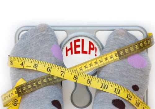
Research shows that obesity makes back pain highly probable.
Research has shown that excess weight and obesity do increase the risk of suffering from low back pain. A meta-analysis of the research on obesity and low back pain found that overweight and obese individuals were most likely to seek care for low back pain, including chronic back pain.
The components of the spine can wear down.
The spine is strong, but it is also delicate. The vertebrae are linked and supported by soft tissues, like discs, ligaments, and tendons.
These soft tissues make it possible to move, flex, twist and turn in a variety of ways, allowing your body to be quite mobile. But these soft tissues are prone to wear and tear.
Even a healthy body will experience wear and tear as it ages. When you add extra weight, though, you increase the wear and tear � and become more susceptible to injuries.
Extra body weight increases damage.
If you have ever carried an object for an extended period of time, like a bag of groceries up and down stairs or a jug of liquid across a parking lot, you know how it can wear you down. While you may feel fine at first, the longer you go, the more tired you become.
Extra body weight is something you carry around with you everywhere you go.
Across the parking lot.
Up the stairs.
Even sitting in a chair.
The structural components of your body, including your joints and the muscles that support them, are constantly forced to handle the extra pressure.
All the extra pressure transfers force through your joints, including your spine. Over time, the force will do damage. Discs will wear out faster, which can lead to degenerative disc disease and back pain.
Injuries become more severe.
The extra weight makes every accident and injury more damaging. Slipping and catching yourself, which may have been okay before, could now pull or tear muscles and tendons. Every time you are in an accident, your body will have a harder time maintaining safe alignment.
Common Injuries Caused by Excess Weight
1. Herniation
A herniated disc occurs when the tough outer layer of the disc is torn, allowing the soft inner layer to protrude. The protrusion can put pressure on nerves in the spine.
2. Pulled tissues.
Excess weight makes it more likely that you will pull or tear muscles, tendons, and ligaments.
Every pound you lose is one less pound your body must carry. The closer you get to your optimal weight, the easier it will be for your body to support your spine. When you get chiropractic adjustments, they will be more likely to remain in place. A healthy weight makes chiropractic care more effective.
If you are suffering from back pain and excess weight, please contact us. Our chiropractic team can help you in your weight loss journey, and we can treat your pain in a way that is both effective and non-invasive.
� Sciatica, or sciatic nerve pain, is a collection of symptoms caused by the compression or impingement of the spinal cord and/or nerve roots, due to disk herniation, spinal health issues like osteoarthritis, spondylolisthesis, and spinal stenosis as well as intraspinal tumors and abscesses. Impingement or compression may typically occur along the spinal canal or intervertebral foramen. Health issues associated with the compression or impingement of the spinal cord and/or nerve roots may commonly occur in the lower back, pelvis, or buttocks. � �
Sciatica Symptoms
� The common symptoms of sciatic nerve pain include pain and discomfort which radiates along the lower back, down the buttocks and posterior aspect of the leg, into the knee and foot. The painful symptoms may affect one or both lower extremities and it may occur with or without low back pain. The pain and discomfort are described as burning, lancinating, or stabbing. Other common symptoms of sciatica include tingling sensations and numbness anywhere along the length of the sciatic nerve, particularly in the lower extremities. � Coughing or the Valsalva maneuver, a specific way of breathing which increases pressure in the chest, may worsen sciatic nerve pain symptoms. Moreover, the compression or impingement of the spinal cord and nerve roots can cause sensory, motor, or reflex deficits, among other health issues. Symptoms of sciatica may depend on which nerves are affected based on the segmental level of the spine. By way of instance, an L5 to S1 disk herniation may affect the ankle jerk reflex while an L3 to L4 disk herniation may affect the knee jerk reflex. �
Sciatica, or sciatic nerve pain, is a collection of symptoms rather than a single health issue, characterized by radiating pain, tingling sensations, and/or numbness which extends from the lower back and buttocks, down into the legs and feet. Sciatica is generally diagnosed through its symptoms and depending on the type of painful symptoms, a healthcare professional can safely and effective treat sciatic nerve pain. It’s important for patients to understand the symptoms of sciatica in order to continue with a diagnosis and follow-up with the appropriate treatment option. – Dr. Alex Jimenez D.C., C.C.S.T. Insight
� Healthcare professionals have determined that straight leg raising may aggravate pain and discomfort which radiates down the length of the leg when gradually raised above 60 degrees or less. According to numerous research studies, this outcome measure is sensitive to sciatic nerve pain. Painful symptoms radiating down the affected leg when the contralateral leg is lifted, also known as crossed straight leg raising, is more common for sciatica. Furthermore, sciatica can ultimately be diagnosed through a series of tests and evaluations. � The straight leg raise test can be performed while patients are sitting with their hip joints flexed at 90 degrees. Then, their leg is carefully raised until the knee is fully extended. If the patient has sciatica, the painful symptoms will most often manifest as the leg is extended. The slump test can be performed like the straight leg raise test, but while the patient is slumping with the thoracic and lumbar spine flexed as well as the neck flexed. The slump test is more accurate but less specific, for disk herniation than the straight leg raise test. �
Sciatic Nerve Pain Diagnosis
� Sciatica is commonly diagnosed through its characteristic, painful symptoms. Once sciatic nerve pain is diagnosed, healthcare professionals should test a patient’s strength, sensations, and reflexes to determine any possible health issues. If painful symptoms persist for more than 6 weeks, or if there are neurologic deficits, imaging and electrodiagnostic studies should be performed. Structural and functional abnormalities which result in sciatica, such as spinal stenosis, can most accurately be diagnosed through MRI or CT scans. � Imaging and electrodiagnostic studies can help confirm the segmental level of the spinal cord and/or nerve root compression and/or impingement, which can exclude health issues that may mimic sciatica, such as polyneuropathy. These studies may help determine whether single or multiple regions of the spinal cord and/or nerve roots are being affected and whether the diagnosis correlates with MRI abnormalities. Abnormalities may not be obvious on imaging and electrodiagnostic studies for up to a few weeks after symptoms manifest. �
Sciatica Treatment
� Patients with sciatica, or sciatic nerve pain, can achieve relief from their painful symptoms through bed rest in a recumbent position with the head of the bed elevated about 30 degrees, also known as the semi-Fowler position. Treatment for low back pain can include nonopioid analgesics, such as NSAIDs and acetaminophen. Drugs and/or medications which decrease neuropathic pain, such as gabapentin or other anticonvulsants and low-dose tricyclic antidepressants, may also help relieve sciatic nerve pain, or sciatica, signs and symptoms. � Muscle spasm associated with low back pain or sciatica can be relieved through the utilization of heat or cold, physical therapy, and chiropractic care, among other alternative treatment options. Whether corticosteroids should be used to treat acute radicular pain remains controversial. Epidural corticosteroids can help achieve pain relief, however, these should not be utilized unless the patient’s painful symptoms are severe or persistent. Many healthcare professionals may utilize oral corticosteroids for these special occasions. � Surgery for sciatic nerve pain, or sciatica, is only recommended for cauda equina syndrome or for unequivocal disk herniation along with the presence of muscular weakness, progressive neurologic deficit and/or intolerable, intractable pain which interferes with regular physical activities in an emotionally stable patient which has not decreased after 6 weeks of conservative treatments. The standard procedure for sciatica caused by disk herniation is through classic diskectomy with a limited laminotomy. If the disk herniation is localized, a microdiscectomy may be performed, where the skin incision and laminotomy are smaller. Chemonucleolysis, which uses an intradiscal injection of chymopapain, is no longer utilized to help treat sciatic nerve pain. � Sciatica is a collection of symptoms caused by the compression or impingement of the spinal cord and/or nerve roots. Understanding the symptoms of sciatic nerve pain is essential towards obtaining a diagnosis in order to follow up with the best treatment. The scope of our information is limited to chiropractic, musculoskeletal and nervous health issues as well as functional medicine articles, topics, and discussions.
To further discuss the subject matter above, please feel free to ask Dr. Alex Jimenez or contact us at 915-850-0900�.
Curated by Dr. Alex Jimenez
Additional Topic Discussion: Severe Sciatica
Back pain�is one of the most prevalent causes of disability and missed days at work worldwide. Back pain attributes to the second most common reason for doctor office visits, outnumbered only by upper-respiratory infections. Approximately 80 percent of the population will experience back pain at least once throughout their life. Your spine is a complex structure made up of bones, joints, ligaments, and muscles, among other soft tissues. Injuries and/or aggravated conditions, such as�herniated discs, can eventually lead to symptoms of sciatica, or sciatic nerve pain. Sports injuries or automobile accident injuries are often the most frequent cause of painful symptoms, however, sometimes the simplest of movements can have these results. Fortunately, alternative treatment options, such as chiropractic care, can help ease sciatic nerve pain, or sciatica, through the utilization of spinal adjustments and manual manipulations, ultimately improving pain relief.
Formulas for Methylation Support
�
XYMOGEN�s Exclusive Professional Formulas are available through select licensed health care professionals. The internet sale and discounting of XYMOGEN formulas are strictly prohibited.
Proudly,�Dr. Alexander Jimenez makes XYMOGEN formulas available only to patients under our care.
Please call our office in order for us to assign a doctor consultation for immediate access.
If you are a patient of Injury Medical & Chiropractic�Clinic, you may inquire about XYMOGEN by calling 915-850-0900.
�
For your convenience and review of the XYMOGEN products please review the following link.*XYMOGEN-Catalog-Download
* All of the above XYMOGEN policies remain strictly in force.
A winged scapula is a debilitating condition left untreated. The condition is often spotted by the protrusion or the sticking out of the scapula from the back. Fortunately, chiropractic treatment can fix the condition. This and associated therapies can help you get relief from your symptoms, and to strengthen your shoulders to avoid further discomfort.
Contents
Symptoms
The symptoms of a winged scapula can include:
Protrusion of the shoulder blade from the back
Pain when sitting from pressure on the scapula
Difficulty moving the shoulder and arm
Weakness in the shoulder and arm
Sometimes the symptoms of a winged scapula are relatively minor in the beginning. You may only have a little discomfort at the start. But symptoms tend to get worse over time, so it is important for you to seek treatment sooner rather than later.
Cause
There are a variety of causes of a winged scapula. Often it is the result of damage to the thoracic nerve in the shoulder, sometimes due to trauma.
The damage to the thoracic nerve can cause muscles in the shoulder to become paralyzed, leading to winged scapula symptoms. Many times the appearance of a winged scapula indicated other problems in the back and shoulder.
Poor posture can also lead to a winged scapula. Years of poor posture can weaken the muscles that hold the shoulder in place to the point where a winged scapula is more likely to occur.
Chiropractic Care Can Help
I.D. The Cause
Because this condition can be caused by several different issues, it is important to determine the exact cause before beginning treatment. A chiropractor is well-qualified to give you a full body examination to locate the source of your injury.
Realigning The Thoracic Nerve
Chiropractors are skilled at aligning the human body, including the thoracic nerve. We can locate the point where misalignment of the nerve has occurred and often correct it.
Strengthening The Shoulders
As a winged scapula is often the result of weakened shoulder muscles, the most effective treatment is usually to strengthen the muscles. Our team can guide you in rehab exercises to return your muscle strength and support your shoulder. We can also help you learn how to activate the right muscles in the right manner to operate your shoulder properly.
The symptoms of a winged scapula can come on slowly, which makes many sufferers feel like they should try fixing the issue themselves before seeking medical care. Unfortunately, trying to heal a winged scapula on your own is not likely to be effective.
In fact, you will need some sort of assistance to even apply ice to the area, You will also need help to identify exactly what is wrong and to treat it. Seeking qualified care is recommended.
Helping You Heal
A winged scapula can be quite painful. You should not have to suffer from should pain and back pain. We encourage you to contact us to schedule an appointment and get the treatment you deserve. We are happy to help you with all your chiropractic needs.
IFM's Find A Practitioner tool is the largest referral network in Functional Medicine, created to help patients locate Functional Medicine practitioners anywhere in the world. IFM Certified Practitioners are listed first in the search results, given their extensive education in Functional Medicine


 �
�