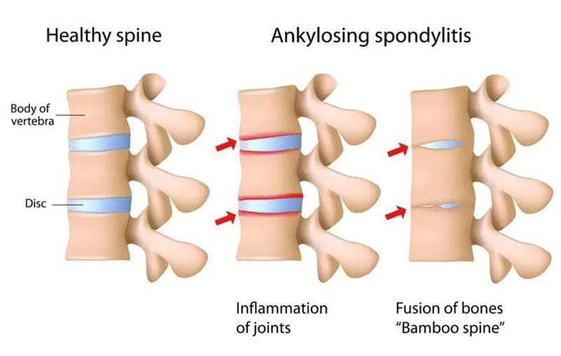
Spondylitis Anti-Inflammation Diet: Individuals who have a chronic back pain condition can be recommended to have two or more vertebrae fused to correct the problem/s and alleviate the pain. However, a form of inflammatory spinal arthritis can cause the vertebrae to fuse by themselves, known as ankylosing spondylitis. One recommended way to bring pain relief is by eating an anti-inflammatory diet. Studies have shown that a low-inflammatory diet can help improve spondylitis symptoms.
Contents
Spondylitis Anti-Inflammation Diet
Ankylosing spondylitis is a progressive inflammatory disease that primarily affects the spine; however, individual symptoms vary. Symptoms include stiffness and pain in the neck, hips, low back, and fatigue. There is no definite pattern meaning:
Symptoms can improve.
Symptoms can worsen or flare up.
Symptoms can stop for a period of time.
Women are affected more often than men with no known cause. There is no cure for ankylosing spondylitis, but treatments and self-care can slow down the disease’s progression and help manage symptoms.
Diet and Inflammation
Diet is not the root cause of inflammatory disease, but eating inflammation-causing foods can worsen symptoms. Reducing inflammation can help alleviate pain.
Eliminating foods that cause or increase inflammation is recommended to help the body become stronger and manage symptoms.
Functional medicine practitioners can help guide individuals on maximizing healthy nutrition and using it to reduce pain and symptoms.
If an individual has a genetic predisposition, their diet can be crucial to calm down the symptoms and help turn the autoimmune disease around.
A spondylitis anti-inflammation diet should be rich in vegetables, fruit, whole grains, and omega-3 fatty acids. Evidence shows that a diet low in starches can lead to less ankylosing spondylitis activity. Low-starch can also help limit the presence of Klebsiella pneumoniae, a bacteria that feeds on starch and is a known trigger for the onset and development of ankylosing spondylitis.
Foods To Eat
Leafy greens
These include spinach, kale, Swiss chard, and collard greens containing magnesiumand polyphenols that reduce inflammation.
These can be raw or cooked with garlic and olive oil added to maximize benefits.
Cruciferous vegetables
These contain sulforaphane, anantioxidantthat includes broccoli cauliflower and can be eaten raw or cooked, roasted with olive oil, sauteed, and stir-fried.
Allium Vegetables
These contain sulfuric compounds and quercetin,a flavonoidthat helps reduce inflammation.
These include red and yellow onions, leeks, garlic, and shallots.
They can be eaten raw or cooked in salads, stir-frys, and sandwiches.
Berries
These contain anthocyanin,an antioxidant flavonoid, and other antioxidants and polyphenols that help with inflammation.
These include strawberries, raspberries, blueberries, blackberries and can be eaten raw, in smoothies, in salads, with oatmeal, or mixed in unsweetened yogurt.
Fruits
Certain fruits contain quercetin and polyphenols to help with inflammation.
These include apples, cherries, oranges.
Healthy oils
Contain oleocanthalwhich acts similar to nonsteroidal anti-inflammatory medications and contains various antioxidants.
These include olive oil for low heat cooking and avocado oil for high heat cooking to replace butter and margarine.
It can be served in dressings and drizzled on foods.
Examples include walnuts, almonds, peanuts, pistachios, chia seeds, and ground flaxseeds.
These can be served as snacks, salads, mixed in side dishes, topping, or added to unsweetened yogurt or oatmeal.
Fatty fish
Omega-3 fatty acids help reduce inflammation.
Examples include salmon, cod, rainbow trout, mackerel, and sardines.
These can be baked, sauteed, grilled, mixed into salads, and stir fry.
Avoid These Foods
When making lifestyle adjustments for a spondylitis anti-inflammation diet, focus on reducing or removing processed foods and saturated fats. These include:
Sugars from all sources like soda, sugary drinks, shakes, candy, and desserts.
Trans fats, like those in fried foods like chips and fries.
Individuals may not be symptomatic with certain foods, but that doesn’t mean the foods should be consumed. Gluten, dairy, and eggs can cause potential problems as they compromise the gut and the immune system. These can set back the individual’s healing or remission.
Body Composition
What Happens To The Body When Eating Fruit
Fruit is made up of simple sugar called fructose, providing the body with a carbohydrate energy source. The natural sugar the body gets from a piece of fruit is not the same as processed fructose added to processed products like fructose corn syrup. Processed products are typically filled with empty calories and very little nutrition. When the body has fruit, the liver processes fructose before getting absorbed through the small intestine. Research shows that exposing the gut to more fiber-rich foods like fruit helps the gut achieve an anti-obese condition by increasing the good bacteria and reducing the obese bacteria. Essential nutrients from fruit include:
Folate
Vitamin C
Vitamin B1
The USDA recommends making half of each meal/plate be fruit and vegetables.
References
Harvard Health Publishing. (November 16, 2021) “Foods that Fight Inflammation.” https://www.health.harvard.edu/staying-healthy/foods-that-fight-inflammation
Macfarlane, Tatiana V et al. “Relationship between diet and ankylosing spondylitis: A systematic review.” European journal of rheumatology vol. 5,1 (2018): 45-52. doi:10.5152/eurjrheum.2017.16103
Nielsen, Forrest H. “Magnesium deficiency and increased inflammation: current perspectives.” Journal of inflammation research vol. 11 25-34. January 18 2018, doi:10.2147/JIR.S136742
Rashid T, Wilson C, Ebringer A. The Link between Ankylosing Spondylitis, Crohn’s Disease, Klebsiella, and Starch Consumption. Clin Dev Immunol. 2013;2013:872632. doi: 10.1155/2013/872632.
Sharma, Satya P et al. “Paradoxical Effects of Fruit on Obesity.” Nutrients vol. 8,10 633. 14 Oct. 2016, doi:10.3390/nu8100633
van Buul, Vincent J et al. “Misconceptions about fructose-containing sugars and their role in the obesity epidemic.” Nutrition research reviews vol. 27,1 (2014): 119-30. doi:10.1017/S0954422414000067
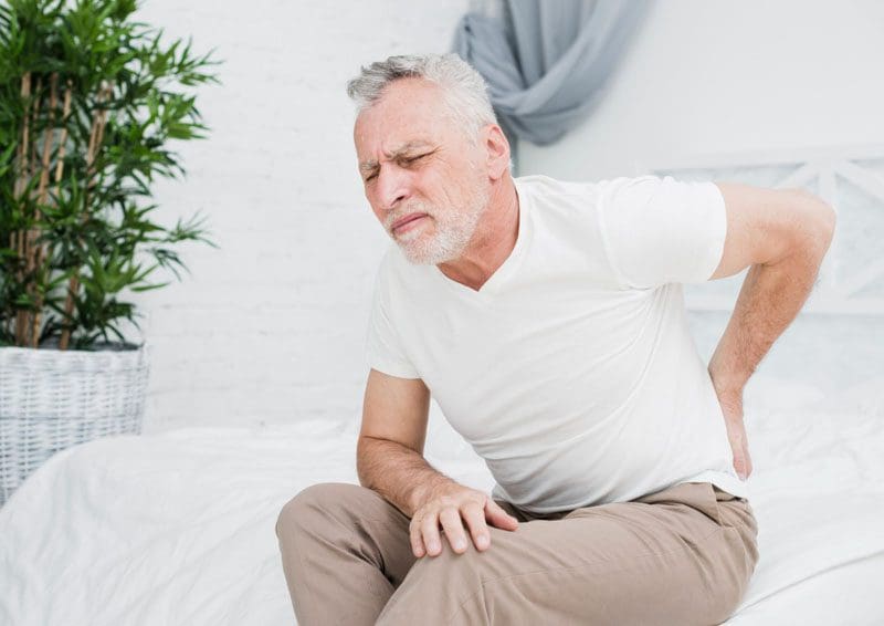
As individual bodies age, the nerves and muscles begin to degenerate, especially in the lower spinal region. This can cause sciatic pain and muscle weakness. Elderly sciatica is very common as the nerves and muscles have gone through a lot. Bending, lifting, carrying, reaching, twisting, and natural wearing and tearing make the sciatic nerve and surrounding muscles prone to injury. For overweight seniors, the risk of developing sciatica is higher.
Contents
Elderly Sciatica
The main reason for elderly sciatica is that as the body ages, the discs/cartilage between the vertebrae/bones in the spine dry out, losing their cushioning ability, which can lead to the bones shifting out of place more easily, rubbing against each other, and compressing nerves. On average, the body loses about 1 centimeter in height every ten years after 40.
Risk Factors
Diabetes
Diabetes affects the entire body.
Not keeping blood sugar in check can cause widespread symptoms that affect the nerves and organs.
Individuals with diabetes have an increased risk of developing sciatica and other nerve-damaging disorders.
Genetics
If spinal conditions are part of family medical history, there is an increased risk of developing sciatica.
With chiropractic, elderly individuals can attain better quality sleep, improved mood, and increased energy levels.
A chiropractic physical therapy team can develop a specialized/customized treatment plan for preventive and palliative care.
Body Composition
Sarcopenia
Sarcopenia affects the elderly population’s mortality, cognitive function, and quality of life. As the elderly population is living longer, preservation of lean mass becomes an integral part of maintaining an individual’s independence. Loss of muscle in the arms and legs is linked to decreased mobility, increased risk of falls, and prolonged hospital stays. Falls and fractures often result in a cycle of muscle deterioration. InBody can help track body composition changes and help to minimize muscle wasting and risk of impaired mobility.
References
Aggarwal, Sameer, and Nityanand. “Calcium and vitamin D in postmenopausal women.” Indian journal of endocrinology and metabolism vol. 17,Suppl 3 (2013): S618-20. doi:10.4103/2230-8210.123549
Dougherty, Paul E et al. “The role of chiropractic care in older adults.” Chiropractic & manual therapies vol. 20,1 3. 21 Feb. 2012, doi:10.1186/2045-709X-20-3
Ferreira, Manuela L, and Andrew McLachlan. “The Challenges of Treating Sciatica Pain in Older Adults.” Drugs & aging vol. 33,11 (2016): 779-785. doi:10.1007/s40266-016-0404-z
Kherad, Mehrsa et al. “Risk factors for low back pain and sciatica in elderly men-the MrOS Sweden study.” Age and aging vol. 46,1 (2017): 64-71. doi:10.1093/ageing/afw152
Anybody can become dehydrated if they don’t take care of themselves and drink plenty of water. Being dehydrated happens when there is insufficient water in the body or increased water loss through sweating, vomiting, and/or diarrhea, along with certain medications, can increase urination and dehydration. Older adults have an increased risk of dehydrating because their body’s fluid reserves decrease, and their body’s ability to signal that they are thirsty does not work as effectively, especially those with memory problems.
Contents
Dehydrated Symptoms
Signs of dehydration include:
Muscle cramps.
Dry mouth
Dry cough.
Tiredness/fatigue.
Flushed red skin.
Swollen feet.
High heart rate but low blood pressure.
Dizziness, weakness, light-headedness.
Headache, delirium, confusion.
Loss of appetite with a sugar craving.
Heat intolerance or chills.
Constipation.
Dark-colored urine. Urine should be a pale clear color.
Dehydration Levels
Dehydration is categorized as:
Mild
The body needs more fluids to be taken in.
Drink water
Drinks containing electrolytes are recommended if experiencing significant sweating or fluid losses from vomiting and diarrhea.
The body should feel better after five or ten minutes.
Bhave, Gautam, and Eric G Neilson. “Volume depletion versus dehydration: how understanding the difference can guide therapy.” American journal of kidney diseases: the official journal of the National Kidney Foundation vol. 58,2 (2011): 302-9. doi:10.1053/j.ajkd.2011.02.395
Centers for Disease Control and Prevention. Drinking-Water. (https://www.cdc.gov/healthywater/drinking/nutrition/index.html)
HealthFirst. What Happens to Your Body When You’re Dehydrated? (https://healthyliving.healthfirst.org/happens-body-youre-dehydrated/)
Kenefick, Robert W, and Michael N Sawka. “Hydration at the worksite.” Journal of the American College of Nutrition vol. 26,5 Suppl (2007): 597S-603S. doi:10.1080/07315724.2007.10719665
Thomas, David R et al. “Understanding clinical dehydration and its treatment.” Journal of the American Medical Directors Association vol. 9,5 (2008): 292-301. doi:10.1016/j.jamda.2008.03.006
Delayed Onset Muscle Soreness – DOMS is when muscle pain or stiffness develops a day or two after playing sports, weight lifting, exercise, or work that involves concentrated physical activity like lifting and carrying objects. DOMS is considered a normal response to extended exertion and is part of the adaptation process that the recovering muscles experience as they undergo hypertrophy or an increase in muscle size. It is common in individuals who have just started exercising, increased the duration or intensity of their workouts, or just beginning a physically demanding job.
Contents
DOMS
When muscle contracts as it lengthens is known as eccentric muscle contractions, which is most associated with DOMS. It is related to increased stress in muscle fibers as they are exerted excessively. This also happens when engaging in movements the muscles are not used to, like a new exercise or helping a friend move heavy boxes, furniture, etc. Examples include:
Individuals will not feel DOMS during the workout or physical activity. Delayed symptoms include:
Swelling in the affected muscles.
Muscles feel tender to the touch.
Muscle fatigue.
Reduced range of motion and movement.
Pain and stiffness when moving.
Decreased muscle strength.
Treatment Options
Time and waiting for the muscles to repair themselves is the natural healing process, but steps can be taken to ease the soreness, stiffness, and pain. This includes:
It is different for everybody; personal experience will determine which works best for the individual.
Active Recovery
Active recovery is a technique that uses low-impact aerobic exercise right after a workout to increase blood flow to the muscles.
The increased blood supply can help relieve the inflammation.
RICE
This technique is used for acute injuries but can be applied to delayed onset muscle soreness. It stands for:
Rest
Ice
Compression
Elevation
Chiropractic
A chiropractic massage is for healing sore muscles, tendons, ligaments after an intense game, workout, etc. Chiropractic increases the blood and nerve circulation around the muscles delivering added oxygen and nutrients. This type of massage helps loosen the muscles/connecting tissues allowing the body to recover and heal quicker.
Body Composition
When Muscles Are Not Rested
Not taking time to recover because of overtraining/working can have consequences on the body. Inflammation that is not given the time to heal can lead to:
Injuries.
Weakened immune system.
Muscle mass loss.
Mental health issues.
The body’s immune system cannot function at total capacity during intense physical stress. This causes difficulty when trying to fight off germs and viruses. Studies have found preventing inflammation and injury requires prioritizing rest. Constantly being on the go and under intense physical stress can take a toll not only on the body but the brain as well. This can lead to irritability, frustration, anger, which leads to other health problems generating a vicious cycle.
References
Cheung, Karoline et al. “Delayed onset muscle soreness: treatment strategies and performance factors.” Sports medicine (Auckland, N.Z.) vol. 33,2 (2003): 145-64. doi:10.2165/00007256-200333020-00005
Guo, Jianmin et al. “Massage Alleviates Delayed Onset Muscle Soreness after Strenuous Exercise: A Systematic Review and Meta-Analysis.” Frontiers in physiology vol. 8 747. 27 Sep. 2017, doi:10.3389/fphys.2017.00747
Reinke, Simon et al. “The influence of recovery and training phases on body composition, peripheral vascular function and immune system of professional soccer players.” PloS one vol. 4,3 (2009): e4910. doi:10.1371/journal.pone.0004910
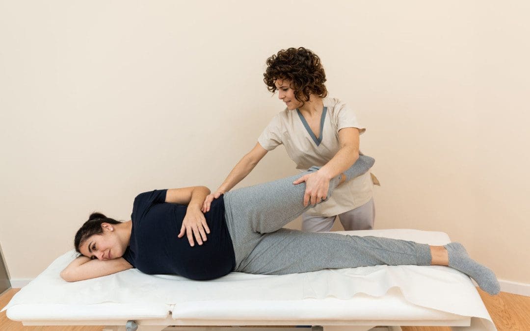
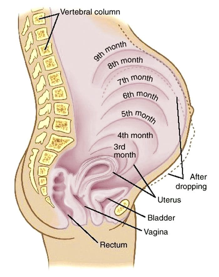
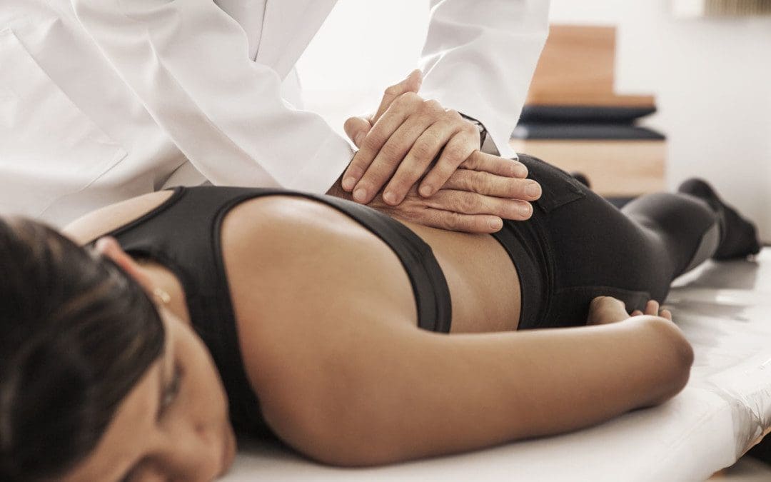
Pregnant and Chiropractic: Many women experience back/pelvis/leg/feet swelling, soreness, achiness, and pain during pregnancy. A growing belly added weight and changes in connective tissue can cause a variety of musculoskeletal strains and misalignments. Chiropractic care provides health maintenance of the spinal column, discs, nerves, joints, muscles, and bones. It is an art and science of adjusting a misaligned body, reducing stress, and promoting health throughout the body.
Contents
Pregnant and Chiropractic
With a primary doctor’s clearance, chiropractic can provide safe adjustments. Chiropractors trained to work with pregnant women utilize techniques that avoid applying pressure on or around the abdomen. Benefits of chiropractic during pregnancy include:
Restores and maintains spinal alignment and balance.
Helps control symptoms of nausea.
Improves energy levels.
Relieves body pain.
Helps reduce labor time and delivery.
Restores pelvic positioning and balance, improving standing, sitting, and walking mechanics.
A chiropractor trained in the needs of pregnant women will also provide exercises and stretches that are safe during pregnancy. A chiropractor will discuss/recommend treatment options, patient concerns, and a complete medical history assessment. They will monitor symptoms to customize treatments to the individual’s specific needs to get the most relief.
Body Composition
Gestational Hypertension
Gestational hypertension develops during pregnancy. It is not preventable and returns to normal levels postpartum. However, there is an increased risk of developing chronic hypertension later if gestational hypertension begins to develop. According to Mayo Clinic, gestational hypertension is diagnosed by the following:
Blood pressure is higher than 140/90 on at least two occasions.
Must be more than four hours apart.
There is no other organ damage present.
References
Gutke, Annelie et al. “Treatments for pregnancy-related lumbopelvic pain: a systematic review of physiotherapy modalities.” Acta Obstetricia et Gynecologica Scandinavica vol. 94,11 (2015): 1156-67. doi:10.1111/aogs.12681
Poděbradská, R et al. “The effect of physiotherapy intervention on the load of the foot and low back pain in pregnancy.” “Vliv fyzioterapeutických postupů na zatížení plosky a bolesti zad v těhotenství.” Ceska gynekologie vol. 84,6 (2019): 450-457.
Schreiner, Lucas et al. “Systematic review of pelvic floor interventions during pregnancy.” International journal of gynecology and obstetrics: the official organ of the International Federation of Gynaecology and Obstetrics vol. 143,1 (2018): 10-18. doi:10.1002/ijgo.12513
Everybody is different in how the body reacts to a chiropractic adjustment. Body misalignment often leads to spinal misalignment or vice versa. Misalignments occur over time; individuals do not notice until soreness and pain begin presenting. Depending on the injury and/or condition, getting the full potential from a chiropractic adjustment means knowing the dos and don’ts following treatment. This involves maintaining a healthy posture, staying hydrated, getting proper rest, and staying active.
Contents
Adjustments
Adjustments are highly effective for the body. Benefits include:
Pain relief.
Restored full range of motion.
Increased strength.
Increased energy.
Improved sleep.
Lowered blood pressure in individuals with hypertension.
It is not recommended to take on intense workouts after an adjustment but to remain active to keep the muscles, tendons, ligaments flexible and strengthen the body during healing.
Activities should be done in moderation and include:
Walking
Jogging
Biking
Swimming
Proper Rest
Getting the proper amount of sleep is essential for the body to heal to the optimal level.
The body getting used to the adjustment can be an exhausting process.
Maintain Healthy Posture
Proper posture is essential to keep the body in healthy alignment and prevent further/new injuries.
A chiropractor and physical therapist will educate and train individuals on maintaining healthy, active postures.
Stretching
Stretching is prescribed as part of the treatment to maintain flexibility and strength.
A chiropractor will recommend and show how to perform specific stretches and exercises between adjustments.
What to Avoid
Recommendations on what to avoid after a chiropractic adjustment.
Explosive Movements
Stay active but limit any explosive movements for a few days after the adjustment.
Avoid Sitting Too Much
Too much sitting, even with a lumbar support chair, can cause the muscles to tighten pulling on the spine.
When sitting, get up and move around every 20 minutes.
Paying attention to the recommended do’s and don’ts will help expedite the healing and create new healthy habits.
Body Composition
Dairy Products
Conventional vs. Organic and Grass-fed Dairy
Studies have found that dairy cows consuming a diet of grass and hay significantly improved nutrient profiles of produced milk.
Milk from grass-fed cows has a higher omega-3 content when compared to organic and conventional grain-fed cows.
Bourrie, Benjamin C T et al. “The Microbiota and Health Promoting Characteristics of the Fermented Beverage Kefir.” Frontiers in microbiology vol. 7 647. 4 May. 2016, doi:10.3389/fmicb.2016.00647
Licciardone, John C et al. “Recovery From Chronic Low Back Pain After Osteopathic Manipulative Treatment: A Randomized Controlled Trial.” The Journal of the American Osteopathic Association vol. 116,3 (2016): 144-55. doi:10.7556/jaoa.2016.031
Maher, C G. “Effective physical treatment for chronic low back pain.” The Orthopedic clinics of North America vol. 35,1 (2004): 57-64. doi:10.1016/S0030-5898(03)00088-9
Will, Joshua Scott et al. “Mechanical Low Back Pain.” American family physician vol. 98,7 (2018): 421-428.
Volleyball is a dynamic game that requires players to be fast on their feet. Players have to be able to quickly shift into various position/s, make quick movements in any direction quickly and reach the ball. Volleyball strength workouts focus on power development and maintaining safe positions when exploding through the plays. Many players include resistance training exercises in their training programs to maximize power and set a solid foundation.
Contents
Volleyball Strength Workout
A well-rounded volleyball workout will help players strengthen and maintain optimal body health.
A recommended exercise that can be done with a resistance band.
The exercise does not require a lot of space, so it can be done almost anywhere.
It is recommended to do two-three sets of 10-15 reps.
It is recommended to consult a professional trainer that can create a diverse fitness program to make exercising/training/working out much more enjoyable.
Body Composition
How Aerobic and Resistance Training Interact
The body adjusts differently to various types of exercise. Aerobic and resistance training each tells the body to adapt in different ways. Both are important for healthy body composition, and when done in combination, it is known as concurrent training. Aerobic is best for losing fat, resistance training builds muscle that keeps the body functioning throughout the day. However, molecular mechanisms involved in aerobic and resistance adaptations can interfere with each other if not appropriately planned. Two steps to minimize any possible interference and maximize aerobic/resistance benefits:
Nutrition
Adequate protein intake is vital for muscular adaptation from resistance training.
It stimulates muscle protein synthesis after concurrent training.
After workout sessions, consume at least 25g of high-quality protein to achieve strength and hypertrophy improvements.
Recovery
When doing both aerobic and resistance training on the same day, maximize recovery time between the sessions.
Strength and aerobic fitness gains are low when the two are separated by 6 hours or less.
Twenty-four hours between sessions is the recommended time, especially if the priority is on endurance.
References
Camera, Donny M et al. “Protein ingestion increases myofibrillar protein synthesis after concurrent exercise.” Medicine and science in sports and exercise vol. 47,1 (2015): 82-91. doi:10.1249/MSS.0000000000000390
Cools, Ann M et al. “Prevention of shoulder injuries in overhead athletes: a science-based approach.” Brazilian journal of physical therapy vol. 19,5 (2015): 331-9. doi:10.1590/bjpt-rbf.2014.0109
Pereira, Ana et al. “Training strategy of explosive strength in young female volleyball players.” Medicina (Kaunas, Lithuania) vol. 51,2 (2015): 126-31. doi:10.1016/j.medici.2015.03.004
Ramirez-Campillo, Rodrigo et al. “Effects of Plyometric Jump Training on Vertical Jump Height of Volleyball Players: A Systematic Review with Meta-Analysis of Randomized-Controlled Trial.” Journal of sports science & medicine vol. 19,3 489-499. 13 Aug. 2020
Seminati, Elena, and Alberto Enrico Minetti. “Overuse in volleyball training/practice: A review on the shoulder and spine-related injuries.” European journal of sports science vol. 13,6 (2013): 732-43. doi:10.1080/17461391.2013.773090
Silva, Ana Filipa et al. “The Effect of Plyometric Training in Volleyball Players: A Systematic Review.” International journal of environmental research and public health vol. 16,16 2960. 17 Aug. 2019, doi:10.3390/ijerph16162960
Villareal, Dennis T et al. “Aerobic or Resistance Exercise, or Both, in Dieting Obese Older Adults.” The New England journal of medicine vol. 376,20 (2017): 1943-1955. doi:10.1056/NEJMoa1616338
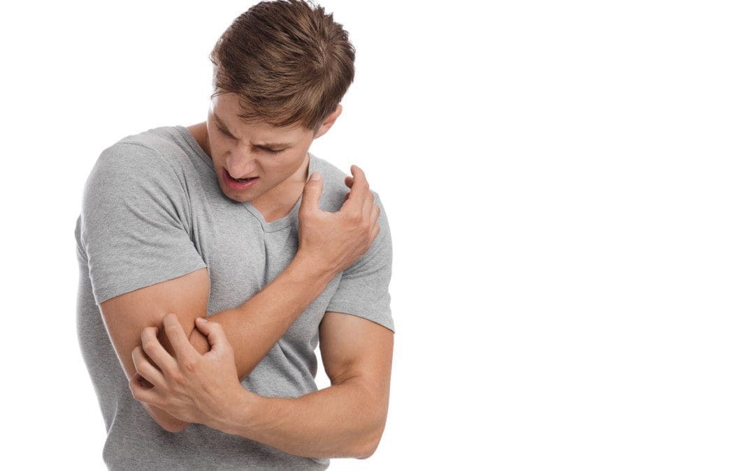
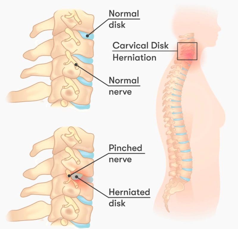
A pinched nerve may not feel like it is healing. This is because of the soreness, aches, discomfort, and tingling feelings/sensations around the affected area. This could be the neck, shoulder, arm, hands, back, legs, and feet. However, when the achiness and tingling move around and shift, it is a sign of the pinched nerve healing.
Contents
Amount of Time For Pinched Nerve Healing
Waiting for the nerve to heal is not a recommended treatment option, as most pinched nerves do not fully recover on their own. A pinched nerve usually takes around six weeks to heal with proper treatment. The longer the nerve stays pinched, the more likely there will be permanent damage. To keep the pinched nerve from returning and getting worse, individuals are recommended to incorporate a pre-habilitation plan that involves continuing rehabilitation exercises to strengthen and keep the muscles, ligaments, and nerves loose, and adjusting posture, work, exercise, and diet habits to prevent re-injuring the nerve or cause new injury/s.
Common Nerve Sites
Nerves run throughout the body, so it’s possible to experience a pinched nerve anywhere. The most common pinched nerve sites occur at joints where there is constant movement. These areas include:
Neck
Shoulders
Lower Back
Arms
Hands
Feet
Healing Signs
Individuals often believe that their pinched nerve is getting worse because of soreness, aches and pains, and weird sensations. When the pain stays in one area, that could be a sign that the nerve has not been fully stretched/released and/or that there is still compression taking place. Treatment and healing include feeling the symptoms but in a different way. The symptoms will move up, down, or around depending on where the pinched nerve is. Treatment takes the nerve/s and stretches/elongates them, but the pinch created a nerve crimp, crease, fold that wants to return to the pinched position. This is why continued treatment and stretching are recommended, as a spasm, trauma, or some awkward movement can cause the nerve to re-fold to the pinched position or cause a whole new pinch.
Chiropractic Release
Chiropractic treats pinched/compressed nerves with several therapeutic modalities. These include:
Body Adjustments
Flexion-distraction
Therapeutic massage
Traction
Inversion
Laser therapy
Ultrasound
Combined, these methods can help heal pinched nerves and keep them from recurring.
Body Composition
Skeletal Muscle
Skeletal muscle is a major muscle group. These muscles are attached to the bone by the tendons. Skeletal muscles incorporate nerves, blood vessels, and connective tissue to operate as a unit. Each skeletal muscle consists of cells that come together that form bundles of skeletal muscle fibers.
Strength training stimulates the muscle fibers. When combined with proper nutrition causes hypertrophy/muscle growth.
Muscles contract and shorten to pull bones and joints, allowing body movement.
The nervous system signals the nerves in the muscle/s and triggers these contractions.
Skeletal muscle helps the body:
Maintain posture
Generate body heat
Stability to the bones and joints
References
Bowley, Michael P, and Christopher T Doughty. “Entrapment Neuropathies of the Lower Extremity.” The Medical clinics of North America vol. 103,2 (2019): 371-382. doi:10.1016/j.mcna.2018.10.013
Campbell, W. “Diagnosis and management of common compression and entrapment neuropathies.” Neurologic clinics vol. 15,3 (1997): 549-67. doi:10.1016/s0733-8619(05)70333-9
England, J D. “Entrapment neuropathies.” Current opinion in neurology vol. 12,5 (1999): 597-602. doi:10.1097/00019052-199910000-00014
Kane, Patrick M et al. “Double Crush Syndrome.” The Journal of the American Academy of Orthopaedic Surgeons vol. 23,9 (2015): 558-62. doi:10.5435/JAAOS-D-14-00176
Healthy sleep plays a vital role in the body’s overall health, as it ensures muscle growth, recovery, and illness prevention. This is especially true for home D.I. Yers’ fitness enthusiasts, weekend warriors, athletes, and physically active individuals. When sleeping, the body goes into recovery mode, releasing hormones and other chemicals to repair and restore muscle. A healthy night’s sleep provides the rest the mind and body need to perform at optimal levels.
Contents
Healthy Sleep
Sleep is vital for recovering from workouts. This could be construction work, exercise, gardening, sports, landscaping, any activity that uses bodyweight or works against some form of resistance. The muscles cannot repair themselves properly without proper sleep. Sleep aids the muscles in releasing protein-building amino acids, helping them grow in size and strength.
Growth hormone is released during non-REM sleep that stimulates tissue growth and repairs muscle.
During REM or rapid eye movement sleep, blood pressure drops, breathing slows and deepens, the brain relaxes, and blood supply to the muscles increases, feeding them oxygen and nutrients.
Unhealthy Sleep
Sleep maintains the muscles’ sharpness, coordination, function, and muscle movement patterns that improve physical performance. The body needs to sleep for at least 7 hours a night for muscles to grow properly. Not getting healthy sleep decreases protein synthesis activity and increases the activity of degradation that leads to muscle loss.
Less Sleep Leads To Eating More
Hormonal changes occur when the body sleeps less, causing individuals to feel hungry more often, increasing the amount of food taken in because after eating, the body does not feel full right away, so the individual continues to eat. Without sleep, the body decreases the production of a hormone that indicates when the body is full and activates a hormone that causes hunger. Insufficient sleep also lowers the body’s sensitivity to insulin. Because of this, the muscle fuel glycogen is not adequately replenished. Without the regular restoration of glycogen, individuals have less energy, insulin sensitivity decreases, increasing the risk of diabetes.
Physical Health
Unhealthy sleep also impacts overall physical health. Individuals that do not get healthy sleep have an increased risk of developing:
Sugar raises blood sugar, which triggers the pancreas to release insulin, fueling the cells causing overstimulation.
Eliminating sugar after dinner can help the body fall asleep.
References
Dattilo, M et al. “Sleep and muscle recovery: endocrinological and molecular basis for a new and promising hypothesis.” Medical hypotheses vol. 77,2 (2011): 220-2. doi:10.1016/j.mehy.2011.04.017
Morselli, Lisa et al. “Role of sleep duration in the regulation of glucose metabolism and appetite.” Best practice & research. Clinical endocrinology & metabolism vol. 24,5 (2010): 687-702. doi:10.1016/j.beem.2010.07.005
Murray, Bob, and Christine Rosenbloom. “Fundamentals of glycogen metabolism for coaches and athletes.” Nutrition reviews vol. 76,4 (2018): 243-259. doi:10.1093/nutrit/nuy001
Automobile accidents and crashes can cause all kinds of damage to the body even when the accident/crash is not severe. Physical symptoms might not present at all for several days, even weeks. This is known as having delayed injury symptoms. These can include:
Swelling.
Stiffness.
Aching.
Pain that radiates all over the body.
Sleep problems.
Headaches.
Brain fog.
Disorientation.
Memory problems.
Chiropractic and physical therapy rehabilitation can restore the body’s alignment, stop inflammation, loosen, stretch and strengthen the musculoskeletal system restoring optimal health.
Contents
Adrenaline
When the body is involved in a dangerous physical situation, it protects itself by releasing a surge of adrenaline. This hormone protects the body, causing the fight or flight response when in danger. Adrenaline causes several preservation responses that include:
Intense increase in energy.
Little or no pain.
Enlarged blood vessels and airways increase oxygen flow.
Increased strength from increased blood flow to the muscles.
Changes in vision and hearing that focus on sights and sounds all around.
Endorphins are released that make the body feel calm and in control.
Endorphins affect the way the body responds to pain and stress.
Individuals don’t start feeling aches and pains until the adrenaline and endorphins wear off. However, because everybody is different and the emergency response has turned off, the body still might not feel the injury symptoms. These are delayed injury symptoms.
Rate of Speed
When riding in a vehicle, the body moves at the same speed as the vehicle. During an impact, the vehicle stops, but the body continues moving until it stops, typically with a lot of force from the seatbelt, airbag, or other barriers. The intense momentum change can cause soft tissue damage and ligament or muscle strains from the stretching, pulling, contracting, and tearing. Also, the intervertebral discs can tear, bulge, or herniate over time, creating pressure on nerves and the surrounding tissues.
Delayed Injury Symptoms
Headaches
Headaches that develop days after an accident/crash are common.
They can signal a possible injury to the neck or head, a blood clot on the brain, or a concussion.
Back pain can be caused by injury to the muscles, ligaments, nerves, or damage to the vertebrae.
Low back pain occurs in more than half of rear-impact collisions and almost three-quarters of side-impact crashes.
Chiropractic Rehabilitation
After an accident, soft tissues can sustain minimal damage; however, the minimal damage left untreated can start to worsen and turn into a painful condition. Emergency room visits are to rule out major injuries like brain/nerve injuries, bleeding, punctures, lacerated organs, fractures that require emergency stabilization. Chiropractors look for other symptoms and mechanisms that indicate damage to the body’s soft tissues and nerves to see if they have been stretched or torn and dysfunction in the nervous system.
Body Composition
Calorie Counting
Counting calories can be a stepping stone to change behavior towards food. Tracking what foods are being taken into the body promotes mindfulness of dietary habits. Studies on the subject reveal a significant association between self-monitoring and weight loss. Takeaways include:
Take small steps by saying no to second portions during dinner or take a healthy sweet snack or piece of fruit instead of a pastry, cookie, etc.
Try to start making a habit of eating less processed foods.
Meals high in protein and fiber are generally more filling, making the body feel fuller from fewer calories.
The more attention there is to the food choices, the more likely reexamination occurs.
References
Burke, Lora E et al. “Self-monitoring in weight loss: a systematic review of the literature.” Journal of the American Dietetic Association vol. 111,1 (2011): 92-102. doi:10.1016/j.jada.2010.10.008
D’Elia, Michael A et al. “Motor vehicle collision with seatbelt sign and traumatic abdominal wall hernia should raise suspicion for hollow viscus injury.” Trauma case reports vol. 22 100206. 25 May. 2019, doi:10.1016/j.tcr.2019.100206
Kacprzynski, Gregory, and Joshua Bucher. “Delayed vertebral artery dissection after mild trauma in a motor vehicle collision.” The American Journal of emergency medicine vol. 45 (2021): 678.e1-678.e2. doi:10.1016/j.ajem.2020.11.028
Olinger, Catherine, and Richard Bransford. “Upper Cervical Trauma.” The Orthopedic clinics of North America vol. 52,4 (2021): 451-479. doi:10.1016/j.ocl.2021.05.013
Sterling, Michele. “Whiplash-associated disorder: musculoskeletal pain and related clinical findings.” The Journal of manual & manipulative therapy vol. 19,4 (2011): 194-200. doi:10.1179/106698111X13129729551949
IFM's Find A Practitioner tool is the largest referral network in Functional Medicine, created to help patients locate Functional Medicine practitioners anywhere in the world. IFM Certified Practitioners are listed first in the search results, given their extensive education in Functional Medicine