Can the Oswestry Low Back Pain Disability Questionnaire help assess how low back pain impacts individuals’ ability to perform everyday tasks and activities and help physical therapists incorporate the outcome measure into an effective treatment plan?
Oswestry Disability Questionnaire
The Oswestry Disability Questionnaire, also known as the Oswestry Disability Index, provides objective data about an individual’s lower back pain. It determines the severity of the pain and how much it limits their daily activities. The questionnaire is a validated measure backed by research that can be used to justify the need for medical treatment. It includes questions regarding the symptoms and severity of low back pain and how these symptoms interfere with regular activities. Lower back pain can result from various causes (National Institute of Neurological Disorders and Stroke, 2020)
Arthritis, including inflammatory types of arthritis like psoriatic arthritis and ankylosing spondylitis.
Lumbar vertebrae compression fractures – usually from trauma or osteoporosis.
Low back surgery – including spinal fusions, discectomies, and laminectomies.
Spinal stenosis
Spondylolisthesis
Scoliosis
How The Questionnaire Works
The Oswestry Disability Questionnaire consists of 10 questions about the impact of lower back pain on daily life. The questions are divided into the following categories: (American Academy of Orthopedic Surgeons, N.D.)
Pain Intensity
How intense is the pain?
If painkillers are used, how much symptom relief do they provide?
Personal Care
Can the patient perform self-care activities like bathing and dressing when experiencing significant pain or limitations?
Whether physical assistance from another person is needed?
Lifting
Can the patient lift objects like weights with or without pain?
Can lifting be performed from the floor or a higher surface like a table if the objects are light, moderate, or heavy?
Walking
If and to what extent does the pain limit the patient’s walking distance and independence?
If an assistive device like a cane or crutches are needed?
Sitting
If so, how much pain limits the patient’s sitting tolerance?
Standing
If so, how much pain limits the patient’s standing tolerance?
Sleeping
If so, how much pain limits a patient’s sleeping duration?
Whether pain medication is needed to help the patient sleep comfortably?
Social Life
If and to what extent a patient’s social activities are limited because of pain symptoms?
Traveling
If so, to what extent does pain limit a patient’s ability to travel?
Employment and/or Homemaking Duties
Does pain limit a patient’s ability to perform job-related and/or household activities, including physically demanding and light duties?
Patients self-report the information and complete it on their own based on their understanding of the extent of their lower back pain and disability.
Each question can be scored between 0 and 5, with 0 indicating no limitations and 5 indicating complete disability.
The scores from all the questions are added together for a cumulative total score of 50 points.
Scores
The Oswestry Disability Questionnaire assesses how much a patient’s lower back pain limits daily activities. This information is used in clinical documentation for medical services. A higher score indicates a greater level of disability, according to the following scoring criteria:
0–4: No disability
5–14: Mild disability
15–24: Moderate disability
25–34: Severe disability
35–50: Completely disabled
Physical therapists must create individualized goals for each patient to develop a treatment plan and receive authorization from insurance companies. One of the most important aspects of a physical therapy goal is that it must be measurable. The Oswestry Disability Questionnaire provides a numerical score to track functional limitations and monitor the range of motion and strength testing. A baseline measurement is taken at the beginning of treatment, and progress is tracked in follow-up visits. A new score is used as a treatment goal. According to a study, the minimal clinically important difference (MCID) for the Oswestry Disability Questionnaire is 12.88. The MCID is the minimum score healthcare providers need to confirm a patient’s progress in function due to treatment. (Johnsen, L. G. et al., 2013)
By tracking changes in the total score before, during, and after treatment, healthcare providers can better assess whether treatment improves symptoms. A decrease in total score by 13 points or more would indicate that treatment is helping to improve a patient’s lower back pain and level of disability. Along with physical examination results, the patient’s score and the severity of symptoms can help healthcare providers determine an appropriate treatment plan.
No Disability
Treatment is unnecessary other than providing advice for lifting mechanics and general physical activity to maintain health.
Mild Disability
Conservative measures, such as physical therapy, exercise, hot or cold therapy, pain medication, and rest, are needed to help alleviate symptoms.
Moderate Disability
More aggressive intervention is needed, which can include extensive physical therapy services and pain management.
Severe Disability
Significant medical intervention is needed, including surgery, pain management, equipment like wheelchairs, and help from a caretaker.
Completely Disabled
Patients are either bedbound or have worsening symptoms, and a caretaker is needed to complete daily activities and self-care tasks.
Injury Medical Chiropractic and Functional Medicine Clinic
Improvements in range of motion, strength, and quality of movement and a decrease in total score can help show the treatment’s positive impact in managing lower back pain. A thorough medical exam and diagnostic tests, such as X-ray, MRI, or EMG, can help determine the underlying causes, discover the cause of the problem, and develop an effective treatment plan. Injury Medical Chiropractic and Functional Medicine Clinic works with primary healthcare providers and specialists to develop personalized treatment programs. Using an integrated approach to treating injuries and chronic pain syndromes to improve flexibility, mobility, and agility and help individuals return to normal activities. Our providers use Functional Medicine, Acupuncture, Electro-Acupuncture, and Sports Medicine principles. If other treatments are needed, Dr. Jimenez has teamed up with top surgeons, clinical specialists, medical researchers, and rehabilitation providers.
Optimizing Your Wellness
References
National Institute of Neurological Disorders and Stroke. (2020). Low Back Pain Fact Sheet. Retrieved from https://www.ninds.nih.gov/sites/default/files/migrate-documents/low_back_pain_20-ns-5161_march_2020_508c.pdf
American Academy of Orthopedic Surgeons. (N.D.). Oswestry Low Back Pain Disability Questionnaire. https://www.aaos.org/globalassets/quality-and-practice-resources/patient-reported-outcome-measures/spine/oswestry-2.pdf
Johnsen, L. G., Hellum, C., Nygaard, O. P., Storheim, K., Brox, J. I., Rossvoll, I., Leivseth, G., & Grotle, M. (2013). Comparison of the SF6D, the EQ5D, and the oswestry disability index in patients with chronic low back pain and degenerative disc disease. BMC musculoskeletal disorders, 14, 148. https://doi.org/10.1186/1471-2474-14-148
Back pain is one of the most common reasons for seeking health care. Individuals dealing with back pain but don’t know the cause may have some inflammatory joint disease or autoimmune condition. Can seeing a rheumatologist help?
Rheumatologist
Depending on what’s causing the back pain, individuals may need to see their primary doctor for a referral. Individuals are recommended to see a rheumatologist if they have back pain that doesn’t come from an injury that doesn’t go away after a few weeks, pain that comes back after treatment, or symptoms that suggest a rheumatic condition. Rheumatologists treat severe or persistent back pain and are experts in autoimmune diseases, including lupus, Sjogren’s syndrome, rheumatoid arthritis, ankylosing spondylitis, axial spondylitis, Psoriatic arthritis, and other forms of inflammatory or autoimmune arthritis.
What Do They Do?
A rheumatologist is an internist or pediatrician who has completed special training in treating conditions that are:
Inflammatory
Autoimmune
Related to painful joint disease
The doctors diagnose, treat, and manage these conditions long-term. Depending on diagnosis and care needs, they may also lead or be part of a team that includes other healthcare providers.
Symptoms
When muscles ache, pain presents, or joints hurt, and especially if there are signs of inflammation that don’t go away, seeing a healthcare provider is recommended. Symptoms of inflammation include:
Redness
Swelling
Pain
Stiffness
Loss of joint function
Usually, to see a rheumatologist, individuals need a referral from their primary care provider and may be referred when:
There is no evidence of a back injury.
At-home therapies like heat application, prescription medications, or physical therapy are unsuccessful.
There is uncertainty about what’s causing the back pain, but I suspect it’s rheumatological.
Blood tests for inflammatory markers or certain antibodies yield abnormal results.
There is a diagnosis of a rheumatic condition and recommend a specialist to manage it.
There is a family history of a rheumatic or autoimmune condition that may cause back pain.
Some types of arthritis can cause permanent, progressive joint damage.
Conditions
Conditions that can affect the spine and cause back pain and are treated by a rheumatologist include: (Johns Hopkins Medicine, 2024)
Rheumatoid arthritis (RA)
This often starts in smaller joints of the hands and feet and later moves to the neck and/or back.
It can also affect different body organs and have systemic symptoms.
Ankylosing Spondylitis (AS)
Primarily a disease of the spine, it may also impact the shoulders, hips, knees, and ankles.
Systemic symptoms, including fever and fatigue, can manifest.
Axial Spondylitis
This primarily affects the spine, chest, and pelvis.
It may also cause problems with the connective tissue, eyes, bowel, and skin.
Psoriatic Arthritis (PsA)
Pain in the lower back is common, especially in severe cases.
It can affect other joints and cause psoriasis.
Reactive Arthritis
This is a reaction to infection.
It is more common in the limbs, hands, and feet joints but can involve the spine.
Enteropathic Arthritis
This mainly affects the spine but can include other joints.
It is associated with inflammatory bowel disease.
Autoimmune diseases that don’t specifically target the spine but can also cause back pain include:
Lupus
Sjögren’s syndrome
Hashimoto’s thyroiditis
Finding a Doctor
Individuals may be fine with their primary healthcare provider’s choice regarding which rheumatologist to see. However, they may want to research other options to ensure the right rheumatologist is chosen. Things to look at include:
Search online medical directories.
Visit the websites of the doctors being considered to learn more about their training, approach, and specialties.
Check online reviews.
Check on health insurance coverage.
Ask members of the healthcare team, friends, and family for recommendations.
Contact rheumatologists’ offices to see if they are accepting new patients.
Once decided, pass along the information to the primary care doctor so they can make the referral.
Preparing For The Initial Visit
Before seeing a new rheumatologist, take a few minutes to prepare so you can make the most of the appointment. Individuals will want to have:
A list of back-related symptoms, including frequency and severity.
A list of what makes symptoms better or worse.
A copy of recent test results and records from other doctors.
Individuals can ask their provider/s to send their medical information to the rheumatologist’s office in advance.
A list of treatments that have been tried and how well they worked.
A list of all medications, over-the-counter and prescription, supplements, and herbal products taken.
A list of medication allergies.
Complete medical history and family history of potentially related diseases.
A list of any questions regarding conditions, treatment, etc.
If possible, fill out any paperwork for the new office beforehand to save time on the appointment day.
Injury Medical Chiropractic and Functional Medicine Clinic
Talking with a healthcare provider is important. Injury Medical Chiropractic and Functional Medicine Clinic works with primary healthcare providers and specialists to develop personalized treatment programs. Using an integrated approach to treating injuries and chronic pain syndromes to improve flexibility, mobility, and agility and help individuals return to normal activities. If other treatments are needed, Dr. Jimenez has teamed up with top surgeons, clinical specialists, medical researchers, and rehabilitation providers.
Quick Patient Initiation Process
References
Hospital for Special Surgery. (2023). What Is a Rheumatologist and What Conditions Do They Treat? https://www.hss.edu/conditions_what-is-a-rheumatologist.asp#when
Yale University School of Medicine. Dee, J. E. (2021). 5 reasons why a patient should see a rheumatologist. https://medicine.yale.edu/news-article/5-reasons-to-see-a-rheumatologist/
National Institute of Arthritis and Musculoskeletal and Skin Diseases. (2023). Autoimmune diseases. Retrieved from https://www.niams.nih.gov/health-topics/autoimmune-diseases
Johns Hopkins Medicine. (2024). Spinal arthritis (arthritis in the back or neck). https://www.hopkinsmedicine.org/health/conditions-and-diseases/spinal-arthritis
It can be challenging for individuals trying to keep their homes clean with chronic back pain. Can learning and proper body mechanics help manage household responsibilities without aggravating pain symptoms?
Household Chores
Household-related back problems usually occur because we don’t take the time to consider how to move and perform the tasks from a musculoskeletal perspective to avoid and prevent injuries. Most ergonomic tips for household chores revolve around the same ideas for athletes and fitness enthusiasts: maintain a neutral spine, avoid twisting when possible, strengthen the body’s core, take regular breaks, stretch, and don’t overdo it. A healthy body mechanics system works for those who garden as well. Using strategies like cleaning a little here and there instead of taking an entire day whenever possible and organizing tools ahead of time along with training oneself how to perform them in a way that the spine, back muscles, and the entire body are protected from injury, pain, sciatica, or re-injury. However, implementing proper body mechanics requires a willingness to become aware of how each task is performed and to retrain the body where necessary to a healthier method/technique and a happier household.
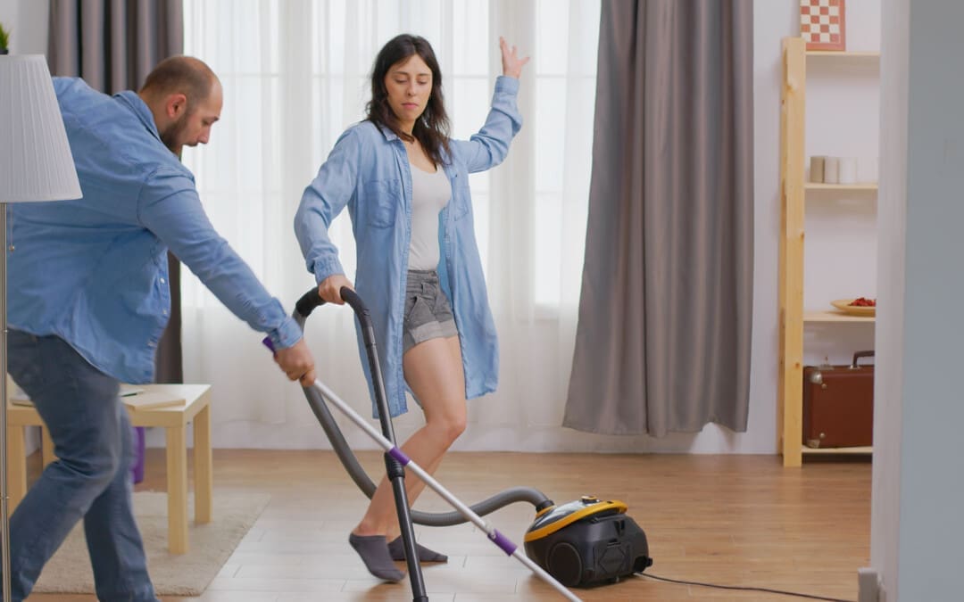
Vacuuming
Vacuuming is one of those chores that can quickly lead to a habitual bent-over posture. This is not recommended for the spine’s health; slouching, whether from a position held for a sustained period of time or an activity that requires repetition, can lead to problems with the intervertebral discs and pain symptoms. (Nazari J., Pope M. H., and Graveling R. A. 2012) Another posture that individuals tend to engage in is vacuuming with an overly straight back. Like slouching, keeping the spine rigidly over-extended while vacuuming can irritate the spine and cause muscle spasms. It can also increase the normal low back curve, which, in turn, may lead to extra tightness and a painful back.
Vacuuming with healthy body mechanics includes employing a minimal lunge that stays in a pain-free position that does not extend beyond the comfortable position. Individuals should place one foot in front of the other for a short distance. The stance is similar to the way fencers position themselves. This allows a shift forward and back during the vacuuming process instead of bending or rounding over at the spine. For those with sacroiliac joint issues, the forward placement of one leg may be more comfortable than the forward placement of the other. Try out and use the side that feels comfortable, and stick with that. Do not work in pain or through the pain. Switching legs and/or arms can help avoid muscle fatigue or injury triggers. Place the non-vacuuming hand on the thigh in front to help take the weight and pressure off the back. Maintain the pelvis in a level position when working. Another strategy for those who can get up and down from the floor without trouble is to vacuum while kneeling on one knee. This brings the body’s center of mass closer to the floor, reducing the degree to which the body has to deal with the force of gravity. Kneeling while vacuuming may also help prevent rounding over at the spine.
Dusting
When dusting, reduce the load off the back by propping the inactive arm on the item or area being cleaned. Alternatively, prop the arm on the thigh.
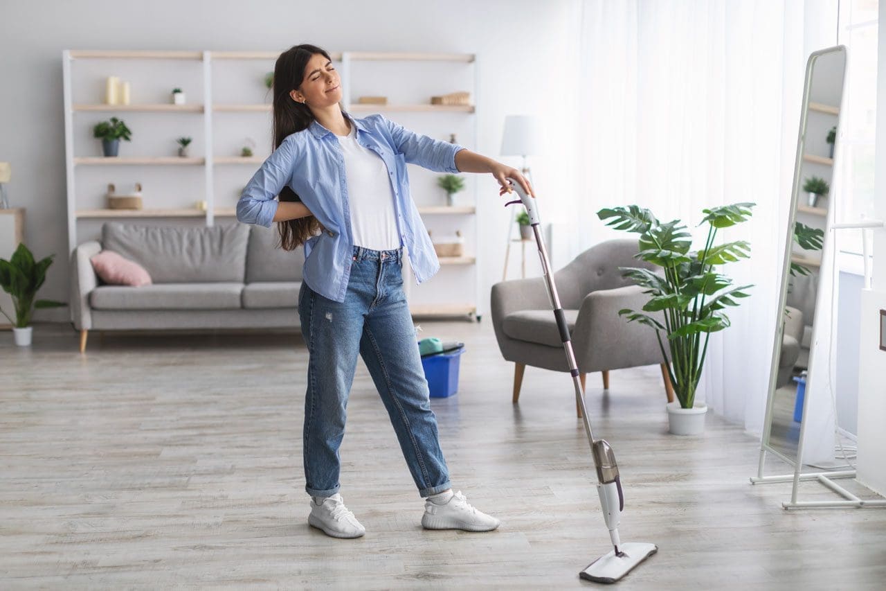
Laundry
In a large household, it is very easy to overdo laundry and trying to finish up as much as possible can lead to pain symptoms and injuries. If possible, break up the loads that have to be lifted or carried into smaller bundles that weigh less. This can mean more loads, but the strategy protects the back and spine. Avoid extremes in the spinal position; don’t round over at the spine or keep it rigid and over-extended. Lift with the legs and protect the discs. Adjustments that can be made to the basic lift with the leg and not the back strategy include putting the laundry basket on a table or chair that is preferably the same height as the washer or dryer. This will minimize bending. To relieve pressure on the back, use one hand to load the washer, dryer, or laundry basket while using one of the appliances to prop the other hand.
Dishes
During dishwashing, use a small step stool or box to help prevent injury and/or relieve pain. Place it in the cabinet under the sink and rest one foot on it. This strategy can work well for those with sacroiliac joint problems, especially if the foot on the pain-free side is the one placed on the box or stool. Ensuring foot placement reduces pain and discomfort and does not cause pain. Using a box or stool can also help with core stability. Core stability is one of the best ways, in general, to prevent injury and keep low back pain away. (Coulombe B. J., Games K. E., Neil E. R., and Eberman L. E. 2017) As the box is directly under the sink, the body has to firmly position itself against the counter, providing stability during the task. The box or stool will contract the pelvic and hip muscles and strengthen the core.
Sweeping
Many sweep, mop, and rake with their spines, which can be counterproductive to health, as twisting and bending simultaneously is a known risk factor for a herniated disc. (Shimia, M. et al., 2013) Use the arms and legs instead of overly involving the back during sweeping and raking. The idea is to reach and pull the broom or sweeper with the arms rather than twisting around to reach all the areas working with one leg in front of the other. When needing to change directions, pivot on the back leg, keeping the trunk relaxed, equivalent to a tai chi movement. Or, turn the whole body in different directions by taking small steps. Taking small steps or pivoting on the back leg to change the direction of the trunk and arms protects from overuse and extensive wear and tear.
Adjustments for a new method of household sweeping and raking include:
Sweeping or raking about 1 to 2 feet in front to avoid overreaching and strain.
Maintaining the spine in one long, flexible, but unbroken line.
The head, shoulders, rib cage, pelvis, knees, and feet should always face the same direction and be vertically balanced relative to one another.
This will mean changing directions by pivoting the back leg or moving the whole body around, taking small steps.
Consider using an ergonomically designed broom, sweeper, mop, rake, and other household tools. This will be a bend in the handle or stem to help avoid bending.
Injury Chiropractic and Functional Medicine Clinic
Injury Medical Chiropractic and Functional Medicine Clinic works with primary healthcare providers and specialists to develop personalized treatment programs. We focus on what works for you and use an integrated approach to treating injuries and chronic pain syndromes to improve flexibility, mobility, and agility, relieve pain, and help individuals return to normal activities. Our providers use Functional Medicine, Acupuncture, Electro-Acupuncture, and Sports Medicine principles. Dr. Jimenez has teamed up with top surgeons, clinical specialists, medical researchers, and rehabilitation providers if other treatments are needed.
Heel Spurs
References
Nazari, J., Pope, M. H., & Graveling, R. A. (2012). Reality about migration of the nucleus pulposus within the intervertebral disc with changing postures. Clinical biomechanics (Bristol, Avon), 27(3), 213–217. https://doi.org/10.1016/j.clinbiomech.2011.09.011
Coulombe, B. J., Games, K. E., Neil, E. R., & Eberman, L. E. (2017). Core Stability Exercise Versus General Exercise for Chronic Low Back Pain. Journal of athletic training, 52(1), 71–72. https://doi.org/10.4085/1062-6050-51.11.16
Shimia, M., Babaei-Ghazani, A., Sadat, B. E., Habibi, B., & Habibzadeh, A. (2013). Risk factors of recurrent lumbar disk herniation. Asian journal of neurosurgery, 8(2), 93–96. https://doi.org/10.4103/1793-5482.116384
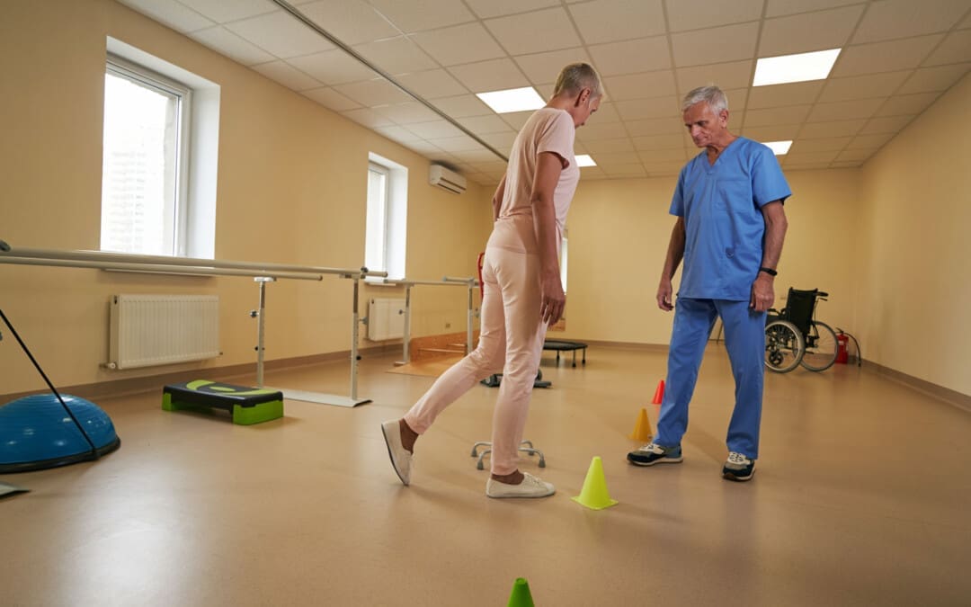
Individuals walking with a limp that results in pain could have an antalgic gait, an abnormal walking pattern commonly seen in emergency clinics and primary care offices. Can recognizing the symptoms help healthcare providers develop an effective treatment for the underlying cause?
Antalgic Gait
Limping and having an antalgic gait usually indicate a larger issue within the leg or lower back. It is the most common type of abnormal gait. There are various causes of antalgic gait, including acute injuries and gradually progressing medical conditions. The most common causes include osteoarthritis in one of the leg’s joints, lumbar radiculopathy, or an injury to a ligament or tendon. Paying attention to when the limping occurs, and any accompanying symptoms can help determine its origins.
Walking
When limping, the stance phase during walking is shorter than the swing phase. Individuals may widen their legs apart to provide a support base to compensate for the imbalance. In severe cases, an individual may swing their leg irregularly or take several side steps.
Causes and Symptoms
Antalgic gait can be caused by pain in any part of the lower extremity. Limping when walking may be a primary concern but is rarely the only complaint. Other associated symptoms may also be present, depending on the cause. These include:
Limited range of motion
Joint stiffness
Muscular weakness
Numbness and tingling
Pain
Swelling
Leg instability or buckling
Clicking or popping
Common Causes include:
Hip, knee, and/or Foot problems
When the hip, knee, ankle, or foot joints are injured or have some issue, walking can be painful and lead to a limp.
Sprains, Strains, or Soft-tissue Injuries
Sprains, strains, and soft-tissue injuries can result from acute injury or chronic, repetitive activities over time. (Pirker W. and Katzenschlager R. 2017) Sprains affect the body’s ligaments, while strains impact muscle tendons. However, sprains and strains occur when the impacted structure is overstretched or partially torn. The damage can lead to pain and antalgic gait. An injury to several other soft-tissue structures, including a bursa or fluid-filled sacs that reduce friction, meniscus, or fat pad, can also lead to limping. Symptoms typically include swelling, pain, and limited range of motion. More severe injuries can also make the leg feel unstable and cause it to give way when walking. Sometimes, bruising can also occur in the area of the injury. (American Academy of Orthopaedic Surgeons, 2020)
Osteoarthritis
Osteoarthritis occurs when the smooth, articular cartilage that lines the ends of bones begins to thin and deteriorate. This can alter the normal movement of a joint and lead to pain. Osteoarthritis symptoms gradually progress, affect individuals over 50, and worsen after periods of sedentary activity (Arthritis Foundation, Osteoarthritis, N.D.) Typically, it results in pain, stiffness, clicking, and occasionally swelling in the affected joint. These symptoms are usually worse in the morning and at the end of a long activity day. Moving around and warming the joint improves osteoarthritis symptoms. (Arthritis Foundation, Osteoarthritis, N.D.)
Lower Back Radiculopathy
Lumbar radiculopathy is when the nerve roots branching off the spine’s lower region become compressed or inflamed. This can occur because of disc issues like bulging, degeneration, herniation, bone spurring, or, rarely, a growth or tumor. (Johns Hopkins Medicine, 2024) Because these nerves control movement, sensation, and strength in the legs and feet, irritation in one can lead to limping. (Yokogawa N. et al., 2015) The antalgic gait from this condition frequently comes on suddenly and is commonly accompanied by back pain symptoms. This can include shooting pain and paresthesia in the leg. Depending on which nerve is involved, individuals may also experience muscular weakness in certain areas of the lower extremity. Sometimes, the affected leg feels like it will buckle while standing or walking. (Johns Hopkins Medicine, 2024)
Other causes include:
Broken bones
Tumors
Infections
Blood clots
Vascular issues
Treatment
Treatment for antalgic gait depends on the underlying cause but can include:
Rest, ice, and elevation are important for injuries. Individuals can control their initial symptoms by icing, elevating the leg, and resting from irritating activities.
Activity modifications
Antibiotics for infections
Pain relievers
Anti-inflammatories
Physical therapy is also frequently initiated to strengthen the core and alleviate walking symptoms.
A spinal injection or surgery can reduce the pressure on the nerve root if conservative interventions fail to improve antalgic gait patterns. (Johns Hopkins Medicine, 2024)
Crutches, canes, walkers, or assistive devices can reduce pressure traveling through an affected joint and improve overall walking quality. A study found that using a cane for two months helped reduce pain and improve function in individuals with knee osteoarthritis. (Fang M. A. et al., 2015)
Injury Medical Chiropractic and Functional Medicine Clinic
Though it can be tempting to ignore the limp and push through it, discussing the condition with a healthcare provider is important. A thorough medical exam and diagnostic tests, such as X-ray, MRI, or EMG, can help determine the underlying causes of a limp, help discover the cause of the problem, and help improve the quality of walking. Injury Medical Chiropractic and Functional Medicine Clinic works with primary healthcare providers and specialists to develop personalized treatment programs. Using an integrated approach to treating injuries and chronic pain syndromes to improve flexibility, mobility, and agility and help individuals return to normal activities. Dr. Jimenez has teamed up with top surgeons, clinical specialists, medical researchers, and rehabilitation providers if other treatments are needed.
Chiropractic and Integrative Healthcare
References
Pirker, W., & Katzenschlager, R. (2017). Gait disorders in adults and the elderly : A clinical guide. Wiener klinische Wochenschrift, 129(3-4), 81–95. https://doi.org/10.1007/s00508-016-1096-4
American Academy of Orthopaedic Surgeons. (2020). Sprains, strains, and other soft-tissue injuries. https://orthoinfo.aaos.org/en/diseases–conditions/sprains-strains-and-other-soft-tissue-injuries/
Yokogawa, N., Toribatake, Y., Murakami, H., Hayashi, H., Yoneyama, T., Watanabe, T., & Tsuchiya, H. (2015). Differences in Gait Characteristics of Patients with Lumbar Spinal Canal Stenosis (L4 Radiculopathy) and Those with Osteoarthritis of the Hip. PloS one, 10(4), e0124745. https://doi.org/10.1371/journal.pone.0124745
Fang, M. A., Heiney, C., Yentes, J. M., Harada, N. D., Masih, S., & Perell-Gerson, K. L. (2015). Effects of contralateral versus ipsilateral cane use on gait in people with knee osteoarthritis. PM & R : the journal of injury, function, and rehabilitation, 7(4), 400–406. https://doi.org/10.1016/j.pmrj.2014.09.018
Can eating white rice as a primary carbohydrate source provide quick energy and glycogen replenishment for athletes and bodybuilders who require high volumes of carbohydrates for fuel and muscle recovery?
Sports Nutrition White Rice
Athletes and bodybuilders often eat white rice to restore mass amounts of glycogen after an intense workout, race, or game. Brown rice is nutritious and recommended for overall health, but athletes and bodybuilders incorporate further nutritional guidelines for added fuel and performance. White rice is a starchy grain used by more than half the world’s population because of its versatility, availability, and ability to adapt to various flavors and seasonings. Its chewiness and soft texture add substance to and complement many meals.
Bodybuilders
Athletes and bodybuilders need macronutrients to fuel extreme training and replenish depleted glycogen stores. Carbohydrate-rich foods like white rice are high on the glycemic index (a score for how foods affect blood sugar and insulin levels) and provide a readily available source of carbohydrates for muscle glycogen synthesis. (Thomas, D. E. et al., 1991) White rice is considered excellent sports nutrition for athletes. (Melin, A. et al., 2016) Although white rice is considered less nutritious than brown rice, athletes and weightlifters consume it as part of their specifically developed nutrition plans. One of the most popular meals is a bowl of white rice combined with grilled chicken breast, providing lean protein. Endurance runners often load up on carbohydrates like white rice before marathons. Strenuous workouts deplete sugar/glycogen in the muscles. Eating the right carbohydrates is important to replenish those stores. Knowing how to improve carbohydrate availability during prolonged exercise is essential for athletes. (Burke, L. M. et al., 2011)
Fuel and Muscle Recovery
The high glycemic value of white rice provides quick fuel for hard workouts and expedites muscle recovery. It does not have the negative effects of potential gastrointestinal issues, allergy symptoms, or blocking the ability to absorb micronutrients. Brown rice contains phytic acid(located in the grain’s bran), an antinutrient that binds to essential minerals like iron, zinc, calcium, and magnesium and prevents the body from absorbing them. The milling process to change brown rice to white removes the phytate. Research is ongoing on degrading phytic acid in brown rice and whole grains, and some studies have found antioxidant benefits in phytate. This could increase the amount of carbohydrates that are safe for athletes. (Liang, J. et al., 2008)
Safe Carbohydrate
Athletes and bodybuilders with food sensitivity may have issues eating whole grains, as brown rice has more fiber. Extreme exercise requires a surplus of carbohydrates. For prolonged exercise lasting more than two hours, athletes should consume 60 grams per hour of carbohydrates. White rice is considered safe to consume before exercise as it is easy on the stomach and has been shown to meet sports nutrition recommendations.
Workouts
Rice contains more carbohydrates than potatoes for the same serving size. Parboiled, converted, and instant white rice can be eaten pre- and post-workout meals, ensuring the body is thoroughly fueled for training, competition, and recovery. However, white rice may not be the best option for sedentary individuals.
Brown Rice
Brown rice is healthier for bodybuilders who train less than four days per week or have a metabolic disease.
Brown rice is a nutrient-dense food recommended for everyday active individuals who can tolerate whole grains and is a rich source of fiber and nutrients essential for a well-balanced healthy diet.
Injury Medical Chiropractic and Functional Medicine Clinic
Injury Medical Chiropractic and Functional Medicine Clinic works with primary healthcare providers and specialists to develop personalized programs. We focus on what works for you to enhance fitness and improve the body through research methods and total wellness programs. These programs strive to achieve improvement goals, and athletes can condition themselves to excel through proper fitness and nutrition. Our providers use Functional Medicine, Acupuncture, Electro-Acupuncture, and Sports Medicine principles. An integrated approach improves flexibility, mobility, and agility. If further training or treatments are needed, Dr. Jimenez has teamed up with top surgeons, clinical specialists, trainers, medical researchers, and rehabilitation providers.
Sports Nutrition and Dietitian
References
Thomas, D. E., Brotherhood, J. R., & Brand, J. C. (1991). Carbohydrate feeding before exercise: effect of glycemic index. International journal of sports medicine, 12(2), 180–186. https://doi.org/10.1055/s-2007-1024664
Melin, A., Tornberg, Å. B., Skouby, S., Møller, S. S., Faber, J., Sundgot-Borgen, J., & Sjödin, A. (2016). Low-energy density and high fiber intake are dietary concerns in female endurance athletes. Scandinavian journal of medicine & science in sports, 26(9), 1060–1071. https://doi.org/10.1111/sms.12516
Burke, L. M., Hawley, J. A., Wong, S. H., & Jeukendrup, A. E. (2011). Carbohydrates for training and competition. Journal of sports sciences, 29 Suppl 1, S17–S27. https://doi.org/10.1080/02640414.2011.585473
Liang, J., Han, B. Z., Nout, M. J., & Hamer, R. J. (2008). Effects of soaking, germination and fermentation on phytic acid, total and in vitro soluble zinc in brown rice. Food chemistry, 110(4), 821–828. https://doi.org/10.1016/j.foodchem.2008.02.064
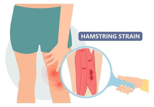
Individuals dealing with symptoms like sudden pain, weakness, and tenderness in the back of the knee could have a hamstring injury. Can knowing the symptoms and performing self-care help bring relief?
Hamstring Pain Behind The Knee
The hamstrings consist of three long muscles that run down the back of the thigh, cross over the back of the knee, and connect to bones in that area. A hamstring injury, such as a strain or tear, tendonitis, or biceps femoris tendinopathy, can cause pain in the back of the knee, difficulty bending the knee, swelling, and bruising. A hamstring strain occurs when the muscle is stretched too far or torn completely. This can happen from sudden, forceful movements or overstretching. Hamstring tendonitis develops over time, usually after a sudden increase in activity, when the hamstring tissue cannot recover from too much loading. Pain is often felt after physical activity and exercise and, in severe cases, during the activity or throughout the day. Biceps femoris tendinopathy can also cause pain in the back of the knee. Strains, tendonitis, bursitis, and muscle tears are all possible explanations for a hamstring injury that leads to pain behind the knee. Discussing pain symptoms with a healthcare provider is recommended, especially if it occurs suddenly during physical activity or exercise. They can help identify the exact cause and offer guidance for rehabilitation, including physical therapy referrals.
Causes and Triggers
Individuals may experience hamstring pain behind the knee when the muscles in that area are overworked, inflamed, or injured, such as from activities like running, walking, dancing, soccer, or basketball. Possible types of injuries and their causes.
A primary cause of muscle strain occurs when the muscle is stretched too far or has to handle a sudden force like sprinting or kicking. (American Academy of Orthopaedic Surgeons, 2021)
Severe Cases
Most causes of pain behind the knee are easily treatable at home with self-care and rest. However, it can be more severe, signaling a blood clot, infection, torn muscle or tendon/ligament. Hamstring knee pain may be serious if any of the following is experienced (American Academy of Orthopaedic Surgeons, 2021)
Sudden pain during physical activity, often during a full stride.
Feeling a pop or sharp pain that causes falling or limping.
Pain that worsens over time and prevents or hinders walking or exercising as normal.
If pain is severe and does not improve with rest and anti-inflammatory medications, evaluation by a healthcare professional is necessary.
Assesses Hamstring Pain
A healthcare provider will ask about symptoms and injury, including what happened when the pain began. They will perform a physical examination, which may include pressing on the back of the thigh to look for swelling, bruising, tenderness, or bunched-up muscles. (American Academy of Orthopaedic Surgeons, 2021) The healthcare provider will ask the patient to perform specific resisted movements, such as the manual muscle test, and measure the range of motion. Diagnostic testing includes an X-ray or MRI to determine the degree of the injury and which soft tissues or bones may be involved.
Self-Care
The first line of treating hamstring knee pain is the RICE protocol, which includes: (Mount Siani, 2024)
Rest
Stop any activity that causes symptoms and pain.
A healthcare provider may recommend crutches or a knee scooter in severe cases.
Ice
Apply cold packs to the swollen or painful area for 20 minutes throughout the day.
Compression
A knee brace, wrap, or bandage that applies gentle pressure to the injured area can help reduce and prevent swelling.
Elevation
Lifting the leg higher than the heart will help reduce swelling and blood accumulation.
Individuals may need to lie on a bed or sofa and elevate their legs with pillows.
Individuals can use at-home pain relievers like acetaminophen or NSAIDs like ibuprofen or naproxen. Over time, and depending on the severity of the injury, a healthcare provider will advise on gentle hamstring stretches and how to ease back into physical activity.
A healthcare provider will advise immobilizing the knee to help with muscle healing, which could involve wearing a knee brace or using crutches.
Physical therapy
A healthcare provider may refer the patient to a physical therapist, who will perform a personalized evaluation and prescribe targeted exercises to heal the injury and regain strength, flexibility, and movement.
Surgery
Tendon avulsion injuries are when the hamstring tendon completely tears away from the bone, and surgery is required to reattach the tendon.
Platelet-rich plasma – PRP
Platelet-rich plasma has become an additional treatment for hamstring muscle strain or tendonitis. (Seow D. et al., 2021)
The treatment involves injecting a solution from the patient’s blood into the muscle to heal the injury.
Recovery
Predicting how long a hamstring injury takes to heal and how long the pain will linger depends on the type, location, and severity. The most severe type is the hamstring coming unattached around the knee. This surgical repair and rehabilitation take at least three months before returning to sports and exercise (American Academy of Orthopaedic Surgeons, 2021). Lesser injuries like tendonitis or a mild strain can take less time to heal. However, it’s essential to avoid reinjuring the area so the condition does not become chronic. This includes: (American Academy of Orthopaedic Surgeons, 2021)
Stretching to encourage and maintain flexibility.
Fixing muscle imbalances between the quadriceps and hamstring.
Endurance and conditioning.
Avoiding overuse.
Injury Medical Chiropractic and Functional Medicine Clinic works with primary healthcare providers and specialists to develop personalized treatment programs. We focus on what works for you and use an integrated approach to treating injuries and chronic pain syndromes to improve flexibility, mobility, and agility, relieving pain and helping individuals return to normal activities. If other treatments are needed, Dr. Jimenez has teamed up with top surgeons, clinical specialists, medical researchers, and rehabilitation providers. Our providers use Functional Medicine, Acupuncture, Electro-Acupuncture, and Sports Medicine principles.
Chiropractic Care for Leg Instability
References
National Library of Medicine. (2017). Tendinitis Also called: Tendonitis. Retrieved from https://medlineplus.gov/tendinitis.html
American Academy of Orthopaedic Surgeons. OrthoInfo. (2020). Sprains, strains, and other soft tissue injuries. https://orthoinfo.aaos.org/en/diseases–conditions/sprains-strains-and-other-soft-tissue-injuries/
American Academy of Orthopaedic Surgeons. OrthoInfo. (2021). Hamstring muscle injuries. https://orthoinfo.aaos.org/en/diseases–conditions/hamstring-muscle-injuries/
American Academy of Orthopaedic Surgeons. OrthoInfo. (2021). Pes aserine (knee tendon) bursitis. https://orthoinfo.aaos.org/en/diseases–conditions/pes-anserine-knee-tendon-bursitis/
Mount Siani. (2024). Hamstring strain – aftercare. https://www.mountsinai.org/health-library/selfcare-instructions/hamstring-strain-aftercare
Seow, D., Shimozono, Y., Tengku Yusof, T. N. B., Yasui, Y., Massey, A., & Kennedy, J. G. (2021). Platelet-Rich Plasma Injection for the Treatment of Hamstring Injuries: A Systematic Review and Meta-analysis With Best-Worst Case Analysis. The American journal of sports medicine, 49(2), 529–537. https://doi.org/10.1177/0363546520916729
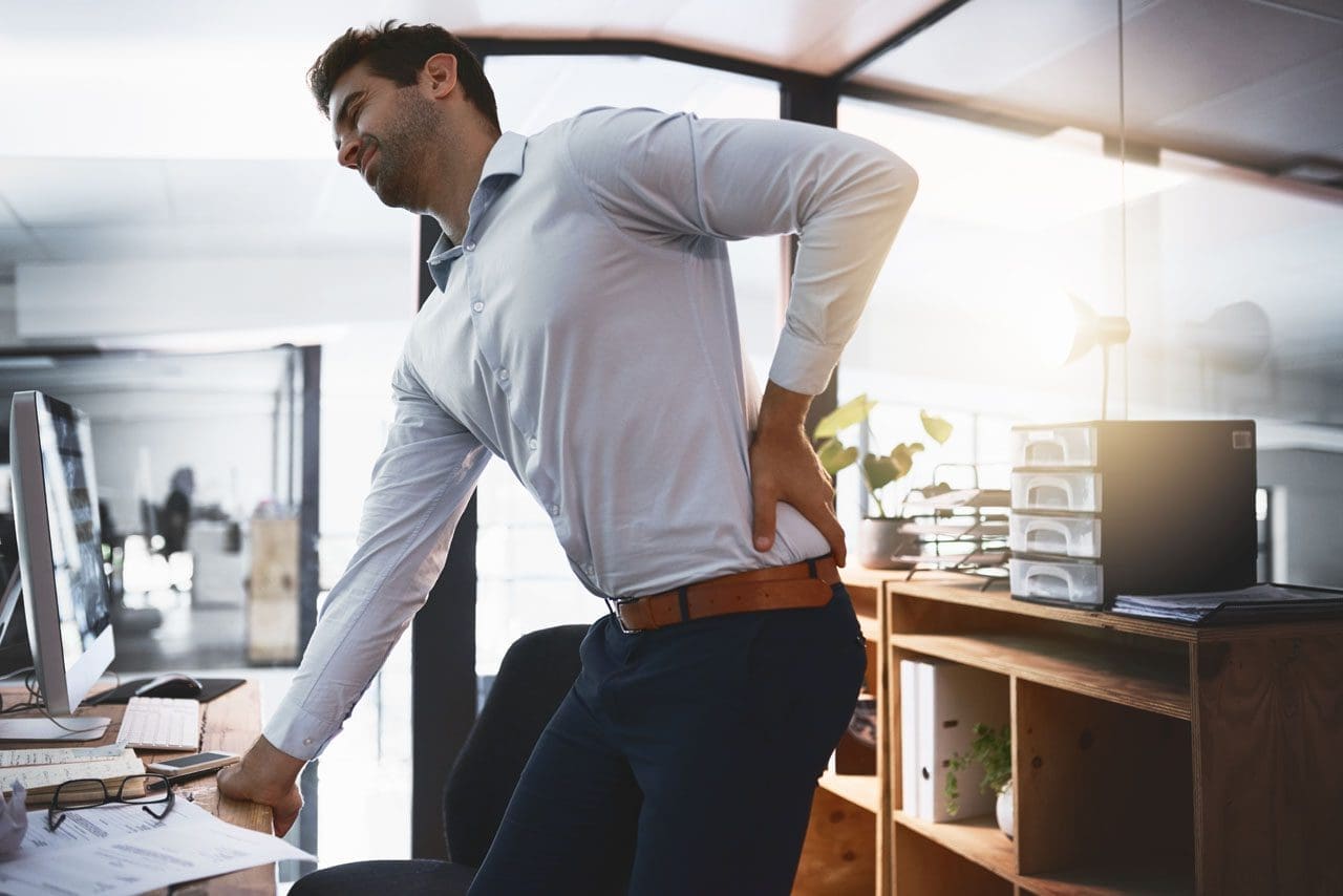
Individuals who sit in an office chair for a long period naturally tend to slouch over or slouch down into the chair. This posture can overstretch the spinal ligaments, strain the discs and surrounding structures in the spine, and contribute to or worsen back pain. Can the right office chair settings help relieve and prevent future injuries?
Work Office Chair
Sitting in an office chair for prolonged periods can cause lower back pain or worsen an existing back problem. The main reason behind this is that sitting is a static posture that increases stress in the back, shoulders, arms, and legs, putting pressure on the back muscles and spinal discs. Over time, incorrect sitting posture can damage the spinal structures. Most office chair customers base their decisions on availability, color, style, and price. However, it’s recommended that individuals learn about fitting seating equipment to their height, frame, or any condition, such as back or hip pain. The controls on or under the armrests and at the bottom of the chair seat, including the levers, paddles, and knobs, are there for a reason. Here are some recommendations for adjusting their work office chair to decrease back and hip pain.
Height Adjustment
The chair’s height affects the quadriceps, psoas, and hamstring muscles, which are important in posture-related back pain. Height adjustment is the primary way to change the angle of the hip joint while sitting. This angle affects the position of the pelvis and the degree of curve in the lower back, which can alter the spine’s normal alignment. (De Carvalho D. et al., 2017) Adjusting the chair’s height can provide a reference for other chair and workstation adjustments.
Back Angle
The hip angle is how close (increased hip flexion) or how far away (less hip flexion) the trunk is to the top of the thigh when sitting. Adjusting chair height can control the angle of the hip joint. When adjusting the height, adjust the degree of flexion at the hip joint to ensure ideal alignment for the individual body frame. A recent study measured the load that sitting has on simulated spinal discs. Researchers concluded that pressure on the spine can be relieved with a more open angle between the trunk and the thigh, that is, the hip joint angle. (Rohlmann A. et al., 2011) The backrest, seat tilt, and lumbar support features help maintain a pain-free back and relieve pressure and tension in the lower back and should be utilized.
Seat Depth
A kitchen chair, for example, may have no back supports or armrests to assist with posture and angle. (Holzgreve F. et al., 2022) When sitting, the individual’s back should be against the back of the work office chair for the best support and a healthy posture. Individuals will want to check and see how concave the seat surface is. If there is a prominent curve, this will cause the back to be rounded when sitting, which can become uncomfortable and lead to back pain. A chair with built-in support or a pillow can help adjust the depth. This means that chair size matters, as different people will need seats of varying depths to match their musculoskeletal structure.
Seat Height
Another way to understand the hip joint angle is to compare the height of the knees to the height of the hips. This is usually the easiest way to assess whether the chair height is right while adjusting. When the seat is right, the feet will be flat on the floor. The feet should reach the floor without causing pressure on the back of the thighs. Individuals with dangling feet, which may be because of their height, should place a footrest or thick book under them. The knees should be approximately level with or lower than the hips. In this case, the level is a 90-degree angle between the hip and trunk, which is stress-free on the hips and back.
Risks
Chair Too High
For individuals who can’t reach their feet to the floor, the chair is probably too high. The Occupational Safety and Health Administration (OSHA) says this is potentially hazardous because it can lead to scooting forward and forgoing the backrest’s support (United States Department of Labor, N.D.) Sitting like this is considered an awkward posture and a risk factor for work-related musculoskeletal disorders (MSD). Musculoskeletal disorders and symptoms caused by awkward sitting posture include muscle fatigue, swelling, pain, numbness, or decreased circulation. (Ng, P. K., Jee, K. S. and Lim, S. Y. 2016)
Chair Too Low
If the knees are higher than the hips, the chair is probably too low, causing extreme flexion in the hip joints. Most individuals’ backs can’t handle this well because their hip muscles are not flexible enough. If sitting with knees higher than hips, the position can cause lower back pain.
Taking Breaks
No matter how comfortable a work office chair is, prolonged static posture is unhealthy for the spine and is a common contributor to back problems and muscle strain. Remember to stand, stretch, and walk for at least a minute or two every half hour to prevent the back from staying in one position for a long period. A quick stretch or minimal movement, like a quick walk, will help. A longer walk will help even more, promoting blood circulation to supply nutrients to all the spinal structures.
Moving and stretching regularly throughout the day will help keep the joints, ligaments, muscles, and tendons loose and promote comfort, relaxation, and the ability to focus productively.
Injury Medical Chiropractic and Functional Medicine Clinic works with primary healthcare providers and specialists to develop personalized treatment programs. An integrated approach to treating injuries and chronic pain syndromes improves flexibility, mobility, and agility, relieving pain and helping individuals return to normal activities. If other treatments are needed, Dr. Jimenez has teamed up with top surgeons, clinical specialists, medical researchers, and rehabilitation providers.
Low Back Pain: Impact and Chiropractic Solutions
References
De Carvalho, D., Grondin, D., & Callaghan, J. (2017). The impact of office chair features on lumbar lordosis, intervertebral joint and sacral tilt angles: a radiographic assessment. Ergonomics, 60(10), 1393–1404. https://doi.org/10.1080/00140139.2016.1265670
Rohlmann, A., Zander, T., Graichen, F., Dreischarf, M., & Bergmann, G. (2011). Measured loads on a vertebral body replacement during sitting. The spine journal : official journal of the North American Spine Society, 11(9), 870–875. https://doi.org/10.1016/j.spinee.2011.06.017
Holzgreve, F., Maurer-Grubinger, C., Fraeulin, L., Bausch, J., Groneberg, D. A., & Ohlendorf, D. (2022). Home office versus ergonomic workstation – is the ergonomic risk increased when working at the dining table? An inertial motion capture based pilot study. BMC musculoskeletal disorders, 23(1), 745. https://doi.org/10.1186/s12891-022-05704-z
United States Department of Labor. (N.D.). Computer workstations eTool. Retrieved from https://www.osha.gov/etools/computer-workstations
Ng, P. K., Jee, K. S. & Lim, S. Y. (2016). Development of Ergonomics Guidelines for Improved Sitting Postures in the Classroom among Malaysian University Students. American Journal of Applied Sciences, 13(8), 907-912. https://doi.org/10.3844/ajassp.2016.907.912
IFM's Find A Practitioner tool is the largest referral network in Functional Medicine, created to help patients locate Functional Medicine practitioners anywhere in the world. IFM Certified Practitioners are listed first in the search results, given their extensive education in Functional Medicine