Chiropractic and Regenerative Joint Pain Care: An Evidence-Based Approach
Abstract
Welcome to our educational series. I’m Dr. Alexander Jimenez, and today, we’ll explore the sophisticated world of regenerative medicine, specifically focusing on platelet-rich plasma (PRP) and its applications in managing joint pain, particularly osteoarthritis. This post translates complex clinical research into practical insights, exploring patient selection, treatment protocols, and the crucial role of integrative care. We will discuss the science behind PRP, including platelet concentration, the debate between leukocyte-rich versus leukocyte-poor preparations, and how these factors influence patient outcomes. Furthermore, we’ll examine how we integrate these advanced biologic treatments with our foundational chiropractic and physical therapy principles to create a comprehensive, patient-centered journey toward healing and restored function. We’ll navigate the nuances of post-injection care, the timing of treatments relative to other interventions, such as cortisone shots, and the latest evidence-based strategies to optimize results. Our goal is to empower you with a clear understanding of how these modern therapies work synergistically with established musculoskeletal care, not just to alleviate pain but to foster true, long-term healing.
Hello, I am Dr. Alexander Jimenez, DC, APRN, FNP-BC, CFMP, IFMCP, ATN, CCST. It’s a privilege to share insights from the forefront of musculoskeletal and regenerative medicine. In my practice, we are dedicated to merging the latest scientific advancements with a holistic, patient-first philosophy. Today, I want to guide you through a fascinating and rapidly evolving area: the use of platelet-rich plasma (PRP) for joint conditions like osteoarthritis. We will explore how we, as integrative practitioners, make clinical decisions based on cutting-edge research and how these decisions fit within a broader chiropractic and physical therapy framework to optimize your recovery.
Optimizing Patient Selection for Regenerative Therapies: Who is the Ideal Candidate?
A common and critical question I encounter is, “Am I a suitable candidate for PRP?” Patients often wonder if there are strict cutoffs based on age, body mass index (BMI), or the severity of their arthritis.
Based on the latest evidence and my clinical experience, the answer is more nuanced than simple metrics. While there can be a subtle bias against higher BMI, the primary predictor of a successful response to PRP is the nature of the patient’s symptoms, not their demographic profile.
Ideal Candidates: Patients who describe their pain as a broad, achy, and inflammatory sensation tend to respond remarkably well. This type of pain often signals an underlying inflammatory process that PRP is uniquely equipped to modulate. In these cases, age and the degree of arthritis seen on an X-ray are less critical factors. We have seen patients in their nineties achieve significant relief.
Less Predictable Candidates: Conversely, individuals who experience sharp, stabbing, or mechanical pressure-type pain often have a more complex clinical picture. This type of pain can indicate issues beyond simple inflammation, such as bone marrow lesions, significant meniscal tears, or other “pain generators” that create mechanical blocks or instability. While these patients may still benefit from PRP, our treatment algorithm must be expanded to address concurrent issues, often through targeted physical therapy and chiropractic adjustments.
It is a matter of managing expectations. For a patient with severe arthritis who is exploring alternatives to knee replacement, we might discuss the potential for a 30-60% improvement over several months. I am always transparent: no treatment is 100% effective. Our approach is to create a personalized, evidence-informed plan that maximizes your chances of success.
The Science of PRP: Leukocyte-Rich vs. Leukocyte-Poor
The conversation around PRP often involves technical terms like leukocyte-rich (LR-PRP) and leukocyte-poor (LP-PRP). Leukocytes are white blood cells, and their presence in the PRP injectate is a subject of significant debate.
A preparation is generally considered “leukocyte-rich” if the concentration of white blood cells exceeds that in the patient’s baseline whole blood. Most commercial PRP systems produce a leukocyte-rich product. The key distinction lies in the types of leukocytes present. The current focus in research is on reducing pro-inflammatory neutrophils while preserving monocytes, which are crucial for tissue remodeling and healing.
Leukocyte-Poor (LP-PRP): This is often preferred for injections near sensitive structures, such as nerves or the spine, where minimizing the initial inflammatory response is paramount.
Leukocyte-Rich (LR-PRP): For most joint and soft tissue applications, a degree of inflammation is not only acceptable but beneficial. This initial inflammatory flare, driven by the leukocytes and platelets, is what kickstarts the healing cascade. Patients receiving LR-PRP might experience more swelling and soreness for a day or two, but this is a sign that the body’s regenerative engine is firing up.
At our clinic, we recognize that the most critical factor in treating conditions like osteoarthritis is the total platelet dose delivered to the joint. Research overwhelmingly supports that a higher platelet count correlates with better clinical outcomes. While we can fine-tune the leukocyte profile, we never want to sacrifice the platelet dose to do so.
The Cornerstone of Recovery: Integrating Chiropractic and Physical Therapy
Regenerative injections like PRP are powerful tools, but they are not a “magic bullet.” True and lasting healing requires a comprehensive approach that addresses the root cause of the joint dysfunction. This is where integrative chiropractic care and physical therapy become indispensable.
The human body is an intricate system of levers and pulleys. If a joint is in pain, it’s often due to improper biomechanics, muscular imbalances, or postural deficits that place abnormal stress on the joint. Injecting PRP can reduce inflammation and stimulate tissue repair, but if the underlying mechanical problems aren’t corrected, the joint will remain under duress, and the pain will likely return.
This is why our protocol is built on a foundation of musculoskeletal care:
Chiropractic Adjustments: We use precise adjustments to restore proper joint alignment and mobility. For a knee, this involves assessing and correcting mechanics not just in the knee itself but also in the hips, ankles, and spine. A misaligned pelvis or a collapsed arch in the foot can profoundly alter the forces acting through the knee joint. By correcting these issues, we ensure that the healing environment stimulated by PRP is not compromised by ongoing mechanical stress.
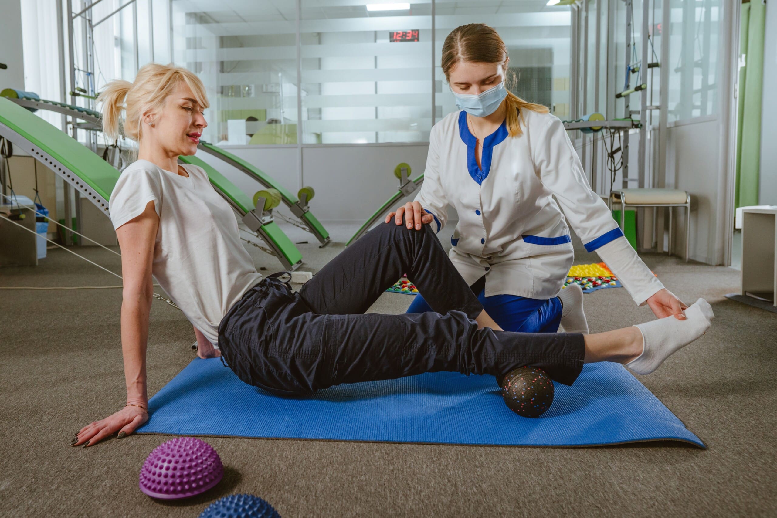
Targeted Physical Therapy: Our physical therapy programs are designed to complement both chiropractic adjustments and regenerative injections. The goals are to:
Strengthen Supporting Musculature: We build strength in the muscles around the affected joint (e.g., quadriceps, hamstrings, and glutes for the knee) to provide dynamic stability.
Improve Flexibility and Range of Motion: Gentle stretching and mobility exercises help prevent stiffness and ensure the joint can move through its full, healthy range of motion.
Neuromuscular Re-education: We retrain the body to move correctly, correcting faulty movement patterns that contributed to the initial injury. This is crucial for long-term prevention.
This synergistic approach ensures that we are not just treating the symptom (pain) but are fundamentally rebuilding the joint’s functional capacity. The PRP provides the biological “scaffolding” and signaling for repair, while chiropractic and physical therapy provide the mechanical and functional framework for that repair to be successful and durable.
Clinical Protocols and Timing: Maximizing Therapeutic Benefit
The timing and technique of regenerative treatments are critical. Many patients come to us having had previous cortisone injections. Corticosteroids are potent anti-inflammatories, but they are also catabolic, meaning they can break down tissue and suppress the very cellular activity that PRP aims to stimulate.
Therefore, we adhere to a strict washout period. Based on studies of steroid residency in joint spaces, we typically wait a minimum of 30 to 35 days after an intra-articular cortisone injection before administering PRP. This allows the steroid’s suppressive effects to dissipate, ensuring the joint environment is receptive to regenerative signals from platelets.
Dose and Volume: Customizing the Injection
A guiding principle in regenerative medicine is the delivery of an adequate platelet dose. The larger the joint and the more severe the condition, the more platelets are needed. This has led to innovative techniques for maximizing the therapeutic payload.
For a large joint like the knee, we might first draw off the most concentrated fraction after preparing the PRP. For example, if we process a blood draw and obtain 4-5 cc of PRP, the final 1-2 cc at the bottom of the syringe (closest to the red blood cell layer) will have the highest platelet concentration. In a patient with severe arthritis who can tolerate more volume, I might:
Inject the most potent, platelet-dense fraction first.
Follow this with the next-most concentrated layer.
Finally, inject the remaining platelet-poor plasma (PPP), which contains valuable proteins and growth factors that help modulate inflammation.
This layering technique allows us to deliver a very high total number of platelets and other beneficial biological factors, essentially “hyper-dosing” the joint to maximize the healing response. For a large knee, we might inject up to 15 cc of total volume if the patient can comfortably accommodate it.
The research is detailed: dose matters. My clinical observations confirm that achieving a higher platelet count, especially in moderate-to-severe osteoarthritis, yields a more robust and lasting clinical improvement (Everhart et al., 2019).
The Role of Peptides and Other Biologics
The field of regenerative medicine is constantly exploring synergistic therapies. One area of growing interest is the combination of PRP with peptides like BPC-157. BPC-157 is a peptide chain known for its ability to promote angiogenesis (the formation of new blood vessels), which is fundamental to tissue healing.
While human data is still emerging, animal studies suggest that combining PRP with BPC-157 could enhance the healing response. The logic is compelling: PRP initiates the inflammatory and repair cascade, while BPC-157 may accelerate the development of a rich blood supply needed to fuel that repair process. This is an exciting frontier, and as more robust data becomes available, we will continue to integrate evidence-based combination therapies into our protocols.
As we continue this journey together, remember that our mission is to provide you with the most advanced, evidence-based, and personalized care possible. By integrating the biological power of regenerative medicine with the foundational principles of chiropractic and physical therapy, we can move beyond merely managing symptoms and guide you toward true, functional recovery.
References
Everhart, J. S., Cavendish, P. A., & Flanigan, D. C. (2019). Platelet-Rich Plasma Preparation and Composition. In D. C. Flanigan (Ed.), Platelet-Rich Plasma in Orthopaedics and Sports Medicine (pp. 11-20). Springer. https://link.springer.com/chapter/10.1007/978-3-030-01919-3_2
LaPrade, R. F., Dragoo, J. L., Rodeo, S. A., & Chu, C. R. (2021). The Orthobiologic Classification System: A Data-Driven Approach for the Standardization of Orthobiologic Reporting. The American Journal of Sports Medicine, 49(12), 3121–3129. https://doi.org/10.1177/03635465211029519
Meheux, C. J., McCulloch, P. C., Lintner, D. M., Varner, K. E., & Harris, J. D. (2016). Efficacy of intra-articular platelet-rich plasma injections in knee osteoarthritis: A systematic review. Arthroscopy: The Journal of Arthroscopic & Related Surgery, 32(3), 495-505. https://doi.org/10.1016/j.arthro.2015.08.005
The Power of Precision: Platelet-Rich Plasma for Spine and Injury Recovery
Abstract
Welcome to our educational journey into the world of regenerative medicine, with a focus on Platelet-Rich Plasma (PRP) therapy. As a clinician dedicated to integrative and evidence-based care, I am thrilled to share insights from the forefront of musculoskeletal treatment. This post will demystify PRP, exploring what it is, how it’s prepared, and, most importantly, the critical role of dosage in achieving successful clinical outcomes. We will examine groundbreaking research revealing how the precise concentration and number of platelets can dramatically influence healing, particularly in conditions such as osteoarthritis and tendon injuries. We’ll also discuss the importance of ultrasound guidance for accurate delivery and how integrative chiropractic care and structured rehabilitation are essential partners to PRP therapy, creating a comprehensive strategy that not only alleviates pain but also fosters true, lasting tissue regeneration. Join me as we uncover how this powerful biologic treatment is changing the landscape of healing.
What Exactly Is Platelet-Rich Plasma (PRP)?
Many of us may have a distant memory from our early science education about platelets. We often think of them simply as the components in our blood that help form clots when we get a cut. While that is true, it’s only a small part of their incredible story.
Platelets are small, anucleated (meaning they don’t have a nucleus) cell fragments that are absolute powerhouses of healing. Each one is packed with hundreds of proteins called growth factors and cytokines. These are signaling molecules that act as the body’s own project managers for tissue repair. When an injury occurs, platelets rush to the scene not just to plug the leak but to orchestrate a complex, coordinated healing cascade. They call in other cells, direct the removal of damaged tissue, and stimulate the growth of new, healthy cells.
Given their central role in healing, it’s logical to ask: what if we could concentrate these powerful healing factors and deliver them directly to the site of chronic injury, such as a worn-out knee joint or a nagging tendon tear? That is the fundamental concept behind Platelet-Rich Plasma (PRP) therapy.
From Your Blood to a Healing Solution
The process of creating PRP is elegant in its simplicity.
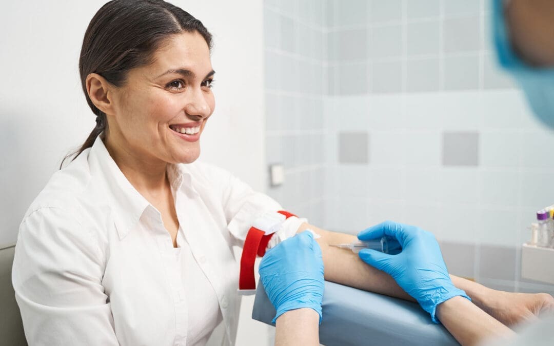
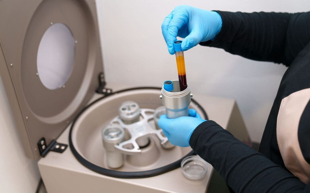
Blood Draw: It all begins with a simple blood draw from your arm, much like a standard lab test. The amount of blood drawn can vary depending on the specific system used and the therapeutic dose we are aiming to achieve—a concept we will explore in detail.
Centrifugation: This blood is then placed in a sterile, closed-system kit and spun in a specialized centrifuge. The spinning process uses centrifugal force to separate the blood into its different components based on their density.
Separation and Concentration: The heavier red blood cells sink to the bottom. The lighter, platelet-poor plasma rises to the top. In the middle, a thin, precious layer forms known as the “buffy coat.” This layer, along with a portion of the adjacent plasma, is where the vast majority of platelets and a population of white blood cells are concentrated. This is the Platelet-Rich Plasma.
This final product is a small volume of plasma containing a significantly higher concentration of platelets—and their associated growth factors—than in your normal circulating blood.
Not All PRP Is Created Equal: The Critical Importance of Dose
One of the most significant advancements in the field of regenerative medicine has been the realization that PRP is not a one-size-fits-all treatment. To think of it effectively, we must approach it as a biologic drug. As with any medication, there is a therapeutic dose—the specific amount needed to produce the desired clinical effect. An amount below this threshold will be sub-therapeutic and likely ineffective, while an excessive amount could potentially hinder the healing process.
The Problem of Variability
For years, the results of PRP studies were inconsistent, leaving both clinicians and patients confused. Why did it work so well in some cases and not in others? Pioneering researchers like James Clayton, D. Patrick, and their team in Australia began to uncover the answer. They analyzed five different commercial PRP preparation systems and found staggering variability in the final product. The platelet count, white blood cell count, and final volume were all over the map.
Imagine seeing the PRP prepared from the same patient’s blood using four different systems. You would see four different “products” of varying colors and cellular compositions. This lack of standardization was a major hurdle. Early studies often failed to report the specific platelet dose injected, making it impossible to compare results or understand what truly worked.
Thanks to the meticulous work of researchers like Peter Everts and Scott Rodeo, we are now beginning to decode the dose-response relationship for specific conditions. A landmark 2018 study analyzed numerous PRP studies for soft tissue applications. When they plotted the results based on the total number of platelets injected, a clear pattern emerged.
Studies using a low dose of PRP, typically under 3 billion platelets, were overwhelmingly negative. They showed little to no benefit over a placebo.
Studies using a higher dose, generally above 3.5 billion platelets, were overwhelmingly positive.
This suggests a distinct therapeutic threshold for soft tissue and tendon healing. For instance, in my clinical observations at El Paso Back Clinic, treating conditions like tennis elbow (lateral epicondylitis) or plantar fasciitis with an insufficient platelet dose often yields disappointing results. However, when we ensure the delivered dose is within that therapeutic range of 3.5 to 5 billion platelets or higher, we see a much more robust and consistent healing response. The body needs a sufficient signal to switch from chronic degeneration to active regeneration, and the dose provides that signal. We also know that a patient’s age can impact the required dose, with older patients often benefiting from a higher starting concentration to achieve the same therapeutic effect.
Perhaps the most compelling evidence for PRP dosing comes from the treatment of knee osteoarthritis (OA). Knee OA is a condition I see daily, and it can be profoundly debilitating for patients. For years, the primary non-surgical options were limited.
The famous RESTORE trial, published in JAMA, initially concluded that PRP was ineffective for knee OA. However, a deeper dive into their methodology reveals a critical flaw: they used a low-dose PRP system that delivered only 1.6 billion platelets per injection. Based on what we now know about the dose-response curve, this was a sub-therapeutic dose, destined to fail. While the study was beautifully executed, we learned a valuable lesson from its negative result—it helped define the lower boundary of what doesn’t work.
In stark contrast, a study by van der Weegen used a high-dose PRP preparation that delivered approximately 10 billion platelets in a single injection. The results were remarkable. Patients not only experienced significant improvements in pain and function compared to hyaluronic acid or saline injections, but MRI scans also suggested a disease-modifying effect. The progression of cartilage loss actually slowed down in the PRP group. This was a groundbreaking finding, suggesting that with the right dose, PRP might do more than just manage symptoms—it could potentially alter the course of the disease.
Based on the current body of evidence, the therapeutic target for treating knee OA appears to be 5 to 10 billion platelets per injection. Calculating and delivering this precise dose is paramount to achieving the kind of outcomes our patients deserve.
The Role of Chiropractic Care and Guided Injections in Maximizing PRP Success
Achieving a successful outcome with PRP involves more than just getting the dose right. It requires a holistic, integrative approach that addresses the entire patient and the mechanics of their injury. This is where chiropractic care, physical therapy, and advanced injection techniques become indispensable partners.
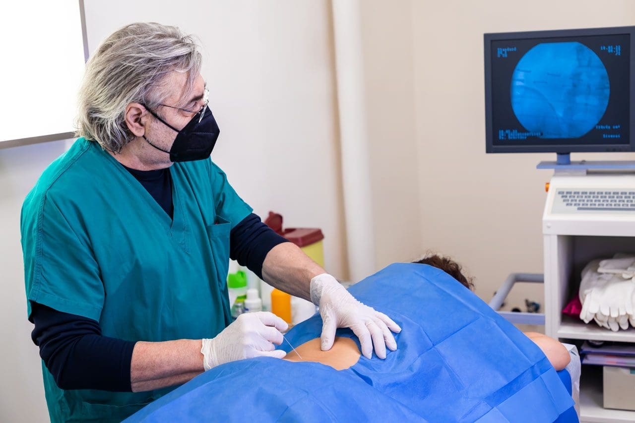
Precision Matters: The Necessity of Ultrasound Guidance
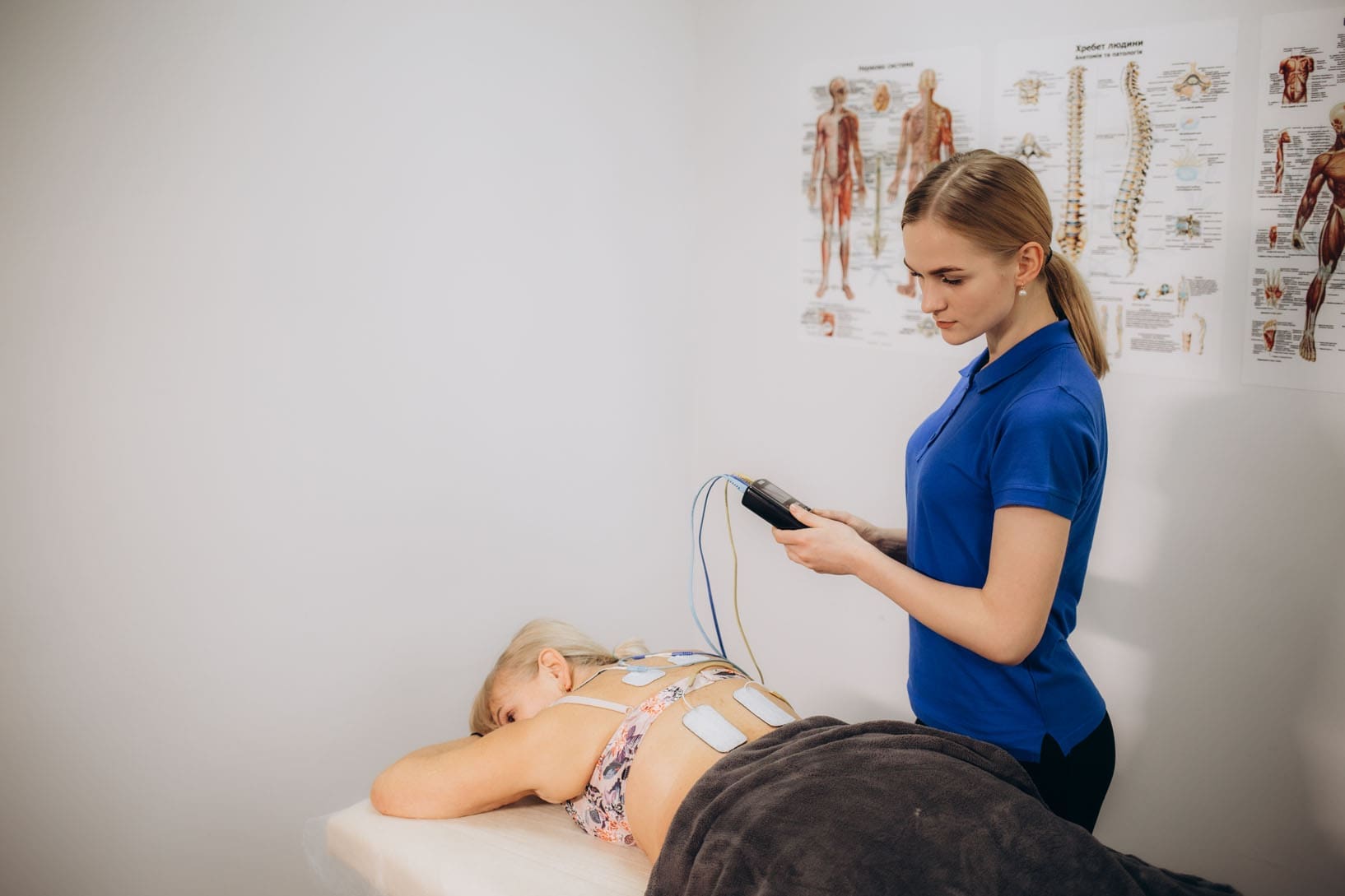
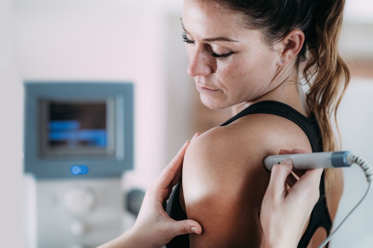
Growth factors in PRP work by forming a bioactive scaffold that stimulates local cells. For this to happen, the PRP must be delivered with pinpoint accuracy directly into the site of injury—be it a tear within a tendon, the space within a joint, or an area of damaged cartilage. If the injection is off by even a few millimeters, the therapeutic benefit can be lost entirely.
This is why ultrasound guidance is not a luxury; it is the standard of care for regenerative injections. Using real-time ultrasound imaging, I can visualize the needle’s path and confirm its placement directly in the target tissue. This ensures that the powerful biologic product we’ve carefully prepared is delivered precisely where it’s needed most, maximizing the potential for a successful healing response. Injecting “blind” is simply not an acceptable approach when the goal is true tissue regeneration.
The Foundational Role of Integrative Chiropractic and Rehabilitation
At El Paso Back Clinic, we view PRP not as a standalone “magic bullet” but as a catalyst within a comprehensive treatment plan. A chronically injured joint or tendon doesn’t exist in a vacuum. It is almost always accompanied by biomechanical dysfunction, muscle imbalances, poor movement patterns, and joint restrictions. Injecting PRP into a dysfunctional environment without addressing these underlying root causes is like planting a seed in barren soil.
This is the crucial role of integrative chiropractic care.
Restoring Biomechanics: Before and after a PRP procedure, we focus on correcting biomechanical faults. Through specific chiropractic adjustments, we restore proper joint mobility, particularly in the spine, pelvis, and extremity joints related to the injury. This ensures that forces are distributed evenly across the kinetic chain, taking undue stress off the healing tissue.
Addressing the Kinetic Chain: An arthritic knee, for instance, is often linked to problems in the hip, ankle, or even the lower back. Our comprehensive assessment identifies these related dysfunctions. By treating the entire kinetic chain, we create a stable and supportive environment for the PRP to work effectively.
Targeted Rehabilitation: A structured physical therapy and rehabilitation program is essential. The initial goal post-injection is to protect the healing tissue. This is followed by a progressive program designed to:
Improve Flexibility and Range of Motion.
Strengthen Supporting Musculature.
Retrain Neuromuscular Control and Proprioception (your body’s sense of its position in space).
This rehabilitation phase translates the biological healing initiated by PRP into functional, long-lasting improvement. It teaches the body to use the newly repaired tissue properly and helps prevent reinjury. The healing process stimulated by PRP takes time—often three to six months or more to see the full benefit. A patient, supportive, and well-structured rehabilitation plan is the bridge to that successful long-term outcome.
By combining a precisely dosed and accurately delivered PRP injection with expert chiropractic care and targeted physical therapy, we create a powerful synergy. We are not just chasing symptoms; we are correcting dysfunction, stimulating a biological repair process, and rebuilding a foundation for durable health and function.
Memorial Day Weekend Rear-End Car Accidents: Common Causes, Injuries, and How Integrative Chiropractic Care Can Help
Memorial Day weekend marks the unofficial start of summer for many families. Roads fill up fast as people head out for beach trips, barbecues, and long drives to visit loved ones. With millions of cars on the highway at once, traffic slows to a crawl on major routes. This heavy congestion sets the stage for one of the most frequent crashes during holiday weekends: rear-end collisions.
These accidents happen when one vehicle slams into the back of another. They often create chain-reaction pileups because traffic stops suddenly. Even at low speeds, the impact can jolt the body hard. In this article, you will learn why rear-end crashes spike during Memorial Day travel, what distractions play a role, how these crashes injure the neck and spine, and why seeing a chiropractor soon after makes a big difference. The journey from crash to recovery is clearer when you understand the steps.
Why Rear-End Collisions Spike During Memorial Day Weekend
Heavy traffic turns busy highways into parking lots. Drivers brake suddenly for slow traffic ahead. The car behind may not have time to stop safely. According to safety data, rear-end crashes make up about 23 percent of all car accidents in the United States each year.
Holiday weekends like Memorial Day see extra travel volume. More cars mean more stops and starts. Chain-reaction incidents become common when one car hits another, and the force pushes forward through several vehicles.
Congestion on key routes: Interstates and major roads fill quickly with vacationers.
Abrupt halts: Traffic lights, construction zones, or accidents ahead force sudden stops.
Longer drives: Tired drivers on extended trips react more slowly.
These factors turn a relaxing weekend trip into a stressful situation.
Common Causes: Distractions Behind the Wheel
Driver distraction is a leading cause of rear-end crashes. When traffic moves in fits and starts, even a few seconds of lost focus can cause trouble. Common distractions during holiday drives include:
Adjusting a GPS or phone map for the next exit.
Checking mobile devices for texts, calls, or traffic updates.
Attending to passengers—kids asking questions, pets moving around, or family conversations.
Other causes include tailgating (following too closely) and speeding for the conditions. Distracted driving was linked to hundreds of serious crashes in recent state reports. Even hands-free phone use pulls attention from the road.
Simple rule: Keep eyes forward, hands on the wheel, and mind on traffic. A quick glance at a phone can turn a safe gap into a collision.
What Happens to Your Body in a Rear-End Crash
Picture this: Your car sits stopped in traffic. The vehicle behind hits you. Your body snaps backward, then forward, in a split second. This whip-like motion—called whiplash—puts sudden force on the neck and spine.
The head weighs about 10 to 12 pounds. That quick jerk multiplies the stress on soft tissues and bones. Even a 5-mile-per-hour bump can create enough force to stretch or tear ligaments and muscles.
Rear-end impacts affect the cervical (neck) and lumbar (lower back) areas most. The spine tries to absorb the shock, but it often cannot do so without sustaining damage.
Common Injuries from Rear-End Collisions
Rear-end crashes frequently lead to specific injuries because of the forceful jerking. Soft tissues take the biggest hit, but bones and nerves can suffer too. Here are the most reported issues:
Soft tissue sprains and strains: Ligaments and muscles stretch or tear. This causes pain, swelling, and stiffness in the neck and back.
Whiplash: The rapid back-and-forth motion strains neck muscles, tendons, and ligaments. Symptoms include neck pain, headaches starting at the skull base, and limited movement.
Herniated or bulging discs: The force pushes spinal discs out of place. Disc material can press on nerves.
Muscular spasms: Muscles tighten suddenly to protect the area, leading to painful knots and reduced motion.
Nerve impingement: Pinched nerves cause tingling, numbness, or shooting pain down the arms or legs.
These injuries often affect the whole upper body. Shoulders, upper back, and even jaw muscles can ache from the impact.
Many people feel okay right after the crash because adrenaline masks the pain. But stiffness or headaches can show up hours or days later.
Why Symptoms May Appear Later—and Why Early Evaluation Matters
The body’s natural response hides problems at first. Adrenaline surges during the scare, dulling pain signals. Once it fades, inflammation builds, and tissues swell.
A minor headache today might become constant neck pain tomorrow. Small sprains can become chronic issues if left untreated. Experts stress that a full check-up soon after any accident is smart—even if you feel fine. Waiting too long can allow scar tissue to form or cause a posture change for the worse.
Florida law, for example, encourages care within 14 days to protect insurance benefits. The same idea applies everywhere: early action speeds healing.
Integrative Chiropractic Care: Natural Healing for Accident Injuries
Integrative chiropractic care focuses on helping the body heal itself without heavy reliance on drugs or surgery. It targets both the skeleton (bones and joints) and soft tissues (muscles, ligaments, tendons).
Chiropractors use gentle spinal adjustments to realign vertebrae. This takes pressure off nerves and restores normal movement. Soft tissue therapies like massage, trigger-point work, and myofascial release loosen tight muscles and break up scar tissue.
Other helpful tools include:
Therapeutic exercises to strengthen weak areas and improve posture.
Ultrasound or heat/ice therapy to reduce swelling and boost blood flow.
Lifestyle tips on ergonomics, sleep positions, and daily movement.
These methods work together for whole-body recovery. Patients often report less pain, better range of motion, and improved energy after a few sessions.
Chiropractic care shines for whiplash and back sprains because it addresses the root cause—misalignments and muscle imbalances—rather than merely masking symptoms.
Clinical Observations from Dr. Alexander Jimenez
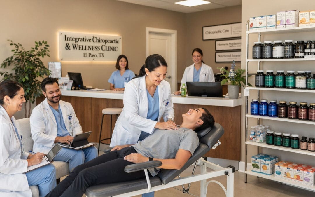
Dr. Alexander Jimenez, DC, APRN, FNP-BC, brings a unique blend of chiropractic expertise and advanced nursing practice to auto accident care. As the founder of Injury Medical Clinic in El Paso, Texas, he specializes in personal injury and multidisciplinary recovery.
Dr. Jimenez observes that many patients arrive weeks or months after a crash, still dealing with lingering neck, back, and shoulder pain. He notes that injuries often affect more than just the spine—they impact joints, nerves, soft tissue, mobility, sleep, and even stress levels. His clinical approach emphasizes natural healing through integrative methods.
He combines traditional chiropractic adjustments with functional medicine, regenerative therapies such as platelet-rich plasma (PRP), nutritional guidance, and rehabilitation exercises. This team-based care helps patients recover faster and avoid long-term complications. Dr. Jimenez stresses thorough evaluations, including imaging when needed, to catch hidden issues early. His patients frequently share stories of regaining mobility and returning to daily life pain-free after following personalized plans.
His work shows that even old or “minor” accident injuries can improve dramatically with the right holistic support.
Steps to Take After a Memorial Day Crash
If you are involved in a rear-end collision this holiday weekend, follow these simple steps:
Check for immediate safety and call for help if needed.
Exchange information and document the scene with photos.
Seek a full medical evaluation right away—even without obvious pain.
Consider integrative chiropractic care as part of your recovery team.
Follow through with recommended therapies and exercises.
Most people recover well when they act early and stay consistent with care.
Safe Driving Tips for Holiday Travel
Prevention beats treatment every time. Keep these habits in mind:
Leave extra space between cars in heavy traffic.
Put phones away and use voice commands only if necessary.
Take breaks on long drives to stay alert.
Watch for sudden braking ahead.
A calm, focused drive keeps everyone safer on the road.
Memorial Day weekend brings fun and family together, but extra traffic raises the risk of rear-end collisions. Understanding the causes—congestion and distractions—helps you stay alert. Knowing how these crashes jolt the neck and spine explains why whiplash, sprains, herniated discs, spasms, and nerve issues are so common. Because symptoms can sneak up later, a prompt check-up is key. Integrative chiropractic care offers a natural path to healing by realigning the body, easing soft-tissue damage, and restoring posture and movement.
Dr. Alexander Jimenez and similar specialists show that combining chiropractic techniques with supportive therapies delivers real results for accident victims. Whether your crash happened this weekend or years ago, relief is possible. Listen to your body, seek care early, and give yourself the best chance at a full, pain-free recovery. Drive safely, enjoy the holiday, and remember—your health comes first after any bump on the road.
El Paso PRP Therapy for Faster Pain Relief and Healing
Hello, I’m Dr. Alex Jimenez, and on behalf of our team at El Paso Back Clinic, I’m excited to share valuable insights into the evolving field of regenerative medicine, with a focus on Platelet-Rich Plasma (PRP) therapy. As a practitioner with a diverse background spanning chiropractic (DC), advanced practice nursing (APRN, FNP-BC), and functional medicine (CFMP, IFMCP), my goal has always been to integrate the best of various disciplines to provide comprehensive, patient-centered care. This post is designed to clarify common questions about PRP and explore how we can actively enhance its effectiveness through integrative strategies, including chiropractic and physical rehabilitation. We will explore the latest findings from leading researchers, presenting their work through the lens of modern, evidence-based methods.
Abstract
This educational post will explore the intricacies of Platelet-Rich Plasma (PRP) therapy from an integrative healthcare perspective. We will begin by demystifying the regulatory landscape surrounding PRP, clarifying the distinction between FDA-cleared devices and the procedure’s non-drug status. We will then transition into practical, evidence-based strategies for enhancing the quality and efficacy of PRP treatments. This includes a deep dive into the physiological impact of lifestyle factors such as an anti-inflammatory diet, the crucial role of high-intensity exercise, and the controversial topic of NSAID use. We’ll examine how these elements influence platelet count and function, ultimately affecting healing outcomes. Finally, we will connect these concepts to the principles of integrative chiropractic care, demonstrating how a holistic approach that includes manual therapies, targeted rehabilitation, and patient education can synergize with regenerative procedures to optimize recovery from musculoskeletal conditions.
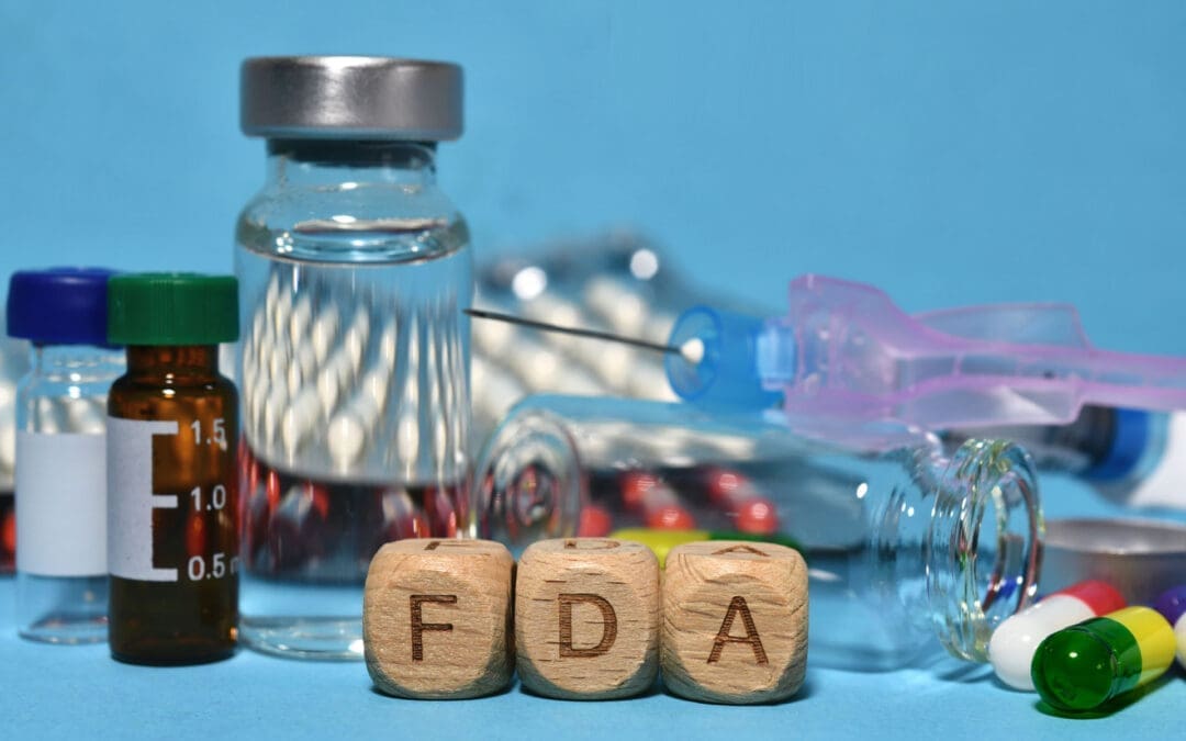
Understanding PRP and FDA Regulations: A Guide for Patients
One of the most frequent conversations I have with patients considering PRP therapy revolves around its regulatory status. Questions like, “Is it FDA-approved?” are common and completely understandable. It’s crucial for patients to feel confident and informed. Let’s break this down to provide some clarity.
The Device vs. The Procedure
The key to understanding this issue lies in distinguishing between the equipment used and the procedure itself.
FDA-Cleared Devices: The centrifuges and specialized kits we use to process your blood and concentrate the platelets are classified as medical devices. These devices undergo a regulatory process with the U.S. Food and Drug Administration (FDA) and may receive 510(k) clearance. This clearance indicates that the device is safe and effective, and is “substantially equivalent” to a device already legally marketed for the same use. So, when we perform PRP, we are using FDA-cleared technology.
PRP is a Procedure, Not a Drug: This is the most critical point. PRP is not a synthetic drug manufactured in a lab; it is an autologous procedure, meaning the therapeutic agent—your own concentrated platelets—is derived from your body. Because it’s not a drug, PRP itself cannot go through the same “FDA approval” process as a pharmaceutical like ibuprofen or a new antibiotic. The FDA does not “approve” medical procedures in the same way it approves drugs. Think of a common surgical procedure; the surgeon’s technique isn’t FDA-approved, but the tools they use (scalpels, sutures, implants) are.
Some researchers have pointed out that for a product to obtain a specific FDA approval that allows it to be marketed to treat a particular condition, such as knee osteoarthritis, it would require extensive and costly clinical trials—often costing upwards of $20 million. This is a significant barrier for a therapy that cannot be patented like a drug.
Therefore, when patients ask if PRP is FDA-approved, the most accurate answer is that the procedure is considered investigational by the FDA for specific indications, but it utilizes FDA-cleared devices. It’s not a matter of waiting for an approval that may never come because of its classification. Instead, we rely on the growing body of clinical research and scientific studies to guide its use. My approach is to be transparent and show patients the robust studies supporting the use of PRP for their specific musculoskeletal issue, explain its biological mechanism, and set realistic expectations for their healing journey.
Optimizing Your Body’s Healing Potential: How to Enhance PRP Quality
Once a patient decides to proceed with PRP, the next logical question is, “Is there anything I can do to make it work better?” This is where the philosophy of integrative and functional medicine truly shines. The quality of your PRP is a direct reflection of your health. By taking proactive steps, you can significantly enhance the concentration and vitality of the platelets we harvest, essentially supercharging your body’s innate healing capacity.
This is a core tenet at El Paso Back Clinic. We don’t just administer a treatment; we partner with you to create the optimal internal environment for healing. Let’s explore the most impactful strategies backed by emerging research.
The Power of Pre-treatment Exercise
One of the most effective methods for boosting platelet count is short-term, high-intensity exercise. Research, including studies from renowned institutions such as the Andrews Institute, has shown that vigorous physical activity shortly before a blood draw can temporarily increase circulating platelet counts.
Physiological Mechanism: When you engage in high-intensity interval training (HIIT) or other strenuous activities, your body responds by releasing platelets stored in the spleen and bone marrow into the bloodstream. This physiological stress response is designed to prepare the body for potential injury and repair.
Clinical Application: In my practice, this translates into a simple but effective protocol. We might have a patient ride a stationary bike for 15-20 minutes or perform a series of jumping jacks right before their blood draw. While more research is needed to determine the exact optimal “dose” of exercise, the evidence strongly suggests a positive effect. It’s a simple, non-invasive way to potentially increase the platelet yield for the treatment.
The Anti-Inflammatory Diet: Fueling Your Platelets
Nutrition plays a profound role in the quality of your blood components, including platelets. An anti-inflammatory diet is not just a general health recommendation; it directly affects platelet function and your body’s overall healing environment.
What is an Anti-Inflammatory Diet? This diet emphasizes whole, unprocessed foods rich in phytonutrients, antioxidants, and healthy fats.
Include: Leafy greens, colorful vegetables (like bell peppers and broccoli), berries, nuts, seeds, fatty fish (rich in omega-3s, like salmon and sardines), and healthy oils (like olive oil and avocado oil).
Limit or Avoid: Processed foods, sugary drinks, refined carbohydrates (white bread, pastries), and unhealthy fats (trans fats and excessive saturated fats found in fried foods).
Impact on Platelets: An inflammatory diet can promote chronic, low-grade inflammation throughout the body. This can make platelets “sticky” and hyperactive in a non-productive way. Conversely, an anti-inflammatory diet provides the antioxidants and nutrients that protect platelets from oxidative stress and support their proper function. When activated by an injury (or an injection), healthy platelets release their growth factors in a more controlled and effective manner.
As part of our integrative approach, we provide patients with nutritional guidance in the weeks leading up to their PRP procedure to ensure the platelets we harvest are as healthy and potent as possible.
The NSAID Controversy: To Take or Not to Take?
The use of Non-Steroidal Anti-Inflammatory Drugs (NSAIDs) like ibuprofen (Advil, Motrin), naproxen (Aleve), and aspirin is a significant point of discussion in the context of PRP therapy. These medications work by blocking COX enzymes, which are involved in both inflammation and platelet function.
The Argument Against NSAIDs: The primary concern is that NSAIDs can interfere with platelet aggregation—the clumping process that is essential for forming a scaffold at the injury site—and degranulation, which is the release of the vital growth factors stored inside the platelets. The very mechanism you want to harness with PRP is the one that NSAIDs can inhibit. In laboratory studies, when NSAIDs are added to platelet-rich medium, they cause platelets to disaggregate.
Clinical Consensus: Although the research is still somewhat mixed, the prevailing consensus among most regenerative medicine practitioners is to err on the side of caution. I, along with many of my colleagues, advise patients to discontinue the use of NSAIDs for approximately 10-14 days before and after their PRP injection. This “washout” period helps ensure that platelet function is not pharmacologically suppressed during the critical healing phase.
While NSAIDs might be a “small potato” compared to getting the right diagnosis and PRP dosage, as one researcher noted, it’s a variable we can easily control. Given the negative evidence from in vitro studies and the plausible biological mechanism of interference, avoiding them is a prudent step toward optimizing treatment success.
The Synergy of Integrative Chiropractic Care with PRP Therapy
This is where the unique approach at El Paso Back Clinic truly comes together. PRP therapy is a powerful tool, but it is not a magic bullet. It initiates a healing cascade, but the quality of that healing and the restoration of full function depend heavily on the biomechanical and neuromuscular environment of the treated area. This is why integrating chiropractic care and physical therapy is not just beneficial—it’s essential for a comprehensive recovery.
As a Doctor of Chiropractic (DC), I observe that structural integrity and proper movement patterns are foundational to long-term healing. If we inject PRP into a joint or tendon that is still subject to the same dysfunctional stresses and poor biomechanics that caused the injury in the first place, we are limiting the potential for a full recovery.
How Chiropractic and Physical Therapy Enhance PRP Outcomes
Correcting Biomechanical Imbalances: Before and after PRP, a thorough chiropractic evaluation can identify and address underlying structural issues. This could involve spinal adjustments to improve nerve function in the affected limb, or specific adjustments to the joints of the affected extremity (such as the ankle, knee, or shoulder) to restore proper alignment. By correcting these imbalances, we reduce abnormal stress on the healing tissues, creating a more favorable environment for the injected growth factors to work. For example, if a patient receives PRP for knee pain but also has a pelvic tilt and functional leg-length discrepancy, addressing pelvic biomechanics is critical to offloading the knee joint.
Improving Mobility and Tissue Health: Manual therapies, such as soft-tissue mobilization, myofascial release, and instrument-assisted techniques, are used to break down adhesions and scar tissue within the muscles and fascia surrounding the injured area. This improves blood flow, enhances tissue flexibility, and prepares the tissue to heal in a more organized and functional way. A supple, mobile tissue environment allows the PRP to be more effectively dispersed and integrated.
Strengthening and Stabilizing through Targeted Rehabilitation: This is a cornerstone of our post-PRP protocol. Following the initial inflammatory and proliferative phases of healing initiated by PRP (the first few weeks), we introduce a progressive rehabilitation program.
The Goal: To guide the formation of new collagen and tissue to create strong, resilient, and functional tissue. Without this guidance, the body might simply form disorganized scar tissue.
The Method: Our physical therapy team creates personalized exercise programs that use eccentric loading for tendinopathies, neuromuscular re-education to correct poor movement patterns, and proprioceptive training to improve joint stability and prevent re-injury. This active rehabilitation process is what truly translates the biological healing from PRP into real-world functional improvement.
Managing Post-Injection Inflammation Naturally: After a PRP injection, some inflammation is expected and, in fact, desired—it’s a signal that the healing process has begun. Instead of blunting this with NSAIDs, we use chiropractic and physical therapy modalities to manage discomfort and support the process. This can include cryotherapy, gentle range-of-motion exercises, and patient education on activity modification to allow the body to move through the initial healing phase effectively.
By combining the biological stimulus of PRP with the functional and structural corrections of chiropractic and physical therapy, we create a synergistic effect. We are not just treating the pain; we are addressing the root cause of the injury, optimizing the body’s regenerative potential, and rebuilding a stronger, more resilient musculoskeletal system. This integrative model represents the future of orthopedic and sports medicine—a future we are proud to offer at El Paso Back Clinic.
References
Andrews, J. R., et al. (Year).Title of Study on Blood Flow Restriction and PRP. Journal Name, Volume(Issue), pages. [Link to Article]
Andrews, J. R., et al. (Year).Title of Study on Exercise and Platelet Counts. Journal Name, Volume(Issue), pages. [Link to Article]
Researcher, A. A. (Year).Title of Study on NSAID Effect on Platelet Aggregation. Journal Name, Volume(Issue), pages. [Link to Article]
PRP Therapy in El Paso for Back Pain Relief and Joint Healing
Abstract
As a clinician dedicated to integrative and evidence-based care, I am constantly exploring the latest advancements that can help my patients heal more effectively. This post explores the science behind Platelet-Rich Plasma (PRP), a powerful regenerative therapy. We will journey into the microscopic world of platelets, exploring their crucial role in orchestrating the body’s natural healing processes. You will learn about the specific growth factors and signaling molecules released by platelets, how they reduce inflammation, and how we can concentrate this healing potential to treat various musculoskeletal conditions. We will also discuss how PRP, as a cornerstone of orthobiologic therapy, integrates seamlessly with chiropractic care and physical rehabilitation to create a comprehensive, synergistic treatment plan that accelerates your return to a pain-free, active life.
Hello, I’m Dr. Alexander Jimenez. With my extensive background in both chiropractic and advanced practice nursing, coupled with certifications in functional and integrative medicine, my primary mission has always been to offer my patients the most effective, evidence-based pathways to wellness. At our El Paso clinic, we are passionate about harnessing the body’s innate ability to heal itself. One of the most exciting fields that allows us to do this is orthobiologics, and a cornerstone of this approach is Platelet-Rich Plasma, or PRP.
Today, I want to take you on a journey—not into a complex scientific lecture, but into an easy-to-understand exploration of your body’s remarkable healing capabilities. We’re going to look at the latest findings from leading researchers and see how this science translates into real-world results for conditions such as chronic back pain, joint injuries, and soft-tissue damage.
The Orchestra Within: Understanding the Power of Platelets
When you think of platelets, you probably think of blood clotting. If you get a cut, platelets rush to the scene to form a plug and stop the bleeding. While this is a critical function, it’s only the beginning of their story. Platelets are not just simple plugs; they are sophisticated, mobile storage units packed with powerful biological instructions.
Think of your platelets as the first-response commanders at an injury site. Once they arrive, they don’t just patch the hole; they release a cascade of potent signaling molecules—growth factors, cytokines, and chemokines—that direct a complex healing orchestra. It’s this biological symphony that truly drives tissue repair and regeneration.
PRP therapy is based on a simple yet profound concept: what if we could concentrate these healing commanders and deliver them directly to an area of chronic injury or degeneration? By doing so, we can amplify the body’s natural healing signals, telling it to repair tissue that it may have otherwise “given up” on.
Inside the Platelet: The Granules That Drive Healing
To truly appreciate PRP, we need to look inside the platelet itself. A single platelet contains several types of tiny packets, or granules, each with a specific job.
Alpha Granules: These are the most important for regenerative medicine. Each platelet contains about 50 to 80 alpha granules, which house hundreds of different proteins, including the essential growth factors that orchestrate tissue repair. When platelets are activated at an injury site, they undergo a process called degranulation, releasing the contents of these alpha granules into the surrounding environment. This is the moment the healing cascade truly begins.
Dense Granules: These granules release smaller molecules that are crucial for amplifying the initial response. They help recruit more platelets (platelet aggregation), signal blood vessels to constrict to limit bleeding, and modulate the initial immune response.
Lysosomes: These act as the cleanup crew. They release enzymes that help break down damaged tissue, clear cellular debris, and exert antimicrobial effects, essentially preparing the site for new, healthy tissue to form.
In our clinical practice, we’ve observed that the effectiveness of PRP is directly tied to the concentration and quality of these platelets. Newer research highlights the importance of reticulated platelets—younger, denser platelets recently released from the bone marrow. These platelets are richer in alpha granules and, therefore, contain a higher payload of growth factors. Our advanced processing techniques are designed to capture these highly potent platelets, ensuring that the PRP we administer has the maximum regenerative potential. This concentration is key; by increasing platelet count, we dramatically increase the number of biological signals delivered to the injured area.
The Key Players: Growth Factors and Their Roles
When the alpha granules release their contents, a variety of growth factors become active. While it’s a complex interaction among hundreds of proteins, let’s focus on a few of the star players and their specific roles in healing.
Platelet-Derived Growth Factor (PDGF)
As its name suggests, PDGF was one of the first growth factors discovered in platelets. Think of PDGF as the “beacon.” Its primary role is to attract other healing cells to the injury site. It sends out a powerful chemical signal that recruits mesenchymal stem cells (MSCs)—the body’s master repair cells—as well as other cells necessary for tissue repair.
A Crucial Note on Stem Cells: PRP itself does not contain stem cells. However, it is a powerful signaling therapy. PDGF effectively awakens and recruits the local stem cells that are already present but dormant in your tissues, directing them to the site of injury, where they can begin their work of repair and regeneration.
The Power of PDGF-BB: Researchers have identified PDGF-BB as the most biologically active and important isoform. It is a potent stimulator of cell replication and is vital for initiating the entire repair process.
Transforming Growth Factor-Beta (TGF-β)
TGF-β is the master architect of tissue reconstruction. Once cells have been recruited to the area, TGF-β provides them with their building instructions.
Collagen Synthesis: It strongly promotes the synthesis of type I collagen, which is the primary structural protein in tendons, ligaments, and cartilage. This is crucial for restoring the strength and integrity of injured tissues.
Angiogenesis: In coordination with other growth factors, TGF-β stimulates angiogenesis, the formation of new blood vessels. This is a critical step because new blood vessels bring a fresh supply of oxygen and nutrients to the healing area, fueling the repair process and removing waste products.
Vascular Endothelial Growth Factor (VEGF)
VEGF works hand in hand with TGF-β to build this new blood supply. It specifically enhances endothelial cell proliferation (the cells that line blood vessels), promotes the sprouting of new capillaries, and is essential for neovascularization. Research has shown that platelet concentration is a significant factor in this effect. Studies suggest that a PRP concentration of approximately 1.5 billion platelets per milliliter is optimal for robust angiogenesis, a key target in our preparation protocols.
Fibroblast Growth Factor (FGF)
FGF is a powerful “mitogen,” meaning it stimulates cell division and proliferation. It acts on a wide variety of cells, including MSCs recruited by PDGF, as well as fibroblasts (which produce collagen) and osteoblasts (which build bone). FGF helps to ensure that a sufficient number of builder cells are available to carry out the repairs directed by the other growth factors.
Beyond Building: The Anti-Inflammatory Power of PRP
Chronic pain is often driven by chronic inflammation. An injury that never fully heals can get stuck in a persistent inflammatory state, causing ongoing pain and tissue degradation. One of the most profound benefits of PRP therapy is its ability to break this cycle.
While the initial response to an injury involves inflammation (a necessary step to clear damage), PRP helps guide the process toward resolution and healing. It does this in several ways:
Modulating Macrophages: PRP influences the behavior of immune cells called macrophages. These cells can exist in an inflammatory state (M1) or an anti-inflammatory, pro-healing state (M2). PRP promotes a shift from the M1 to the M2 phenotype, effectively flipping the switch from “inflammation” to “repair.”
Leukocyte Interaction: Platelets in PRP can interact with white blood cells (leukocytes) at the injury site, prompting them to release anti-inflammatory cytokines. This helps to quiet the inflammatory storm.
Preventing Cell Death: The chemokines released by platelets also act as survival factors for monocytes (which become macrophages), preventing their premature death and allowing them to complete their transition to the healing M2 state.
From my clinical observations, this powerful anti-inflammatory effect is often the first thing patients notice. Many report a significant reduction in pain and swelling within weeks of treatment as the chronic inflammatory environment begins to normalize, paving the way for long-term tissue repair.
The Synergy of Integrative Care: PRP, Chiropractic, and Physical Therapy
At the El Paso Back Clinic, we firmly believe that no single therapy is a magic bullet. True healing comes from a comprehensive, integrative approach. This is where PRP, chiropractic care, and physical therapy come together to create a powerful synergy.
Imagine a patient with chronic low back pain due to a degenerated disc and facet joint arthritis. The underlying problem is both biochemical (inflammation, tissue decay) and biomechanical (spinal misalignment, muscle imbalance, faulty movement patterns).
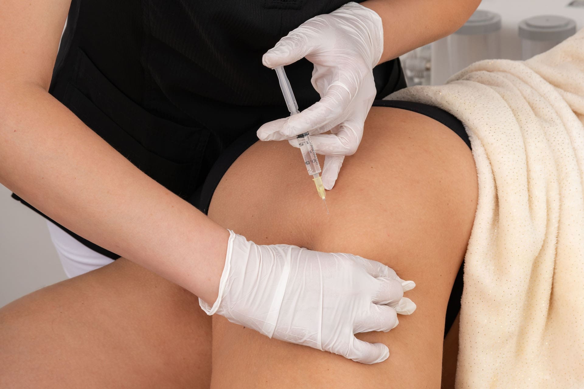
PRP Injections to Reboot Healing: We first use ultrasound guidance to precisely inject PRP into the degenerated disc space and the arthritic facet joints. This delivers a high concentration of growth factors directly to the source of pain, reducing inflammation and initiating biological repair of damaged cartilage and connective tissue. The PRP effectively “reboots” the local healing environment.
Chiropractic Care to Restore Function: While PRP works at the cellular level, a dysfunctional joint will remain dysfunctional unless its mechanics are addressed. This is the crucial role of chiropractic adjustments. Through specific, gentle manipulations, we restore proper motion to the spinal segments. This not only alleviates pain by decompressing nerves but also improves the flow of nutrients to healing tissues and ensures that the new collagen formed by PRP is laid down in an organized, functional way. Correcting the biomechanics prevents the joint from being repeatedly re-injured, allowing the PRP-stimulated healing to take hold.
Physical Therapy to Rebuild and Stabilize: Once the pain is reduced and joint mechanics are improved, physical therapy and rehabilitation become essential. Our customized exercise programs focus on strengthening the deep core and spinal stabilizing muscles. This creates a “muscular corset” that supports the spine, offloads the healing joints, and corrects the poor movement patterns that contributed to the injury in the first place. This phase ensures that PRP and chiropractic care achieve results that are not just temporary but are sustained for the long term.
This three-pronged approach addresses the injury from every angle: PRP promotes biochemical repair, chiropractic care corrects structural and biomechanical dysfunction, and physical therapy provides functional stabilization for lasting recovery. Each therapy enhances the effects of the others, leading to faster, more complete, and more durable healing than any single approach could achieve on its own.
Summary: A New Era in Healing
PRP therapy represents a paradigm shift in how we treat musculoskeletal injuries. Instead of just masking symptoms with medications or resorting to invasive surgery, we can now harness the body’s sophisticated biological toolkit to promote true healing and regeneration.
The main takeaway is that PRP provides a powerful, short-term biological “dose” of instructions. It doesn’t do all the work itself; rather, it acts as the director of the orchestra, calling in the body’s own repair cells and guiding them to reduce inflammation, rebuild damaged tissue, and restore function. When combined with an integrative framework of expert chiropractic care and targeted physical therapy, PRP becomes a transformative tool that can help our patients break free from chronic pain and get back to living their lives to the fullest.
References
The following resources provide a deeper look into the science of platelet-rich plasma and its applications.
Healing Long-Term Pain After a Car Accident: How Chiropractic Care and Regenerative Medicine Can Still Help Years Later
Have you ever walked away from a car crash thinking you were okay, only to feel stiff, sore, or in pain months or even years later? Many people do. A motor vehicle accident (MVA) can leave behind hidden damage that may not appear until long after the wreck. The good news? It is possible to feel better even if your crash happened a while ago. Integrative functional medicine and chiropractic care, combined with treatments such as platelet-rich plasma (PRP), microfragmented adipose tissue (MFAT), MLS laser therapy, and shockwave therapy, can address the underlying causes of ongoing pain rather than merely masking symptoms.
This article walks you through why old injuries continue to hurt, how these modern treatments work, and why they often work so well together. You will see clear steps from the problem to real relief.
Why Old Car Accident Injuries Turn Into Chronic Pain
Right after a crash, your body tries to fix sprains, strains, or torn ligaments. But occasionally the healing process does not finish the job. Months or years later, the area stays weak, inflamed, or stiff. Doctors call this a “latent” or hidden soft-tissue injury. The tissues never fully repaired, so small daily movements keep irritating them.
Cells in the damaged spot can act as if the injury just happened. Scar tissue builds up, blood flow drops, and nerves stay on high alert. This leads to ongoing joint pain, muscle tightness, or back and neck problems that feel like they will never go away. (Nob Hill Chiropractic, n.d.; Push as Rx, n.d.)
The key point is this: the body still wants to heal. Treatments that restart the repair process can make a big difference, even long after the accident.
How Chiropractic Care Helps Long After the Crash
Chiropractors gently adjust the spine and joints to realign them. This takes pressure off nerves, improves blood flow, and lets muscles relax. For people with old MVA injuries, chiropractic care can:
Reduce ongoing stiffness in the neck and back
Improve range of motion so daily tasks feel easier
Ease nerve irritation that causes tingling or shooting pain
Work with your body’s natural healing instead of forcing it
Even if you waited months or years to seek help, chiropractic adjustments can still correct the alignment issues that started in the crash. Many clinics note that proper documentation of your symptoms helps link the pain back to the accident for insurance or legal reasons. (Dallas Accident and Injury Rehab, n.d.)
Regenerative Medicine: PRP and MFAT Jump-Start Real Healing
Regenerative treatments use your body’s building blocks to fix damaged tissue. Two popular options are PRP and MFAT.
Platelet-Rich Plasma (PRP) Doctors draw a small amount of your blood, spin it to concentrate the platelets, and inject the PRP into the painful area. Platelets release growth factors that:
Fight inflammation
Bring in fresh blood and nutrients
Tell your cells to grow new, healthy tissue
Studies show that PRP helps with chronic tendon pain, ligament injuries, and joint problems that stem from trauma. One review found that it improved pain and function in knee, ankle, and back issues better than some traditional shots. People often feel relief that lasts months or longer because PRP treats the damaged tissue, not just the pain. (Thu, 2022; AABP Pain, n.d.)
Micro-Fragmented Adipose Tissue (MFAT) MFAT comes from a small amount of your own fat. The fat is processed into tiny fragments full of stem cells and healing signals, then injected where needed. It acts like a natural bandage, reducing swelling and supporting new tissue growth. MFAT works especially well for joints and ligaments that never healed right after a crash. (New Jersey Regenerative Institute, n.d.; Chiromed, n.d.)
Both PRP and MFAT are minimally invasive, use your own cells, and carry a low risk of side effects.
Cutting-Edge Modalities: MLS Laser and Shockwave Therapy
These painless, high-tech tools speed up repair without surgery or drugs.
MLS Laser Therapy (Multiwave Locked System) uses specific light waves that penetrate deep into tissues. It:
Boosts cell energy so repairs happen faster
Lowers swelling and redness
Eases pain by calming overactive nerves
Improves blood flow to bring oxygen and nutrients
Patients with old whiplash, muscle strains, or ligament sprains often notice reduced stiffness and improved mobility after just a few sessions. The therapy is safe, relaxing, and works well alongside other treatments. (Nob Hill Chiropractic, n.d.; Drelham Nemat, n.d.; CARS Medical, n.d.)
Shockwave Therapy: This uses sound waves to break up scar tissue and stimulate the growth of new blood vessels. When paired with PRP, it can provide faster pain relief and better long-term results in chronic tendon problems. (Jhan et al., 2024)
Why These Treatments Work Best Together
The real magic happens when chiropractic care, regenerative injections, and laser or shockwave therapy team up. Here is why:
Chiropractic aligns the body so the injected healing cells can reach the right spots.
Regenerative medicine (PRP and MFAT) rebuilds the damaged tissue at the cellular level.
MLS laser and shockwave reduce inflammation and scar tissue, so the new repairs can take hold.
Together they address the root cause—poorly healed soft tissue and ligament damage—rather than masking symptoms with pain pills. Many patients report less chronic pain, stronger joints, and a return to normal activities without surgery. (Push as Rx, n.d.; Chiromed, n.d.)
Clinical Observations from Dr. Alexander Jimenez
Dr. Alexander Jimenez, DC, APRN, FNP-BC, sees this pattern every day in his El Paso practice. As a chiropractor and family nurse practitioner trained in functional medicine, he treats hundreds of people with old MVA injuries. His clinical observations show that crashes often create a “chain reaction” of problems: one tight muscle pulls on another, nerves get irritated, and inflammation lingers for years if not fully addressed.
Dr. Jimenez uses a full evaluation—digital motion X-rays, nerve tests, and functional assessments—to identify the exact root causes. He then combines gentle chiropractic adjustments, PRP and shockwave therapy, MLS laser, and personalized nutrition plans. Patients with whiplash, chronic back pain, or unresolved ligament issues often regain mobility and feel stronger months after starting care. He stresses that even long-standing injuries respond when the whole body is supported, not just the painful spot. His approach aligns with research: early, or even delayed, integrative care can prevent arthritis and chronic disability. (Dr. Alexander Jimenez, n.d.)
Real Benefits You Can Expect
Here are some common improvements people notice:
Less daily pain and fewer pain pills needed
Better movement in the neck, back, shoulders, or knees
Stronger ligaments and tendons that feel more stable
Improved sleep because pain no longer keeps you awake
Faster return to work, sports, or family activities
Lower chance of needing surgery later
Results vary from person to person, but starting with a thorough exam helps create a plan that fits your exact needs.
Taking the First Step Toward Lasting Relief
If you have lived with pain from a car accident that happened months or years ago, you do not have to accept it as “just the way it is.” Integrative functional medicine and chiropractic care, paired with PRP, MFAT, MLS laser, and shockwave therapy, give your body the tools to finish the healing it started long ago. These approaches focus on the root cause—unresolved soft tissue and ligament damage—so you can move, work, and live with less pain.
Talk to a qualified provider who understands MVA injuries and regenerative options. A simple consultation can show whether these treatments are right for you. Many people discover that real relief is still possible, no matter how much time has passed.
MLS Laser and Chiropractic Care for Back and Joint Pain
Abstract
In this educational post, I walk you through how we integrate modern photobiomodulation (MLS laser therapy) with chiropractic care, manual therapy, and active rehabilitation for spinal and joint pain. You will learn how we set up treatment for low back facet pain, why patient comfort and precise dosing matter, and how we target both the painful site and the connective tissue network to drive better outcomes. I explain energy density (joules per cm²), the Arndt–Schulz dose-response principle, tissue optics, and how pulsed dual-wavelength lasers engage mitochondrial and neuroimmune pathways to reduce pain and enhance recovery. We will also explore how robotic and handheld delivery complement each other, how we schedule acute and chronic care plans, how we combine laser with shockwave, PRP, and movement therapy, and when this approach can delay surgery by improving pain and function. Throughout, I share clinical observations from our El Paso Back Clinic and highlight evidence from leading researchers using rigorous, evidence-based methods. The emphasis is on integrative chiropractic and physical therapy, with medications and hormones kept in the background.
At El Paso Back Clinic, our mission is to merge hands-on chiropractic care, targeted physical therapy, and precision technologies that safely accelerate healing. One modality we employ is MLS laser therapy, a form of photobiomodulation that uses synchronized near-infrared wavelengths to influence cellular energy, microcirculation, and neuroinflammatory signaling. In this post, I reframe a recent procedural walkthrough from my perspective and expand on the physiology, clinical reasoning, and practical protocols we use every day with patients presenting with low back pain, knee osteoarthritis, plantar fasciitis, and other musculoskeletal conditions. The star is not the device; it is the integrated plan that places your spine and movement at the center of care.
Optimizing patient comfort and precision: Why setup matters
Key concepts:
Patient positioning
Direct-to-skin contact when appropriate
Targeting by symptoms and anatomy
Stability during unattended robotic delivery
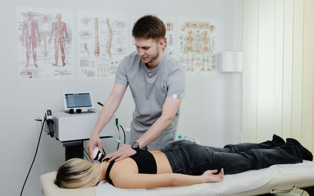
When I set up laser therapy—especially with a robotic head—my first priority is patient comfort and stability. If a patient shifts during an unattended cycle, the beam may drift from the intended target. For lumbar facet-mediated pain at L4–L5, I position the patient comfortably prone, ensure the treatment field is exposed with direct skin access when using a contact handpiece, and confirm the exact region of maximal tenderness and referral (e.g., right-sided zygapophyseal joint pain with proximal radiation).
To minimize error, I zero the device’s X and Y axes, center the beam over the primary pain generator, then expand the field to include adjacent connective tissue tracks. This is our clinical multimodal approach: treat the source, the site, and the surrounding soft tissue network. By caring for the paraspinal fascia, intermuscular septa, and periarticular tissues, we respect that pain is rarely a single-point phenomenon. Fascia transmits load and communicates mechanosensory signals; addressing it improves regional glide and reduces nociceptive drive.
Why direct skin contact? Tissue optics favor minimal reflection and refraction losses. Air-skin interfaces reflect more energy, especially at certain angles. When we must avoid contact—such as at post-surgical sites or in cases of allodynia—we employ a non-contact, collimated robotic head positioned at an optimal focal distance, measured with a calibrated ruler.
Robotic plus handheld delivery: Complementary tools
Robotic head:
Non-contact, collimated beam; ideal for broad areas, post-surgical sensitivity
Software auto-recalculates dose time when X-Y field size changes
Handheld contact piece:
Tactile feedback for focal trigger points and joint spaces
Allows dynamic, movement-based application during active care
In practice, I often run both channels simultaneously. The robot delivers a uniform, programmable energy density across a defined area while I probe and treat focal trigger points or facet capsules with the handheld. This mirrors how we layer manual therapy with exercise: a global reset paired with local precision.
Dosing by energy density: The language of photobiomodulation
Target dose: typically 4–10 joules/cm², depending on condition and depth
Why density matters more than total joules: tissue dose equals energy per unit area
Auto-time calibration: changing the field size while maintaining the same J/cm² adjusts the total joules and time automatically
We dose by energy density, not just total energy. For example, a lumbar facet region might be set to 6 J/cm². On a larger field, total joules increase, but the cellular dose per square centimeter remains constant, aligning with literature-supported ranges that optimize photobiomodulation responses without tipping into bioinhibition. This reflects the Arndt–Schulz principle: too little energy yields no change, optimal energy stimulates, and excessive energy can dampen biological activity.
The physiology behind pain relief and tissue recovery
Mitochondrial activation:
Photons at near-infrared wavelengths interact with cytochrome c oxidase, improving electron transport and boosting ATP production
Enhanced ATP supports ion pump function, cytoskeletal remodeling, and protein synthesis required for tissue repair
Nitric oxide and microcirculation:
Photo-dissociation of nitric oxide from cytochrome c oxidase and endothelial effects promotes vasodilation and microvascular perfusion, aiding oxygen delivery and metabolite clearance
Neuroinflammatory modulation:
Downregulation of pro-inflammatory cytokines and modulation of glial activity reduce peripheral and central sensitization
Neural effects and immediate analgesia:
Modulation of small-diameter nociceptive fibers and gate-control mechanisms can provide early symptom relief
Collagen and connective tissue remodeling:
Changes in fibroblast activity and collagen organization may improve tendon/ligament structure over time when paired with load-specific rehab
In our clinic, patients sometimes report warmth or a faint tingling, but with synchronized pulsed delivery and short pulse durations, surface heat remains low while energy is effectively absorbed at depth. When tissue temperature stays stable over time, we know we are within the desired window: enough photons to trigger biochemical cascades without superficial overheating.
Why pulsed, dual-wavelength delivery matters
Wavelength pairing:
808 nm: deeper penetration for mitochondrial and vascular effects
905 nm: high peak power in short pulses adds neuromodulatory and analgesic benefits while protecting against thermal buildup
Synchronized pulse trains:
High peak, short duration pulses deliver energy in “packets,” allowing absorption periods between bursts and reducing superficial heat accumulation
These engineering choices align with clinical goals: delivering energy to deeper targets, such as facet capsules or the posterior knee compartment, while preserving patient comfort.
Chiropractic integration: Adjustments, motor control, and fascia
Spinal adjustments:
Restoring joint play at hypomobile segments reduces aberrant mechanoreceptor input and reflex muscle guarding
Fascial glide and soft-tissue work:
Instrument-assisted or hands-on release improves shear planes; laser primes fibroblasts and microcirculation for better tissue response
We pair laser sessions with graded movement to convert biochemical gains into functional patterns
Laser does not replace chiropractic care; it helps us reach the dose of movement sooner by lowering pain and stiffness that otherwise block progress. For example, after an MLS session over L4–L5 facets and paraspinals, we cue diaphragmatic breathing and segmental stabilization to capitalize on reduced nociception and improved circulation.
Case walk-through: Low back facet pain (L4–L5)
Assessment:
Right-sided facet loading pain with limited extension and paraspinal tenderness
No red flags; neurological exam stable
Laser setup:
Patient prone, area exposed; robot field centered over right L4–L5 facet region
Density: 6 J/cm², field expanded to capture paraspinal fascia and myofascial referral zones
Handheld: contact sweeps over identified trigger points
Session length:
Robot 6–10 minutes, depending on field size; handheld 20–30 seconds per trigger point
Immediate follow-up:
Prone press-ups to reassess extension tolerance
Gentle lumbar stabilization exercises to lock in gains
Home plan:
Extension-biased mobility as tolerated, core endurance drills, ergonomic cues
What my patients often notice is not just pain relief within hours but improved ease of movement—the kind of change that allows us to progress from passive care to active loading.
Knee osteoarthritis: Accessing the joint intelligently
Beam access matters:
Anterior patella reflects substantial energy; flexing the knee opens the joint space and reduces reflection
Posterior and medial/lateral approaches improve delivery to synovium and periarticular tissues
Dosing strategy:
Target 4–8 J/cm² per compartment; treat multiple compartments in the same session by apportioning field time
Integration with PT:
Laser to modulate pain and effusion
Progressive quadriceps and hip strengthening, gait retraining, and balance work
Manual therapy for capsular mobility as indicated
While no laser regrows cartilage in advanced bone-on-bone disease, many of our patients experience reduced pain and swelling and better function, which can delay the need for surgery. The goal is to expand the movement envelope required for strength and neuromuscular control.
Acute vs. chronic protocols: Cumulative effects and scheduling
Acute conditions:
Six treatments delivered as close to daily as feasible (e.g., Monday–Wednesday–Friday pattern), aiming for rapid symptom control
Chronic conditions:
Twelve treatments, ideally within four weeks, to build cumulative neuroimmune and mitochondrial effects
Why packages:
Effects are additive; stopping after early relief risks relapse before tissue remodeling and motor reeducation are complete
Reassessment points:
After 3–4 sessions: evaluate pain and function
After 6–12 sessions: progress exercise intensity, reduce passive modalities
Our patients often report noticeable changes 4–6 hours after a session; we encourage them to “test” function later the same day (for example, stair climbing or walk tolerance) to anchor improvements to real-life tasks.
Combining laser with orthobiologics and shockwave
With PRP:
Two to three pre-injection laser sessions to improve local perfusion and tissue readiness
Day-of-injection: protocol tailored to avoid blunting intended inflammatory signaling while supporting analgesia
Six post-injection sessions to enhance microcirculation and cellular energy during proliferative phases
With shockwave:
Laser can reduce pain and prime tissues for mechanical signaling from shockwave
Sequence depends on goals; we often laser first for analgesia, then apply focused shockwave for mechanotransduction, followed by graded loading
Rationale:
Photobiomodulation and mechanotherapy act on complementary pathways—bioenergetics and microcirculation (laser) plus tenocyte activation and neovascular remodeling (shockwave)
Hormonal or medication considerations remain in the background for us; when appropriate, we coordinate with the patient’s prescribing providers to avoid interventions (e.g., routine NSAIDs immediately after PRP) that might dampen desired signaling. Our primary emphasis remains movement-based rehabilitation supported by laser and manual care.
Bone and postoperative considerations
Bone healing:
The evidence base for photobiomodulation in fracture healing exists but varies by device and parameters; in clinical experience, early application within 7–10 days post-fracture may support the inflammatory and early reparative phases. This is commonly considered off-label for certain devices and requires case-by-case judgment and collaboration with the treating orthopedic team
Post-surgical care:
Non-contact robotic delivery allows dosing without skin contact when sensitivity is high
Goals include edema control, pain reduction, and earlier initiation of therapeutic exercise
Dose ceilings and the bioinhibition paradox
Arndt–Schulz law:
Insufficient dose yields no effect; optimal dose stimulates; excessive dose may inhibit
Practical application:
If more time is desired, we distribute energy across multiple approaches (e.g., anterior-posterior or medial-lateral fields) instead of stacking excessive dose on one spot
Patients frequently report a “melting” of stiffness within the same day after an MLS session paired with extension-bias exercise; repeated sessions lower baseline pain and improve extension tolerance, allowing us to progress to anti-rotation and hip hinge training
Knee osteoarthritis:
Combining posterior-compartment laser dosing with patellar mobilization and quadriceps strengthening reduces pain during sit-to-stand and stair negotiation within two to three weeks; gains consolidate when patients adhere to home-based strength and balance work
Plantar fasciitis:
Laser applied to the medial calcaneal region and along the plantar fascia with calf mobility and foot intrinsics training shortens the “first-step” pain window and speeds return to walking programs
Post-injection care:
In patients receiving PRP from collaborative providers, pre- and post-injection laser often reduces pain spikes and supports earlier initiation of controlled loading, which in turn improves functional outcomes at 6–12 weeks
Safety, reliability, and patient communication
Safety profile:
Proper eyewear, attention to reflective surfaces, and adherence to dosing ranges keep risk low
Device reliability:
Modern systems include field service support; routine calibration and training ensure consistent delivery
Expectations:
We counsel that pain did not develop in ten minutes and will not vanish in ten; however, many feel better within hours, see consistent improvement after three sessions, and sustain gains with a full plan of care
Putting it all together: A typical plan
Evaluation:
History, movement assessment, palpation, neurological screen, and imaging if indicated
Plan creation:
Define primary pain generators and movement deficits
Choose laser parameters (wavelengths, pulsing, J/cm²) and field geometry
Integrate manual therapy and exercise blocks within each visit
Visit flow:
Laser (robotic field + handheld focal points)
Manual therapy for joint and soft tissue restrictions
Targeted exercises (mobility, motor control, strength)
Education and home program
Progression:
Increase exercise intensity as pain decreases
Taper passive modalities
Reassess goals every 3–4 sessions
Why these techniques work, in plain terms
Pain is both chemical and mechanical. Laser modifies the chemical environment (reduces inflammatory signaling, increases ATP, improves microcirculation). Chiropractic and rehab address the mechanical side (joint motion, tissue glide, strength, coordination). Combining them tackles the problem from both angles
The nervous system adapts to pain by inhibiting movement. Rapid analgesia from laser helps unlock motor patterns so we can retrain stability and strength sooner
Tissues heal under the right load. Once pain is controlled and circulation improved, progressive loading guides collagen alignment and muscle conditioning for durable outcomes
Evidence-based grounding
Photobiomodulation has a growing body of research demonstrating analgesic, anti-inflammatory, and pro-recovery effects in musculoskeletal conditions. Rigorous, modern methodologies—randomized controlled trials, dose–response investigations, and consensus guidelines—support dosing in the 4–10 J/cm² range for many superficial-to-moderate-depth targets and highlight the importance of wavelength, pulse structure, and treatment frequency. Clinical effectiveness is maximized when photobiomodulation is embedded within active rehabilitation rather than used in isolation.
If you are considering care at El Paso Back Clinic, our team will assess your unique presentation and craft an integrative plan that prioritizes spinal mechanics, movement, and function—leveraging laser therapy where it adds value and always keeping the emphasis on your long-term resilience.
IFM's Find A Practitioner tool is the largest referral network in Functional Medicine, created to help patients locate Functional Medicine practitioners anywhere in the world. IFM Certified Practitioners are listed first in the search results, given their extensive education in Functional Medicine