ClickCease
+1-915-850-0900 [email protected]
Select Page
Assessment and Treatment of Upper Trapezius

Assessment and Treatment of Upper Trapezius

These assessment and treatment recommendations represent a synthesis of information derived from personal clinical experience and from the numerous sources which are cited, or are based on the work of researchers, clinicians and therapists who are named (Basmajian 1974, Cailliet 1962, Dvorak & Dvorak 1984, Fryette 1954, Greenman 1989, 1996, Janda 1983, Lewit 1992, 1999, Mennell 1964, Rolf 1977, Williams 1965).


Clinical Application of Neuromuscular Techniques: Upper Trapezius


Lewit (1999) simplifies the need to assess for shortness by stating, �The upper trapezius should be treated if tender and taut.� Since this is an almost universal state in modern life, it seems that everyone requires MET application to this muscle. Lewit also notes that a characteristic mounding of the muscle can often be observed when it is very short, producing the effect of �Gothic shoulders�, similar to the architectural supports of a Gothic church tower (see Fig. 2.13).


Assessment for Shortness of Upper Trapezius (13) (Fig. 4.30)


Figure 4 30 Assessment of the Relative Shortness of the Right Side Upper Trapezius Image 1


Figure 4.30 Assessment of the relative shortness of the right side upper trapezius. One side is compared with the other (for both the range of unforced motion and the nature of the end-feel of motion) to ascertain the side most in need of MET attention.


Test for upper trapezius for shortness (a) See scapulohumeral rhythm test (Ch. 5) which helps identify excessive activity or inappropriate tone in levator scapula and upper trapezius, which, because they are postural muscles, indicates shortness (Fig 5.13A, B). Greenman (1996) describes a functional �firing sequence� assessment which identifies general imbalance and dysfunction involving the upper and lower fixators of the shoulder (Fig. 4.31).


Figure 4 31 Palpation Assessment for Upper and Lower Fixators of the Shoulder Image 2


Figure 4.31 Palpation assessment for upper and lower fixators of the shoulder, including upper trapezius (Greenman 1996).


The patient is seated and the practitioner stands behind. The practitioner rests his right hand over the right shoulder area to assess firing sequence of muscles. The other hand can be placed either on the mid-thoracic region, mainly on the side being assessed, or spanning the lower back to palpate quadratus firing. The assessment should be performed at least twice so that various hand positions are used for different muscles (as in Fig. 4.31).


Greenman bases his description on Janda (1983), who notes the �correct� sequence for shoulder abduction, when seated, as involving: supraspinatus, deltoid, infraspinatus, middle and lower trapezius and finally contralateral quadratus. In dysfunctional states the most common substitutions are said to involve: shoulder elevation by levator scapulae and upper trapezius, as well as early firing by quadratus lumborum, ipsilateral and contralateral.


Inappropriate activity of the upper fixators results in shortness, and of the lower fixators in weakness and possible lengthening (see Ch. 2 for discussion of postural/phasic, etc. muscle characteristics).


Test for upper trapezius for shortness (b) The patient is seated and the practitioner stands behind with one hand resting on the shoulder of the side to be tested and stabilising it. The other hand is placed on the ipsilateral side of the head and the head/neck is taken into contralateral sidebending without force while the shoulder is stabilised (see Fig. 4.30).


The same procedure is performed on the other side with the opposite shoulder stabilised. A comparison is made as to which sidebending manoeuvre produced the greater range and whether the neck can easily reach 45� of side-flexion in each direction, which it should. If neither side can achieve this degree of sidebend, then both trapezius muscles may be short. The relative shortness of one, compared with the other, is evaluated.


Test for upper trapezius for shortness (c) The patient is seated and the practitioner stands behind with a hand resting over the muscle on the side to be assessed. The patient is asked to extend the arm at the shoulder joint, bringing the flexed arm/elbow backwards. If the upper trapezius is stressed on that side it will inappropriately activate during this movement. Since it is a postural muscle, shortness in it can then be assumed (see discussion of postural muscle characteristics in Ch. 3).


Test of upper trapezius for shortness (d) The patient is supine with the neck fully (but not forcefully) sidebent contralaterally (away from the side being assessed). The practitioner is standing at the head of the table and uses a cupped hand contact on the ipsilateral shoulder (i.e. on the side being tested) to assess the ease with which it can be depressed (moved caudally) (Fig. 4.32).


Figure 4 32 MET Treatment of Right Side Upper Trapezius Muscle Image 3


Figure 4.32 MET treatment of right side upper trapezius muscle. A Posterior fibres, B middle fibres, C anterior fibres. Note that stretching in this (or any of the alternative positions which access the middle and posterior fibres) is achieved following the isometric contraction by means of an easing of the shoulder away from the stabilised head, with no force being applied to the neck and head itself.


There should be an easy �springing� sensation as the practitioner pushes the shoulder towards the feet, with a soft end-feel to the movement. If depression of the shoulder is difficult or if there is a harsh, sudden end-point, upper trapezius shortness is confirmed.


This same assessment (always with full lateral flexion) should be performed with the head fully rotated away from the side being treated, half turned away from the side being treated, and slightly turned towards the side being treated, in order to respectively assess the relative shortness and functional efficiency of posterior, middle and anterior subdivisions of the upper portion of trapezius.


MET Treatment of Chronically Shortened Upper Trapezius


MET treatment of upper trapezius, method (a) (Fig. 4.32) In order to treat all the fibres of upper trapezius, MET needs to be applied sequentially. The upper trapezius is subdivided here into anterior, middle and posterior fibres. The neck should be placed into different positions of rotation, coupled with the sidebending as described in the assessment description above, for precise treatment of the various fibres.


The patient lies supine, arm on the side to be treated lying alongside the trunk, head/neck sidebent away from the side being treated to just short of the restriction barrier, while the practitioner stabilises the shoulder with one hand and cups the ear/mastoid area of the same side of the head with the other:


  • With the neck fully sidebent and fully rotated contralaterally, the posterior fibres of upper trapezius are involved in the contraction (see below). This will facilitate subsequent stretching of this aspect of the muscle.
  • With the neck fully sidebent and half rotated, the middle fibres are involved in the contraction.
  • With the neck fully sidebent and slightly rotated towards the side being treated the anterior fibres of upper trapezius are being treated.


The various contractions and subsequent stretches can be performed with practitioner�s arms crossed, hands stabilising the mastoid area and shoulder.


The patient introduces a light resisted effort (20% of available strength) to take the stabilised shoulder towards the ear (a shrug movement) and the ear towards the shoulder. The double movement (or effort towards movement) is important in order to introduce a contraction of the muscle from both ends simultaneously. The degree of effort should be mild and no pain should be felt. The contraction is sustained for 10 seconds (or so) and, upon complete relaxation of effort, the practitioner gently eases the head/neck into an increased degree of sidebending and rotation, where it is stabilised, as the shoulder is stretched caudally.


When stretching is introduced the patient can usefully assist in this phase of the treatment by initiating, on instruction, the stretch of the muscle (�as you breathe out please slide your hand towards your feet�). This reduces the chances of a stretch reflex being initiated. Once the muscle is being stretched, the patient relaxes and the stretch is held for 10�30 seconds.


CAUTION: No stretch should be introduced from the cranial end of the muscle as this could stress the neck. The head is stabilised at its side-flexion and rotation barrier.


Disagreement


There is some disagreement as to the head/neck rotation position as described in the treatment method above, which calls (for posterior and middle fibres) for sidebending and rotation away from the affected side.


Liebenson (1996), suggests that the patient �lies supine with the head supported in anteflexion and laterally flexed away and rotated towards the side of involvement�.


Lewit (1985b) suggests: �The patient is supine � the therapist fixes the shoulder from above with one hand, sidebending the head and neck with the other hand so as to take up the slack. He then asks the patient to look towards the side away from which the head is bent, resisting the patient�s automatic tendency to move towards the side of the lesion.� (This method is described below.)


The author has used the methods described above with good effect and urges readers to try these approaches as well as those of Liebenson and Lewit, and to evaluate results for themselves.


MET treatment of acutely shortened upper trapezius, method (b) Lewit suggests the use of eye movements to facilitate initiation of PIR before stretching, an ideal method for acute problems in this region.


The patient is supine, while the practitioner fixes the shoulder and the sidebent (away from the treated side) head and neck at the restriction barrier and asks the patient to look, with the eyes only (i.e. not to turn the head), towards the side away from which the neck is bent.


This eye movement is maintained, as is a held breath, while the practitioner resists the slight isometric contraction that these two factors (eye movement and breath) will have created.


On exhalation and complete relaxation, the head/neck is taken to a new barrier and the process repeated. If the shoulder is brought into the equation, this is firmly held as it attempts to lightly push into a shrug. After this 10 second contraction the muscle will have released somewhat and slack can again be taken out as the head is repositioned before a repetition of the procedure commences.


Dr. Alex Jimenez offers an additional assessment and treatment of the hip flexors as a part of a referenced clinical application of neuromuscular techniques by Leon Chaitow and Judith Walker DeLany. The scope of our information is limited to chiropractic and spinal injuries and conditions. To discuss the subject matter, please feel free to ask Dr. Jimenez or contact us at 915-850-0900 .


By Dr. Alex Jimenez


Green-Call-Now-Button-24H-150x150-2-3.png


Additional Topics: Wellness


Overall health and wellness are essential towards maintaining the proper mental and physical balance in the body. From eating a balanced nutrition as well as exercising and participating in physical activities, to sleeping a healthy amount of time on a regular basis, following the best health and wellness tips can ultimately help maintain overall well-being. Eating plenty of fruits and vegetables can go a long way towards helping people become healthy.


blog picture of cartoon paperboy big news


WELLNESS TOPIC: EXTRA EXTRA: Managing Workplace Stress


Blank
References
1.�Ballou SK, Keefer L. Multicultural considerations in the diagnosis and management of irritable bowel syndrome: a selective summary.�Eur J Gastroenterol Hepatol.�2013;25:1127�1133.�[PubMed]
2.�Gonzales Gamarra RG, Ruiz S�nchez JG, Le�n Jim�nez F, Cubas Benavides F, D�az V�lez C. [Prevalence of irritable bowel syndrome in the adult population of the city of Chiclayo in 2011]�Rev Gastroenterol Peru.�2012;32:381�386.�[PubMed]
3.�Gwee KA, Lu CL, Ghoshal UC. Epidemiology of irritable bowel syndrome in Asia: something old, something new, something borrowed.�J Gastroenterol Hepatol.�2009;24:1601�1607.�[PubMed]
4.�Krogsgaard LR, Engsbro AL, Bytzer P. The epidemiology of irritable bowel syndrome in Denmark. A population-based survey in adults ? 50 years of age.�Scand J Gastroenterol.�2013;48:523�529.�[PubMed]
5.�Ibrahim NK, Battarjee WF, Almehmadi SA. Prevalence and predictors of irritable bowel syndrome among medical students and interns in King Abdulaziz University, Jeddah.�Libyan J Med.�2013;8:21287.[PMC free article][PubMed]
6.�Lee YY, Waid A, Tan HJ, Chua AS, Whitehead WE. Rome III survey of irritable bowel syndrome among ethnic Malays.�World J Gastroenterol.�2012;18:6475�6480; discussion p. 6479.�[PMC free article][PubMed]
7.�Ghoshal UC, Abraham P, Bhatt C, Choudhuri G, Bhatia SJ, Shenoy KT, Banka NH, Bose K, Bohidar NP, Chakravartty K, et al. Epidemiological and clinical profile of irritable bowel syndrome in India: report of the Indian Society of Gastroenterology Task Force.�Indian J Gastroenterol.�2008;27:22�28.�[PubMed]
8.�Bures J, Cyrany J, Kohoutova D, F�rstl M, Rejchrt S, Kvetina J, Vorisek V, Kopacova M. Small intestinal bacterial overgrowth syndrome.�World J Gastroenterol.�2010;16:2978�2990.�[PMC free article][PubMed]
9.�Dibaise JK, Young RJ, Vanderhoof JA. Enteric microbial flora, bacterial overgrowth, and short-bowel syndrome.�Clin Gastroenterol Hepatol.�2006;4:11�20.�[PubMed]
10.�Bouhnik Y, Alain S, Attar A, Flouri� B, Raskine L, Sanson-Le Pors MJ, Rambaud JC. Bacterial populations contaminating the upper gut in patients with small intestinal bacterial overgrowth syndrome.�Am J Gastroenterol.�1999;94:1327�1331.�[PubMed]
11.�Dukowicz AC, Lacy BE, Levine GM. Small intestinal bacterial overgrowth: a comprehensive review.�Gastroenterol Hepatol (N Y)�2007;3:112�122.�[PMC free article][PubMed]
12.�Posserud I, Stotzer PO, Bj�rnsson ES, Abrahamsson H, Simr�n M. Small intestinal bacterial overgrowth in patients with irritable bowel syndrome.�Gut.�2007;56:802�808.�[PMC free article][PubMed]
13.�Ghoshal U, Ghoshal UC, Ranjan P, Naik SR, Ayyagari A. Spectrum and antibiotic sensitivity of bacteria contaminating the upper gut in patients with malabsorption syndrome from the tropics.�BMC Gastroenterol.�2003;3:9.�[PMC free article][PubMed]
14.�Carrara M, Desideri S, Azzurro M, Bulighin GM, Di Piramo D, Lomonaco L, Adamo S. Small intestine bacterial overgrowth in patients with irritable bowel syndrome.�Eur Rev Med Pharmacol Sci.�2008;12:197�202.�[PubMed]
15.�Ghoshal UC, Ghoshal U, Das K, Misra A. Utility of hydrogen breath tests in diagnosis of small intestinal bacterial overgrowth in malabsorption syndrome and its relationship with oro-cecal transit time.�Indian J Gastroenterol.�2006;25:6�10.�[PubMed]
16.�Ghoshal UC, Kumar S, Mehrotra M, Lakshmi C, Misra A. Frequency of small intestinal bacterial overgrowth in patients with irritable bowel syndrome and chronic non-specific diarrhea.�J Neurogastroenterol Motil.�2010;16:40�46.�[PMC free article][PubMed]
17.�Pimentel M, Chow EJ, Lin HC. Eradication of small intestinal bacterial overgrowth reduces symptoms of irritable bowel syndrome.�Am J Gastroenterol.�2000;95:3503�3506.�[PubMed]
18.�Sachdeva S, Rawat AK, Reddy RS, Puri AS. Small intestinal bacterial overgrowth (SIBO) in irritable bowel syndrome: frequency and predictors.�J Gastroenterol Hepatol.�2011;26 Suppl 3:135�138.�[PubMed]
19.�Park H. The role of small intestinal bacterial overgrowth in the pathophysiology of irritable bowel syndrome.�J Neurogastroenterol Motil.�2010;16:3�4.�[PMC free article][PubMed]
20.�Ghoshal UC, Shukla R, Ghoshal U, Gwee KA, Ng SC, Quigley EM. The gut microbiota and irritable bowel syndrome: friend or foe?�Int J Inflam.�2012;2012:151085.�[PMC free article][PubMed]
21.�Brown AC. Ulcerative colitis, Crohn�s disease and irritable bowel syndrome patients need fecal transplant research and treatment.�J Crohns Colitis.�2014;8:179.�[PubMed]
22.�Sampath K, Levy LC, Gardner TB. Fecal transplantation: beyond the aesthetic.�Gastroenterology.�2013;145:1151�1153.�[PubMed]
23.�Grace E, Shaw C, Whelan K, Andreyev HJ. Review article: small intestinal bacterial overgrowth–prevalence, clinical features, current and developing diagnostic tests, and treatment.�Aliment Pharmacol Ther.�2013;38:674�688.�[PubMed]
24.�Yakoob J, Abbas Z, Khan R, Hamid S, Awan S, Jafri W. Small intestinal bacterial overgrowth and lactose intolerance contribute to irritable bowel syndrome symptomatology in Pakistan.�Saudi J Gastroenterol.�2011;17:371�375.�[PMC free article][PubMed]
25.�Gwee KA, Bak YT, Ghoshal UC, Gonlachanvit S, Lee OY, Fock KM, Chua AS, Lu CL, Goh KL, Kositchaiwat C, et al. Asian consensus on irritable bowel syndrome.�J Gastroenterol Hepatol.�2010;25:1189�1205.�[PubMed]
26.�Lacy BE, Gabbard SL, Crowell MD. Pathophysiology, evaluation, and treatment of bloating: hope, hype, or hot air?�Gastroenterol Hepatol (N Y)�2011;7:729�739.�[PMC free article][PubMed]
27.�Harder H, Serra J, Azpiroz F, Passos MC, Aguad� S, Malagelada JR. Intestinal gas distribution determines abdominal symptoms.�Gut.�2003;52:1708�1713.�[PMC free article][PubMed]
28.�Kumar S, Misra A, Ghoshal UC. Patients with irritable bowel syndrome exhale more hydrogen than healthy subjects in fasting state.�J Neurogastroenterol Motil.�2010;16:299�305.�[PMC free article][PubMed]
29.�Hungin AP, Mulligan C, Pot B, Whorwell P, Agr�us L, Fracasso P, Lionis C, Mendive J, Philippart de Foy JM, Rubin G, Winchester C, de Wit N. Systematic review: probiotics in the management of lower gastrointestinal symptoms in clinical practice — an evidence-based international guide.�Aliment Pharmacol Ther.�2013;38:864�886.�[PMC free article][PubMed]
30.�Attar A, Flouri� B, Rambaud JC, Franchisseur C, Ruszniewski P, Bouhnik Y. Antibiotic efficacy in small intestinal bacterial overgrowth-related chronic diarrhea: a crossover, randomized trial.�Gastroenterology.�1999;117:794�797.�[PubMed]
31.�Marcelino RT, Fagundes-Neto U. [Hydrogen test (H2) in the air expired for the diagnosis of small bowel bacterial overgrowth]�Arq Gastroenterol.�1995;32:191�198.�[PubMed]
32.�Santavirta J. Lactulose hydrogen and [14C]xylose breath tests in patients with ileoanal anastomosis.�Int J Colorectal Dis.�1991;6:208�211.�[PubMed]
33.�Ghoshal UC. How to interpret hydrogen breath tests.�J Neurogastroenterol Motil.�2011;17:312�317.[PMC free article][PubMed]
34.�Yang CY, Chang CS, Chen GH. Small-intestinal bacterial overgrowth in patients with liver cirrhosis, diagnosed with glucose H2 or CH4 breath tests.�Scand J Gastroenterol.�1998;33:867�871.�[PubMed]
35.�Ghoshal UC, Ghoshal U, Ayyagari A, Ranjan P, Krishnani N, Misra A, Aggarwal R, Naik S, Naik SR. Tropical sprue is associated with contamination of small bowel with aerobic bacteria and reversible prolongation of orocecal transit time.�J Gastroenterol Hepatol.�2003;18:540�547.�[PubMed]
36.�Lu CL, Chen CY, Chang FY, Lee SD. Characteristics of small bowel motility in patients with irritable bowel syndrome and normal humans: an Oriental study.�Clin Sci (Lond)�1998;95:165�169.�[PubMed]
37.�Sciarretta G, Furno A, Mazzoni M, Garagnani B, Malaguti P. Lactulose hydrogen breath test in orocecal transit assessment. Critical evaluation by means of scintigraphic method.�Dig Dis Sci.�1994;39:1505�1510.�[PubMed]
38.�Rao SS, Camilleri M, Hasler WL, Maurer AH, Parkman HP, Saad R, Scott MS, Simren M, Soffer E, Szarka L. Evaluation of gastrointestinal transit in clinical practice: position paper of the American and European Neurogastroenterology and Motility Societies.�Neurogastroenterol Motil.�2011;23:8�23.[PubMed]
39.�Yu D, Cheeseman F, Vanner S. Combined oro-caecal scintigraphy and lactulose hydrogen breath testing demonstrate that breath testing detects oro-caecal transit, not small intestinal bacterial overgrowth in patients with IBS.�Gut.�2011;60:334�340.�[PubMed]
40.�Rana SV, Sharma S, Sinha SK, Kaur H, Sikander A, Singh K. Incidence of predominant methanogenic flora in irritable bowel syndrome patients and apparently healthy controls from North India.�Dig Dis Sci.�2009;54:132�135.�[PubMed]
41.�Dridi B, Henry M, El Kh�chine A, Raoult D, Drancourt M. High prevalence of Methanobrevibacter smithii and Methanosphaera stadtmanae detected in the human gut using an improved DNA detection protocol.�PLoS One.�2009;4:e7063.�[PMC free article][PubMed]
42.�Chatterjee S, Park S, Low K, Kong Y, Pimentel M. The degree of breath methane production in IBS correlates with the severity of constipation.�Am J Gastroenterol.�2007;102:837�841.�[PubMed]
43.�Lunia MK, Sharma BC, Sachdeva S. Small intestinal bacterial overgrowth and delayed orocecal transit time in patients with cirrhosis and low-grade hepatic encephalopathy.�Hepatol Int.�2013;7:268�273.[PubMed]
44.�Resmini E, Parodi A, Savarino V, Greco A, Rebora A, Minuto F, Ferone D. Evidence of prolonged orocecal transit time and small intestinal bacterial overgrowth in acromegalic patients.�J Clin Endocrinol Metab.�2007;92:2119�2124.�[PubMed]
45.�Hamilton I, Worsley BW, Cobden I, Cooke EM, Shoesmith JG, Axon AT. Simultaneous culture of saliva and jejunal aspirate in the investigation of small bowel bacterial overgrowth.�Gut.�1982;23:847�853.[PMC free article][PubMed]
46.�Corazza GR, Sorge M, Strocchi A, Benati G, Di Sario A, Treggiari EA, Brusco G, Gasbarrini G. Non-absorbable antibiotics and small bowel bacterial overgrowth.�Ital J Gastroenterol.�1992;24:4�9.�[PubMed]
47.�Kuwahara T, Ogura Y, Oshima K, Kurokawa K, Ooka T, Hirakawa H, Itoh T, Nakayama-Imaohji H, Ichimura M, Itoh K, et al. The lifestyle of the segmented filamentous bacterium: a non-culturable gut-associated immunostimulating microbe inferred by whole-genome sequencing.�DNA Res.�2011;18:291�303.�[PMC free article][PubMed]
48.�Beumer RR, de Vries J, Rombouts FM. Campylobacter jejuni non-culturable coccoid cells.�Int J Food Microbiol.�1992;15:153�163.�[PubMed]
49.�Fromm H, Sarva RP, Ravitch MM, McJunkin B, Farivar S, Amin P. Effects of jejunoileal bypass on the enterohepatic circulation of bile acids, bacterial flora in the upper small intestine, and absorption of vitamin B12.�Metabolism.�1983;32:1133�1141.�[PubMed]
50.�Yoshida T, McCormick WC, Swell L, Vlahcevic ZR. Bile acid metabolism in cirrhosis. IV. Characterization of the abnormality in deoxycholic acid metabolism.�Gastroenterology.�1975;68:335�341.[PubMed]
51.�Bj�rneklett A, Fausa O, Midtvedt T. Bacterial overgrowth in jejunal and ileal disease.�Scand J Gastroenterol.�1983;18:289�298.�[PubMed]
52.�Vanner S. The small intestinal bacterial overgrowth. Irritable bowel syndrome hypothesis: implications for treatment.�Gut.�2008;57:1315�1321.�[PubMed]
53.�Kinross JM, von Roon AC, Holmes E, Darzi A, Nicholson JK. The human gut microbiome: implications for future health care.�Curr Gastroenterol Rep.�2008;10:396�403.�[PubMed]
54.�Clauw DJ. Fibromyalgia: an overview.�Am J Med.�2009;122:S3�S13.�[PubMed]
55.�Pimentel M, Wallace D, Hallegua D, Chow E, Kong Y, Park S, Lin HC. A link between irritable bowel syndrome and fibromyalgia may be related to findings on lactulose breath testing.�Ann Rheum Dis.�2004;63:450�452.�[PMC free article][PubMed]
56.�Simr�n M, Stotzer PO. Use and abuse of hydrogen breath tests.�Gut.�2006;55:297�303.[PMC free article][PubMed]
57.�Gasbarrini A, Lauritano EC, Gabrielli M, Scarpellini E, Lupascu A, Ojetti V, Gasbarrini G. Small intestinal bacterial overgrowth: diagnosis and treatment.�Dig Dis.�2007;25:237�240.�[PubMed]
58.�Ghoshal UC, Srivastava D, Verma A, Misra A. Slow transit constipation associated with excess methane production and its improvement following rifaximin therapy: a case report.�J Neurogastroenterol Motil.�2011;17:185�188.�[PMC free article][PubMed]
59.�Bala L, Ghoshal UC, Ghoshal U, Tripathi P, Misra A, Gowda GA, Khetrapal CL. Malabsorption syndrome with and without small intestinal bacterial overgrowth: a study on upper-gut aspirate using 1H NMR spectroscopy.�Magn Reson Med.�2006;56:738�744.�[PubMed]
60.�Haboubi NY, Lee GS, Montgomery RD. Duodenal mucosal morphometry of elderly patients with small intestinal bacterial overgrowth: response to antibiotic treatment.�Age Ageing.�1991;20:29�32.�[PubMed]
61.�Shindo K, Machida M, Koide K, Fukumura M, Yamazaki R. Deconjugation ability of bacteria isolated from the jejunal fluid of patients with progressive systemic sclerosis and its gastric pH.�Hepatogastroenterology.�1998;45:1643�1650.�[PubMed]
62.�Wanitschke R, Ammon HV. Effects of dihydroxy bile acids and hydroxy fatty acids on the absorption of oleic acid in the human jejunum.�J Clin Invest.�1978;61:178�186.�[PMC free article][PubMed]
63.�Shanab AA, Scully P, Crosbie O, Buckley M, O�Mahony L, Shanahan F, Gazareen S, Murphy E, Quigley EM. Small intestinal bacterial overgrowth in nonalcoholic steatohepatitis: association with toll-like receptor 4 expression and plasma levels of interleukin 8.�Dig Dis Sci.�2011;56:1524�1534.�[PubMed]
64.�Ghoshal UC, Park H, Gwee KA. Bugs and irritable bowel syndrome: The good, the bad and the ugly.�J Gastroenterol Hepatol.�2010;25:244�251.�[PubMed]
65.�Barbara G, Stanghellini V, Brandi G, Cremon C, Di Nardo G, De Giorgio R, Corinaldesi R. Interactions between commensal bacteria and gut sensorimotor function in health and disease.�Am J Gastroenterol.�2005;100:2560�2568.�[PubMed]
66.�Cherbut C, Aub� AC, Blotti�re HM, Galmiche JP. Effects of short-chain fatty acids on gastrointestinal motility.�Scand J Gastroenterol Suppl.�1997;222:58�61.�[PubMed]
67.�Ramakrishna BS, Roediger WE. Bacterial short chain fatty acids: their role in gastrointestinal disease.�Dig Dis.�1990;8:337�345.�[PubMed]
68.�Dumoulin V, Moro F, Barcelo A, Dakka T, Cuber JC. Peptide YY, glucagon-like peptide-1, and neurotensin responses to luminal factors in the isolated vascularly perfused rat ileum.�Endocrinology.�1998;139:3780�3786.�[PubMed]
69.�Balsari A, Ceccarelli A, Dubini F, Fesce E, Poli G. The fecal microbial population in the irritable bowel syndrome.�Microbiologica.�1982;5:185�194.�[PubMed]
70.�Nobaek S, Johansson ML, Molin G, Ahrn� S, Jeppsson B. Alteration of intestinal microflora is associated with reduction in abdominal bloating and pain in patients with irritable bowel syndrome.�Am J Gastroenterol.�2000;95:1231�1238.�[PubMed]
71.�Cummings JH, Macfarlane GT. The control and consequences of bacterial fermentation in the human colon.�J Appl Bacteriol.�1991;70:443�459.�[PubMed]
72.�Camilleri M. Probiotics and irritable bowel syndrome: rationale, mechanisms, and efficacy.�J Clin Gastroenterol.�2008;42 Suppl 3 Pt 1:S123�S125.�[PubMed]
73.�Spiller R. Probiotics: an ideal anti-inflammatory treatment for IBS?�Gastroenterology.�2005;128:783�785.�[PubMed]
74.�Ford AC, Spiegel BM, Talley NJ, Moayyedi P. Small intestinal bacterial overgrowth in irritable bowel syndrome: systematic review and meta-analysis.�Clin Gastroenterol Hepatol.�2009;7:1279�1286.�[PubMed]
75.�Cuoco L, Salvagnini M. Small intestine bacterial overgrowth in irritable bowel syndrome: a retrospective study with rifaximin.�Minerva Gastroenterol Dietol.�2006;52:89�95.�[PubMed]
76.�Di Stefano M, Corazza GR. Treatment of small intestine bacterial overgrowth and related symptoms by rifaximin.�Chemotherapy.�2005;51 Suppl 1:103�109.�[PubMed]
77.�Pimentel M, Lembo A, Chey WD, Zakko S, Ringel Y, Yu J, Mareya SM, Shaw AL, Bortey E, Forbes WP. Rifaximin therapy for patients with irritable bowel syndrome without constipation.�N Engl J Med.�2011;364:22�32.�[PubMed]
78.�Hwang L, Low K, Khoshini R, Melmed G, Sahakian A, Makhani M, Pokkunuri V, Pimentel M. Evaluating breath methane as a diagnostic test for constipation-predominant IBS.�Dig Dis Sci.�2010;55:398�403.�[PubMed]
79.�Low K, Hwang L, Hua J, Zhu A, Morales W, Pimentel M. A combination of rifaximin and neomycin is most effective in treating irritable bowel syndrome patients with methane on lactulose breath test.�J Clin Gastroenterol.�2010;44:547�550.�[PubMed]
80.�Bengmark S. Colonic food: pre- and probiotics.�Am J Gastroenterol.�2000;95:S5�S7.�[PubMed]
81.�Quigley EM, Quera R. Small intestinal bacterial overgrowth: roles of antibiotics, prebiotics, and probiotics.�Gastroenterology.�2006;130:S78�S90.�[PubMed]
82.�Xiao SD, Zhang DZ, Lu H, Jiang SH, Liu HY, Wang GS, Xu GM, Zhang ZB, Lin GJ, Wang GL. Multicenter, randomized, controlled trial of heat-killed Lactobacillus acidophilus LB in patients with chronic diarrhea.�Adv Ther.�2003;20:253�260.�[PubMed]
83.�O’Mahony L, McCarthy J, Kelly P, Hurley G, Luo F, Chen K, O�Sullivan GC, Kiely B, Collins JK, Shanahan F, et al. Lactobacillus and bifidobacterium in irritable bowel syndrome: symptom responses and relationship to cytokine profiles.�Gastroenterology.�2005;128:541�551.�[PubMed]
84.�Tsuchiya J, Barreto R, Okura R, Kawakita S, Fesce E, Marotta F. Single-blind follow-up study on the effectiveness of a symbiotic preparation in irritable bowel syndrome.�Chin J Dig Dis.�2004;5:169�174.[PubMed]
85.�Kim YG, Moon JT, Lee KM, Chon NR, Park H. [The effects of probiotics on symptoms of irritable bowel syndrome]�Korean J Gastroenterol.�2006;47:413�419.�[PubMed]
86.�Park JS, Yu JH, Lim HC, Kim JH, Yoon YH, Park HJ, Lee SI. [Usefulness of lactulose breath test for the prediction of small intestinal bacterial overgrowth in irritable bowel syndrome]�Korean J Gastroenterol.�2010;56:242�248.�[PubMed]
87.�Mann NS, Limoges-Gonzales M. The prevalence of small intestinal bacterial vergrowth in irritable bowel syndrome.�Hepatogastroenterology.�2009;56:718�721.�[PubMed]
88.�Scarpellini E, Giorgio V, Gabrielli M, Lauritano EC, Pantanella A, Fundar� C, Gasbarrini A. Prevalence of small intestinal bacterial overgrowth in children with irritable bowel syndrome: a case-control study.�J Pediatr.�2009;155:416�420.�[PubMed]
89.�Nucera G, Gabrielli M, Lupascu A, Lauritano EC, Santoliquido A, Cremonini F, Cammarota G, Tondi P, Pola P, Gasbarrini G, et al. Abnormal breath tests to lactose, fructose and sorbitol in irritable bowel syndrome may be explained by small intestinal bacterial overgrowth.�Aliment Pharmacol Ther.�2005;21:1391�1395.�[PubMed]
90.�Reddymasu SC, Sostarich S, McCallum RW. Small intestinal bacterial overgrowth in irritable bowel syndrome: are there any predictors?�BMC Gastroenterol.�2010;10:23.�[PMC free article][PubMed]
91.�Lombardo L, Foti M, Ruggia O, Chiecchio A. Increased incidence of small intestinal bacterial overgrowth during proton pump inhibitor therapy.�Clin Gastroenterol Hepatol.�2010;8:504�508.�[PubMed]
92.�Parodi A, Dulbecco P, Savarino E, Giannini EG, Bodini G, Corbo M, Isola L, De Conca S, Marabotto E, Savarino V. Positive glucose breath testing is more prevalent in patients with IBS-like symptoms compared with controls of similar age and gender distribution.�J Clin Gastroenterol.�2009;43:962�966.[PubMed]
93.�Rana SV, Sinha SK, Sikander A, Bhasin DK, Singh K. Study of small intestinal bacterial overgrowth in North Indian patients with irritable bowel syndrome: a case control study.�Trop Gastroenterol.�2008;29:23�25.�[PubMed]
94.�Majewski M, McCallum RW. Results of small intestinal bacterial overgrowth testing in irritable bowel syndrome patients: clinical profiles and effects of antibiotic trial.�Adv Med Sci.�2007;52:139�142.[PubMed]
95.�Lupascu A, Gabrielli M, Lauritano EC, Scarpellini E, Santoliquido A, Cammarota G, Flore R, Tondi P, Pola P, Gasbarrini G, et al. Hydrogen glucose breath test to detect small intestinal bacterial overgrowth: a prevalence case-control study in irritable bowel syndrome.�Aliment Pharmacol Ther.�2005;22:1157�1160.[PubMed]
Close Accordion
Research Studies on SIBO in Irritable Bowel Syndrome

Research Studies on SIBO in Irritable Bowel Syndrome

Irritable bowel syndrome, or IBS, is a prevalent condition characterized by abdominal pain or discomfort, bloating, connected to altered stool form (such as diarrhea and constipation) as well as passage. Approximately 4 percent to 30 percent of individuals world-wide suffer from IBS. Small intestinal bacterial overgrowth, or SIBO, which was clinically demonstrated in patients with structural abnormalities in the gut, such as ileo-transverse anastomosis, stricture, fistula, slow motility and reduced gut defense, may also be characterized by abdominal pain or discomfort, bloating, flatulence and loose motion. It’s been recognized that SIBO may occur in the absence of structural abnormalities. These patients may be incorrectly diagnosed with IBS, or irritable bowel syndrome.


How common is SIBO diagnosed in IBS?


Small intestinal bacterial overgrowth has been described as the excessive presence of bacteria, forming 105 units per milliliter on culture of their intestine aspirate. As this is an invasive test, lots of noninvasive techniques like lactulose and glucose hydrogen breath tests (LHBT and GHBT) are broadly used to diagnose SIBO. This issue has been recognized among people with IBS. In a variety of research studies, frequency of SIBO among patients presenting IBS varied from 4 percent to 78 percent, according to Table ?1, more so among patients with diarrhea-predominant IBS. Not only quantitative increase (SIBO) but qualitative change from the gut bacteria (dysbiosis) was reported among patients with IBS. Research studies utilizing antibiotics and probiotics have caused disagreement to care for this disease with lately transplantation which led to a paradigm shift. Nonetheless, it’s essential to understand the wide-variability in frequency of SIBO among people with IBS. A wide-variability in frequency may indicate it is vital to evaluate the evidence carefully to determine whether the association between IBS and SIBO is under-projected in previous research studies.


Table 1 Summary of Prevalence of SIBO in IBS Image 1


The research studies are examined by people on discordance with the connection between IBS and SIBO as well as their strength and weakness, such as evidence on exploitation of gut flora on indications of IBS and other issues.


Assessment of Studies on SIBO in IBS


Table ?1 summarizes the outcomes among patients with IBS from research studies on individuals with SIBO. As can be noted in the table, the frequency of people with IBS and SIBO varied from 4 percent to 78 percent and from 1 percent and 40 percent among controls. Frequency of individuals with SIBO and IBS was greater than among controls. It might be concluded that SIBO is correlated with IBS. It’s essential to assess the explanations in various research studies.


Critical Evaluation of Studies on SIBO in IBS


Can IBS phenotype determine frequency of SIBO?


IBS is a state that’s heterogeneous. The sub-types may be diarrhea or constipation-predominant or may be alternating. Patients with diarrhea-predominant IBS have organic cause including SIBO compared to other types of IBS. In a study on 129 patients with non-diarrheal IBS, 73 with long-term diarrhea, for example diarrhea-predominant IBS, and 51 healthy controls, frequency of SIBO with GHBT was 11 (8.5 percent), 16 (22 percent) and 1 (2%), respectively. Similar findings are reported in various studies. Diarrheal IBS needs to be evaluated in comparison to other sorts of IBS for SIBO. Research studies that contained percentage of individuals are extremely likely to reveal frequency of SIBO.


Bloating is a symptom commonly reported among patients with IBS. Frequency of bloating has been reported to vary from Asia by 26 percent to 83 percent in research studies on IBS. The pathogenesis of bloating may be correlated with increased quantity of gas in the gut, its abnormal source and improved gut sense in response to distension of the gut. Patients with SIBO may have increased amount of gas inside the gut, so it’s plausible to believe IBS patients with bloating that is noticeable are expected to have SIBO. There is limited data with this specific circumstance. Evidence also demonstrated that both fasting along with post-substrate (e.g., sugar, lactulose) breath hydrogen is considerably higher compared to individuals with IBS compared to controls. Probiotics and antibiotics, which are demonstrated to reduce gas, are demonstrated to ease bloating. It has been noted that treatment can revert hydrogen breath tests back to normal. Patients with IBS, flatulence and bloating should be evaluated for SIBO. More data is involved with this issue.


Can techniques used to diagnose SIBO determine its frequency?


Several techniques are used to diagnose SIBO; including GHBT LHBT,14C breath test, and culture of aspirate. The principle of hydrogen breath tests is summarized in Figure 1. Dietary carbohydrates produce hydrogen in the gut. In patients with SIBO, the bacteria in the small bowel ferment these carbohydrates, producing hydrogen, which gets absorbed and is exhaled in the breath.


Figure 1 Outline of Principle of Method and Interpretation of Glucose and Lactulose Hydrogen Breath Tests Image 2

Figure 1


Hydrogen breath test involves giving patients a load of carbohydrate (generally in the sort of glucose and lactulose) and measuring expired hydrogen concentrations in a period of time. Identification of SIBO using hydrogen breath test depends upon the bodily principle of patients with SIBO, glucose may be fermented by bacteria in the intestine resulting in production of hydrogen gas that is consumed and exhaled in expired air (Figure ?1, A1). By contrast, lactulose, which may function as a non-absorbable disaccharide, will produce an early summit due to fermentation in the small intestine (normally within 90-min) or two summit (as a consequence of small intestine fermentation and minute from colon), if SIBO is present (Figure ?1, B2 and B3). There are limits in hydrogen breath test for identification of SIBO. There may be similarities in patients with problems and SIBO employing rapid transit making differentiation difficult. An ancient summit can be positive in people with gut transit time. By way of instance, in a study from India, median oro-cecal transit interval was 65 minutes (variety 40-110 moments) in healthy subjects. In another study from Taiwan, mean transit interval was 85 min. It’s been substantiated in Western individuals recently by simultaneously using LHBT and radio-nuclide method to gauge gut transport. Double summit standards for evaluation of SIBO using LHBT is quite insensitive. Sensitivity of GHBT to diagnose SIBO is 44 percent contemplating the culture of gut aspirate as a regular standard. As a result, it’s estimated that the researchers who used a historic summit standards in LHBT could discover a greater frequency of SIBO among people with IBS along with controls. In contrast, those who would use either GHBT or double summit benchmark in LHBT might locate a minimum frequency of SIBO alike in patients with IBS and controls. It is well worth noting from Table ?1 that the frequency of SIBO among people with IBS and controls on LHBT (early summit standards) varied from 34.5 percent to 78 percent and 7 percent to 40 percent, respectively; in contrast with the frequency GHBT varied from 8.5 percent to 46 percent and 2 percent to 18percent.


Fifteen percent of people might have methanogenic flora in the gut. Methanobrevibacter smithii, Methanobrevibacter stadmanae and perhaps several of those coliform bacteria are methanogens. In these areas, only hydrogen breath tests may not diagnose SIBO, estimation of methane may also be demanded (Figure ?1). Table ?1 shows that 8.5 percent to 26 percent of IBS sufferers and 0 percent to 25 percent of controls exhaled methane inside their breath. Whether methane was not expected in them, SIBO could not have been diagnosed. Methane was not estimated, which could have resulted in underestimation of frequency of SIBO as outlined in a proportion of the research study. Methane production in excess is connected to constipation. Consequently, methane estimation in breath, which is inaccessible in several commercially available hydrogen breath test machines, is particularly vital in patients employing constipation-predominant IBS. Some could have slow transit through the small intestine making prolonged testing as a lot of hours required and many people may not want to undergo such testing. However, a period of testing for them may overlook SIBO’s identification.


The jejunal aspirate culture has traditionally been used as the gold standard to diagnose SIBO, according to Figure ?2. On the other hand, the limitations of this test include invasiveness in addition to the challenges posed by attempting to civilization all strains and species. In fact, usage of air during endoscopy might lead to a false negative impact as anaerobes do not rise when these are exposed to oxygen. Furthermore, a massive percentage of germs are not cultured. By contrast, single lumen catheter passed through the nose or through the biopsy channel of endoscope, may lead to contamination with oro-pharyngeal flora supplying false positive result. Therefore, we left a double-lumen catheter to prevent these oro-pharyngeal contamination (Figure ?2). Studies on SIBO one of patients with IBS using qualitative civilization of small bowel aspirate are scanty (Table ?1). A study by Posserud et al reported that a frequency of SIBO of 4 percent among people with IBS. Taking the result of study using GHBT, the sensitivity of 44 percent to diagnose the intestine aspirate appears to have the incidence of SIBO . More studies are essential on this issue.


Figure 2 Outline of Method of Culturing Bacteria and Counting the Colonies Image 3

Figure 2


Figure 3 Bile Acid Breath Test Involving Bile Acid and Glycocholic Acid Image 4

Figure 3


13C and�14C based tests have also been developed based on the bacterial metabolism of D-xylose (Figure ?3). Of acids containing13C and�14C may be used to diagnose SIBO. The glycocholic acid breath test contains the managing of the bile acid14C glycocholic acid, as well as the discovery of14CO2, which may be increased in SIBO (Figure 3), according to the clinical and experimental data from the various research studies on SIBO associated with IBS. While evidence may appear conclusive, further research studies may be required to properly determine the results.


Information referenced from the National Center for Biotechnology Information (NCBI) and the National University of Health Sciences. The scope of our information is limited to chiropractic and spinal injuries and conditions. To discuss the subject matter, please feel free to ask Dr. Jimenez or contact us at 915-850-0900 .


By Dr. Alex Jimenez


Green-Call-Now-Button-24H-150x150-2-3.png


Additional Topics: Wellness


Overall health and wellness are essential towards maintaining the proper mental and physical balance in the body. From eating a balanced nutrition as well as exercising and participating in physical activities, to sleeping a healthy amount of time on a regular basis, following the best health and wellness tips can ultimately help maintain overall well-being. Eating plenty of fruits and vegetables can go a long way towards helping people become healthy.


blog picture of cartoon paperboy big news


WELLNESS TOPIC: EXTRA EXTRA: Managing Workplace Stress


Blank
References
1.�Ballou SK, Keefer L. Multicultural considerations in the diagnosis and management of irritable bowel syndrome: a selective summary.�Eur J Gastroenterol Hepatol.�2013;25:1127�1133.�[PubMed]
2.�Gonzales Gamarra RG, Ruiz S�nchez JG, Le�n Jim�nez F, Cubas Benavides F, D�az V�lez C. [Prevalence of irritable bowel syndrome in the adult population of the city of Chiclayo in 2011]�Rev Gastroenterol Peru.�2012;32:381�386.�[PubMed]
3.�Gwee KA, Lu CL, Ghoshal UC. Epidemiology of irritable bowel syndrome in Asia: something old, something new, something borrowed.�J Gastroenterol Hepatol.�2009;24:1601�1607.�[PubMed]
4.�Krogsgaard LR, Engsbro AL, Bytzer P. The epidemiology of irritable bowel syndrome in Denmark. A population-based survey in adults ? 50 years of age.�Scand J Gastroenterol.�2013;48:523�529.�[PubMed]
5.�Ibrahim NK, Battarjee WF, Almehmadi SA. Prevalence and predictors of irritable bowel syndrome among medical students and interns in King Abdulaziz University, Jeddah.�Libyan J Med.�2013;8:21287.[PMC free article][PubMed]
6.�Lee YY, Waid A, Tan HJ, Chua AS, Whitehead WE. Rome III survey of irritable bowel syndrome among ethnic Malays.�World J Gastroenterol.�2012;18:6475�6480; discussion p. 6479.�[PMC free article][PubMed]
7.�Ghoshal UC, Abraham P, Bhatt C, Choudhuri G, Bhatia SJ, Shenoy KT, Banka NH, Bose K, Bohidar NP, Chakravartty K, et al. Epidemiological and clinical profile of irritable bowel syndrome in India: report of the Indian Society of Gastroenterology Task Force.�Indian J Gastroenterol.�2008;27:22�28.�[PubMed]
8.�Bures J, Cyrany J, Kohoutova D, F�rstl M, Rejchrt S, Kvetina J, Vorisek V, Kopacova M. Small intestinal bacterial overgrowth syndrome.�World J Gastroenterol.�2010;16:2978�2990.�[PMC free article][PubMed]
9.�Dibaise JK, Young RJ, Vanderhoof JA. Enteric microbial flora, bacterial overgrowth, and short-bowel syndrome.�Clin Gastroenterol Hepatol.�2006;4:11�20.�[PubMed]
10.�Bouhnik Y, Alain S, Attar A, Flouri� B, Raskine L, Sanson-Le Pors MJ, Rambaud JC. Bacterial populations contaminating the upper gut in patients with small intestinal bacterial overgrowth syndrome.�Am J Gastroenterol.�1999;94:1327�1331.�[PubMed]
11.�Dukowicz AC, Lacy BE, Levine GM. Small intestinal bacterial overgrowth: a comprehensive review.�Gastroenterol Hepatol (N Y)�2007;3:112�122.�[PMC free article][PubMed]
12.�Posserud I, Stotzer PO, Bj�rnsson ES, Abrahamsson H, Simr�n M. Small intestinal bacterial overgrowth in patients with irritable bowel syndrome.�Gut.�2007;56:802�808.�[PMC free article][PubMed]
13.�Ghoshal U, Ghoshal UC, Ranjan P, Naik SR, Ayyagari A. Spectrum and antibiotic sensitivity of bacteria contaminating the upper gut in patients with malabsorption syndrome from the tropics.�BMC Gastroenterol.�2003;3:9.�[PMC free article][PubMed]
14.�Carrara M, Desideri S, Azzurro M, Bulighin GM, Di Piramo D, Lomonaco L, Adamo S. Small intestine bacterial overgrowth in patients with irritable bowel syndrome.�Eur Rev Med Pharmacol Sci.�2008;12:197�202.�[PubMed]
15.�Ghoshal UC, Ghoshal U, Das K, Misra A. Utility of hydrogen breath tests in diagnosis of small intestinal bacterial overgrowth in malabsorption syndrome and its relationship with oro-cecal transit time.�Indian J Gastroenterol.�2006;25:6�10.�[PubMed]
16.�Ghoshal UC, Kumar S, Mehrotra M, Lakshmi C, Misra A. Frequency of small intestinal bacterial overgrowth in patients with irritable bowel syndrome and chronic non-specific diarrhea.�J Neurogastroenterol Motil.�2010;16:40�46.�[PMC free article][PubMed]
17.�Pimentel M, Chow EJ, Lin HC. Eradication of small intestinal bacterial overgrowth reduces symptoms of irritable bowel syndrome.�Am J Gastroenterol.�2000;95:3503�3506.�[PubMed]
18.�Sachdeva S, Rawat AK, Reddy RS, Puri AS. Small intestinal bacterial overgrowth (SIBO) in irritable bowel syndrome: frequency and predictors.�J Gastroenterol Hepatol.�2011;26 Suppl 3:135�138.�[PubMed]
19.�Park H. The role of small intestinal bacterial overgrowth in the pathophysiology of irritable bowel syndrome.�J Neurogastroenterol Motil.�2010;16:3�4.�[PMC free article][PubMed]
20.�Ghoshal UC, Shukla R, Ghoshal U, Gwee KA, Ng SC, Quigley EM. The gut microbiota and irritable bowel syndrome: friend or foe?�Int J Inflam.�2012;2012:151085.�[PMC free article][PubMed]
21.�Brown AC. Ulcerative colitis, Crohn�s disease and irritable bowel syndrome patients need fecal transplant research and treatment.�J Crohns Colitis.�2014;8:179.�[PubMed]
22.�Sampath K, Levy LC, Gardner TB. Fecal transplantation: beyond the aesthetic.�Gastroenterology.�2013;145:1151�1153.�[PubMed]
23.�Grace E, Shaw C, Whelan K, Andreyev HJ. Review article: small intestinal bacterial overgrowth–prevalence, clinical features, current and developing diagnostic tests, and treatment.�Aliment Pharmacol Ther.�2013;38:674�688.�[PubMed]
24.�Yakoob J, Abbas Z, Khan R, Hamid S, Awan S, Jafri W. Small intestinal bacterial overgrowth and lactose intolerance contribute to irritable bowel syndrome symptomatology in Pakistan.�Saudi J Gastroenterol.�2011;17:371�375.�[PMC free article][PubMed]
25.�Gwee KA, Bak YT, Ghoshal UC, Gonlachanvit S, Lee OY, Fock KM, Chua AS, Lu CL, Goh KL, Kositchaiwat C, et al. Asian consensus on irritable bowel syndrome.�J Gastroenterol Hepatol.�2010;25:1189�1205.�[PubMed]
26.�Lacy BE, Gabbard SL, Crowell MD. Pathophysiology, evaluation, and treatment of bloating: hope, hype, or hot air?�Gastroenterol Hepatol (N Y)�2011;7:729�739.�[PMC free article][PubMed]
27.�Harder H, Serra J, Azpiroz F, Passos MC, Aguad� S, Malagelada JR. Intestinal gas distribution determines abdominal symptoms.�Gut.�2003;52:1708�1713.�[PMC free article][PubMed]
28.�Kumar S, Misra A, Ghoshal UC. Patients with irritable bowel syndrome exhale more hydrogen than healthy subjects in fasting state.�J Neurogastroenterol Motil.�2010;16:299�305.�[PMC free article][PubMed]
29.�Hungin AP, Mulligan C, Pot B, Whorwell P, Agr�us L, Fracasso P, Lionis C, Mendive J, Philippart de Foy JM, Rubin G, Winchester C, de Wit N. Systematic review: probiotics in the management of lower gastrointestinal symptoms in clinical practice — an evidence-based international guide.�Aliment Pharmacol Ther.�2013;38:864�886.�[PMC free article][PubMed]
30.�Attar A, Flouri� B, Rambaud JC, Franchisseur C, Ruszniewski P, Bouhnik Y. Antibiotic efficacy in small intestinal bacterial overgrowth-related chronic diarrhea: a crossover, randomized trial.�Gastroenterology.�1999;117:794�797.�[PubMed]
31.�Marcelino RT, Fagundes-Neto U. [Hydrogen test (H2) in the air expired for the diagnosis of small bowel bacterial overgrowth]�Arq Gastroenterol.�1995;32:191�198.�[PubMed]
32.�Santavirta J. Lactulose hydrogen and [14C]xylose breath tests in patients with ileoanal anastomosis.�Int J Colorectal Dis.�1991;6:208�211.�[PubMed]
33.�Ghoshal UC. How to interpret hydrogen breath tests.�J Neurogastroenterol Motil.�2011;17:312�317.[PMC free article][PubMed]
34.�Yang CY, Chang CS, Chen GH. Small-intestinal bacterial overgrowth in patients with liver cirrhosis, diagnosed with glucose H2 or CH4 breath tests.�Scand J Gastroenterol.�1998;33:867�871.�[PubMed]
35.�Ghoshal UC, Ghoshal U, Ayyagari A, Ranjan P, Krishnani N, Misra A, Aggarwal R, Naik S, Naik SR. Tropical sprue is associated with contamination of small bowel with aerobic bacteria and reversible prolongation of orocecal transit time.�J Gastroenterol Hepatol.�2003;18:540�547.�[PubMed]
36.�Lu CL, Chen CY, Chang FY, Lee SD. Characteristics of small bowel motility in patients with irritable bowel syndrome and normal humans: an Oriental study.�Clin Sci (Lond)�1998;95:165�169.�[PubMed]
37.�Sciarretta G, Furno A, Mazzoni M, Garagnani B, Malaguti P. Lactulose hydrogen breath test in orocecal transit assessment. Critical evaluation by means of scintigraphic method.�Dig Dis Sci.�1994;39:1505�1510.�[PubMed]
38.�Rao SS, Camilleri M, Hasler WL, Maurer AH, Parkman HP, Saad R, Scott MS, Simren M, Soffer E, Szarka L. Evaluation of gastrointestinal transit in clinical practice: position paper of the American and European Neurogastroenterology and Motility Societies.�Neurogastroenterol Motil.�2011;23:8�23.[PubMed]
39.�Yu D, Cheeseman F, Vanner S. Combined oro-caecal scintigraphy and lactulose hydrogen breath testing demonstrate that breath testing detects oro-caecal transit, not small intestinal bacterial overgrowth in patients with IBS.�Gut.�2011;60:334�340.�[PubMed]
40.�Rana SV, Sharma S, Sinha SK, Kaur H, Sikander A, Singh K. Incidence of predominant methanogenic flora in irritable bowel syndrome patients and apparently healthy controls from North India.�Dig Dis Sci.�2009;54:132�135.�[PubMed]
41.�Dridi B, Henry M, El Kh�chine A, Raoult D, Drancourt M. High prevalence of Methanobrevibacter smithii and Methanosphaera stadtmanae detected in the human gut using an improved DNA detection protocol.�PLoS One.�2009;4:e7063.�[PMC free article][PubMed]
42.�Chatterjee S, Park S, Low K, Kong Y, Pimentel M. The degree of breath methane production in IBS correlates with the severity of constipation.�Am J Gastroenterol.�2007;102:837�841.�[PubMed]
43.�Lunia MK, Sharma BC, Sachdeva S. Small intestinal bacterial overgrowth and delayed orocecal transit time in patients with cirrhosis and low-grade hepatic encephalopathy.�Hepatol Int.�2013;7:268�273.[PubMed]
44.�Resmini E, Parodi A, Savarino V, Greco A, Rebora A, Minuto F, Ferone D. Evidence of prolonged orocecal transit time and small intestinal bacterial overgrowth in acromegalic patients.�J Clin Endocrinol Metab.�2007;92:2119�2124.�[PubMed]
45.�Hamilton I, Worsley BW, Cobden I, Cooke EM, Shoesmith JG, Axon AT. Simultaneous culture of saliva and jejunal aspirate in the investigation of small bowel bacterial overgrowth.�Gut.�1982;23:847�853.[PMC free article][PubMed]
46.�Corazza GR, Sorge M, Strocchi A, Benati G, Di Sario A, Treggiari EA, Brusco G, Gasbarrini G. Non-absorbable antibiotics and small bowel bacterial overgrowth.�Ital J Gastroenterol.�1992;24:4�9.�[PubMed]
47.�Kuwahara T, Ogura Y, Oshima K, Kurokawa K, Ooka T, Hirakawa H, Itoh T, Nakayama-Imaohji H, Ichimura M, Itoh K, et al. The lifestyle of the segmented filamentous bacterium: a non-culturable gut-associated immunostimulating microbe inferred by whole-genome sequencing.�DNA Res.�2011;18:291�303.�[PMC free article][PubMed]
48.�Beumer RR, de Vries J, Rombouts FM. Campylobacter jejuni non-culturable coccoid cells.�Int J Food Microbiol.�1992;15:153�163.�[PubMed]
49.�Fromm H, Sarva RP, Ravitch MM, McJunkin B, Farivar S, Amin P. Effects of jejunoileal bypass on the enterohepatic circulation of bile acids, bacterial flora in the upper small intestine, and absorption of vitamin B12.�Metabolism.�1983;32:1133�1141.�[PubMed]
50.�Yoshida T, McCormick WC, Swell L, Vlahcevic ZR. Bile acid metabolism in cirrhosis. IV. Characterization of the abnormality in deoxycholic acid metabolism.�Gastroenterology.�1975;68:335�341.[PubMed]
51.�Bj�rneklett A, Fausa O, Midtvedt T. Bacterial overgrowth in jejunal and ileal disease.�Scand J Gastroenterol.�1983;18:289�298.�[PubMed]
52.�Vanner S. The small intestinal bacterial overgrowth. Irritable bowel syndrome hypothesis: implications for treatment.�Gut.�2008;57:1315�1321.�[PubMed]
53.�Kinross JM, von Roon AC, Holmes E, Darzi A, Nicholson JK. The human gut microbiome: implications for future health care.�Curr Gastroenterol Rep.�2008;10:396�403.�[PubMed]
54.�Clauw DJ. Fibromyalgia: an overview.�Am J Med.�2009;122:S3�S13.�[PubMed]
55.�Pimentel M, Wallace D, Hallegua D, Chow E, Kong Y, Park S, Lin HC. A link between irritable bowel syndrome and fibromyalgia may be related to findings on lactulose breath testing.�Ann Rheum Dis.�2004;63:450�452.�[PMC free article][PubMed]
56.�Simr�n M, Stotzer PO. Use and abuse of hydrogen breath tests.�Gut.�2006;55:297�303.[PMC free article][PubMed]
57.�Gasbarrini A, Lauritano EC, Gabrielli M, Scarpellini E, Lupascu A, Ojetti V, Gasbarrini G. Small intestinal bacterial overgrowth: diagnosis and treatment.�Dig Dis.�2007;25:237�240.�[PubMed]
58.�Ghoshal UC, Srivastava D, Verma A, Misra A. Slow transit constipation associated with excess methane production and its improvement following rifaximin therapy: a case report.�J Neurogastroenterol Motil.�2011;17:185�188.�[PMC free article][PubMed]
59.�Bala L, Ghoshal UC, Ghoshal U, Tripathi P, Misra A, Gowda GA, Khetrapal CL. Malabsorption syndrome with and without small intestinal bacterial overgrowth: a study on upper-gut aspirate using 1H NMR spectroscopy.�Magn Reson Med.�2006;56:738�744.�[PubMed]
60.�Haboubi NY, Lee GS, Montgomery RD. Duodenal mucosal morphometry of elderly patients with small intestinal bacterial overgrowth: response to antibiotic treatment.�Age Ageing.�1991;20:29�32.�[PubMed]
61.�Shindo K, Machida M, Koide K, Fukumura M, Yamazaki R. Deconjugation ability of bacteria isolated from the jejunal fluid of patients with progressive systemic sclerosis and its gastric pH.�Hepatogastroenterology.�1998;45:1643�1650.�[PubMed]
62.�Wanitschke R, Ammon HV. Effects of dihydroxy bile acids and hydroxy fatty acids on the absorption of oleic acid in the human jejunum.�J Clin Invest.�1978;61:178�186.�[PMC free article][PubMed]
63.�Shanab AA, Scully P, Crosbie O, Buckley M, O�Mahony L, Shanahan F, Gazareen S, Murphy E, Quigley EM. Small intestinal bacterial overgrowth in nonalcoholic steatohepatitis: association with toll-like receptor 4 expression and plasma levels of interleukin 8.�Dig Dis Sci.�2011;56:1524�1534.�[PubMed]
64.�Ghoshal UC, Park H, Gwee KA. Bugs and irritable bowel syndrome: The good, the bad and the ugly.�J Gastroenterol Hepatol.�2010;25:244�251.�[PubMed]
65.�Barbara G, Stanghellini V, Brandi G, Cremon C, Di Nardo G, De Giorgio R, Corinaldesi R. Interactions between commensal bacteria and gut sensorimotor function in health and disease.�Am J Gastroenterol.�2005;100:2560�2568.�[PubMed]
66.�Cherbut C, Aub� AC, Blotti�re HM, Galmiche JP. Effects of short-chain fatty acids on gastrointestinal motility.�Scand J Gastroenterol Suppl.�1997;222:58�61.�[PubMed]
67.�Ramakrishna BS, Roediger WE. Bacterial short chain fatty acids: their role in gastrointestinal disease.�Dig Dis.�1990;8:337�345.�[PubMed]
68.�Dumoulin V, Moro F, Barcelo A, Dakka T, Cuber JC. Peptide YY, glucagon-like peptide-1, and neurotensin responses to luminal factors in the isolated vascularly perfused rat ileum.�Endocrinology.�1998;139:3780�3786.�[PubMed]
69.�Balsari A, Ceccarelli A, Dubini F, Fesce E, Poli G. The fecal microbial population in the irritable bowel syndrome.�Microbiologica.�1982;5:185�194.�[PubMed]
70.�Nobaek S, Johansson ML, Molin G, Ahrn� S, Jeppsson B. Alteration of intestinal microflora is associated with reduction in abdominal bloating and pain in patients with irritable bowel syndrome.�Am J Gastroenterol.�2000;95:1231�1238.�[PubMed]
71.�Cummings JH, Macfarlane GT. The control and consequences of bacterial fermentation in the human colon.�J Appl Bacteriol.�1991;70:443�459.�[PubMed]
72.�Camilleri M. Probiotics and irritable bowel syndrome: rationale, mechanisms, and efficacy.�J Clin Gastroenterol.�2008;42 Suppl 3 Pt 1:S123�S125.�[PubMed]
73.�Spiller R. Probiotics: an ideal anti-inflammatory treatment for IBS?�Gastroenterology.�2005;128:783�785.�[PubMed]
74.�Ford AC, Spiegel BM, Talley NJ, Moayyedi P. Small intestinal bacterial overgrowth in irritable bowel syndrome: systematic review and meta-analysis.�Clin Gastroenterol Hepatol.�2009;7:1279�1286.�[PubMed]
75.�Cuoco L, Salvagnini M. Small intestine bacterial overgrowth in irritable bowel syndrome: a retrospective study with rifaximin.�Minerva Gastroenterol Dietol.�2006;52:89�95.�[PubMed]
76.�Di Stefano M, Corazza GR. Treatment of small intestine bacterial overgrowth and related symptoms by rifaximin.�Chemotherapy.�2005;51 Suppl 1:103�109.�[PubMed]
77.�Pimentel M, Lembo A, Chey WD, Zakko S, Ringel Y, Yu J, Mareya SM, Shaw AL, Bortey E, Forbes WP. Rifaximin therapy for patients with irritable bowel syndrome without constipation.�N Engl J Med.�2011;364:22�32.�[PubMed]
78.�Hwang L, Low K, Khoshini R, Melmed G, Sahakian A, Makhani M, Pokkunuri V, Pimentel M. Evaluating breath methane as a diagnostic test for constipation-predominant IBS.�Dig Dis Sci.�2010;55:398�403.�[PubMed]
79.�Low K, Hwang L, Hua J, Zhu A, Morales W, Pimentel M. A combination of rifaximin and neomycin is most effective in treating irritable bowel syndrome patients with methane on lactulose breath test.�J Clin Gastroenterol.�2010;44:547�550.�[PubMed]
80.�Bengmark S. Colonic food: pre- and probiotics.�Am J Gastroenterol.�2000;95:S5�S7.�[PubMed]
81.�Quigley EM, Quera R. Small intestinal bacterial overgrowth: roles of antibiotics, prebiotics, and probiotics.�Gastroenterology.�2006;130:S78�S90.�[PubMed]
82.�Xiao SD, Zhang DZ, Lu H, Jiang SH, Liu HY, Wang GS, Xu GM, Zhang ZB, Lin GJ, Wang GL. Multicenter, randomized, controlled trial of heat-killed Lactobacillus acidophilus LB in patients with chronic diarrhea.�Adv Ther.�2003;20:253�260.�[PubMed]
83.�O’Mahony L, McCarthy J, Kelly P, Hurley G, Luo F, Chen K, O�Sullivan GC, Kiely B, Collins JK, Shanahan F, et al. Lactobacillus and bifidobacterium in irritable bowel syndrome: symptom responses and relationship to cytokine profiles.�Gastroenterology.�2005;128:541�551.�[PubMed]
84.�Tsuchiya J, Barreto R, Okura R, Kawakita S, Fesce E, Marotta F. Single-blind follow-up study on the effectiveness of a symbiotic preparation in irritable bowel syndrome.�Chin J Dig Dis.�2004;5:169�174.[PubMed]
85.�Kim YG, Moon JT, Lee KM, Chon NR, Park H. [The effects of probiotics on symptoms of irritable bowel syndrome]�Korean J Gastroenterol.�2006;47:413�419.�[PubMed]
86.�Park JS, Yu JH, Lim HC, Kim JH, Yoon YH, Park HJ, Lee SI. [Usefulness of lactulose breath test for the prediction of small intestinal bacterial overgrowth in irritable bowel syndrome]�Korean J Gastroenterol.�2010;56:242�248.�[PubMed]
87.�Mann NS, Limoges-Gonzales M. The prevalence of small intestinal bacterial vergrowth in irritable bowel syndrome.�Hepatogastroenterology.�2009;56:718�721.�[PubMed]
88.�Scarpellini E, Giorgio V, Gabrielli M, Lauritano EC, Pantanella A, Fundar� C, Gasbarrini A. Prevalence of small intestinal bacterial overgrowth in children with irritable bowel syndrome: a case-control study.�J Pediatr.�2009;155:416�420.�[PubMed]
89.�Nucera G, Gabrielli M, Lupascu A, Lauritano EC, Santoliquido A, Cremonini F, Cammarota G, Tondi P, Pola P, Gasbarrini G, et al. Abnormal breath tests to lactose, fructose and sorbitol in irritable bowel syndrome may be explained by small intestinal bacterial overgrowth.�Aliment Pharmacol Ther.�2005;21:1391�1395.�[PubMed]
90.�Reddymasu SC, Sostarich S, McCallum RW. Small intestinal bacterial overgrowth in irritable bowel syndrome: are there any predictors?�BMC Gastroenterol.�2010;10:23.�[PMC free article][PubMed]
91.�Lombardo L, Foti M, Ruggia O, Chiecchio A. Increased incidence of small intestinal bacterial overgrowth during proton pump inhibitor therapy.�Clin Gastroenterol Hepatol.�2010;8:504�508.�[PubMed]
92.�Parodi A, Dulbecco P, Savarino E, Giannini EG, Bodini G, Corbo M, Isola L, De Conca S, Marabotto E, Savarino V. Positive glucose breath testing is more prevalent in patients with IBS-like symptoms compared with controls of similar age and gender distribution.�J Clin Gastroenterol.�2009;43:962�966.[PubMed]
93.�Rana SV, Sinha SK, Sikander A, Bhasin DK, Singh K. Study of small intestinal bacterial overgrowth in North Indian patients with irritable bowel syndrome: a case control study.�Trop Gastroenterol.�2008;29:23�25.�[PubMed]
94.�Majewski M, McCallum RW. Results of small intestinal bacterial overgrowth testing in irritable bowel syndrome patients: clinical profiles and effects of antibiotic trial.�Adv Med Sci.�2007;52:139�142.[PubMed]
95.�Lupascu A, Gabrielli M, Lauritano EC, Scarpellini E, Santoliquido A, Cammarota G, Flore R, Tondi P, Pola P, Gasbarrini G, et al. Hydrogen glucose breath test to detect small intestinal bacterial overgrowth: a prevalence case-control study in irritable bowel syndrome.�Aliment Pharmacol Ther.�2005;22:1157�1160.[PubMed]
Close Accordion
Psoriasis: Conventional And Alternative Treatment

Psoriasis: Conventional And Alternative Treatment

Psoriasis Abstract

Psoriasis is a common T-cell-mediated immune disorder characterized by circumscribed, red, thickened plaques with an overlying silver-white scale. It occurs worldwide, although the incidence is lower in warmer, sunnier climates. The primary cause of psoriasis is unknown. During an active disease state, an underlying inflammatory mechanism is frequently involved. Many conventional treatments focus on suppressing symptoms associated with psoriasis and have significant side effects. This article reviews several of the researched natural approaches to psoriasis treatment, while addressing its underlying cause. (Altern Med Rev 2007;12(4):319-330)

Introduction

Recent genetic and immunological advances have greatly increased understanding of the pathogenesis of psoriasis as a chronic, immune-mediated inflammatory disorder. The primary immune defect in psoriasis appears to be an increase in cell signaling via chemokines and cytokines that act on upregulated gene expression and cause hyper-proliferation of keratinocytes. A new understanding of this complex disease has catalyzed the development of targeted biological treatments. These revolutionary therapies are not without potential risk, however. A review of alternative natural therapies provides some options for increasing safety and efficacy in the management of psoriasis. Psoriasis � Pathophysiology, Conventional, and Alternative Approaches to Treatment Michael Traub, ND, and Keri Marshall MS, ND

Epidemiology

The prevalence of psoriasis varies widely depending on ethnicity. Psoriasis occurs most commonly in Caucasians, with an estimated occurrence of 60 cases per 100,000/year in this population. Its prevalence in the United States is 2-4 percent, although it is rare or absent in Native American and certain African-American populations. While common in Japan, it is much less common in China, with an estimated incidence of 0.3 percent. The prevalence in the general population of Northern Europe and Scandinavia is 1.5-3 percent. Women and men are equally affected by this condition. The observation that latitude affects prevalence is most likely related to the beneficial effect of sunlight on the disease.1 Although psoriasis can occur at any age, the mean age of onset for chronic plaque psoriasis is estimated at 33 years, with 75 percent of cases initiated before age 46.2 The age of onset appears to be slightly earlier in women than men. Longitudinal studies suggest spontaneous remission may occur in about one-third of patients with psoriasis.3

Pathophysiology

Until recently psoriasis was considered a disorder of epidermal keratinocytes; however, it is now recognized primarily as an immune-mediated disorder. In order to properly understand the immune dysfunction present in psoriasis, it is imperative to understand the normal immune response of skin. Skin is a primary lymphoid organ with an effective immunological surveillance system equipped with antigen presenting cells, cytokine synthesizing keratinocytes, epidermotropic T cells, dermal capillary endothelial cells, draining nodes, mast cells, tissue macrophages, granulocytes, fibroblasts, and non-Langerhans cells. Skin also has lymph nodes and circulating T lymphocytes. Together these cells communicate by means of cytokine secretion and respond accordingly via stimulation by bacteria, chemical, ultraviolet (UV) light, and other irritating factors. The primary cytokine released in response to antigen presentation is tumor necrosis factor-alpha (TNF-?). Generally, this is a controlled process unless the insult to the skin is prolonged, in which case imbalanced cytokine production leads to a pathological state such as psoriasis.

Debate continues whether psoriasis is an autoimmune disorder or a T-helper 1 (Th1) immune dysfunction. T-cell activation, TNF-?, and dendritic cells are pathogenic factors stimulated in response to a triggering factor, such as a physical injury, inflammation, bacteria, virus, or withdrawal of corticosteroid medication. Initially, immature dendritic cells in the epidermis stimulate T-cells from lymph nodes in response to as yet unidentified antigen stimulation. The lymphocytic infiltrate in psoriasis is predominately CD4 and CD8 T cells. Adhesion molecules that promote leukocyte adherence are highly expressed in psoriatic lesions.4 After T cells receive primary stimulation and activation, a resulting synthesis of mRNA for interleukin-2 (IL-2) occurs, resulting in a subsequent increase in IL-2 receptors. Psoriasis is considered a Th1-dominant disease due to the increase in cytokines of the Th1 pathway � interferon gamma (IFN-?), IL-2, and interleukin 12 (IL-12) � found in psoriatic plaques.

The increased IL-2 from activated T cells and IL-12 from Langerhans cells ultimately regulate genes that code for the transcription of cytokines such as IFN-?, TNF-?, and IL-2, responsible for differentiation, maturation, and proliferation of T cells into memory effector cells. Ultimately, T cells migrate to the skin, where they accumulate around dermal blood vessels. These are the first in a series of immunologic changes that result in the formation of acute psoriatic lesions. Because the above-described immune response is a somewhat normal response to antigen stimulation, it remains unclear why the T-cell activation that occurs, followed by subsequent migration of leukocytes into the epidermis and dermis, creates accelerated cellular proliferation. Upregulated gene regulation may be a causative factor. Vascular endothelial growth factor (VEGF) and interleukin-8 released from keratinocytes may contribute to the vascularization seen in psoriasis.5

Dendritic cells appear to be involved in the pathogenesis of psoriasis. One type of dendritic cell involved is the Langerhans cells, the outermost sentinel of the immune system that recognizes and captures antigens, migrates to local lymph nodes, and presents them to T cells. The activation of T lymphocytes releases pro-inflammatory cytokines such as TNF-? that lead to keratinocyte proliferation. This hyperproliferative response decreases epidermal transit time (the approximate time it takes for normal maturation of skin cells) from 28 days to 2-4 days and produces the typical erythematous scaly plaques of psoriasis. This understanding of pathogenic mechanisms has led to the development and therapeutic use of TNF-? blocking agents.

About 30 percent of individuals with psoriasis have a family history of the disease in a first- or seconddegree relative. At least nine chromosomal susceptibility loci have been elucidated (PSORS1-9). HLA-Cw6 is a major determinant of phenotypic expression. An association with the PSORS has been found with functional polymorphisms in modifier genes that mediate inflammation (e.g., TNF-?) and vascular growth (e.g., VEGF).6

It is known that psoriasis develops in bone marrow transplant recipients from donors with psoriasis, clears in recipients from donors without psoriasis, and that immunosuppressive drugs are effective in reducing psoriasis.7,8 Given the genetic predisposition to this disease, what can be done to reduce the genetic expression besides resorting to immunosuppressive therapies? A naturopathic approach consists of dietary modification,�therapeutic fasting, omega-3 supplementation, topical natural medicines, herbal medicine, and stress management.

Pizzorno and Murray propose the above-mentioned �unidentified antigens� result from incomplete protein digestion, increased intestinal permeability, and food allergies; bowel toxemia (endotoxins); impaired liver detoxification; bile acid deficiencies; alcohol consumption; excessive consumption of animal fats; nutrient deficiencies (vitamins A and E, zinc, and selenium); and stress.9 These hypotheses, although plausible, have not been adequately tested.

Co-Morbidities

Psoriasis is associated with several co-morbidities, including decreased quality of life, depression, increased cardiovascular risk, type 2 diabetes mellitus, metabolic syndrome, cancer, Crohn�s disease, and psoriatic arthritis. It remains unclear whether cancers, in particular skin cancer and lymphoma, are related to psoriasis or to its treatment. Phototherapy and immunosuppressive therapy can increase the risk of non-melanoma skin cancer, for example.10

Of particular concern is the observed link between psoriasis and cardiovascular disease. Evidence indicates psoriasis is an independent risk factor for cardiovascular disease.11 Dyslipidemia, coronary calcification, increased highly sensitive C-reactive protein (CRP), decreased folate, and hyperhomocysteinemia are found with significantly higher frequency in psoriasis patients.12 Inflammation is the common theme underlying both conditions, characterized by the presence of pro-inflammatory cytokines and endothelial activation.

The inflammatory processes underlying psoriasis also suggest the possibility of omega-3 fatty acid, folate, and vitamin B12 deficiencies, which are also often found in cardiovascular disease.13 High homocysteine and decreased folate levels correlate with Psoriasis Area and Severity Index (PASI). A rapid skin cell turnover rate in psoriasis may result in increased folate utilization and subsequent deficiency.14 The author of one study concludes: �Dietary supplementation of folic acid, B6, and B12 appears reasonable in psoriasis patients, particularly those with elevated homocysteine, low folate and additional cardiovascular risk factors.�15

Psoriatic arthritis is a clinical condition occurring in 25 percent of individuals afflicted with psoriasis.16 In approximately 10 percent of this population, the arthritic symptoms precede the skin lesions. Psoriatic arthritis often presents as seronegative inflammatory arthritis, with a classic presentation consisting of oligoarthritis, distal interphalangeal joint involvement, dactylitis (inflammation of the digits), and calcaneal inflammation.

Opinions conflict whether the skin condition and arthritis represent a differing manifestation of the same disease. Genetic evidence, immunological studies, and treatment response variability suggest they may be two different conditions, perhaps with similar underlying inflammation and immune irregularity.17,18

Although palmoplantar pustulosis (PP) is often described as a subtype of psoriasis, different demographics and genetic analysis suggests a different etiology than psoriasis. On appearance, PP has yellowbrown sterile pustules that appear on palms and soles. Only 25 percent of those affected report chronic plaque psoriasis. PP occurs more frequently in women (9:1/ female:male) and 95 percent of affected people have a current or previous history of smoking. As a result, PP may be considered a co-morbid condition rather than a distinct form of psoriasis.19

Diagnostic Criteria

Psoriasis is classified into several subtypes, with the chronic plaque (psoriasis vulgaris) form comprising approximately 90 percent of cases. Sharply demarcated erythematous silvery scaling plaques occur most commonly on the extensor surface of the elbows, knees, scalp, sacral, and groin regions. Other involved areas include the ears, glans penis, perianal region, and sites of repeated trauma. An active inflammatory case of psoriasis can demonstrate the Koebner phenomenon in which new lesions form at a site of trauma or pressure.

In the future, chronic plaque psoriasis might be found to consist of several related conditions with distinct phenotypical and genotypical characteristics, providing an explanation for its variable response to therapy, especially with biologic agents.

Inverse psoriasis occurs in intertriginous sites and skin folds and is red, shiny, and usually without scaling. Sebopsoriasis, which is often confused with seborrheic dermatitis, is characterized by greasy scales�in the eyebrows, nasolabial folds, and postauricular and presternal areas.

Acute guttate psoriasis occurs in children, adolescents, and young adults approximately two weeks after an acute beta-hemolytic streptococcal infection, such as tonsillitis or pharyngitis, or a viral infection. It manifests as an erythematous, papular eruption with lesions less than 1 cm in diameter on the trunk and extremities. Acute guttate psoriasis is usually self-limited, resolving within 3-4 months. One study indicated only one-third of individuals with guttate psoriasis develop classic plaque psoriasis.20

Pustular psoriasis (von Zumbusch) is also an acute psoriatic eruption. The patient presents with fever and small, monomorphic, painful, sterile pustules, often precipitated by an intercurrent infection or the abrupt withdrawal of systemic or superpotent topical steroids. It can be localized to the palms and soles (palmar-plantar psoriasis) or it can be generalized and potentially life-threatening.

Erythrodermic psoriasis, also life threatening, involves the entire body surface and can result in hypothermia, hypoalbuminemia, anemia, infection, and high-output cardiac failure.

Psoriatic nail disease occurs in approximately 50 percent of psoriasis patients and most commonly manifests as pitting. Other nail changes can include onycholysis, discoloration, thickening, and dystrophy.

Risk Factors

Development of psoriasis involves interaction of multiple genetic risk factors with environmental factors, such as beta-hemolytic streptococcal infection, HIV, stress, and medications (e.g., beta-blockers and lithium). As previously mentioned, folate and vitamin B12 deficiency can also predispose. In addition, there is evidence that alcoholism, cigarette smoking, obesity, type 2 diabetes mellitus, and metabolic syndrome increase risk for developing psoriasis.

With the exception of VEGF, no biomarkers have been found as reliable predictors of psoriasis activity. CRP, soluble adhesion molecules, and soluble cytokine receptors have been investigated but do not correlate with severity.21

Conventional Treatment

Conventional treatment of psoriasis is based on the degree of severity. Mild and limited psoriasis treatment includes topical corticosteroids, tars, anthralin, calcipotriene (a vitamin D3 analog), tazarotene (a retinoid), and phototherapy. Physicians can set realistic expectations for therapy, giving the patient control over the disease without expectation of complete cure. Scalp psoriasis usually responds to salicylic acid shampoos.

Narrow-band UVB is less effective but safer than psoralen plus ultraviolet A (PUVA), which carries with it an increased risk of skin cancer. Sun exposure is another form of phototherapy. UV exposure reduces antigen presenting and affects cell signaling, favoring development of T-helper 2 (Th2) immune responses. Antigen-presenting Langerhans cells are decreased in both number and function.22

A topical combination of calcipotriene and betamethasone (Taclonex�) has shown greater efficacy in severe psoriasis than monotherapy with either alone.23

Patient compliance must be considered when developing a treatment plan. The use of less messy topical solution and foam preparations of topical corticosteroids and calcipotriene (compared to ointments and creams) can improve compliance.

Systemic treatment of severe psoriasis usually involves the use of oral retinoids, methotrexate, cyclosporine, and biological agents that can significantly impact other bodily systems.

The oral retinoid acitretin is teratogenic and is converted to etretinate with concomitant alcohol ingestion. Etretinate has a longer half-life and is more teratogenic than acitretin. Female patients must use two forms of birth control and must not become pregnant for at least three years after treatment. Because of possible interaction with oral contraceptives, St. John�s wort (Hypericum perfoliatum) should be avoided. Other adverse effects include mucocutaneous effects, elevated triglycerides, alopecia, and hepatitis. Treatment with acitretin requires frequent monitoring of blood counts, comprehensive metabolic profiles, and urinalysis. Strategies to reduce acitretin toxicity include intermittent use, reduction of maintenance dose to every other day or every third day, combination treatment with PUVA or topical calcipotriene, low-fat diet, aerobic exercise, fish oil supplementation, and as stated above, alcohol avoidance.

Methotrexate (MTX) is the most commonly used systemic agent for psoriasis and, because it has been available for 35 years, most dermatologists are comfortable with its use. Methotrexate inhibits dihydrofolate reductase (resulting in a deficiency of active folic acid) and induces adenosine A1, a potent anti-inflammatory agonist. Its mechanism of action may be even more complex, evidenced by the fact that caffeine inhibits MTX�s anti-inflammatory effects in rheumatoid arthritis but not in psoriasis or psoriatic arthritis.24 The most common serious adverse effects of MTX are myelosuppression and liver fibrosis. While myelosuppression does not frequently occur, patients using MTX often report symptoms of headache, fatigue, and nausea. Folate supplementation reduces the incidence of megaloblastic anemia, hepatotoxicity, and gastrointestinal intolerance. Although folic acid and folinic acid appear to be equally effective, folic acid is more cost effective.25 However, a recent double-blind study of 22 psoriasis patients stable on long-term MTX therapy revealed folic acid reduced MTX�s efficacy in controlling psoriasis. Patients were randomly assigned to receive 5 mg/day folic acid or placebo for 12 weeks. The mean PASI increased (worsened) in the folic acid group, from 6.4 at baseline to 10.8 at 12 weeks. In the placebo group, the mean PASI fell from 9.8 at baseline to 9.2 at 12 weeks (p<0.05 for the difference in the change between groups).26

Cyclosporine, a potent and toxic drug, is sometimes considered for cases not controlled with acitretin, PUVA, or MTX, but is contraindicated in patients with abnormal renal function, poorly controlled hypertension, hepatic dysfunction, or immunosuppression. Prolonged use inevitably results in renal damage. Blood pressure and creatinine monitoring is essential.

Biological agents block T-cell activation and TNF-?. Alefacept (Amevive�) interferes with T-cell activation and reduces circulating CD 45 RO+ T cells. This drug is a fusion protein of the Fc receptor of human IgG1 and LFA3, a co-stimulatory ligand, which interacts with CD2 on the surface of T-cells. CD4 cells must be monitored weekly during treatment with this agent.

Efalizumab (Raptiva�) is a humanized antibody to CD11 that interferes with T-cell trafficking into inflamed tissues and prevents T-cell activation. Although it is rapidly effective, rebound may occur.

TNF-? blockers downregulate proinflammatory gene expression and reverse the psoriatic phenotype. Etanercept (Enbrel�) is a fusion protein directed against soluble TNF-?. Infliximab (Remicade�) is a mouse/human chimeric monoclonal antibody against soluble and cell-bound TNF-?, while adalimumab (Humira�) is a human monoclonal antibody against TNF-?. These TNF-? inhibitors are administered by injection and have been associated with the induction of various autoimmune phenomena. Like TNF-? itself, TNF-? inhibitors can have both proinflammatory and anti-inflammatory activities. Just because a particular agent blocks TNF-?, it does not necessarily benefit psoriasis. If a patient is genetically predisposed to overproducing TNF-?, blocking it may not be sufficient to produce benefit.27 Possible risks of TNF-? blockers include reactivation of latent tuberculosis, hepatotoxicity, lymphoma, and congestive heart failure.

Challenges that remain with biologics for psoriasis include: (1) understanding the predominant mechanism in psoriasis and psoriatic arthritis; (2) understanding different patient responses to therapy; (3) predicting clinical response before or early in therapy; (4) developing oral, inhaled, and topical formulations; and (5) determining whether treatment alters longterm outcome.

Fumaric acid is the primary psoriasis therapy in Germany. It decreases T-cell dependent cytokines, but is not as effective as other conventional treatments, and carries a high risk of toxicity and gastrointestinal intolerance.

Providing rotational and combination therapies increases efficacy and decreases toxicity of treatment. The future may bring stem-cell therapy and gene-based therapies, including �antisense� treatments that directly inhibit psoriasis-specific genes. However, the adverse effects and toxicity of conventional psoriasis treatments necessitate safer and effective natural treatments that can be used as alternatives or in an integrative fashion.

Natural Treatments For Psoriasis

Diet

An evidence-based approach suggests psoriasis, essentially an inflammatory disorder, should benefit from an anti-inflammatory diet, identification, elimination and/or rotation of allergenic foods, and therapeutic fasting.28-30 Although there is no published data on food allergy avoidance, many psoriasis patients show increased sensitivity to gluten and their psoriasis symptoms improve on a gluten-free diet.31 Measurement of antibodies to tissue transglutaminase and gliadin can help identify this subgroup. Evidence also suggests maintaining a healthy weight benefits psoriasis patients, since psoriasis positively correlates with increased body mass index.32

The balance between proinflammatory and anti-inflammatory eicosanoids is influenced in large part by the type of dietary fatty acids consumed. An antiinflammatory diet consists basically of an emphasis on �good fats� (cold water fish, nuts, seeds, olive oil, other high quality oils), whole grains, legumes, vegetables, and fruits and the avoidance of �bad fats� (saturated animal fats, trans fats, fried and processed foods, poor quality oils) and refined carbohydrates. In addition, an excessive amount of omega-6 fatty acids in the diet can contribute to an inflammatory response.33 The primary sources of dietary omega-6 oils are vegetable oils such as corn, soy, safflower, and sunflower, while the primary sources of arachidonic acid are meat, eggs, and dairy.

Prostaglandin E2 (PGE2) is a prominent eicosanoid derived from the omega-6 fatty acid arachidonic acid. A dominant action of PGE2 as a messenger molecule is to enhance sensitivity in pain neurons, increase swelling, and constrict blood vessels. Over-consumption of omega-6 oils provides excess substrate for the synthesis of PGE2, which drives an aggressive and sustained inflammatory response. Prostaglandin E3 (PGE3) is�derived from the omega-3 fatty acid, eicosapentaenoic acid (EPA). Higher levels of PGE3 reduce sensitivity to pain, relax blood vessels, increase blood flow, and support the body�s natural anti-inflammatory response (Figure 1).

psoriasis illustration

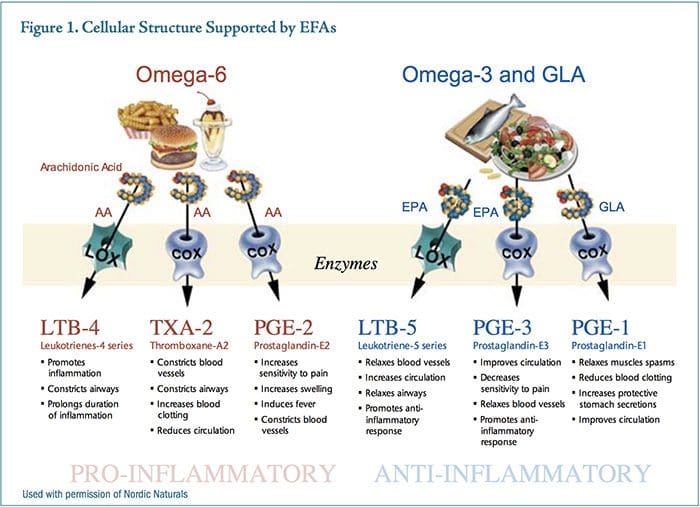
While both PGE2 and PGE3 are necessary for proper homeostasis, the relative amounts of these competing messenger molecules either contribute to or mitigate chronic inflammatory syndromes. EPA is thought to act by competing with arachidonic acid for binding sites on cyclooxygenase-2 (COX-2), producing a less potent inflammatory mediator, therefore reducing inflammation.34

Prior to the Industrial Revolution, there were no significant sources of omega-6 vegetable oils in the diet. Most cultures consumed diets low in these oils and high in fish or range-fed beef or bison higher in omega-3s, creating a ratio of omega-6:omega-3 that was approximately 3:1. The Industrial Revolution brought with it the knowledge and tools to refine vegetable oils, resulting in a rapid shift in dietary habits for most Western cultures. The ratio of omega-6:omega-3 was quickly pushed toward the current estimate of as high as 11:1 omega-6:omega-3.35 The human body has not been able to genetically adapt to this dramatic shift in fatty acid consumption.

Many modern cultures consume copious amounts of vegetable oils, mostly in processed foods. For example, soy oil production for food consumption increased 1,000-fold between 1909 and 1999.36 In addition, livestock, poultry, and farmed fish are being fed cornmeal and soy-based feed, which raises the omega-6 content of the meat and fish. When farm animals are raised on grass, worms, or other natural diets, the tissues are naturally higher in omega-3 fatty acids.37

The beef industry touts �marbling� in finished beef products, which is due to the corn and soy feed. Corn- and soy-fed cattle have a higher omega-6 fatty acid content compared to grass-fed cattle. While grassfed cattle can contain up to 4-percent omega-3 fatty acids, corn-fed cattle typically contains 0.5-percent omega-3s.37

The standard American diet supplies an average omega-6:omega-3 ratio of approximately 11:1. A vegetarian-based diet may put an individual at risk for�eating high amounts of vegetable oils and soy products, and low amounts of fish, which can tip the balance toward a pro-inflammatory state. Reducing dietary vegetable oils and increasing the omega-3 fats EPA and docosahexaenoic acid (DHA) by consuming fatty fish such as cod, salmon, mackerel, and sardines can benefit individuals experiencing chronic inflammatory conditions.33

Several herbs used as seasonings, including turmeric, red pepper, cloves, ginger, cumin, anise, fennel, basil, rosemary, garlic, and pomegranate, can block nuclear factor-kappaB (NF?B) activation of inflammatory cytokines.38

Dietary approaches that modify fatty acid intake can influence the eicosanoid profile in such a way that inflammatory processes such as arachidonic acid production and T-cell activation are dampened, while cytokines such as interleukin-4 (the primary cytokine responsible for stimulating a Th2 immune response) are upregulated.34

Nutritional Supplementation

Essential Fatty Acids

Essential fatty acids (EFAs) influence the pathophysiology of psoriasis in three ways: first, EFAs impact the kinetics of cell membranes; second, EFAs impact dermal and epidermal blood flow via improved endothelial function; and third, EFAs act as an immunomodulating agent through their impact on eicosanoids. EFAs are used as basic substrates in the development of the phospholipid bi-layer in virtually every cell in the human body, including the dermis and epidermis. They create structural integrity that regulates fluidity, which impacts cell transport, messenger binding, and cell communication. Omega-3 fatty acids can act both directly and indirectly on endothelial function by reducing mononuclear cell cytokines such as IL-1 and TNF?, 39 decreasing formation of chemo-attractant protein platelet-derived growth factor (PDGF), increasing bioavailability of nitric oxide, and reducing expression of adhesion molecules. The cumulative effect modulating these bioactive mediators is to prevent vascularization, or new blood vessel growth within the psoriatic plaque, while simultaneously allowing improved perfusion of dermal tissue.

Components of both natural and acquired immunity, including the production of key immune modulators, can be affected by omega-3 and -6 fatty acid intake, as discussed above. Immunomodulatory effects of omega-3 fatty acids include suppression of lymphoproliferation, CD4+ cells, antigen presentation, adhesion molecule presentation, Th1 and Th2 responses, and pro-inflammatory cytokine production.34

Several studies have demonstrated the benefit of intravenous or oral supplementation of fish oil for psoriasis.40-42 In a study by Mayser et al, intravenous infusions of omega-3 fatty acids led to an increase in the anti-inflammatory leukotriene B5 (LTB5) within 4-7 days of starting treatment, when compared to control patients.43 In this trial, patients received either an omega-3 or omega-6 preparation twice daily for 10 days. No side effects were noted.

EPA competes with arachidonic acid (AA) for 5-lipoxygenase and produces LTB5, which is only one-tenth as potent as the inflammatory mediator leukotriene B4 (LTB4). Levels of LTB4 have been shown to be elevated in psoriatic plaques and demonstrate chemotactic properties necessary for infiltration of leukocyte and keratinocyte proliferation.43

Ziboh�s review article on omega-3s and psoriasis references six studies conducted using oral fish oil supplementation with mixed results. Unfortunately, original references cannot be found. Two studies were double-blind and placebo-controlled, using 1.8 g EPA and DHA over courses of eight weeks and 12 weeks. The eight-week study demonstrated benefit in itching, scaling, and erythema, while the 12-week study showed no benefit.44

Three open studies were conducted, providing 10-18 g EPA and DHA daily for eight weeks. All studies showed improvement, with two studies demonstrating mild-to-moderate and one study demonstrating moderate-to-excellent improvement in scaling, itching, and lesion thickness. One open study combined with a low-fat diet showed a significant reduction in psoriatic symptoms.44,45

Several studies have explored the use of topical fish oil at varying EPA concentrations. Some studies reported benefits, including a reduction in plaque thickness and scaling.46,47 In one study by Puglia et al, fish oil extracts and ketoprofen were applied topically to�psoriatic lesions, with an observed reduction in erythema.48 The most significant drawback to topical fish oil application is compliance due to the odor.

Fish oil has also proven to be beneficial in autoimmune joint conditions such as rheumatoid arthritis (RA).49 While fish oil supplementation has not been used in clinical trials for the treatment of psoriatic arthritis, it may be beneficial in treating this condition, which has many similarities to RA, including a common underlying inflammatory mechanism and immune dysfunction.

Folate

Methotrexate therapy results in a folate deficiency. As mentioned above, in patients receiving MTX for psoriasis, folate supplementation reduced the incidence of hepatotoxicity and gastrointestinal intolerance but might impair the efficacy of MTX.24 When supplementing with folic acid or the active forms, folinic acid or 5-methyltetrahydrofolate, the recommended dose is 1-5 mg/day.

Bioactive Whey Protein Isolate

XP-828L is a novel dietary supplement made of a protein extract derived from bovine whey that has recently been shown to be beneficial in psoriasis.50,51 The bioactive profile of XP-828L is likely due to the presence of growth factors, immunoglobulins, and active peptides found in this specific whey extract. An in vitro study demonstrated XP-828L has immune-regulating effects, including inhibiting the production of Th1 cytokines such as IFN-g and IL-2, which may make it effective in treating T-helper 1-related disorders, such as psoriasis.52

An open-label study was conducted on 11 adult patients with chronic, stable plaque psoriasis on two percent or more of total body surface area. Study participants received 5 g twice daily of XP-828L for 56 days. Evaluations using PASI and Physician�s Global Assessment (PGA) scores were made on the initial screening day and again on days 1, 28, and 56. At the conclusion of the study, seven of the 11 subjects had a reduced PASI score that ranged from 9.5 percent to 81.3 percent.50 The results of a larger double-blind,�placebo-controlled study of 84 individuals with mildto-moderate psoriasis showed XP-828L (5 g/day for 56 days) significantly reduced the PGA score compared to placebo (p<0.05). No adverse affects were noted from any study participants in either study.50,51

Vitamin D

It has been established that patients with disseminated psoriasis have significantly decreased serum levels of the biologically active form of vitamin D, 1-alpha,25-dihydroxyvitamin D3 (1-?,25(OH)2D3; calcitriol) compared to age- and sex-matched controls and also compared to patients with moderate psoriasis.53 Whether this is a contributing factor to psoriasis or a result of the disorder has not been elucidated.

Keratinocytes in the epidermis convert 7-dehydrocholesterol to vitamin D3 in the presence of UVB. Sunlight, UVB phototherapy, oral calcitriol, and topical vitamin D analogs are effective therapy for psoriasis due to vitamin D�s anti-proliferative and pro-differentiating actions on keratinocytes.54-56

Calcitriol-binding to vitamin D receptors (VDR) in the skin modulates the expression of a large number of genes including cell cycle regulators, growth factors, and their receptors. Polymorphisms of the VDR gene are associated with psoriasis and may predispose to the development of psoriasis and resistance to calcipotriol therapy, as well as contribute to liver dysfunction in patients with psoriasis.57

Given vitamin D�s importance in psoriasis, cancer, inflammatory diseases, and other conditions, it has been suggested by some investigators that recommendations for sun protection and skin cancer prevention may need to be re-evaluated to allow for sufficient vitamin D status. A recent study showed abundant sun exposure in a sample of adults in Hawaii did not necessarily ensure vitamin D adequacy, which points to the need for vitamin D supplementation to achieve optimal blood levels.58

Studies have demonstrated that oral vitamin D can be safely taken in daily doses of up to 5,000 IU, with some experts recommending up to 10,000 IU daily to correct a deficiency.59-61 Oral and topical vitamin D, sunlight, and UVB phototherapy have shown considerable efficacy in the treatment of psoriasis.56

Topical Treatments Of Psoriasis

Several topical treatments for psoriasis may provide benefit, including calcipotriene (Dovonex�; a synthetic vitamin D3 analogue), Berberis aquifolium cream (10%)62 (Psoriaflora�; Relieva�), curcumin gel (1%), Aloe vera, and a flavonoid-rich salve (Flavsalve�).

Curcumin gel yielded 90-percent resolution of plaques in 50 percent of patients within 2-6 weeks; the remainder of the study subjects showed 50- to 85-percent improvement. Curcumin was found to be twice as effective as calcipotriol cream (which generally takes three months to exert its full effect). The mechanism of curcumin is as a selective phosphorylase kinase inhibitor, thereby reducing inflammation through inhibition of NF?B.63

A controlled trial of Aloe vera extract cream (0.5%) in 60 patients for 4-12 months demonstrated a significant clearing of psoriatic plaques (82.8%) compared to placebo (7.7%) (p<0.001). In addition, the PASI decreased to a mean of 2.2.64

The scaliness of psoriasis benefits from the use of emollients. Intercellular lipids such as ceramides (lipid molecules composed of fatty acids and sphingosine) play an important role in the regulation of skin-water barrier homeostasis and water-holding capacity. It has been shown that ceramides are decreased in the psoriatic epidermis. Newer ceramide-containing emollients (e.g., CeraVe�, Mimyx�, Aveeno Eczema Care) have shown benefit in psoriasis and may improve skin barrier function and decrease water loss.65

Botanical Influences

A Chinese herbal formula (Herose� Psoria Capsule) has demonstrated safety and efficacy in the treatment of severe plaque psoriasis.66 Herose consists of rhizoma Zingiberis, radix Salviae miltiorrhizae, radix Astragali, ramulus Cinnamomi, radix Paeoniae alba, radix Codonopsis pilosula, and semen Coicis. In an openlabel trial, 15 subjects took four Herose capsules (450 mg each) three times daily for 10 months. The investigator evaluated the PASI and therapeutic response to Herose for each patient. The formula is intended for warming the yang and promoting blood circulation.

Lifestyle Interventions

Lifestyle factors such as cigarette smoking and alcohol consumption are associated with severity of psoriasis.67 Physical activity and outdoor activities (taking precautions not to sunburn) are beneficial.68 Bathing and sunbathing at the Dead Sea for four weeks resulted in a decrease of PASI of 81.5 percent, a 78-percent decrease in keratinocyte hyperplasia, and almost total elimination of T lymphocytes from the epidermis, with a low number remaining in the dermis.69

Stress management can benefit individuals with psoriasis. Subjects who listened to a guided meditation tape while undergoing phototherapy cleared four times faster than those who received phototherapy only, as judged by two independent dermatologists. Psoriasis status was assessed in three ways: direct inspection by clinic nurses; direct inspection by physicians blinded to the patient�s study condition (tape or no-tape); and blinded physician evaluation of photographs of psoriasis lesions. Four sequential indicators of skin status were monitored during the study: a First Response Point, a Turning Point, a Halfway Point, and a Clearing Point. Subjects in the tape groups reached the Halfway Point (p= 0.013) and the Clearing Point (p=0.033) significantly more rapidly than those in the no-tape condition, for both UVB and PUVA treatments.70 Finally, psychotherapy can be an essential adjunct for individuals with persistent unresolved psychological issues such as anxiety, depression, and the psychosocial stress of this chronic skin disease.

Discussion

Psoriasis is characterized by T-cell activation that releases pro-inflammatory cytokines such as TNF-?, leading to keratinocyte proliferation and the typical skin lesions of psoriasis.

The conventional approach to psoriasis consists of utilizing topical and/or oral corticosteroids, other immunosuppressant drugs, oral retinoids, UV light, and several (not necessarily novel, having been used previously for Crohn�s and RA) biological agents. Although these treatments can be highly effective at controlling the disease, none are universally safe and effective, and each carries a considerable risk profile.

There is some evidence for the use of dietary modification and fish oil to decrease inflammation in psoriasis. More research is warranted to clarify the use�of these and various topical botanical therapies and lifestyle interventions for improving clinical symptoms, decreasing the phenotypic expression of psoriasis, and providing safe and effective treatments.


blank
References:

1. Griffiths CEM, Camp RDR, Barker JNWN.
Psoriasis. In: Burns DA, Breathnach SM, Cox N,
Griffiths CE, eds. Rook�s Textbook of Dermatology. 7th
ed. Oxford: Blackwell; 2005:35.1-35.69.
2. Nevitt GJ, Hutchinson PE. Psoriasis in the
community; prevalence, severity and patients belief
and attitudes towards the disease. Br J Dermatol
1996;135:533-537.
3. Farber EM, Nall ML. The natural history of psoriasis
in 5600 patients. Dermatologica 1974;148:1-18.
4. Robert C, Kupper TS. Inflammatory skin diseases,
T cells and immune surveillance. N Engl J Med
1999;341:1817-1828.
5. Simonetti O, Lucarini G, Goteri G, et al. VEGF is
likely a key factor in the link between inflammation
and angiogenesis in psoriasis: results of an
immunohistochemical study. Int J Immunopathol
Pharmacol 2006;19:751-760.
6. Capon F, Munro M, Barker J, Trembath R. Searching
for the major histocompatibility complex psoriasis
susceptibility gene. J Invest Dermatol 2002;118:745-
751.
7. Wahie S, Alexandroff A, Reynolds NJ, Meggit SJ.
Psoriasis occurring after myeloablative therapy and
autologous stem cell transplantation. Br J Dermatol
2006;154:194-195.
8. Eedy DJ, Burrows D, Bridges JM, Jones FG.
Clearance of severe psoriasis after allogenic bone
marrow transplantation. BMJ 1990;300:908.
9. Pizzorno JE, Murray MT. Textbook of Natural
Medicine. 3rd ed. St. Louis, MO: Churchill
Livingstone; 2006:2080.
10. Lindelof B, Eklund G, Liden S, Stern RS. The
prevalence of malignant tumors in patients with
psoriasis. J Am Acad Dermatol 1990;22:1056-1060.
11. Mrowietz U, Elder JT, Barker J. The importance of
disease associations and concomitant therapy for the
long-term management of psoriasis patients. Arch
Dermatol Res 2006;298:309-319.
12. Rocha-Pereira P, Santos-Silva A, Rebelo I, et al.
Dyslipidemia and oxidative stress in mild and in
severe psoriasis as a risk for cardiovascular disease.
Clin Chim Acta 2001;303:33-39.
13. Ludwig RJ, Herzog C, Rostock A, et al. Psoriasis:
a possible risk factor for development of coronary
artery calcification. Br J Dermatol 2007;156:271-276.

14. Vanizor Kural B, Orem A, Cimsit G, et al.
Plasma homocysteine and its relationships with
atherothrombotic markers in psoriatic patients. Clin
Chim Acta 2003;332:23-30.
15. Malerba M, Gisondi P, Radaeli A, et al. Plasma
homocysteine and folate levels in patients
with chronic plaque psoriasis. Br J Dermatol
2006;155:1165-1169.
16. Zachariae H. Prevalence of joint disease in patients
with psoriasis: implications for therapy. Am J Clin
Dermatol 2003;4:441-447.
17. Ho P, Bruce IN, Silman A, et al. Evidence for
common genetic control in pathways of inflammation
for Crohn�s disease and psoriatic arthritis. Arthritis
Rheum 2005;52:3596-3602.
18. Pitzalis C, Cauli A, Pipitone N, et al. Cutaneous
lymphocyte antigen-positive T lymphocytes
preferentially migrate to the skin but not to the joint
in psoriatic arthritis. Arthritis Rheum 1996;39:137-
145.
19. Asumalahti K, Ameen M, Suomela S, et al. Genetic
analysis of PSORS1 distinguishes guttate psoriasis
and palmoplantar pustulosis. J Invest Dermatol
2003;120:627-632.
20. Martin BA, Chalmers RJ, Telfer NR. How great
is the risk of further psoriasis following a single
episode of acute guttate psoriasis? Arch Dermatol
1996:132:717-718.
21. Creamer D, Allen MH, Groves RW, Barker JN.
Circulating vascular permeability factor/vascular
endothelial growth factor in erythroderma. Lancet
1996;348:1101.
22. Zanolli MD, Camisa C, Feldman S, et al. Psoriasis:
the high notes on current treatment. Program of the
American Academy of Dermatology, Academy 2000;
August 5, 2000; Nashville, TN.
23. Kaufmann R, Bibby AJ, Bissonnette R, et al. A new
calcipotriol/betamethasone dipropionate formulation
(Daivobet) is an effective once-daily treatment for
psoriasis vulgaris. Dermatology 2002;205:389-393.
24. Swanson DL, Barnes SA, Mengden Koon SJ, elAzhary
RA. Caffeine consumption and methotrexate
dosing requirement in psoriasis and psoriatic arthritis.
Int J Dermatol 2007;46:157-159.
25. Strober BE, Menon K. Folate supplementation during
methotrexate therapy for patients with psoriasis. J
Am Acad Dermatol 2005;53:652-659.
26. Salim A, Tan E, Ilchyshyn A, Berth-Jones J. Folic acid
supplementation during treatment of psoriasis with
methotrexate: a randomized, double-blind, placebocontrolled
trial. Br J Dermatol 2006;154:1169-1174.
27. Fiorentino D. The yin and yang of TNF-(alpha)
inhibition. Arch Dermatol 2007;143:233-236.
28. Wolters M. Diet and psoriasis: experimental data and
clinical evidence. Br J Dermatol 2005;153:706-714.
29. Brown AC, Hairfield M, Richards DG, et al. Medical
nutrition therapy as a potential complementary
treatment for psoriasis � five case reports. Altern Med
Rev 2004;9:297-307.
30. Lithell H, Bruce A, Gustafsson IB, et al. A fasting
and vegetarian diet treatment trial on chronic
inflammatory disorders. Acta Derm Venereol
1983;63:397-403.
31. Chalmers RJ, Kirby B. Gluten and psoriasis. Br J
Dermatol 2000;142:5-7.
32. Naldi L, Parazzini F, Peli L, et al. Dietary factors and
the risk of psoriasis. Results of an Italian case-control
study. Br J Dermatol 1996;134:101-106.
33. Adam O, Beringer C, Kless T, et al. Antiinflammatory
effects of a low arachidonic acid diet
and fish oil in patients with rheumatoid arthritis.
Rheumatol Int 2003;23:27-36.
34. Calder PC. n-3 Polyunsaturated fatty acids,
inflammation, and inflammatory diseases. Am J Clin
Nutr 2006;83:1505S-1519S.
35. Yehuda S.Omega-6/omega-3 ratio and brain-related
functions. World Rev Nutr Diet 2003;92:37-56.
36. Sirtori CR. Risks and benefits of soy phytoestrogens
in cardiovascular diseases, cancer, climacteric
symptoms and osteoporosis. Drug Saf 2001;24:665-
682.
37. Marchello MJ, Driskell JA. Nutrient composition of
grass- and grain-finished bison. Great Plains Research
2001;11:65-82.
38. Aggarwal BB, Shishodia S. Suppression of the
nuclear factor-kappaB activation pathway by spicederived
phytochemicals: reasoning for seasoning. Ann
N Y Acad Sci 2004;1030:434-441.
39. Yaqoob P. Fatty acids as gatekeepers of immune cell
regulation. Trends Immunol 2003;24:639-645.
40. Bittiner SB, Tucker WF, Cartwright I, Bleehen SS. A
double-blind, randomised, placebo-controlled trial of
fish oil in psoriasis. Lancet 1988;1:378-380.
41. Gupta AK, Ellis CN, Tellner DC, et al. Double-blind,
placebo-controlled study to evaluate the efficacy
of fish oil and low-dose UVB in the treatment of
psoriasis. Br J Dermatol 1989;120:801-807.
42. Mayser P, Mrowietz U, Arenberger P, et al. Omega-3
fatty acid-based lipid infusion in patients with
chronic plaque psoriasis: results of a double-blind,
randomized, placebo-controlled, multicenter trial. J
Am Acad Dermatol 1998;38:539-547.
43. Mayser P, Grimm H, Grimminger F. n-3 fatty acids in
psoriasis. Br J Nutr 2002;87:S77-S82.
44. Ziboh VA. The role of n-3 fatty acids in psoriasis. In:
Kremer J, ed. Medicinal Fatty Acids in Inflammation.
Basel, Switzerland: Birkhauser Verlag; 1998:45-53.

45. Calder PC. n-3 Polyunsaturated fatty acids,
inflammation and immunity: pouring oil on troubled
waters or another fishy tale? Nutr Res 2001;21:309-
341.
46. Zulfakar MH, Edwards M, Heard CM. Is there a role
for topically delivered eicosapentaenoic acid in the
treatment of psoriasis? Eur J Dermatol 2007;17:284-
291.
47. Richards H, Thomas CP, Bowen JL, Heard CM.
In vitro transcutaneous delivery of ketoprofen and
polyunsaturated fatty acids from a pluronic lecithin
organogel vehicle containing fish oil. J Pharm
Pharmacol 2006;58:903-908.
48. Puglia C, Tropea S, Rizza L, et al. In vitro
percutaneous absorption studies and in vivo
evaluation of anti-inflammatory activity of essential
fatty acids (EFA) from fish oil extracts. Int J Pharm
2005;299:41-48.
49. Cleland LG, James MJ. Fish oil and rheumatoid
arthritis: antiinflammatory and collateral health
benefits. J Rheumatol 2000;27:2305-2307.
50. Poulin Y, Pouliot Y, Lamiot E, et al. Safety and
efficacy of a milk-derived extract in the treatment of
plaque psoriasis: an open-label study. J Cutan Med
Surg 2005;9:271-275.
51. Poulin Y, Bissonnette R, Juneau C, et al. XP-828L
in the treatment of mild to moderate psoriasis:
randomized, double-blind, placebo-controlled study. J
Cutan Med Surg 2006;10:241-248.
52. Aattouri N, Gauthier SF, Santure M, et al.
Immunosuppressive effect of a milk-derived extract.
12th International Congress of Immunology and 4th
Annual Conference of FOCIS. Montreal, Canada;
July 18-23, 2004.
53. Staberg B, Oxholm A, Klemp P, Christiansen C.
Abnormal vitamin D metabolism in patients with
psoriasis. Acta Derm Venereol 1987;67:65-68.
54. Reichrath J. Vitamin D and the skin: an ancient
friend, revisited. Exp Dermatol 2007;16:618-625.
55. Osmancevic A, Landin-Wilhelmsen K, Larko O,
et al. UVB therapy increases 25(OH) vitamin D
syntheses in postmenopausal women with psoriasis.
Photodermatol Photoimmunol Photomed 2007;23:172-
178.
56. Perez A, Raab R, Chen TC, et al. Safety and efficacy
of oral calcitriol (1,25-dihydroxyvitamin D3) for the
treatment of psoriasis. Br J Dermatol 1996;134:1070-
1078.
57. Okita H, Ohtsuka T, Yamakage A, Yamazaki
S. Polymorphism of the vitamin D(3) receptor
in patients with psoriasis. Arch Dermatol Res
2002;294:159-162.
58. Binkley N, Novotny R, Krueger D, et al. Low vitamin
D status despite abundant sun exposure. J Clin
Endocrinol Metab 2007;92:2130-2135.
59. Grant WB, Holick MF. Benefits and requirements of
vitamin D for optimal health: a review. Altern Med
Rev 2005;10:94-111.
60. Hollis BW. Circulating 25-hydroxyvitamin
D levels indicative of vitamin D sufficiency:
implications for establishing a new effective dietary
intake recommendation for vitamin D. J Nutr
2005;135:317-322.
61. Vieth R, Bischoff-Ferrari H, Boucher BJ, et al. The
urgent need to recommend an intake of vitamin D
that is effective. Am J Clin Nutr 2007;85:649-650.
62. Gulliver WP, Donsky HJ. A report on three recent
clinical trials using Mahonia aquifolium 10% topical
cream and a review of the worldwide clinical
experience with Mahonia aquifolium for the treatment
of plaque psoriasis. Am J Ther 2005;12:398-406.
63. Heng MC, Song MK, Harker J, Heng MK. Druginduced
suppression of phosphorylase kinase activity
correlates with resolution of psoriasis as assessed
by clinical, histological and immunohistochemical
parameters. Br J Dermatol 2000;143:937-949.
64. Syed TA, Ahmad SA, Holt AH, et al. Management
of psoriasis with Aloe vera extract in a hydrophilic
cream: a placebo-controlled, double-blind study. Trop
Med Int Health 1996;1:505-509.
65. Lew BL, Cho Y, Kim J, et al. Ceramides and cell
signaling molecules in psoriatic epidermis: reduced
levels of ceramides, PKC-alpha, and JNK. J Korean
Med Sci 2006;21:95-99.
66. Yuqi TT. Review of a treatment for psoriasis using
Herose, a botanical formula. J Dermatol 2005;32:940-
945.
67. Chodorowska G, Kwiatek J. Psoriasis and cigarette
smoking. Ann Univ Mariae Curie Sklodowska [Med]
2004;59:535-538.
68. Schiener R, Brockow T, Franke A, et al. Bath PUVA
and saltwater baths followed by UV-B phototherapy
as treatments for psoriasis: a randomized controlled
trial. Arch Dermatol 2007;143:586-596.
69. Hodak E, Gottlieb AB, Segal T, et al. Climatotherapy
at the Dead Sea is a remittive therapy for psoriasis:
combined effects on epidermal and immunologic
activation. J Am Acad Dermatol 2003;49:451-457.
70. Kabat-Zinn J, Wheeler E, Light T, et al. Influence
of a mindfulness meditation-based stress reduction
intervention on rates of skin clearing in patients
with moderate to severe psoriasis undergoing
phototherapy (UVB) and photochemotherapy
(PUVA). Psychosom Medicine 1998;60:625-632.

Close Accordion
Suffer From Sciatica: Chiropractic Care Reduces Pain, Promotes Healing

Suffer From Sciatica: Chiropractic Care Reduces Pain, Promotes Healing

Suffer Sciatica: Are you experiencing pain along one side of your body from your lower back down through your hip and the back of your leg? If so, you could be suffering from a condition called sciatica.

According to the Mayo Clinic, sciatica can best be described as “most commonly occurring when a herniated disk or a bone spur on the spine compresses part of the nerve. This causes inflammation, pain and often some numbness in the affected leg.”

A variety of issues weigh in on an individual’s likelihood of ending up with sciatica. Most of them deal with increased pressure on the spine.

Suffer Sciatica: Causes

Obesity: carrying too much weight is instrumental in bringing on a number of health related issues. Extra pounds overload the spine, causing damage that results in sciatica.

Improper Lifting: Individuals who frequently twist the bodies and lift heavy loads are more likely to suffer from sciatica. Certain jobs that require these movements are a key cause of the condition.

Sedentary Lifestyle. A person’s job does not have to involve lifting to be responsible for this condition. Sitting for extended periods without stretching or standing puts excess pressure on the spine and can cause sciatica.

Too Many Birthdays. Getting older can affect all of our body’s joints and bones in a negative manner, especially if we never committed to an exercise routing. An individual’s back often deteriorates with age, causing bone spurs and herniated disks that sometimes result in sciatica.

Treatment options for sciatica are varied, and the choice depends on the severity of the condition.

Pain Medication: A common and easy way to treat sciatica is with drug therapy. Anti-inflammatory drugs are frequently used to reduce�the inflammation around the nerve, which is a big contributor of the pain. Over-the-counter pain medicines, as well as codeine, may also help with pain management.

suffer medical diagnosis of a pinched nerve

Acupuncture. Alternative therapies like acupuncture have shown positive results in the treatment of sciatica. If a drug-free treatment option appeals to you, find an experienced acupuncturist in your area and talk to them about treatment options.

Strengthening Exercises. A consistent exercise program strengthens your muscles and helps the body function effectively. Ask your doctor which exercises assist the body with bouncing back from sciatica.

Supplements. Supplying the body with vital vitamins and minerals assists in overall health in general, including improvement from sciatica. Daily doses of supplements such as calcium, magnesium, St. John’s Wort, and Vitamin B12 have shown to treat sciatica effectively.

Chiropractic Care. Chiropractors understand all things spine-related, and work with the body as a whole to help it heal itself. Chiropractic treatment for sciatica works to align the spine and reduce the stress to the lower back. Treatment helps alleviate the underlying causes of the condition, and shows positive results in a short amount of time.

Cortisone Injections. Most of the time, sciatica can be treated by the less invasive measures mentioned above. However, severe bouts of sciatica may require a shot of cortisone directly into the inflamed area. Individuals generally choose this option when other treatments have garnered no relief.

Dealing with sciatica is painful and irritating, as the condition often sidelines the sufferer from daily activities. By knowing the treatment options that are effective in combating both the underlying causes and the pain of sciatica, sufferers can begin a regimen that will help them get back on their feet, pain-free in the shortest period of time possible and no longer have to suffer.

If you are suffering from sciatica and would like to talk to an experienced chiropractor about how to treat the condition, contact us today.

Sciatica

This article is copyrighted by Blogging Chiros LLC for its Doctor of Chiropractic members and may not be copied or duplicated in any manner including printed or electronic media, regardless of whether for a fee or gratis without the prior written permission of Blogging Chiros, LLC.

Expectant Mothers Benefit From Chiropractic Care

Expectant Mothers Benefit From Chiropractic Care

Expectant Mothers: Pregnancy is an exciting, precious time in a woman’s life, full of new experiences. Unfortunately, the baby’s development brings about bodily changes that often wreak havoc on the back and joints, and end up causing pain. These issues also frequently cause issues during delivery, and increase the time it takes for the body to recover post-pregnancy.

Expectant moms benefit from chiropractic care in a number of ways. Here are five key ways chiropractic care helps alleviate the toll pregnancy takes on a woman’s body.

#1: Expectant Mothers: Chiropractic Keeps The Spine In Alignment.

Pregnancy adds significant additional weight to a woman’s body in a short amount of time. This change bears on the spine, frequently pulling it out of alignment.

When this happens, the pain can be quite severe. Chiropractic care during pregnancy works to keep the spine in alignment and all supporting tendons working optimally, to be better prepared and able to adequately support the extra weight.

#2: Chiropractic Reduces Need For Pain Relievers.

Most times, individuals experiencing moderate pain pop a couple of over the counter pain relievers and think nothing of it. However, pregnant women strive to avoid medications when possible.

Chiropractic adjustments decrease the underlying issues that cause pain, so the patient relies less on medications. Experiencing less pain as well as eliminating the need for pain killers is a win-win situation for expectant mothers.

expectant#3: Chiropractic Strengthens And Repairs Joints.

Pregnancy really beats up an expectant mothers joints. Chiropractic care for expectant mothers is a productive way to minimize the effect the large, protruding abdomen has on her hips, legs, and ankles.

Treating the body as a whole, chiropractic treatment works to strengthen the body and promotes healing of injured or strained areas.

#4: Chiropractic Helps Achieve Pelvic Alignment.

An aligned pelvis is critical to the birthing process, and increases the chances of being able to give birth naturally. According to the American Pregnancy Association,

“When the pelvis is misaligned it may reduce the amount of room available for the developing baby. This restriction is called intrauterine constraint. A misaligned pelvis may also make it difficult for the baby to get into the best possible position for delivery. This can affect the mother�s ability to have a natural, non-invasive birth.”

An experienced chiropractor can effectively align the pelvis before delivery, so the mother is able to deliver with little incident.

#5: Chiropractic Increases The Body’s Ability To Bounce Back.

Let’s face it, every pregnant woman thinks “will I ever fit in the clingy red dress again?” The healthier and stronger a woman’s body is before and during pregnancy, the easier it is to get back into shape once the baby is born. Eating right and safely exercising are effective ways to accomplish this.

Chiropractic care is also a valuable component to fitness. Expectant mothers who choose chiropractic enjoy better posture, less pain, and increased mobility, especially late in the third trimester.

This allows them to maintain exercise routines and be active longer than those suffering from back pain and achy joints. After the delivery, it’s easier to get back into a fitness routine, and into that red dress, if the new mother’s joints, back, and hips are aligned and functioning properly.

Chiropractic care can serve to reduce pain and increase the overall heath of expectant mothers, letting her relax and focus on the more pleasant aspects of pregnancy. Expecting women who commit to chiropractic care can look forward to a stronger body, the chances of a smoother delivery, and an easier recovery after the baby comes.

Pregnancy & Chiropractic Care

This article is copyrighted by Blogging Chiros LLC for its Doctor of Chiropractic members and may not be copied or duplicated in any manner including printed or electronic media, regardless of whether for a fee or gratis without the prior written permission of Blogging Chiros, LLC.

Chronic Ear Infections And Children: The Chiropractic Answer

Chronic Ear Infections And Children: The Chiropractic Answer

Ear Infections: “Mom, it hurts. It hurts!”

Parents of children suffering from ear infections are all too familiar with this cry. Tugging at their ears, crying, and trouble sleeping are all signs of an ear infection in your little one.

Children are more susceptible to ear infections than adults because of their smaller Eustachian tubes, and their less effective immune systems. Unfortunately, chronic ear infections can quickly become a painful, ongoing problem.

Parents have a variety of options at their disposal to treat their child’s ear infections. Drops, antibiotics, and ventilation tubes placed by surgical procedure are all commonly used treatments for chronic infections. Chiropractic care is another option available and is rapidly gaining acceptance and popularity for the effective treatment of ear infections.

According to HealthyChild.com, ear infections are the reason for 35% of pediatrician visits, and ear pain is the number one reason a child visits a chiropractor. Children who suffer from recurring ear infections benefit from chiropractic care in several ways that are not available through other, more traditional forms of treatment.

Here are a few of the benefits chiropractic care offer for ear infections are:

Ear Infections: It Is Non-Invasive

Unlike the surgical procedure that is often used to combat ear infections by inserting tubes in the child’s ears, chiropractic adjustments provide a non-invasive form of treatment.

This treatment option works well because a little one’s spine may be moved out of alignment from the birthing process, or from any number of spills he or she makes learning to walk, run, or navigate stairs. An experienced chiropractor can map out a treatment plan that includes gentle adjustments of the child’s neck and spine. The goal is to decrease fluid build up within the ear canals that bring on ear infections and pain.

It Is A Painless Option Of Treatment

The last thing parents want in any medical treatment is to cause their child more pain. A negative experience at a doctor’s office can create a fear that lasts for years to come.

Children with ear infections have nothing to fear from visiting a chiropractor. During a chiropractic visit, the child will participate in adjustments that are pain free. The treatments will be able to reduce the pain in a short amount of time in many instances, and reduce the chance of dealing with another ear infection down the road.

ear infectionsIt Treats The Underlying Cause

Other options of treatment may decrease the symptoms of an ear infection, but never get to the actual root issue. Chiropractic care strives to return the body in its entirety to its normal function.

This is the main building block concept for chiropractic in general. By re-aligning the spine, the body as a whole can be healthier and more resistant to infection and disease. Chiropractic care helps adjust the child’s body back to a normal state, thus providing a way for it to heal itself and fight off future ear (and other) infections better than simply using drops and antibiotics.

If your child is susceptible to ear infections, there are steps you can take to minimize the chances of an occurrence in the first place. Frequent hand washing to prevent the spread of germs and avoiding cigarette smoke are two simple ways to combat ear infections.

Chiropractic treatment continues to show promise in treating the underlying issues that cause a child to suffer from ear infections. If you are looking for a treatment that is pain free, not intimidating to the child, and an alternative to invasive surgery, enlisting the help of a chiropractor is a fantastic option.

So if your child is experiencing chronic ear infections, give us a call. Our Doctor of Chiropractic is here to help!

Doctors Of Chiropractic Ensure Patients Have Strong Posture

This article is copyrighted by Blogging Chiros LLC for its Doctor of Chiropractic members and may not be copied or duplicated in any manner including printed or electronic media, regardless of whether for a fee or gratis without the prior written permission of Blogging Chiros, LLC.

Muscle Energy Techniques (MET): Introduction

Muscle Energy Techniques (MET): Introduction

Muscle Energy Techniques: A revolution has taken place in manipulative therapy involving a movement away from high velocity/low amplitude thrusts (HVT � now commonly known as �mobilization with impulse� and characteristic of most chiropractic and, until recently, much osteopathic manipulation) towards gentler methods which take far more account of the soft tissue component (DiGiovanna 1991, Lewit 1999, Travell & Simons 1992).

Greenman (1996) states that: �Early [osteopathic] techniques did speak of muscle relaxation with soft tissue procedures, but specific manipulative approaches to muscle appear to be 20th century phenomena.� One such approach � which targets the soft tissues primarily, although it also makes a major contribution towards joint mobilization � has been termed muscle energy technique (MET) in osteopathic medicine. There are a variety of other terms used to describe this approach, the most general (and descriptively accurate) of which was that used by chiropractor Craig Liebenson (1989, 1990) when he described muscle energy techniques as �active muscular relaxation techniques�. Muscle energy techniques evolved out of osteopathic procedures developed by pioneer practitioners such as T. J. Ruddy (1961), who termed his approach �resistive duction�, and Fred Mitchell Snr (1967). As will become clear in this chapter, there also exists a commonality between Muscle energy techniques and various procedures used in orthopaedic and physiotherapy methodology, such as proprioceptive neuromuscular facilitation (PNF). Largely due to the work of experts in physical medicine such as Karel Lewit (1999), MET has evolved and been refined, and now crosses all interdisciplinary boundaries.

MET has as one of its objectives the induced relaxation of hypertonic musculature and, where�appropriate (see below), the subsequent stretching of the muscle. This objective is shared with a number of �stretching� systems, and it is necessary to examine and to compare the potential benefits and drawbacks of these various methods (see Box 1.1).

MET, as presented in this book, owes most of its development to osteopathic clinicians such as T. J. Ruddy (1961) and Fred Mitchell Snr (1967), with more recent refinements deriving from the work of people such as Karel Lewit (1986, 1999) and Vladimir Janda (1989) of the former Czechoslovakia, both of whose work will be referred to many times in this text.

T. J. Ruddy (1961)

In the 1940s and 50s, osteopathic physician T. J. Ruddy developed a treatment method involving patient-induced, rapid, pulsating contractions against resistance which he termed �rapid resistive duction�. It was in part this work which Fred Mitchell Snr used as the basis for the evolution of MET (along with PNF methodology, see Box 1.1). Ruddy�s method called for a series of rapid, low amplitude muscle contractions against resistance, at a rate a little faster than the pulse rate. This approach is now known as pulsed MET, rather than the tongue-twisting �Ruddy�s rapid resistive duction�.

As a rule, at least initially, these patient-directed pulsating contractions involve an effort towards the barrier, using antagonists to shortened structures. This approach can be applied in all areas where sustained contraction muscle energy technique procedures are appropriate, and is particularly useful for self-treatment, following instruction from a skilled practitioner. Ruddy suggests that the effects include improved local oxygenation, venous and lymphatic circulation, as well as a positive influence on both static and kinetic posture, because of the effects on proprioceptive and interoceptive afferent pathways.

Ruddy�s work formed part of the base on which Mitchell Snr and others constructed MET and aspects of its clinical application are described in Chapter 3.

Fred Mitchell Snr

No single individual was alone responsible for MET, but its inception into osteopathic work must be credited to F. L. Mitchell Snr, in 1958. Since then his son F. Mitchell Jnr (Mitchell et al 1979) and many others have evolved a highly sophisticated system of manipulative methods (F. Mitchell Jnr, tutorial on biomechanical procedures, American Academy of Osteopathy, 1976) in which the patient �uses his/her muscles, on request, from a precisely controlled position in a specific direction, against a distinctly executed counterforce�.

Philip Greenman

Professor of biomechanics Philip Greenman (1996) states that:

The function of any articulation of the body which can be moved by voluntary muscle action, either directly or indirectly, can be influenced by muscle energy procedures … . Muscle energy techniques can be used to lengthen a shortened, contractured or spastic muscle; to strengthen a physiologically weakened muscle or group of muscles; to reduce localized edema, to relieve passive congestion, and to mobilize an articulation with restricted mobility.

Sandra Yale

Osteopathic physician Sandra Yale (in DiGiovanna 1991) extols MET�s potential in even fragile and severely ill patients:

Muscle energy techniques are particularly effective in patients who have severe pain from acute somatic dysfunction, such as those with a whiplash injury from a car accident, or a patient with severe muscle spasm from a fall. MET methods are also an excellent treatment modality for hospitalized or bedridden patients. They can be used in older patients who may have severely restricted motion from arthritis, or who have brittle osteoporotic bones.

muscle energy techniquesEdward Stiles

Among the key MET clinicians is Edward Stiles, who elaborates on the theme of the wide range of MET application (Stiles 1984a, 1984b). He states that:

Basic science data suggests the musculoskeletal system plays an important role in the function of other systems. Research indicates that segmentally related somatic and visceral structures may affect one another directly, via viscerosomatic and somaticovisceral reflex pathways. Somatic dysfunction may increase energy demands, and it can affect a wide variety of bodily processes; vasomotor control, nerve impulse patterns (in facilitation), axionic flow of neurotrophic proteins, venous and lymphatic circulation and ventilation. The impact of somatic dysfunction on various combinations of these functions may be associated with myriad symptoms and signs. A possibility which could account for some of the observed clinical effects of manipulation.

As to the methods of manipulation he now uses clinically, Stiles states that he employs muscle energy methods on about 80% of his patients, and functional techniques (such as strain/counterstrain) on 15�20%. He uses high velocity thrusts on very few cases. The most useful manipulative tool available is, he maintains, muscle energy techniques.

J. Goodridge and W. Kuchera

Modern osteopathic refinements of MET � for example the emphasis on very light contractions which has strongly influenced this text � owe much to physicians such as John Goodridge and William Kuchera, who consider that (Goodridge & Kuchera 1997):

Localization of force is more important than intensity. Localization depends on palpatory proprioceptive perception of movement (or resistance to movement) at or about a specific articulation … . Monitoring and confining forces to the muscle group or level of somatic dysfunction involved are important for achieving desirable changes. Poor results are most often due to improperly localized forces, often with excessive patient effort.

Early Sources Of Muscle Energy Techniques

MET emerged squarely out of osteopathic tradition, although a synchronous evolution of treatment methods, involving isometric contraction and stretching, was taking place independently in physical therapy, called PNF (see Box 1.1).

Fred Mitchell Snr (1958) quoted the words of the developer of osteopathy, Andrew Taylor Still: �The attempt to restore joint integrity before soothingly restoring muscle and ligamentous normality was putting the cart before the horse.� As stated earlier, Mitchell�s work drew on the methods developed by Ruddy; however, it is unclear whether Mitchell Snr, when he was refining MET methodology in the early 1950s, had any awareness of proprioceptive neuromuscular facilitation (PNF), a method which had been developed a few years earlier, in the late 1940s, in a physical therapy context (Knott & Voss 1968).

PNF method tended to stress the importance of rotational components in the function of joints and muscles, and employed these using resisted (isometric) forces, usually involving extremely strong contractions. Initially, the focus of PNF related to the strengthening of neurologically weakened muscles, with attention to the release of muscle spasticity following on from this, as well as to improving range of motion at intervertebral levels (Kabat 1959, Levine et al 1954) (see Box 1.1).

Postisometric Relaxation & Reciprocal Inhibition: Two Forms Of MET (Box 1.2)

A term much used in more recent developments of muscle energy techniques is postisometric relaxation (PIR), especially in relation to the work of Karel Lewit (1999). The term postisometric relaxation refers to the effect of the subsequent reduction in tone experienced by a muscle, or group of muscles, after brief periods during which an isometric contraction has been performed.

The terms proprioceptive neuromuscular facilitation (PNF) and postisometric relaxation (PIR) (the latent hypotonic state of a muscle following isometric activity) therefore represent variations on the same theme. A further variation involves the physiological response of the antagonists of a muscle which has been isometrically contracted � reciprocal inhibition (RI).

muscle energy techniques

When a muscle is isometrically contracted, its antagonist will be inhibited, and will demonstrate reduced tone immediately following this. Thus the antagonist of a shortened muscle, or group of muscles, may be isometrically contracted in order to achieve a degree of ease and additional movement potential in the shortened tissues.

Sandra Yale (in DiGiovanna 1991) acknowledges that, apart from the well understood processes of reciprocal inhibition, the precise reasons for the effectiveness of MET remain unclear � although in achieving PIR the effect of a sustained contraction on the Golgi tendon organs seems pivotal, since their response to such a contraction seems to be to set the tendon and the muscle to a new length by inhibiting it (Moritan 1987). Other variations on this same theme include �hold�relax� and �contract�relax� techniques (see Box 1.1).

Lewit & Simons (1984) agree that while reciprocal inhibition is a factor in some forms of therapy related to postisometric relaxation techniques, it is not a factor in PIR itself, which is a phenomenon resulting from a neurological loop, probably involving the Golgi tendon organs (see Figs 1.1 and 1.2).

muscle energy techniquesmuscle energy techniques
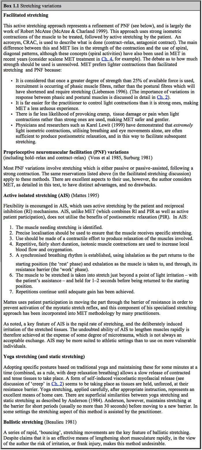
Liebenson (1996) discusses both the benefits of, and the mechanisms involved in, use of muscle energy techniques (which he terms �manual resistance techniques�, or MRT):

Two aspects to MRT [i.e. MET by another name] are their ability to relax an overactive muscle … and their ability to enhance stretch of a shortened muscle or its associated fascia when connective tissue or viscoelastic changes have occurred.

Two fundamental neurophysiological principles account for the neuromuscular inhibition that occurs during application of these techniques. The first is postcontraction inhibition [also known as postisometric relaxation, or PIR], which states that after a muscle is contracted, it is automatically in a relaxed state for a brief, latent, period. The second is reciprocal inhibition (RI) which states that when one muscle is contracted, its antagonist is automatically inhibited.

Liebenson suggests that there is evidence that the receptors responsible for PIR lie within the muscle and not in the skin or associated joints (Robinson 1982).

Where pain of an acute or chronic nature makes controlled contraction of the muscles involved difficult, the therapeutic use of the antagonists can patently be of value. Thus modern MET incorporates both postisometric relaxation and reciprocal inhibition methods, as well as aspects unique to itself, such as isokinetic techniques, described later.

A number of researchers, including Karel Lewit of Prague (Lewit 1999), have reported on the usefulness of aspects of MET in the treatment of trigger points, and this is seen by many to be an excellent method of treating these myofascial states, and of achieving the restoration of a situation where the muscle in which the trigger lies is once more capable of achieving its full resting length, with no evidence of shortening.

Travell & Simons (1992) mistakenly credited Lewit with developing MET, stating that �The concept of applying post-isometric relaxation in the treatment of myofascial pain was presented for the first time in a North American journal in 1984 [by Lewit]�. In fact Mitchell Snr had described the method some 25 years previously, a fact acknowledged by Lewit (Lewit & Simons 1984).

Key Points About Modern Muscle Energy Techniques

MET methods all employ variations on a basic theme. This primarily involves the use of the patient�s own muscular efforts in one of a number of ways, usually in association with the efforts of the therapist:

1. The operator�s force may exactly match the effort of the patient (so producing an isometric contraction) allowing no movement to occur � and producing as a result a physiological neurological response (via the Golgi tendon organs) involving a combination of:

� reciprocal inhibition of the antagonist(s) of the muscle(s) being contracted, as well as

� postisometric relaxation of the muscle(s) which are being contracted.

  1. The operator�s force may overcome the effort of the patient, thus moving the area or joint in the direction opposite to that in which the patient is attempting to move it (this is an isotonic eccentric contraction, also known as an isolytic contraction).
  2. The operator may partially match the effort of the patient, thus allowing, although slightly retarding, the patient�s effort (and so producing an isotonic concentric, isokinetic, contraction).

Other variables may be also introduced, for example involving:

l Whether the contraction should commence with the muscle or joint held at the resistance barrier or short of it � a factor decided largely on the basis of the degree of chronicity or acuteness of the tissues involved

  • How much effort the patient uses � say, 20% of strength, or more, or less
  • The length of time the effort is held � 7�10 seconds, or more, or less (Lewit (1999) favours 7� 10 seconds; Greenman (1989), Goodridge & Kuchera (1997) all favour 3�5 seconds)
  • Whether, instead of a single maintained contraction, to use a series of rapid, low amplitude contractions (Ruddy�s rhythmic resisted duction method, also known as pulsed muscle energy techniques)
  • The number of times the isometric contraction (or its variant) is repeated � three repetitions are thought to be optimal (Goodridge & Kuchera 1997)
  • The direction in which the effort is made � towards the resistance barrier or away from it, thus involving either the antagonists to the muscles or the actual muscles (agonists) which require �release� and subsequent stretching (these variations are also known as �direct� and �indirect� approaches, see p. 8)
  • Whether to incorporate a held breath and/or specific eye movements to enhance the effects of the contraction � desirable if possible, it is suggested (Goodridge & Kuchera 1997, Lewit 1999)
  • What sort of resistance is offered (for example by the operator, by gravity, by the patient, or by an immovable object)
  • Whether the patient�s effort is matched, overcome or not quite matched � a decision based on the precise needs of the tissues � to achieve relaxation, reduction in fibrosis or tonifying/ reeducation
  • Whether to take the muscle or joint to its new barrier following the contraction, or whether or not to stretch the area/muscle(s) beyond the barrier � this decision is based on the nature of�the problem being addressed (does it involve shortening? fibrosis?) and its degree of chronicity
  • Whether any subsequent (to a contraction) stretch is totally passive, or whether the patient should participate in the movement, the latter being thought by many to be desirable in order to reduce danger of stretch reflex activation (Mattes 1995)
  • Whether to utilize Muscle energy techniques alone, or in a sequence with other modalities such as the positional release methods of strain/counterstrain, or the ischaemic compression/inhibitory pressure techniques of neuromuscular technique (NMT) � such decisions will depend upon the type of problem being addressed, with myofascial trigger point treatment frequently benefiting from such combinations (see description of integrated neuromuscular inhibition (INIT), p. 197 (Chaitow 1993)).

Greenman summarises the requirements for the successful use of MET in osteopathic situations as �control, balance and localisation�. His suggested basic elements of MET include the following:

  • A patient/active muscle contraction, which
    � commences from a controlled position
    � is in a specific direction (towards or away from a restriction barrier)
  • The operator applies distinct counterforce (to meet, not meet, or to overcome the patient�s force)
  • The degree of effort is controlled (sufficient to obtain an effect but not great enough to induce trauma or difficulty in controlling the effort).

What is done subsequent to the contraction may involve any of a number of variables, as will be explained.

The essence of MET then is that it uses the energy of the patient, and that it may be employed in one or other of the manners described above with any combination of variables depending upon the particular needs of the case. Goodridge (one of the first osteopaths to train with Mitchell Snr in 1970) summarises as follows: �Good results [with MET] depend on accurate diagnosis, appropriate levels of force, and sufficient localization. Poor results are most often caused by inaccurate diagnosis, improperly localized forces, or forces that are too strong� (Goodridge & Kuchera 1997) (see also Box 1.3).

muscle energy techniques
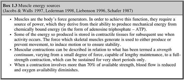
Using agonist or antagonist? (Box 1.4)

As mentioned, a critical consideration in MET, apart from degree of effort, duration and frequency of use, involves the direction in which the effort is made. This may be varied, so that the operator�s�force is directed towards overcoming the restrictive barrier (created by a shortened muscle, restricted joint, etc.); or indeed opposite forces may be used, in which the operator�s counter-effort is directed away from the barrier.

There is general consensus among the various osteopathic experts already quoted that the use of postisometric relaxation is more useful than reciprocal inhibition in normalizing hypertonic musculature. This, however, is not generally held to be the case by experts such as Lewit and Janda, who see specific roles for the reciprocal inhibition variation.

muscle energy techniques

Osteopathic clinicians such as Stiles and Greenman believe that the muscle which requires stretching (the agonist) should be the main source of �energy� for the isometric contraction, and suggest that this achieves a more significant degree of relaxation, and so a more useful ability to subsequently stretch the muscle, than would be the case were the relaxation effect being achieved via use of the antagonist (i.e. using reciprocal inhibition).

Following on from an isometric contraction � whether agonist or antagonist is being used � there is a refractory, or latency, period of approximately 15 seconds during which there can be an easier (due to reduced tone) movement towards the new position (new resistance barrier) of a joint or muscle.

Variations On The Muscle Energy Techniques Theme

Liebenson (1989, 1990) describes three basic variations which are used by Lewit and Janda as well as by himself in a chiropractic rehabilitation setting.

Lewit�s (1999) modification of MET, which he calls postisometric relaxation, is directed towards relaxation of hypertonic muscle, especially if this relates to reflex contraction or the involvement of myofascial trigger points. Liebenson (1996) notes that �this is also a suitable method for joint mobilisation when a thrust is not desirable�.

Lewit�s postisometric relaxation method

(Lewit 1999)

  1. The hypertonic muscle is taken, without force or �bounce�, to a length just short of pain, or to the point where resistance to movement is first noted (Fig. 1.3).
  2. The patient gently contracts the affected hypertonic muscle away from the barrier (i.e. the agonist is used) for between 5 and 10 seconds, while the effort is resisted with an exactly equal counterforce. Lewit usually has the patient inhale during this effort.
  3. This resistance involves the operator holding the contracting muscle in a direction which would stretch it, were resistance not being offered.
  4. The degree of effort, in Lewit�s method, is minimal. The patient may be instructed to think in terms of using only 10 or 20% of his available strength, so that the manoeuvre is never allowed to develop into a contest of strength between the operator and the patient.
  5. After the effort, the patient is asked to exhale and to let go completely, and only when this is achieved is the muscle taken to a new barrier with all slack removed but no stretch � to the extent that the relaxation of the hypertonic muscles will now allow.
  6. Starting from this new barrier, the procedure is repeated two or three times.
  7. In order to facilitate the process, especially where trunk and spinal muscles are involved, Lewit usually asks the patient to assist by looking with his eyes in the direction of the contraction during the contracting phase, and in the direction of stretch during the stretching phase of the procedure.

The key elements in this approach, as in most MET, involve precise positioning, as well as taking out slack and using the barrier as the starting and ending points of each contraction.

muscle energy techniquesWhat Is Happening?

Karel Lewit, discussing MET methods (Lewit 1999), states that medullary inhibition is not capable of explaining their effectiveness. He considers that the predictable results obtained may relate to the following facts:

  • During resistance using minimal force (isometric contraction) only a very few fibers are active, the others being inhibited
  • During relaxation (in which the shortened musculature is taken gently to its new limit without stretching) the stretch reflex is avoided � a reflex which may be brought about even by passive and non-painful stretch (see Mattes� views p. 3).

He concludes that this method demonstrates the close connection between tension and pain, and between relaxation and analgesia.

The use of eye movements as part of the methodology is based on research by Gaymans (1980) which indicates, for example, that flexion is enhanced by the patient looking downwards, and extension by the patient looking upwards. Similarly, sidebending is facilitated by looking towards the side involved. These ideas are easily proved by self-experiment: an attempt to flex the spine while maintaining the eyes in an upwards (towards the forehead) looking direction will be found to be less successful than an attempt made to flex while looking downwards. These eye-direction aids are also useful in manipulation of the joints.

Effects of Muscle energy techniques

Lewit (1999) discusses the element of passive muscular stretch in MET and maintains that this factor does not always seem to be essential. In some areas, self-treatment, using gravity as the resistance factor, is effective, and such cases sometimes involve no element of stretch of the muscles in question. Stretching of muscles during MET, according to Lewit (1999), is only required when contracture due to fibrotic change has occurred, and is not necessary if there is simply a disturbance in function. He quotes results in one series of patients in his own clinic in which 351 painful muscle groups, or muscle attachments, were treated by MET (using postisometric�relaxation) in 244 patients. Analgesia was immediately achieved in 330 cases and there was no effect in only 21 cases. These are remarkable results by any standards.

Lewit suggests, as do many others, that trigger points and �fibrositic� changes in muscle will often disappear after MET contraction methods. He further suggests that referred local pain points, resulting from problems elsewhere, will also disappear more effectively than where local anaesthesia or needling (acupuncture) methods are employed.

Janda�s postfacilitation stretch method

Janda�s variation on this approach (Janda 1993), known as �postfacilitation stretch�, uses a different starting position for the contraction and also a far stronger isometric contraction than that suggested by Lewit and most osteopathic users of Muscle energy techniques:

  1. The shortened muscle is placed in a mid-range position about halfway between a fully stretched and a fully relaxed state.
  2. The patient contracts the muscle isometrically, using a maximum degree of effort for 5�10 seconds while the effort is resisted completely.
  3. On release of the effort, a rapid stretch is made to a new barrier, without any �bounce�, and this is held for at least 10 seconds.
  4. The patient relaxes for approximately 20 seconds and the procedure is repeated between three and five times more.

Some sensations of warmth and weakness may be anticipated for a short while following this more vigorous approach.

Reciprocal inhibition variation

This method, which forms a component of PNF methodology (see Box 1.1) and Muscle energy techniques, is mainly used in acute settings, where tissue damage or pain precludes the use of the more usual agonist contraction, and also commonly as an addition to such methods, often to conclude a series of stretches whatever other forms of MET have been used (Evjenth & Hamberg 1984):

  1. The affected muscle is placed in a mid-range position.
  2. The patient is asked to push firmly towards the restriction barrier and the operator either completely resists this effort (isometric) or allows a movement towards it (isotonic). Some degree of rotational or diagonal movement may be incorporated into the procedure.
  3. On ceasing the effort, the patient inhales and exhales fully, at which time the muscle is passively lengthened.

Liebenson notes that �a resisted isotonic effort towards the barrier is an excellent way in which to facilitate afferent pathways at the conclusion of treatment with active muscular relaxation techniques or an adjustment (joint). This can help reprogram muscle and joint proprioceptors and thus re-educate movement patterns.� (See Box 1.2.)

Strengthening variation

Another major muscle energy variation is to use what has been called isokinetic contraction (also known as progressive resisted exercise). In this the patient starts with a weak effort but rapidly progresses to a maximal contraction of the affected muscle(s), introducing a degree of resistance to the operator�s effort to put the joint, or area, through a full range of motion. The use of isokinetic contraction is reported to be a most effective method of building strength, and to be superior to high repetition, lower resistance exercises (Blood 1980). It is also felt that a limited range of motion, with good muscle tone, is preferable (to the patient) to having a normal range with limited power. Thus the strengthening of weak musculature in areas of permanent limitation of mobility is seen as�an important contribution in which isokinetic contractions may assist.

Isokinetic contractions not only strengthen the fibres which are involved, but also have a training effect which enables them to operate in a more coordinated manner. There is often a very rapid increase in strength. Because of neuromuscular recruitment, there is a progressively stronger muscular effort as this method is repeated. Isokinetic contractions, and accompanying mobilisation of the region, should take no more than 4 seconds at each contraction in order to achieve maximum benefit with as little fatiguing as possible, either of the patient or the operator. Prolonged contractions should be avoided. The simplest, safest, and easiest-to-handle use of isokinetic methods involves small joints, such as those in the extremities. Spinal joints may be more difficult to mobilise while muscular resistance is being fully applied.

The options available in achieving increased strength via these methods therefore involve a choice between either a partially resisted isotonic contraction, or the overcoming of such a contraction, at the same time as the full range of movement is being introduced (note that both isotonic concentric and eccentric contractions will take place during the isokinetic movement of a joint). Both of these options should involve maximum contraction of the muscles by the patient. Home treatment of such conditions is possible, via self-treatment, as in other MET methods.

Isolytic Muscle Energy Techniques

Another application of the use of isotonic contraction occurs when a direct contraction is resisted and overcome by the operator (Fig. 1.4). This has been termed isolytic contraction, in that it involves the stretching, and sometimes the breaking down, of fibrotic tissue present in the affected muscles. Adhesions of this type are reduced by the application of force by the operator which is just greater than that being exerted by the patient. This procedure can be uncomfortable, and the patient should be advised of this. Limited degrees of effort are therefore called for at the outset of isolytic contractions. This is an isotonic eccentric contraction, in that the origins and insertions of the muscles involved will become further separated, despite the patient�s effort to approximate them. In order to achieve the greatest degree of stretch (in the condition of myofascial fibrosis, for example), it is necessary for the largest number of fibers possible to be involved in the isotonic contraction. Thus there is a contradiction in that, in order to achieve this large involvement, the degree of contraction should be a maximal one, and yet this is likely to produce pain, which is contraindicated. It may also, in many instances, be impossible for the operator to overcome.

muscle energy techniquesThis stretches the muscles which are contracting (TFL shown in example) thereby inducing a degree of controlled microtrauma, with the aim of increasing the elastic potential of shortened or fibrosed tissues.

The patient should be instructed to use about 20% of possible strength on the first contraction, which is resisted and overcome by the operator, in a contraction lasting 3�4 seconds. This is then repeated, but with an increased degree of effort on the part of the patient (assuming the first effort was relatively painless). This continuing increase in the amount of force employed in the contracting musculature may be continued until, hopefully, a maximum contraction effort is possible, again to be overcome by the operator. In some muscles, of course, this may require a heroic degree of effort on the part of the operator, and alternative methods are therefore desirable. Deep tissue techniques, such as neuromuscular technique, would seem to offer such an alternative. The isolytic manoeuvre should have as its ultimate aim a fully relaxed muscle, although this will not always be possible.

Why Fibrosis Occurs Naturally

An article in the Journal of the Royal Society of Medicine (Royal Society of Medicine 1983) discusses connective tissue changes:

Aging affects the function of connective tissue more obviously than almost any organ system. Collagen fibrils thicken, and the amounts of soluble polymer decrease. The connective tissue cells tend to decline in number, and die off. Cartilages become less elastic, and their complement of proteoglycans changes both quantitatively and qualitatively. The interesting question is how many of these processes are normal, that contribute blindly and automatically, beyond the point at which they are useful? Does prevention of aging, in connective tissues, simply imply inhibition of cross linking in collagen fibrils, and a slight stimulation of the production of chondroitin sulphate proteoglycan?

The effects of various soft tissue approaches such as NMT and Muscle energy techniques will impact directly on these tissues as well as on the circulation and drainage of the affected structures, which suggests that the ageing process can be influenced. Destruction of collagen fibrils, however, is a serious matter (for example when using isolytic stretches), and although the fibrous tissue may be replaced in the process of healing, scar-tissue formation is possible, and this makes repair inferior to the original tissues, both in functional and structural terms. An isolytic contraction has the ability to break down tight, shortened tissues and the replacement of these with superior material will depend, to a large extent, on the subsequent use of the area (exercise, etc.), as well as the nutritive status of the individual. Collagen formation is dependent on adequate vitamin C, and a plentiful supply of amino acids such as proline, hydroxyproline and arginine. Manipulation, aimed at the restoration of a degree of normality in connective tissues, should therefore take careful account of nutritional requirements.

The range of choices in stretching, irrespective of the form of prelude to this � strong or mild isometric contraction, starting at or short of the barrier � therefore covers the spectrum from all- passive to all-active, with many variables in between.

Putting It Together

Many may prefer to use the variations, as described above, within individual settings. The recommendation of this text, however, is that they should be �mixed and matched� so that elements of all of them may be used in any given setting, as appropriate. Lewit�s (1999) approach seems ideal for more acute and less chronic conditions, while Janda�s (1989) more vigorous methods seem�ideal for hardy patients with chronic muscle shortening.

Muscle energy techniques offers a spectrum of approaches which range from those involving hardly any active contraction at all, relying on the extreme gentleness of mild isometric contractions induced by breath-holding and eye movements only, all the way to the other extreme of full-blooded, total- strength contractions. Subsequent to isometric contractions � whether strong or mild � there is an equally sensitive range of choices, involving either energetic stretching or very gentle movement to a new restriction. We can see why Sandra Yale (in DiGiovanna 1991) speaks of the usefulness of MET in treating extremely ill patients.

Many patients present with a combination of recent dysfunction (acute in terms of time, if not in degree of pain or dysfunction) overlaid on chronic changes which have set the scene for their acute current problems. It seems perfectly appropriate to use methods which will deal gently with hypertonicity, and more vigorous methods which will help to resolve fibrotic change, in the same patient, at the same time, using different variations on the theme of MET. Other variables can be used which focus on joint restriction, or which utilise RI should conditions be too sensitive to allow PIR methods, or variations on Janda�s more vigorous stretch methods (see Box 1.1).

Discussion of common errors in application of Muscle energy techniques will help to clarify these thoughts.

Why Muscle Energy Techniques Might Be Ineffective At Times

Poor results from use of Muscle energy techniques may relate to an inability to localize muscular effort sufficiently, since unless local muscle tension is produced in the precise region of the soft tissue dysfunction, the method is likely to fail to achieve its objectives. Also, of course, underlying pathological changes may have taken place, in joints or elsewhere, which make such an approach of short-term value only, since such changes will ensure recurrence of muscular spasms, sometimes almost immediately.

MET will be ineffective, or cause irritation, if excessive force is used in either the contraction phase or the stretching phase.

The keys to successful application of Muscle energy techniques therefore lie in a precise focusing of muscular activity, with an appropriate degree of effort used in the isometric contraction, for an adequate length of time, followed by a safe movement through the previous restriction barrier, usually with patient assistance.

Use of variations such as stretching chronic fibrotic conditions following an isometric contraction and use of the integrated approach (INIT) mentioned earlier in this chapter represent two examples of further adaptations of Lewit�s basic approach which, as described above, is ideal for acute situations of spasm and pain.

To Stretch Or To Strengthen?

Marvin Solit (1963), a former pupil of Ida Rolf, describes a common error in application of Muscle energy techniques � treating the �wrong� muscles the �wrong� way:

As one looks at a patient�s protruding abdomen, one might think that the abdominal muscles are weak, and that treatment should be geared towards strengthening them. By palpating the abdomen, however, one would not feel flabby, atonic muscles which would be the evidence of weakness; rather, the muscles are tight, bunched and shortened. This should not be surprising because here is an example of muscle working overtime maintaining body equilibrium. In addition these muscles are supporting the sagging viscera, which normally would be supported by their individual ligaments. As�the abdominal muscles are freed and lengthened, there is a general elevation of the rib cage, which in turn elevates the head and neck.

Attention to tightening and hardening these supposedly weak muscles via exercise, observes Solit, results in no improvement in posture, and no reduction in the �pot-bellied� appearance. Rather, the effect is to further depress the thoracic structures, since the attachments of the abdominal muscles, superiorly, are largely onto the relatively mobile, and unstable, bones of the rib cage. Shortening these muscles simply achieves a degree of pull on these structures towards the stable pelvic attachments below.

The approach to this problem adopted by Rolfers is to free and loosen these overworked and only apparently weakened tissues. This allows for a return to some degree of normality, freeing the tethered thoracic structures, and thus correcting the postural imbalance. Attention to the shortened, tight musculature, which will also be inhibiting their antagonist muscles, should be the primary aim. Exercise is not suitable at the outset, before this primary goal is achieved.

The common tendency in some schools of therapy to encourage the strengthening of weakened muscle groups in order to normalise postural and functional problems is also discussed by Vladimir Janda (1978). He expresses the reasons why this approach is �putting the cart before the horse�: �In pathogenesis, as well as in treatment of muscle imbalance and back problems, tight muscles play a more important, and perhaps even primary, role in comparison to weak muscles� (Fig. 1.5). He continues with the following observation:

Clinical experience, and especially therapeutic results, support the assumption that (according to Sherrington�s law of reciprocal innervation) tight muscles act in an inhibitory way on their antagonists. Therefore, it does not seem reasonable to start with strengthening of the weakened muscles, as most exercise programmes do. It has been clinically proved that it is better to stretch tight muscles first. It is not exceptional that, after stretching of the tight muscles, the strength of the weakened antagonists improves spontaneously, sometimes immediately, sometimes within a few days, without any additional treatment.

This sound, well-reasoned, clinical and scientific observation, which directs our attention and efforts towards the stretching and normalizing of those tissues which have shortened and tightened, seems irrefutable, and this theme will be pursued further in Chapter 2.

muscle energy techniquesMuscle energy techniques are designed to assist in this endeavor and, as discussed above, also provides an excellent method for assisting in the toning of weak musculature, should this still be required, after the stretching of the shortened antagonists, by use of isotonic methods.

Tendons

Aspects of the physiology of muscles and tendons are worthy of a degree of review, in so far as Muscle energy techniques and its effects are concerned (see also Box 1.5). The tone of muscle is largely the job of the Golgi tendon organs. These detect the load applied to the tendon, via muscular contraction. Reflex effects, in the appropriate muscles, are the result of this information being passed from the Golgi tendon organ back along the cord. The reflex is an inhibitory one, and thus differs from the muscle spindle stretch reflex. Sandler (1983) describes some of the processes involved:

When the tension on the muscles, and hence the tendon, becomes extreme, the inhibitory effect from the tendon organ can be so great that there is sudden relaxation of the entire muscle under stretch. This effect is called the lengthening reaction, and is probably a protective reaction to the force which, if unprotected, can tear the tendon from its bony attachments. Since the Golgi tendon organs, unlike the [muscle] spindles, are in series with the muscle fibres, they are stimulated by both passive and active contractions of the muscles.

Pointing out that muscles can either contract with constant length and varied tone (isometrically), or with constant tone and varied length (isotonically), he continues: �In the same way as the gamma efferent system operates as a feedback to control the length of muscle fibers, the tendon reflex serves as a reflex to control the muscle tone�.

muscle energy techniques
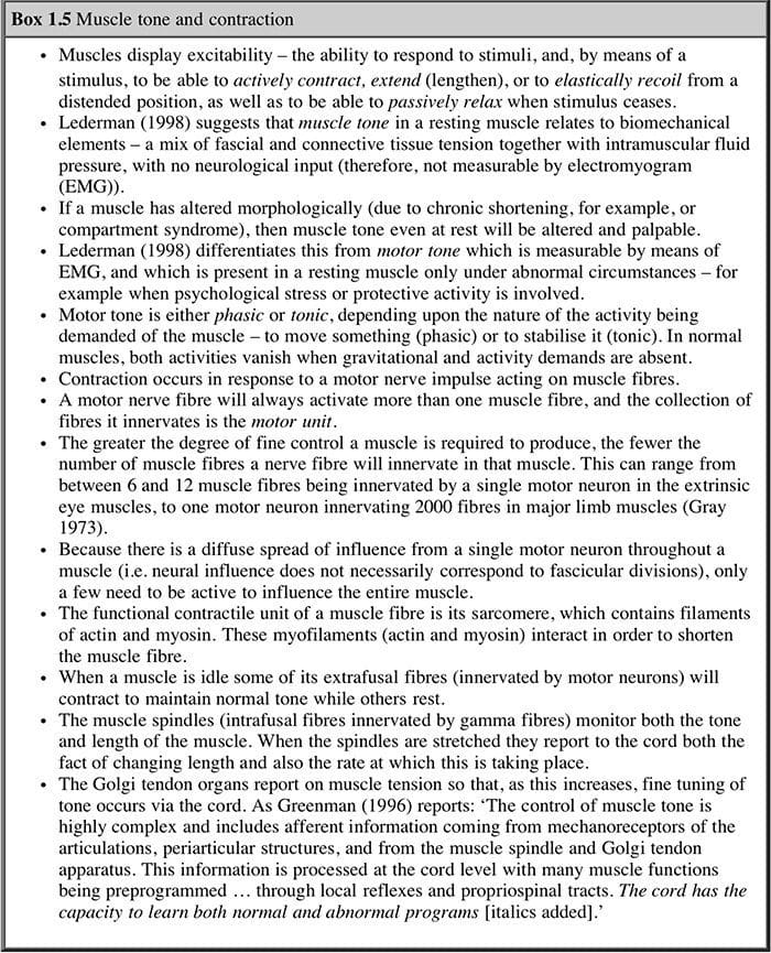
The relevance of this to soft tissue techniques is explained as follows:

In terms of longitudinal soft tissue massage, these organs are very interesting indeed, and it is perhaps the reason why articulation of a joint, passively, to stretch the tendons that pass over the joint, is often as effective in relaxing the soft tissues as direct massage of the muscles themselves. Indeed, in some cases, where the muscle is actively in spasm, and is likely to object to being pummelled directly, articulation, muscle energy techniques, or functional balance techniques, that make use of the tendon organ reflexes, can be most effective.

The use of this knowledge in therapy is obvious and Sandler explains part of the effect of massage on muscle: �The [muscle] spindle and its reflex connections constitute a feedback device which can�operate to maintain constant muscle length, as in posture; if the muscle is stretched the spindle discharges increase, but if the muscle is shortened, without a change in the rate of gamma discharge, then the spindle discharge will decrease, and the muscle will relax.�

Sandler believes that massage techniques cause a decrease in the sensitivity of the gamma efferent, and thus increase the length of the muscle fibers rather than a further shortening of them; this produces the desired relaxation of the muscle. Muscle energy techniques provides for the ability to influence both the muscle spindles and also the Golgi tendon organs.

Joints & Muscle Energy Techniques

Bourdillon (1982) tells us that shortening of muscle seems to be a self-perpetuating phenomenon which results from an over-reaction of the gamma-neuron system. It seems that the muscle is incapable of returning to a normal resting length as long as this continues. While the effective length of the muscle is thus shortened, it is nevertheless capable of shortening further. The pain factor seems related to the muscle�s inability thereafter to be restored to its anatomically desirable length. The conclusion is that much joint restriction is a result of muscular tightness and shortening. The opposite may also apply where damage to the soft or hard tissues of a joint is a factor. In such cases the periarticular and osteophytic changes, all too apparent in degenerative conditions, are the major limiting factor in joint restrictions. In both situations, however, Muscle energy techniques may be useful, although more useful where muscle shortening is the primary factor.

The restriction which takes place as a result of tight, shortened muscles is usually accompanied by some degree of lengthening and weakening of the antagonists. A wide variety of possible permutations exists in any given condition involving muscular shortening which may be initiating, or be secondary to, joint dysfunction combined with weakness of antagonists. A combination of isometric and isotonic methods can effectively be employed to lengthen and stretch the shortened groups, and to strengthen and shorten the weak, overlong muscles.

Paul Williams (1965) stated a basic truth which is often neglected by the professions which deal with musculoskeletal dysfunction:

The health of any joint is dependent upon a balance in the strength of its opposing muscles. If for any reason a flexor group loses part, or all of its function, its opposing tensor group will draw the joint into a hyperextended position, with abnormal stress on the joint margins. This situation exists in the lumbar spine of modern man.

Lack of attention to the muscular component of joints in general, and spinal joints in particular, results in frequent inappropriate treatment of the joints thus affected. Correct understanding of the role of the supporting musculature would frequently lead to normalisation of these tissues, without the need for heroic manipulative efforts. Muscle energy techniques and other soft tissue approaches focus attention on these structures and offer the opportunity to correct both the weakened musculature and the shortened, often fibrotic, antagonists.

More recently, Norris (1999) has pointed out that:

The mixture of tightness and weakness seen in the muscle imbalance process alters body segment alignment and changes the equilibrium point of a joint. Normally the equal resting tone of the agonist and antagonist muscles allows the joint to take up a balanced position where the joint surfaces are evenly loaded and the inert tissues of the joint are not excessively stressed. However if the muscles on one side of a joint are tight and the opposing muscles relax, the joint will be pulled out of alignment towards the tight muscle(s).

Such alignment changes produce weight-bearing stresses on joint surfaces, and result also in shortened soft tissues chronically contracting over time. Additionally such imbalances result in reduced segmental control with chain reactions of compensation emerging (see Ch. 2).

Several studies will be detailed (Chs 5 and 8) showing the effectiveness of Muscle energy techniques application in diverse population groups, including a Polish study on the benefits of Muscle energy techniques in joints damaged by haemophilia, and a Swedish study on the effects of Muscle energy techniques in treating lumbar spine dysfunction, as well as an American/Czech study involving myofascial pain problems. In the main, the results indicate a universal role in providing resolution or relief of such problems by means of the application of safe and effective muscle energy techniques.

blank
References:

Anderson B 1984 Stretching. Shelter Publishing, Nolinas, California
Beaulieu J 1981 Developing a stretching program. Physician and Sports Medicine 9(11): 59�69
Blood S 1980 Treatment of the sprained ankle. Journal of the American Osteopathic Association
79(11): 689
Bourdillon J 1982 Spinal manipulation, 3rd edn. Heinemann, London
Chaitow L 1993 Integrated neuromuscular inhibition technique (INIT) in treatment of pain and
trigger points. British Journal of Osteopathy 13: 17�21
DiGiovanna E 1991 Osteopathic approach to diagnosis and treatment. Lippincott, Philadelphia
Evjenth O, Hamberg J 1984 Muscle stretching in manual therapy. Alfta, Sweden
Gaymans F 1980 Die Bedeuting der atemtypen fur mobilisation der werbelsaule maanuelle. Medizin
18: 96
Goodridge J P 1981 Muscle energy technique: definition, explanation, methods of procedure.
Journal of the American Osteopathic Association 81(4): 249�254
Goodridge J, Kuchera W 1997 Muscle energy treatment techniques. In: Ward R (ed) Foundations of
osteopathic medicine. Williams and Wilkins, Baltimore
Gray�s Anatomy 1973 Churchill Livingstone, Edinburgh
Greenman P 1989 Manual therapy. Williams and Wilkins, Baltimore
Greenman P 1996 Principles of manual medicine, 2nd edn. Williams and Wilkins, Baltimore
Grieve G P 1985 Mobilisation of the spine. Churchill Livingstone, Edinburgh, p 190
Jacobs A, Walls W 1997 Anatomy. In: Ward R (ed) Foundations of osteopathic medicine. Williams
and Wilkins, Baltimore
Janda V 1978 Muscles, central nervous regulation and back problems. In: Korr I (ed)
Neurobiological mechanisms in manipulative therapy. Plenum Press, New York
Janda V 1989 Muscle function testing. Butterworths, London
Janda V 1993 Presentation to Physical Medicine Research Foundation, Montreal, Oct 9�11Kabat H 1959 Studies of neuromuscular dysfunction. Kaiser Permanente Foundation Medical
Bulletin 8: 121�143
Knott M, Voss D 1968 Proprioceptive neuromuscular facilitation, 2nd edn. Harper and Row, New
York
Lederman E 1998 Fundamentals of manual therapy. Churchill Livingstone, Edinburgh
Levine M et al 1954 Relaxation of spasticity by physiological techniques. Archives of Physical
Medicine 35: 214�223
Lewit K 1986 Muscular patterns in thoraco-lumbar lesions. Manual Medicine 2: 105
Lewit K 1999 Manipulative therapy in rehabilitation of the motor system, 3rd edn. Butterworths,
London
Lewit K, Simons D 1984 Myofascial pain: relief by post isometric relaxation. Archives of Physical
Medical Rehabilitation 65: 452�456
Liebenson C 1989 Active muscular relaxation techniques (part 1). Journal of Manipulative and
Physiological Therapeutics 12(6): 446�451
Liebenson C 1990 Active muscular relaxation techniques (part 2). Journal of Manipulative and
Physiological Therapeutics 13(1): 2�6
Liebenson C (ed) 1996 Rehabilitation of the spine. Williams and Wilkins, Baltimore
McAtee R, Charland J 1999 Facilitated stretching, 2nd edn. Human Kinetics, Champaign, Illinois
Mattes A 1995 Flexibility � active and assisted stretching. Mattes, Sarasota
Mitchell F L Snr 1958 Structural pelvic function. Yearbook of the Academy of Osteopathy 1958,
Carmel, p 71 (expanded in references in 1967 yearbook)
Mitchell F L Snr 1967 Motion discordance. Yearbook of the Academy of Applied Osteopathy 1967,
Carmel, pp 1�5
Mitchell F Jnr, Moran P S, Pruzzo N 1979 An evaluation and treatment manual of osteopathic
muscle energy procedures. Valley Park, Illinois
Moritan T 1987 Activity of the motor unit during concentric and eccentric contractions. American
Journal of Physiology 66: 338�350
Norris C 1999 Functional load abdominal training (part 1). Journal of Bodywork and Movement
Therapies 3(3): 150�158
Robinson K 1982 Control of soleus motoneuron excitability during muscle stretch in man. Journal
of Neurology and Neurosurgical Psychiatry 45: 699
Royal Society of Medicine 1983 Connective tissues: the natural fibre reinforced composite material.
Journal of the Royal Society of Medicine 76
Ruddy T 1961 Osteopathic rhythmic resistive duction therapy. Yearbook of Academy of Applied
Osteopathy 1961, Indianapolis, p 58Sandler S 1983 Physiology of soft tissue massage. British Osteopathic Journal 15: 1�6
Schafer R 1987 Clinical biomechanics, 2nd edn. Williams and Wilkins, Baltimore
Solit M 1963 A study in structural dynamics. Yearbook of Academy of Applied Osteopathy 1963
Stiles E 1984a Patient Care May 15: 16�97
Stiles E 1984b Patient Care August 15: 117�164
Surburg P 1981 Neuromuscular facilitation techniques in sports medicine. Physician and Sports
Medicine 9(9): 115�127
Travell J, Simons D 1992 Myofascial pain and dysfunction, vol. 2, Williams and Wilkins, Baltimore
Voss D, Ionta M, Myers B 1985 Proprioceptive neuromuscular facilitation, 3rd edn. Harper and
Row, Philadelphia
Williams P 1965 The lumbo-sacral spine. McGraw Hill, New York

Close Accordion
Mastodon