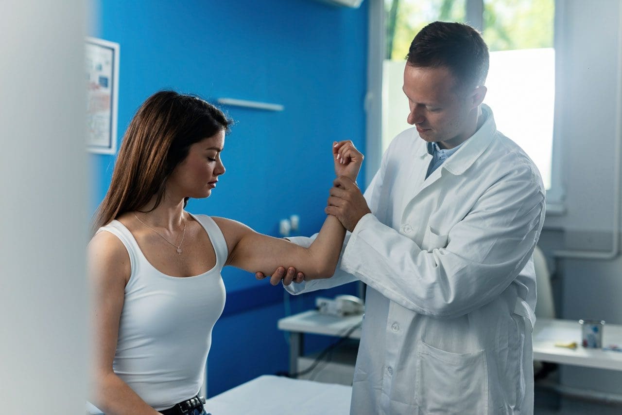
Knee injuries can present in physically active individuals that lift weights. Can understanding the types of weightlifting knee injuries help in prevention?
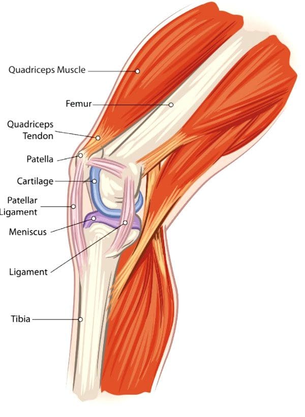
Weightlifting Knee Injuries
Weight training is very safe for the knees as regular weight training can improve knee strength and prevent injury as long as the correct form is followed. For Individuals with knee injuries from other activities, incorrect weight-training exercises could worsen the injury. (Ulrika Aasa et al., 2017) As well as, sudden twisting movements, poor alignment, and pre-existing injuries can increase the risk of worsening or creating further injuries. (Hagen Hartmann et al, 2013) The body and the knees are designed to support vertical forces on the joints.
Common Injuries
Weightlifting knee injuries occur as the knee joints endure a wide range of stresses and strains. In weight training, the ligaments that attach to the complex bone system of the knee joint can be damaged by incorrect movements, overloading the weight, and increasing the weight too soon. These injuries can result in pain, swelling, and immobility that can range from minor to severe, from a sprain or a slight tear to a complete tear in serious cases.
Anterior Cruciate Ligament – ACL – Injury
This ligament attaches the thigh’s femur bone to the lower leg’s shin bone/tibia and controls excessive rotation or extension of the knee joint. (American Academy of Family Physicians. 2024)
Anterior means front.
ACL injuries are seen mostly in athletes but can happen to anybody.
Severe damage to the ACL usually means surgical reconstruction and up to 12 months of rehabilitation.
When weightlifting, try to avoid twisting knee movements, intentionally or accidentally, under excessive load.
Posterior Cruciate Ligament – PCL – Injury
The PCL connects the femur and tibia at different points to the ACL.
It controls any backward motion of the tibia at the joint.
Injuries occur most with high-impact forces as a result of accidents and sometimes in activities where forceful trauma to the knee occurs.
Medial Collateral Ligament – MCL – Injury
This ligament maintains the knee from bending too far to the inside/medially.
Injuries mostly occur from impact to the outside of the knee or from accidental bodyweight force on the leg that bends at an unusual angle.
Lateral Collateral Ligament – LCL – Injury
This ligament connects the smaller bone of the lower leg/fibula to the femur.
It is opposite to the MCL.
It maintains excessive outward movement.
LCL injuries occur when a force pushes the knee out.
Cartilage Injury
Cartilage prevents bones from rubbing together and cushions impact forces.
Knee menisci are cartilage that cushions the knee joints inside and outside.
Other types of cartilage protect the thigh and shin bones.
When cartilage gets torn or damaged, surgery may be required.
Tendonitis
Aggravated and overused knee tendons can lead to weightlifting knee injuries.
A related injury known as iliotibial band syndrome/ITB causes pain to the outside of the knee, usually in runners, but it can occur from overuse.
Rest, stretching, physical therapy, and anti-inflammatory medication are a common treatment plan.
The condition causes the cartilage to deteriorate and bones to rub together, resulting in pain and stiffness.
Prevention
Individuals can minimize their risk of weightlifting knee injuries and pain by following their doctor’s and personal trainers’ recommendations.
Individuals with an existing knee injury should follow their doctor’s or physical therapist’s recommendations.
A knee sleeve can keep the muscles and joints secure, providing protection and support.
Stretching the leg and knee muscles can maintain joint flexibility.
Avoid sudden lateral movements.
Possible recommendations can include:
Avoiding Certain Exercises
Isolation exercises like leg curls, standing, or on a bench, as well as using the leg extension machine, can stress the knee.
Deep Squat Training
Research shows that the deep squat can protect against lower leg injury if the knee is healthy. However, this is when done with proper technique, under expert supervision, and with a gradual progressive load. (Hagen Hartmann et al, 2013)
Individuals should talk to their doctor before beginning a new exercise routine. A personal trainer can provide training in learning the proper technique and weightlifting form.
How I Tore my ACL Part 2
References
Aasa, U., Svartholm, I., Andersson, F., & Berglund, L. (2017). Injuries among weightlifters and powerlifters: a systematic review. British journal of sports medicine, 51(4), 211–219. https://doi.org/10.1136/bjsports-2016-096037
Hartmann, H., Wirth, K., & Klusemann, M. (2013). Analysis of the load on the knee joint and vertebral column with changes in squatting depth and weight load. Sports medicine (Auckland, N.Z.), 43(10), 993–1008. https://doi.org/10.1007/s40279-013-0073-6
American Academy of Family Physicians. ACL injury. (2024). ACL injury (Diseases and Conditions, Issue. https://familydoctor.org/condition/acl-injuries/
Mellinger, S., & Neurohr, G. A. (2019). Evidence based treatment options for common knee injuries in runners. Annals of translational medicine, 7(Suppl 7), S249. https://doi.org/10.21037/atm.2019.04.08
Driban, J. B., Hootman, J. M., Sitler, M. R., Harris, K. P., & Cattano, N. M. (2017). Is Participation in Certain Sports Associated With Knee Osteoarthritis? A Systematic Review. Journal of athletic training, 52(6), 497–506. https://doi.org/10.4085/1062-6050-50.2.08
For individuals experiencing lower back pain can understanding the anatomy and function of the multifidus muscle help in injury prevention and in the development of a highly effective treatment plan?
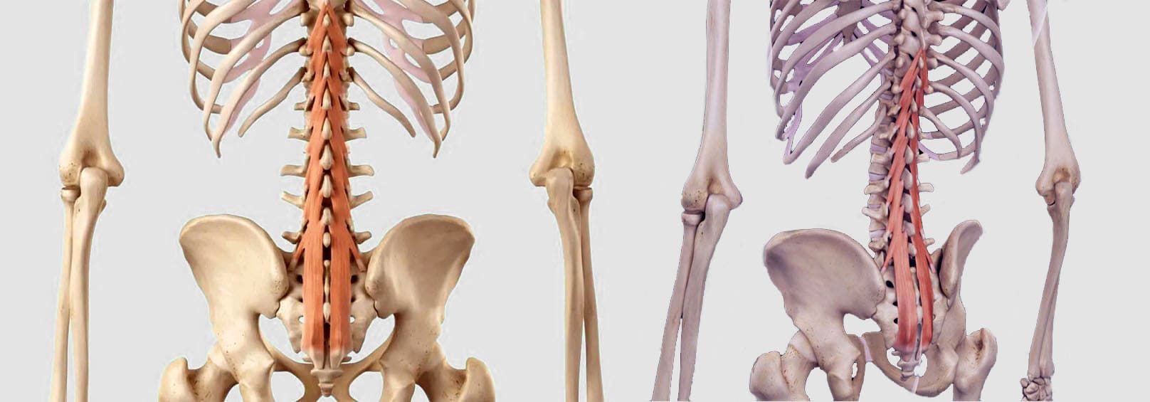
Multifidus Muscle
The multifidus muscles are long and narrow on either side of the spinal column, which helps stabilize the lower region of the spine or lumbar spine. (Maryse Fortin, Luciana Gazzi Macedo 2013) Sitting too much, practicing unhealthy postures, and lack of movement can progress to the multifidus muscle weakening or atrophy, which can lead to spinal instability, vertebral compression, and back pain. (Paul W. Hodges, Lieven Danneels 2019)
Anatomy
Known as the deep layer, it is the innermost layer of the three muscle layers of the back and controls the movement of the spine. The other two layers, known as the intrinsic and superficial, are responsible for the thoracic cage/rib cage and shoulder movement. (Anouk Agten et al., 2020) The multifidus has attachment points at:
The thoracic spine of the middle back.
The lumbar spine of the lower back.
The iliac spine – the base of the wing-shaped iliac bone of the pelvis.
Sacrum – series of bones at the base of the spine connected to the tailbone.
When standing or moving, the multifidus muscle works with the transversus abdominus and pelvic floor muscles to stabilize the lumbar spine. (Christine Lynders 2019)
Muscle Function
The main function is to stabilize the lower back, but it also helps extend the lower spine whenever reaching or stretching. (Jennifer Padwal et al., 2020) Because the muscle has numerous attachment points and is serviced by a specific branch of nerves known as the posterior rami, it allows each vertebra to work individually and more efficiently.
The multifidus muscle works with two other deep muscle groups to stabilize and move the spine. (Jeffrey J Hebert et al., 2015)
The rotatores muscle enables unilateral rotation, turning from side to side, and bilateral extension or bending backward and forward.
The semispinalis muscle above the multifidus allows extension and rotation of the head, neck, and upper back.
The multifidus muscle ensures spinal strength because it has more attachment points to the spine than the other layers, which reduces spinal flexibility and rotation but increases strength and stability. (Anouk Agten et al., 2020)
Lower Back Pain
A weak multifidus muscle destabilizes the spine and provides less support to the vertebra. This adds pressure on muscles and connective tissues between and adjacent to the spinal column, increasing the risk of lower back pain symptoms. (Paul W. Hodges, Lieven Danneels 2019) The loss of muscle strength and stability can cause atrophy or wasting away. This can cause compression and other back problems. (Paul W. Hodges et al., 2015) Back problems associated with multifidus muscle deterioration include (Paul W. Hodges, Lieven Danneels 2019)
Herniated discs – also bulging or slipped discs.
Nerve entrapment or compression pinched nerve.
Sciatica
Referred pain – nerve pain originating from the spine felt in other areas.
Osteoarthritis – wear-and-tear arthritis
Spinal osteophytes – bone spurs
Weak abdominal or pelvic floor muscles can compromise the core, increasing the risk of chronic lower back pain and injury.
Individuals are recommended to consult a physical therapist and chiropractor who can help develop the appropriate treatment, rehabilitation, and strengthening plan based on age, injury, underlying conditions, and physical abilities.
Can Core Exercises Help with Back Pain?
References
Fortin, M., & Macedo, L. G. (2013). Multifidus and paraspinal muscle group cross-sectional areas of patients with low back pain and control patients: a systematic review with a focus on blinding. Physical therapy, 93(7), 873–888. https://doi.org/10.2522/ptj.20120457
Hodges, P. W., & Danneels, L. (2019). Changes in Structure and Function of the Back Muscles in Low Back Pain: Different Time Points, Observations, and Mechanisms. The Journal of orthopaedic and sports physical therapy, 49(6), 464–476. https://doi.org/10.2519/jospt.2019.8827
Agten, A., Stevens, S., Verbrugghe, J., Eijnde, B. O., Timmermans, A., & Vandenabeele, F. (2020). The lumbar multifidus is characterised by larger type I muscle fibres compared to the erector spinae. Anatomy & cell biology, 53(2), 143–150. https://doi.org/10.5115/acb.20.009
Lynders C. (2019). The Critical Role of Development of the Transversus Abdominis in the Prevention and Treatment of Low Back Pain. HSS journal : the musculoskeletal journal of Hospital for Special Surgery, 15(3), 214–220. https://doi.org/10.1007/s11420-019-09717-8
Padwal, J., Berry, D. B., Hubbard, J. C., Zlomislic, V., Allen, R. T., Garfin, S. R., Ward, S. R., & Shahidi, B. (2020). Regional differences between superficial and deep lumbar multifidus in patients with chronic lumbar spine pathology. BMC musculoskeletal disorders, 21(1), 764. https://doi.org/10.1186/s12891-020-03791-4
Hebert, J. J., Koppenhaver, S. L., Teyhen, D. S., Walker, B. F., & Fritz, J. M. (2015). The evaluation of lumbar multifidus muscle function via palpation: reliability and validity of a new clinical test. The spine journal : official journal of the North American Spine Society, 15(6), 1196–1202. https://doi.org/10.1016/j.spinee.2013.08.056
Hodges, P. W., James, G., Blomster, L., Hall, L., Schmid, A., Shu, C., Little, C., & Melrose, J. (2015). Multifidus Muscle Changes After Back Injury Are Characterized by Structural Remodeling of Muscle, Adipose and Connective Tissue, but Not Muscle Atrophy: Molecular and Morphological Evidence. Spine, 40(14), 1057–1071. https://doi.org/10.1097/BRS.0000000000000972
For fitness and sports enthusiasts, weekend warriors, and athletes looking to improve physical performance, can incorporating acupuncture for sports performance be effective?
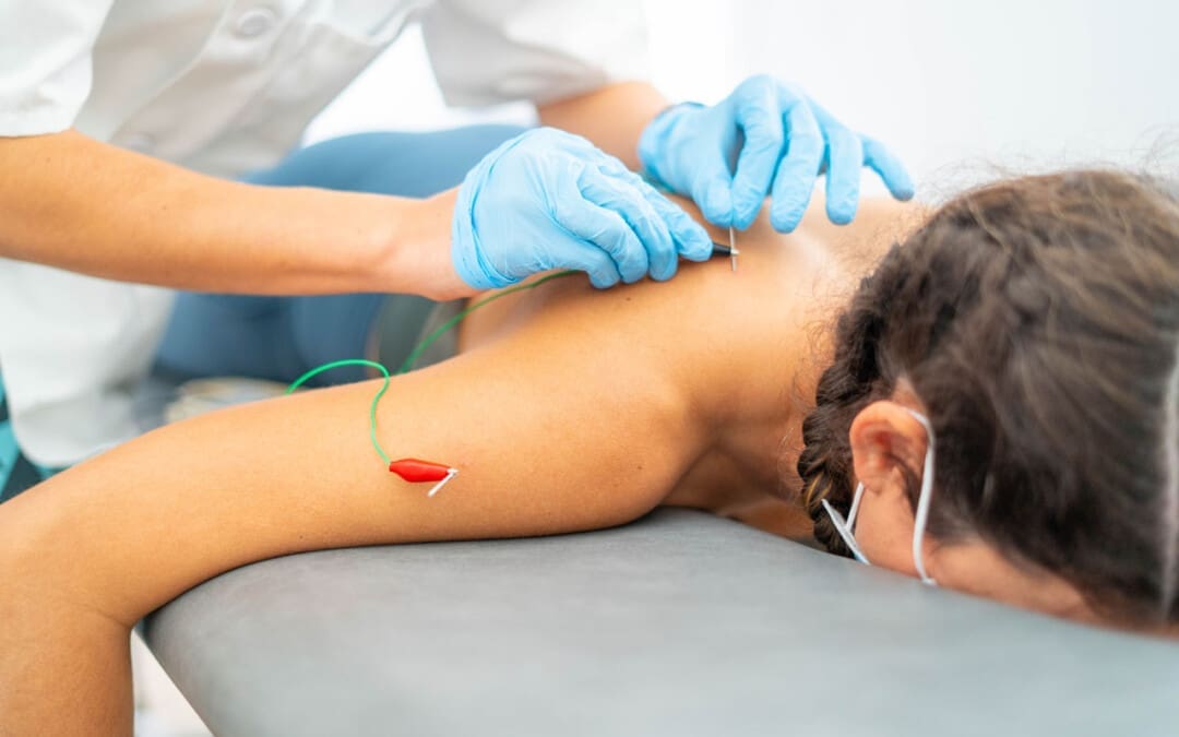
Acupuncture For Sports Performance
Acupuncture for sports performance follows the same needle insertion for specific points to treat pain symptoms, alleviate inflammation and fatigue, and enhance blood circulation to improve physical and athletic performance. Acupuncture is based on traditional Chinese medicine principles that focus on restoring the balance of the nervous system and body to activate natural healing and increase energy circulation. (Johns Hopkins Medicine. 2024).
Acupuncture has become a popular alternative treatment for sports injuries as it has shown positive outcomes and recovery from injuries. (George G. A. Pujalte et al., 2023)
The body’s blood and energy pathways, known as meridians, become blocked by inflammation because of illness, injury, or overuse, resulting in pain, stress, and various symptoms. The acupuncture needles stimulate the pathways to clear the blockages, allowing optimal circulation of energy and blood to reduce inflammation and restore balance. (Jiajie Zhu et al., 2021)
Sports acupuncture works by maintaining optimal circulation of blood and energy through meridians through the arteries, tendons, muscles, and organs for enhanced productivity and ability. (Liang Kang et al., 2021)
Electroacupuncture involves connecting electrical stimulation from a tens machine to specific points over an area to enhance the needle treatment. (Keitaro Kubo et al., 2020)
Acupuncture Can Help
Ways that acupuncture can help include:
Increase Range of Motion
Acupuncture can help loosen tight muscles, tendons, and ligaments overused during training or games.(Chi-Tsai Tang, 2023)
This allows athletes to perform at peak levels without risking worsening or causing further injury.
Increase Flexibility
Acupuncture helps increase elasticity in joints by releasing adhesions within connective tissue for increased mobility.
Improve Reflexes
Targeting key points stimulates nerve activity, which can improve quicker reflexes and improve coordination.(Chi-Tsai Tang, Bo Song. 2022)
Increase Circulation
Acupuncture increases blood circulation to areas lacking oxygen.
Acupuncture releases endorphins, which reduce pain and also provide an overall sense of calmness and relaxation.
This enables athletes to stay focused and motivated throughout training and games. (Chi-Tsai Tang, 2023)
Reduce Fatigue
Regular acupuncture for sports performance can help maintain energy levels to help prevent burnout and maintain optimal performance during practice and games. (George G. A. Pujalte et al., 2023)
Relieve Muscle Tension
Acupuncture treatment can help relax tense muscles caused by repetitive use as well as from stress tension that could be caused by anxiety before a game or tournament.
For individuals who want to improve their physical performance, sports acupuncture can provide a natural, non-invasive alternative that can help improve athletic performance mentally and physically.
Lumbar Spine Injuries in Sports: Chiropractic Healing
Zhu, J., Li, J., Yang, L., & Liu, S. (2021). Acupuncture, from the ancient to the current. Anatomical record (Hoboken, N.J. : 2007), 304(11), 2365–2371. https://doi.org/10.1002/ar.24625
Kang, L., Liu, P., Peng, A., Sun, B., He, Y., Huang, Z., Wang, M., Hu, Y., & He, B. (2021). Application of traditional Chinese therapy in sports medicine. Sports medicine and health science, 3(1), 11–20. https://doi.org/10.1016/j.smhs.2021.02.006
Tang, C. T., & Song, B. (2022). Acupuncture and Dry Needling for Sports Performance and Recovery. Current sports medicine reports, 21(6), 213–218. https://doi.org/10.1249/JSR.0000000000000968
Kubo, K., Iizuka, Y., Yajima, H., Takayama, M., & Takakura, N. (2020). Changes in Blood Circulation of the Tendons and Heart Rate Variability During and After Acupuncture. Medical acupuncture, 32(2), 99–107. https://doi.org/10.1089/acu.2019.1397
Tang C. T. (2023). Practicing Outside the Lines: Using Acupuncture in the Athletic Training Room and on the Field. Medical acupuncture, 35(5), 266–269. https://doi.org/10.1089/acu.2023.0043
During a fall individuals tend to automatically outstretch their hands to help break a fall, which can slam onto the ground causing a falling onto an outstretched hand or FOOSH injury. Should individuals get checked by a healthcare provider if they believe there is no injury?
FOOSH Injuries
Falling down usually results in minor injuries. A FOOSH injury occurs when falling down and trying to break the fall by reaching out with the hand/s. This can result in an upper extremity injury like a sprain or a fracture. But sometimes, falling on one’s hands can lead to serious injuries and/or create future musculoskeletal issues. Individuals who have fallen or suffered a FOOSH injury should consult their healthcare provider and then a physical therapist or chiropractor to safely develop a treatment plan to rehabilitate, strengthen, and expedite recovery.
After The Injury
For individuals who have fallen down and landed on their hand, wrist, or arm, here are a few things to ensure the proper care for the injury, including:
Follow the R.I.C.E. protocol for acute injuries
Visit a healthcare provider or local emergency clinic
Contact a physical therapist
A FOOSH injury could be or become serious, so to avoid letting small issues become big problems, get examined by a musculoskeletal specialist. The healthcare provider will obtain an imaging scan of the injured and surrounding areas. They will perform a physical examination to determine the type of injury, like a sprain or muscle strain. Not getting appropriate medical treatment after a fall can result in chronic pain and loss of function. (J. Chiu, S. N. Robinovitch. 1998)
Common Injuries
A FOOSH injury can injure different areas. These usually involve the wrist and hand, but the elbow or shoulder can also be injured. Common injuries include:
Colles’ fracture
A wrist fracture where the end of the arm bone is displaced backward.
Smith’s fracture
A wrist fracture, similar to a Colles’ fracture, is where the end of the arm bone is displaced towards the front of the wrist.
Boxer’s fracture
A fracture of the small bones in the hand.
Typically, it occurs after punching something, but it can happen from falling on an outstretched fist.
Elbow dislocation or fracture
The elbow can pop out of the joint or can break a bone in the elbow.
Collarbone fracture
The force from falling with the hands and arms outstretched can travel up to the collarbone, causing a fracture.
Proximal humeral fracture
Falling onto an outstretched hand injury can cause the arm bone to get jammed into the shoulder, causing a proximal humeral fracture.
Shoulder dislocation
The shoulder can pop out of the joint.
This can cause a rotator cuff tear or labrum injury.
Regardless of the injury, individuals should visit a healthcare provider to evaluate the damage. If the injury is serious, the practitioner can make an accurate or differential diagnosis and develop a treatment plan. (William R. VanWye et al., 2016)
Physical Therapy
Individuals can benefit from physical therapy to help recover and return to their previous level of function. Physical therapy varies depending on the specific injury, but generally, a physical therapist can help individuals return to function after a fall on an outstretched hand. (William R. VanWye et al., 2016) Common treatments can include:
Treatments and modalities to decrease pain, inflammation, and swelling.
Instruction on how to wear an arm sling properly.
Exercises and stretches to improve the range of motion, strength, and functional mobility.
Balance exercises.
Scar tissue management if surgery was necessary.
The therapy team will ensure the proper treatment is utilized to quickly and safely return to normal activities.
Chiropractic Care For Healing After Trauma
References
Chiu, J., & Robinovitch, S. N. (1998). Prediction of upper extremity impact forces during falls on the outstretched hand. Journal of biomechanics, 31(12), 1169–1176. https://doi.org/10.1016/s0021-9290(98)00137-7
VanWye, W. R., Hoover, D. L., & Willgruber, S. (2016). Physical therapist screening and differential diagnosis for traumatic-onset elbow pain: A case report. Physiotherapy theory and practice, 32(7), 556–565. https://doi.org/10.1080/09593985.2016.1219798
Individuals who participate in physical and sports activities could suffer an Achilles tendon tear. Can understanding the symptoms and risks help in treatment and return the individual back to their sports activity sooner?
Achilles Tendon
This is a common injury that occurs when the tendon attaching the calf muscle to the heel gets torn.
About the Tendon
The Achilles tendon is the largest tendon in the body.
In sports and physical activities, intense explosive movements like running, sprinting, quickly shifting positions, and jumping are exerted on the Achilles.
The injury often occurs without any contact or collision but rather the running, starting, stopping, and pulling actions placed on the feet.
Certain antibiotics and cortisone shots can increase the likelihood of Achilles tear injuries.
A specific antibiotic, fluoroquinolones, has been shown to increase the risk of Achilles tendon problems.
Cortisone shots are also associated with Achilles tears, which is why many healthcare providers don’t recommend cortisone for Achilles tendonitis. (Anne L. Stephenson et al., 2013)
Symptoms
A tendon tear or rupture causes sudden pain behind the ankle.
Individuals may hear a pop or a snap and often report the feeling as being kicked in the calf or heel.
Individuals have difficulty pointing their toes downward.
Individuals may have swelling and bruising around the tendon.
A healthcare provider will examine the ankle for continuity of the tendon.
Squeezing the calf muscle is supposed to cause the foot to point downwards, but in individuals with a tear, the foot will not move, resulting in positive results on the Thompson test.
A defect in the tendon can usually be felt after a tear.
X-rays may be used to rule out other conditions, including ankle fracture or ankle arthritis.
Fluoroquinolone antibiotics are commonly used for the treatment of respiratory infections, urinary tract infections, and bacterial infections. These antibiotics are associated with Achilles tendon rupture, but further research is needed to determine how they affect the Achilles tendon. Individuals taking these medications are advised to consider an alternative medication if Achilles tendon problems begin to develop. (Anne L. Stephenson et al., 2013)
Treatment
Depending on the severity of the injury, treatment can consist of non-surgical techniques or surgery.
The benefit of surgery is there is usually less immobilization.
Individuals can often return to sports activities sooner, and there is less chance of re-rupturing the tendon.
Thevendran, G., Sarraf, K. M., Patel, N. K., Sadri, A., & Rosenfeld, P. (2013). The ruptured Achilles tendon: a current overview from biology of rupture to treatment. Musculoskeletal surgery, 97(1), 9–20. https://doi.org/10.1007/s12306-013-0251-6
Stephenson, A. L., Wu, W., Cortes, D., & Rochon, P. A. (2013). Tendon Injury and Fluoroquinolone Use: A Systematic Review. Drug safety, 36(9), 709–721. https://doi.org/10.1007/s40264-013-0089-8
Pedowitz, D., & Kirwan, G. (2013). Achilles tendon ruptures. Current reviews in musculoskeletal medicine, 6(4), 285–293. https://doi.org/10.1007/s12178-013-9185-8
Yasui, Y., Tonogai, I., Rosenbaum, A. J., Shimozono, Y., Kawano, H., & Kennedy, J. G. (2017). The Risk of Achilles Tendon Rupture in the Patients with Achilles Tendinopathy: Healthcare Database Analysis in the United States. BioMed research international, 2017, 7021862. https://doi.org/10.1155/2017/7021862
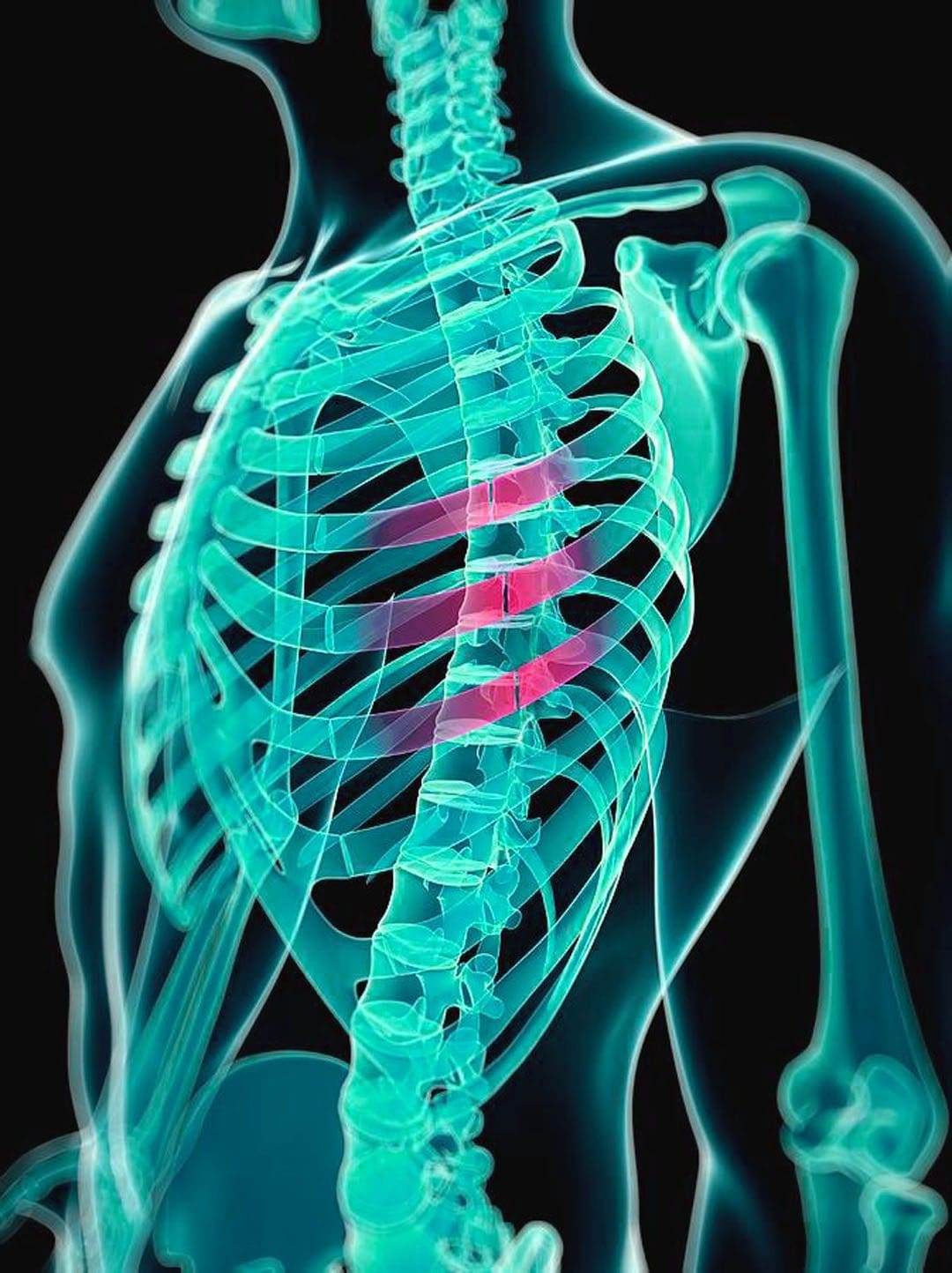
Individuals may not realize they have a cracked rib until symptoms like pain when taking in a deep breath begin to present. Can knowing the symptoms and causes of cracked or broken ribs help in diagnosis and treatment?
Cracked Rib
A broken/fractured rib describes any break in the bone. A cracked rib is a type of rib fracture and is more a description than a medical diagnosis of a rib that has been partially fractured. Any blunt impact to the chest or back can cause a cracked rib, including:
Falling
Vehicle collision
Sports injury
Violent coughing
The main symptom is pain when inhaling.
The injury typically heals within six weeks.
Symptoms
Cracked ribs are usually caused by a fall, trauma to the chest, or intense violent coughing. Symptoms include:
Swelling or tenderness around the injured area.
Chest pain when breathing/inhaling, sneezing, laughing, or coughing.
Chest pain with movement or when lying down in certain positions.
Possible bruising.
Although rare, a cracked rib can cause complications like pneumonia.
See a healthcare provider immediately if experiencing difficulty breathing, severe chest pain, or a persistent cough with mucus, high fever, and/or chills.
Types
In most cases, a rib usually gets broken in one area, causing an incomplete fracture, which means a crack or break that does not go through the bone. Other types of rib fractures include:
Displaced and Nondisplaced Fractures
Completely broken ribs may or may not shift out of place.
If the rib does move, this is known as a displaced rib fracture and is more likely to puncture lungs or damage other tissues and organs. (Yale Medicine. 2024)
A rib that stays in place usually means the rib is not completely broken in half and is known as a nondisplaced rib fracture.
Flail Chest
A section of the ribcage can break away from the surrounding bone and muscle, although this is rare.
If this happens, the ribcage will lose stability, and the bone will move freely as the individual inhales or exhales.
This broken ribcage section is called a flail segment.
This is dangerous as it can puncture the lungs and cause other serious complications, like pneumonia.
Causes
Common causes of cracked ribs include:
Vehicle collisions
Pedestrian accidents
Falls
Impact injuries from sports
Overuse/Repetitive stress brought on by work or sports
Severe coughing
Older individuals can experience a fracture from a minor injury due to the progressive loss of bone minerals. (Christian Liebsch et al., 2019)
The Commonality of Rib Fractures
Rib fractures are the most common type of bone fracture.
They account for 10% to 20% of all blunt trauma injuries seen in emergency rooms.
In cases where an individual seeks care for a blunt injury to the chest, 60% to 80% involve a broken rib. (Christian Liebsch et al., 2019)
Diagnosis
A cracked rib is diagnosed with a physical exam and imaging tests. During the examination, a healthcare provider will listen to the lungs, press gently on the ribs, and watch as the rib cage moves. The imaging test options include: (Sarah Majercik, Fredric M. Pieracci 2017)
X-rays – These are for detecting recently cracked or broken ribs.
CT Scan – This imaging test comprises multiple X-rays and can detect smaller cracks.
MRI – This imaging test is for soft tissues and can often detect smaller breaks or cartilage damage.
Bone Scan – This imaging test uses a radioactive tracer to visualize the structure of bones and can show smaller stress fractures.
Treatment
In the past, treatment used to involve wrapping the chest with a band known as a rib belt. These are rarely used today as they can restrict breathing, increasing the risk of pneumonia or even a partial lung collapse. (L. May, C. Hillermann, S. Patil 2016). A cracked rib is a simple fracture that requires the following:
Rest
Over-the-counter or prescription medications can help manage pain symptoms.
Nonsteroidal anti-inflammatory drugs – NSAIDs like ibuprofen or naproxen are recommended.
If the break is extensive, individuals may be prescribed stronger pain medication depending on the severity and underlying conditions.
Physical therapy can expedite the healing process and help maintain the range of motion of the chest wall.
For patients who are frail and elderly individuals, physical therapy can help the patient walk and normalize certain functions.
A physical therapist can train the individual to transfer between bed and chairs safely while maintaining awareness of any movements or positioning that make the pain worse.
A physical therapist will prescribe exercises to keep the body as strong and limber as possible.
For example, lateral twists can help improve the range of motion in the thoracic spine.
During the early stages of recovery, it is recommended to sleep in an upright position.
Lying down can add pressure, causing pain and possibly worsen the injury.
Use pillows and bolsters to help support sitting up in bed.
What may feel like a cracked rib may be a similar condition, which is why it’s important to get checked out. Other possible symptom causes can include:
Bruised ribs – This occurs when the ribs are not cracked, but the smaller blood vessels around the region burst and leak into surrounding tissues. (Sarah Majercik, Fredric M. Pieracci 2017)
Pulled muscle – A muscle strain, or pulled muscle, occurs when the muscle gets overstretched, which can lead to a tear. The ribs are not affected, but it can feel like they are. (Sarah Majercik, Fredric M. Pieracci 2017)
Emergency
The most common complication is being unable to take a deep breath because of the pain. When the lungs cannot breathe deeply enough, mucous and moisture can build up and lead to an infection like pneumonia. (L. May, C. Hillermann, S. Patil 2016). Displaced rib fractures can also damage other tissues or organs, increasing the risk of a collapsed lung/pneumothorax or internal bleeding. It is recommended to seek immediate medical attention if symptoms develop like:
Shortness of breath
Difficulty breathing
A bluish color of the skin caused by lack of oxygen
A persistent cough with mucus
Chest pain when breathing in and out
Fever, sweating, and chills
Rapid heart rate
The Power of Chiropractic Care In Injury Rehabilitation
Liebsch, C., Seiffert, T., Vlcek, M., Beer, M., Huber-Lang, M., & Wilke, H. J. (2019). Patterns of serial rib fractures after blunt chest trauma: An analysis of 380 cases. PloS one, 14(12), e0224105. https://doi.org/10.1371/journal.pone.0224105
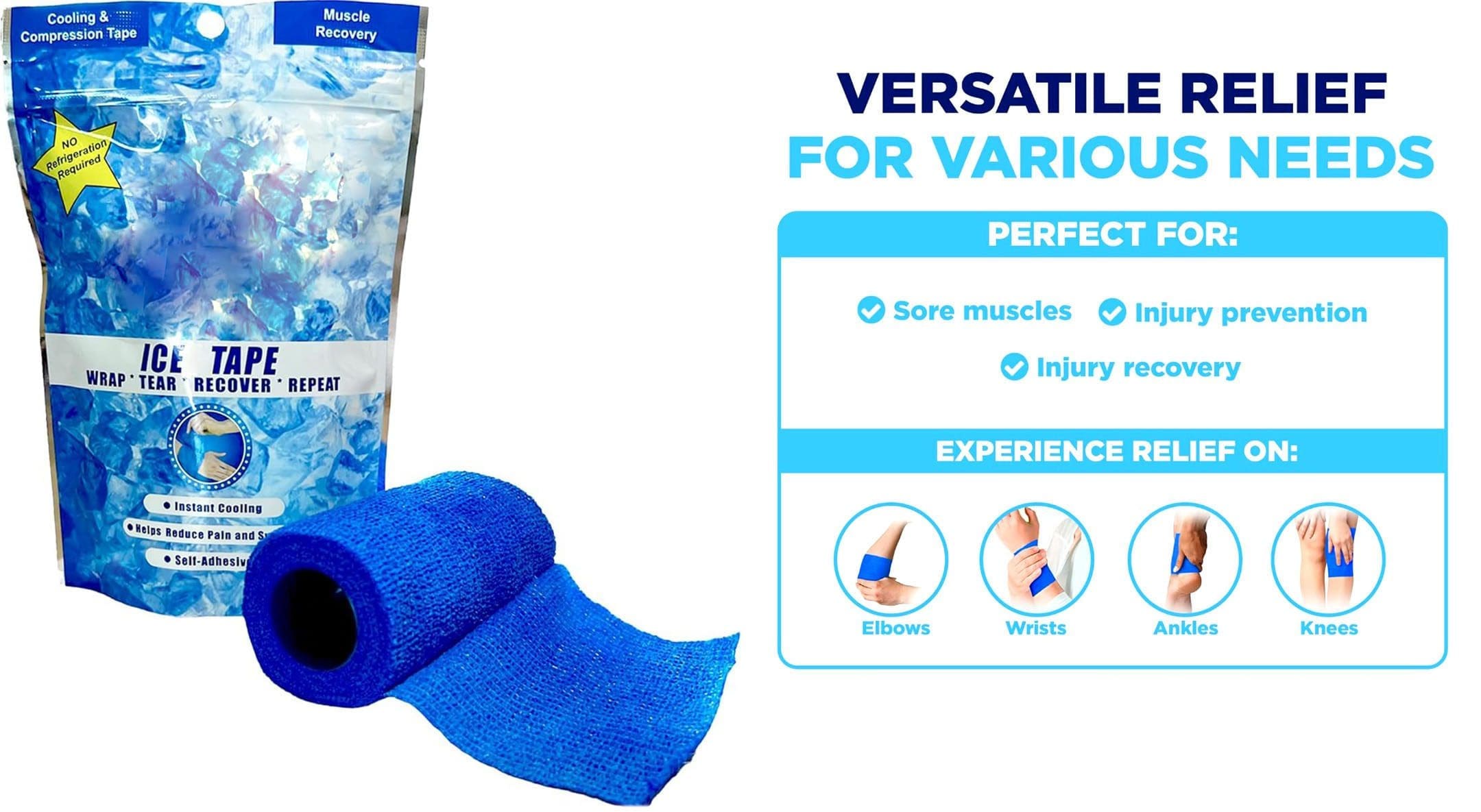
For individuals into sports, fitness enthusiasts, and those that engage in physical activities, musculoskeletal injuries are common. Can using ice tape help during the initial or acute phase of injury decrease inflammation and swelling to expedite recovery and return to activities sooner?
Ice Tape
After a musculoskeletal injury, individuals are recommended to follow the R.I.C.E. method to help reduce swelling and inflammation. R.I.C.E. is the acronym for Rest, Ice, Compression, and Elevation. (Michigan Medicine. University of Michigan. 2023) The cold helps to decrease pain, lower tissue temperature, and decrease swelling around the site of the injury. By controlling the inflammation with ice and compression early after injury, individuals can maintain the appropriate range of motion and mobility around the injured body part. (Jon E. Block. 2010) There are different ways to apply ice to an injury.
Store-bought ice bags and cold packs.
Soaking the injured body part in a cold whirlpool or tub.
Making reusable ice packs.
A compression bandage can be used together with the ice.
Ice Tape is a compression bandage that provides cold therapy all at once. After an injury, applying it can help decrease the pain and swelling during the acute inflammatory phase of healing. (Matthew J. Kraeutler et al., 2015)
How The Tape Works
The tape is a flexible bandage that is infused with therapeutic cooling gel. When applied to an injured body part and exposed to air, the gel activates, generating a cold sensation around the area. The therapeutic medicinal effect can last five to six hours. Combined with a flexible bandage, it provides ice therapy and compression. The ice tape can be used straight out of the package but can also be stored in the refrigerator to increase the cold effect. Depending on the maker’s instructions, the tape should not be stored in the freezer as this can make it too hard to wrap around the injured area.
Advantages
The benefits include the following:
Easy to Use
The product is easy to use.
Take out the tape, and start wrapping it around the injured body part.
Fasteners Not Required
The wrap sticks to itself, so the tape stays in place without using clips or fasteners.
Easy to Cut
The standard roll is 48 inches long by 2 inches wide.
Most injuries require enough to wrap around the injured area.
Scissors cut the exact amount needed, and store the rest in the resealable bag.
Reusable
After 15 to 20 minutes of application, the product can be easily removed, rolled up, stored in the bag, and used again.
The tape can be used multiple times.
The tape begins to lose its cooling quality after several uses.
Portable
The tape does not need to be placed in a cooler when traveling.
It is easily portable and perfect for a quick ice and compression application immediately after an injury.
It can decrease pain and inflammation and kept at the workplace.
Disadvantages
A few disadvantages include the following:
Chemical Odor
The gel on the flexible wrap can have a medicine odor.
It is not quite as powerful smelling as pain creams, but the chemical odor could bother some individuals.
Might Not Be Cold Enough
The tape works for immediate pain relief and inflammation, but it may not get cold enough for the user when applied right from the package at room temperature.
However, it can be placed in a refrigerator to increase the coldness and may provide a more therapeutic cooling effect, especially for those dealing with tendinitis or bursitis.
Stickiness Could Be Distracting
The tape could be a bit sticky for some.
This sticky factor can be a minor annoyance.
However, it just feels sticky when being applied.
A couple of flecks of the gel may get left behind when removed.
The ice tape can also stick to clothing.
For individuals looking for a quick, on-the-go cooling therapy for injured or aching body parts, ice tape may be an option. It could be good to have on hand to provide cooling compression if a minor injury occurs while participating in athletics or physical activities and relief for overuse or repetitive strain injuries.
Block J. E. (2010). Cold and compression in the management of musculoskeletal injuries and orthopedic operative procedures: a narrative review. Open access journal of sports medicine, 1, 105–113. https://doi.org/10.2147/oajsm.s11102
Kraeutler, M. J., Reynolds, K. A., Long, C., & McCarty, E. C. (2015). Compressive cryotherapy versus ice-a prospective, randomized study on postoperative pain in patients undergoing arthroscopic rotator cuff repair or subacromial decompression. Journal of shoulder and elbow surgery, 24(6), 854–859. https://doi.org/10.1016/j.jse.2015.02.004
IFM's Find A Practitioner tool is the largest referral network in Functional Medicine, created to help patients locate Functional Medicine practitioners anywhere in the world. IFM Certified Practitioners are listed first in the search results, given their extensive education in Functional Medicine