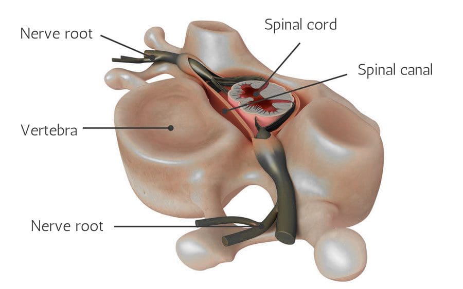
Spinal nerves send motor, sensory, and autonomic signals between the central nervous system and the body and are part of the peripheral nervous system. They are essential for carrying information that controls body movements and sensations to the brain. When a nerve gets injured, compressed, or damaged, it can cause discomfort, increased sensitivity, numbness, muscle weakness, and pain.
Damaged Nerve Roots
Nerve root pain is often caused by other underlying conditions that have caused compression or damage to the nerve root. Causes of nerve root pain can include:
Spinal nerves impacted by injuries or infection can lose their ability to control the body areas, lose their functional capacity, lose sensation, and die.
Spinal Imaging
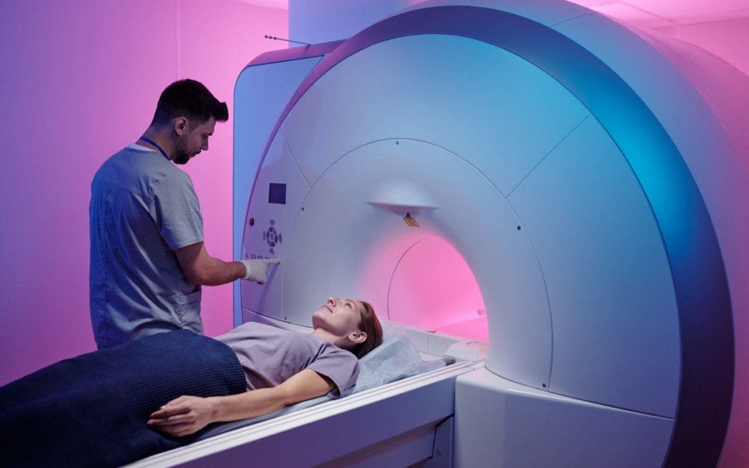
Nerve damage can be diagnosed on a neurological exam and correlated with MRI and X-ray imaging. Conditions that MRI can identify include herniated discs, spinal cord compression or fracture, arthritic development, tumors, or cysts pressing on a nerve.
MRI images are obtained with a magnetic field and radio waves.
MRI shows spine images from the side/sagittal view and cross-sectional/axial views.
This allows the chiropractic doctor to see the vertebrae and discs and identify abnormalities.
The spinal cord is a gray area in the middle surrounded by the spinal fluid, which appears white.
Little white channels on either side of the spinal cord are where the nerve roots branch off.
X-rays can show the alignment of the bones along the spine and determine any narrowing or damage to the discs.
It is important to be evaluated and diagnosed for signs and symptoms of nerve injury as soon as possible, as nerve damage accelerates and worsens.
Function Restoration
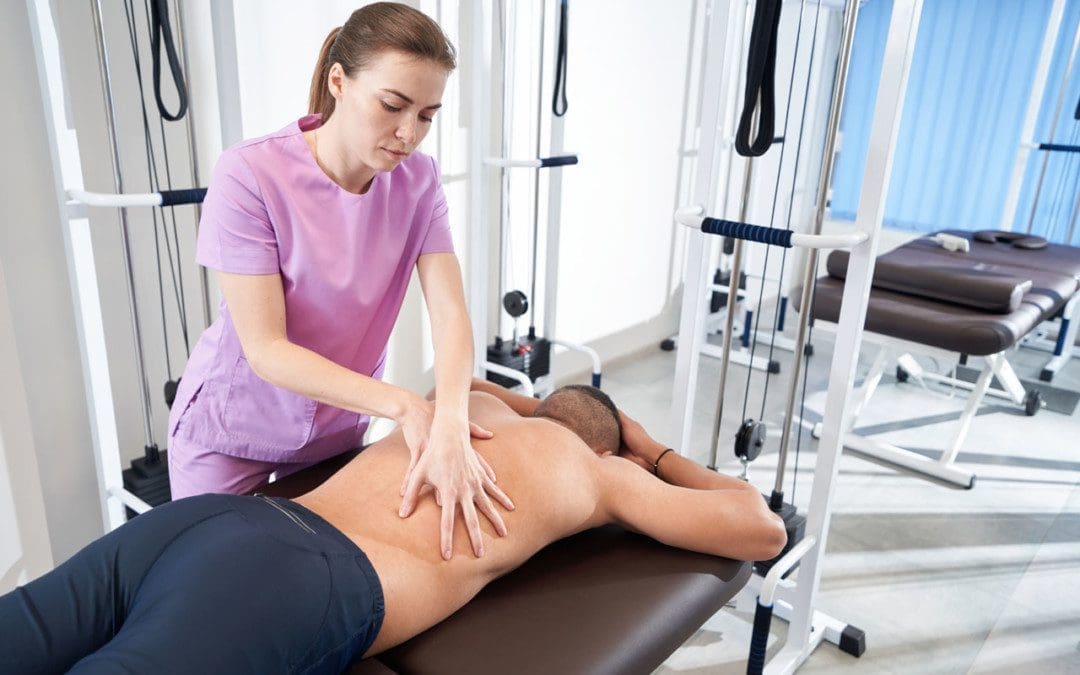
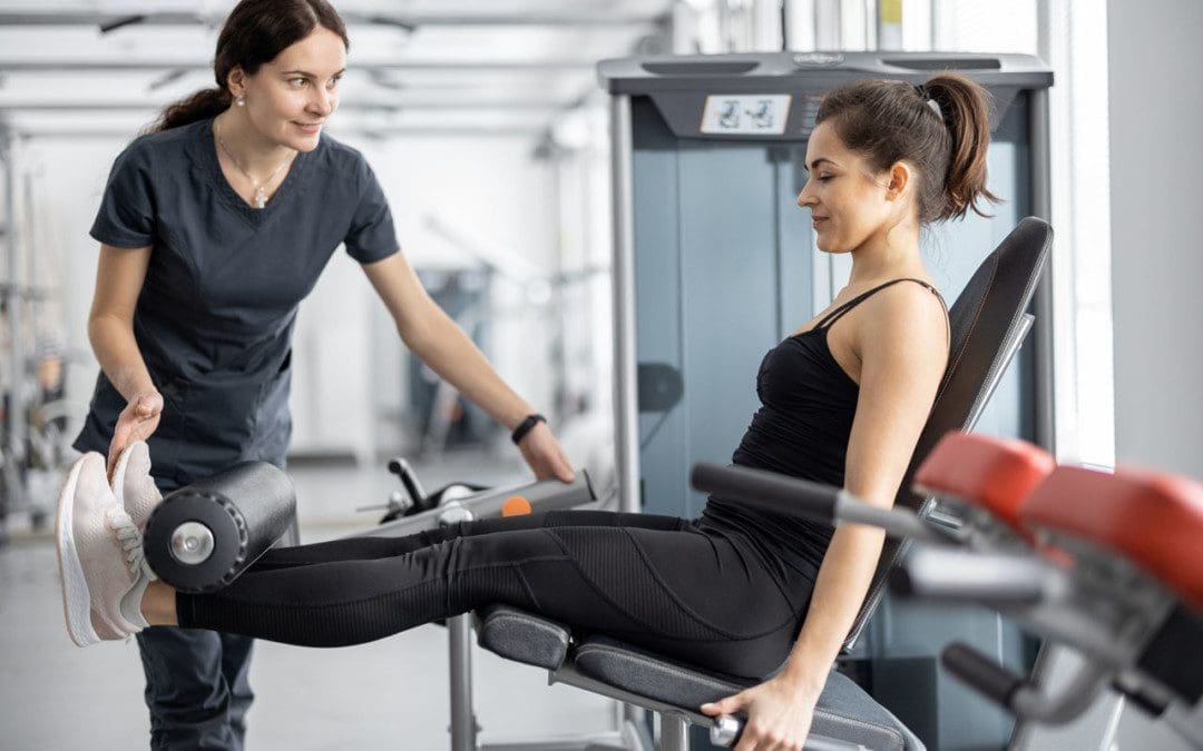
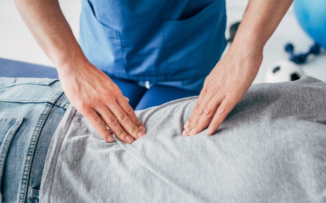
Sometimes, the symptoms improve by themselves and do not require treatment. Nonetheless, physicians begin with conservative, non-surgical approaches to treat nerve root pain. Chiropractic and physical massage therapy involves specific movements, stretches, and exercises to keep the affected muscles and joints active,prevent stiffness and help restore function and feeling. Treatment can include:
Therapeutic massage
Manual adjustment/resistance treatment
Trigger point therapy
Instrument-assisted soft tissue therapy
Decompression
Traction
Joint stretching
Electrical stimulation
Ultrasound
Specialized exercise
Activity modification
Anti-inflammatory diet
Nerve Chiropractor
References
Liu, Yan, and Huan Wang. “Peripheral nerve injury-induced changes in the spinal cord and strategies to counteract/enhance the changes to promote nerve regeneration.” Neural regeneration research vol. 15,2 (2020): 189-198. doi:10.4103/1673-5374.265540
Menorca, Ron M G, et al. “Nerve physiology: mechanisms of injury and recovery.” Hand clinics vol. 29,3 (2013): 317-30. doi:10.1016/j.hcl.2013.04.002
Shehab, Safa Al-Deen Saudi. “Fifth lumbar spinal nerve injury causes neurochemical changes in corresponding and adjacent spinal segments: a possible mechanism underlying neuropathic pain.” Journal of chemical neuroanatomy vol. 55 (2014): 38-50. doi:10.1016/j.jchemneu.2013.12.002
Stoll, G, and H W Müller. “Nerve injury, axonal degeneration, and neural regeneration: basic insights.” Brain pathology (Zurich, Switzerland) vol. 9,2 (1999): 313-25. doi:10.1111/j.1750-3639.1999.tb00229.x
Ye, Xuan, et al. “Nerve fascicle transfer using a part of the C-7 nerve for spinal accessory nerve injury.” Journal of neurosurgery. Spine vol. 28,5 (2018): 555-561. doi:10.3171/2017.8.SPINE17582
Individuals that have experienced a muscle strain, pull, spasm, etc., that has healed can begin to behave overly cautious, avoiding putting full weight on the area or using full motion out of fear of re-injuring it. This can and does strain other body areas because of the imbalance and awkward positioning. It also leads to anxiety, emotional distress, and decreased self-confidence in everyday movement. Adjustments, massage, and decompression therapy can maintain musculoskeletal health, and a chiropractor can help retrain individuals on healthy posture and confident movement.
Muscle Guarding
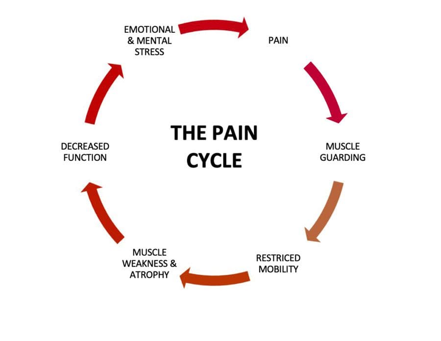
Muscles can be held in a position of readiness to act, like the stress response of fight or flight. When this happens, the muscles are partially contracted in preparation for action and is a form of muscle-guarding. But once the fight or flight passes, the muscles relax into their normal position. With injury muscle guarding, the fears and stresses after recovering from an injury can cause the injured and non-injured muscles to stay in the guarded/semi-contracted position. The longer the muscle guarding continues, fatigue begins to set in, decreasing function, restricting mobility, and making the body more vulnerable to damage and injury.
The Brain
The discomfort, pain, or just the thought reinforces the need to guard the area. The brain will find a way to move without causing pain and create compensating but unhealthy movement patterns that strain the other areas of the body. The body adapts to not using the formerly injured muscles and now relies on the other muscles to perform the functions in a non-relaxed state that can become normal, causing stiffness, soreness, tenderness, tendon tension, and pain.
An example is a hip strain, pull or spasm that has been treated and has healed, but the individual is fearful of another injury or going through the painful experience again and begins walking by shifting all their weight to the other side and steps with a limp or some abnormal motion that strains and/or injures the rest of the body.
Chiropractic Treatment and Retraining
Individuals experiencing muscle guarding can find help through chiropractic to retrain their muscles to return to their normal position and regain confidence in their movements. The body will be rebalanced by releasing and relaxing the tight muscles. Then therapeutic repetitive movements, specialized exercises, stretches, and relaxation techniques will help the individual relearn to use the muscles without fear.
Protective Muscle Guarding
References
Hanlon, Shawn et al. “Examining Ankle-Joint Laxity Using 2 Knee Positions and With Simulated Muscle Guarding.” Journal of athletic training vol. 51,2 (2016): 111-7. doi:10.4085/1062-6050-51.3.06
Olugbade, Temitayo et al. “The relationship between guarding, pain, and emotion.” Pain reports vol. 4,4 e770. 22 Jul. 2019, doi:10.1097/PR9.0000000000000770
Prkachin, Kenneth M et al. “Pain behavior and the development of pain-related disability: the importance of guarding.” The Clinical journal of pain vol. 23,3 (2007): 270-7. doi:10.1097/AJP.0b013e3180308d28
Spinal disc deterioration from aging is normal, but health issues or injuries can advance the degenerative process. Disc protrusions are related to herniated discs but are the mildest form of the condition and are a common form of spinal disc deterioration that can cause neck and back issues. However, individuals may have a small protruding disc that can go undetected unless it irritates or compresses the surrounding nerves. Chiropractic care, decompression, and massage therapy can realign the disc back into position, relieving discomfort and pain.
Disc Protrusion
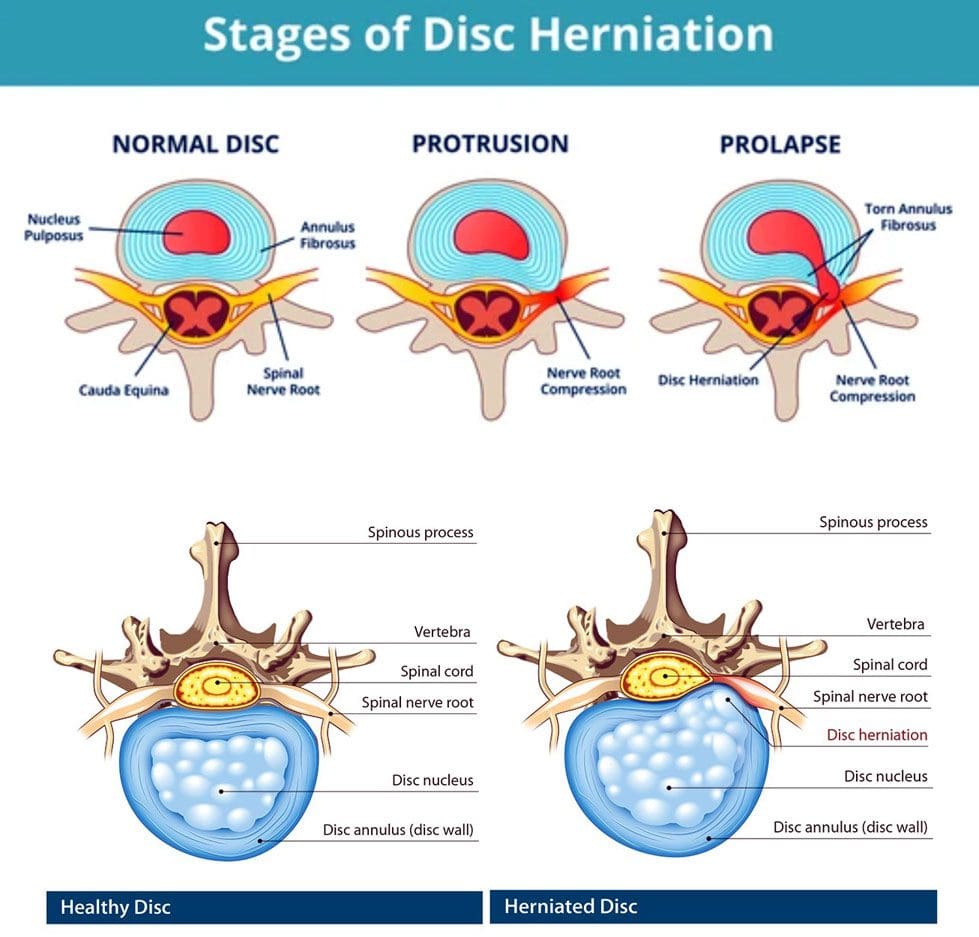
A disc is like a sturdy soft rubber shock absorber/cushion with added gel inside. The gel acts as a shock absorber. When the gel begins to protrude out slightly, this is a disc protrusion. Once a protruding disc begins to develop, it usually remains in that position. The disc can sometimes reabsorb on its own and realign back into position, but there is no way of knowing that will happen or how long it will take. With age and/or injuries, the body’s parts change. The spine’s discs dehydrate and lose elasticity weakening the discs and making them more vulnerable to herniation stages:
First Stage
Following natural weakening can be classified as a disc protrusion when the disc’s core begins pushing into the spinal column.
Disc protrusions can be tiny or push out an entire side of the disc.
Second Stage
Disc deterioration often consists of a bulging disc when the core pushes out farther around the circumference beyond the disc’s outer layer, called the annulus fibrosus, creating the telltale bulge.
A bulging disc involves more than 180 degrees of the disc’s circumference.
Third Stage
The third stage is a herniated disc, meaning the disc’s outer wall has torn, allowing the inner gel to leak out, usually irritating the surrounding nerves.
Fourth Stage
The fourth stage is sequestration, a herniated disc in which a piece of the nucleus breaks free of the vertebral disc fragments and falls into the spinal canal.
Types
A disc protrusion is one type of disc herniation that pushes out but remains connected. Different types compress and irritate the discs differently and produce various symptoms, including:
Paracentral
This is the most common, where the disc protrusion jams the space between the central canal and the foramen.
Central
This is where the disc protrusion impinges into the spinal canal, with or without spinal cord compression.
Foraminal
The disc intrudes into the foramen, the space through which nerve roots branch off the spinal cord and exit the vertebrae.
Symptoms, Diagnosis, and Chiropractic Care
Individuals with a disc protrusion can have symptoms similar to sciatica, which includes back, buttock, and leg discomfort, numbness, and pain sensations.
Treatment for disc protrusion will be based on the individual’s symptoms.
A chiropractor will take a detailed medical history and perform a physical examination.
A spinal MRI test could be ordered depending on the injury or condition.
A customized treatment plan will be developed to fit the individual’s medical needs.
Most disc protrusions improve after a few weeks of rest, avoiding strenuous activities, activity modification, an anti-inflammatory diet, and gentle exercises that the chiropractic team will provide.
True Spinal Decompression
References
Fardon, David F et al. “Lumbar disc nomenclature: version 2.0: Recommendations of the combined task forces of the North American Spine Society, the American Society of Spine Radiology and the American Society of Neuroradiology.” The spine journal: official journal of the North American Spine Society vol. 14,11 (2014): 2525-45. doi:10.1016/j.spinee.2014.04.022
Mysliwiec, Lawrence Walter, et al. “MSU classification for herniated lumbar discs on MRI: toward developing objective criteria for surgical selection.” The European spine journal: official publication of the European Spine Society, the European Spinal Deformity Society, and the European Section of the Cervical Spine Research Society vol. 19,7 (2010): 1087-93. doi:10.1007/s00586-009-1274-4
The game of baseball takes a toll on the body, especially when players advance from little league to high school, college, minor league, and the pros. The most common baseball injuries can range from mild to severe, from normal wear and tear on the joints and muscles to repetitive stress injuries, collisions with other players, getting hit with the ball, or bodily trauma. A chiropractor can provide ideal treatment for players of all ages and levels with decreased downtime and expedited healing and recovery.
Baseball Injuries
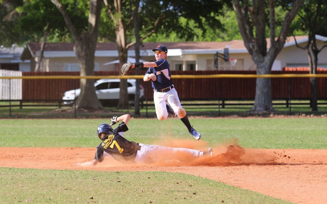
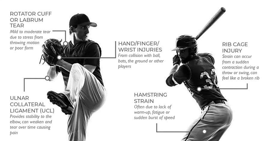
Although there have been a lot of advances in player safety and health, from helmets with face guards to shin and arm padding, the equipment lessens the impact and risks of injury. The game still involves running, sliding, twisting, and jumping, causing the body to maneuver awkwardly. Players often report sliding into first, feeling a pop or twisting to catch a fly ball, and feeling something snap. The most common injuries include:
Torn Labrum
Cartilage surrounding the shoulder joint socket, known as the labrum, often gets torn.
The soft tissue keeps the bones in place and provides stability.
Pitching and throwing motions stress the labrum.
With time, the cartilage begins to overstretch and tear, leading to swelling, shoulder pain, weakness, and overall instability.
Rotator Cuff Tears
The rotator cuff structure involves a complex set of tendons and muscles that stabilize the shoulder.
Pitchers are the most vulnerable, but all players are susceptible.
Cases are caused by not warming up and stretching correctly and repetitive/overuse movements.
Swelling and pain are the most common symptoms.
With a severe tear, a player will lose the ability to rotate the shoulder correctly.
Shoulder Instability or Dead Arm
This is when the shoulder muscles become overly fatigued, and the joint becomes unstable, losing the ability to throw precisely.
The condition is called dead arm by players and trainers.
This type of injury is caused by overuse and repeated stress.
Healing involves letting the shoulder rest for an extended period, but treatment, like chiropractic or physical therapy, could be recommended depending on the severity.
Pitchers Elbow
A pitcher’s elbow injury is caused by overuse and sustained/repeated damage to the tendons that rotate the wrist.
Pain and swelling occur along the inside of the elbow and forearm.
Wrist Tendonitis and Trauma
Wrist Tendonitis or tenosynovitis happens when the ligaments and tendons become tender, swollen, ruptured, or torn.
This causes inflammation, pain, and weakness.
Trauma injuries can result from collisions with another player, the ground, or a ball.
Knee Tears and Trauma
Knee injuries can be caused by normal wear and tear, overuse, or traumatic impact.
The fibrous bands are what stabilize and cushion the knee.
Overuse and any awkward movement can cause the tearing of the various ligaments.
The bands can develop micro-tears or complete ruptures, causing inflammation, pain, and instability.
Chiropractic Care and Rehabilitation
Chiropractic treatment and physical therapy have been found to help athletes maintain flexibility and range of motion, rehabilitate the body after an injury, and prevent new injuries or worsening of current injuries.
Chiropractic helps stretch and flex the muscles to stay limber and less prone to injury.
Chiropractic is a natural pain reliever for sore muscles and joint pain.
Physical therapy can strengthen an injured area during recovery and educate on proper form and techniques.
Taping and strapping can help support the elbows, wrists, ankles, and knees, reducing stress.
A combination of treatment approaches can help decrease recovery time so players can get back on the field.
Shoulder Adjustment Baseball Injuries
References
Bullock, Garrett S et al. “Shoulder Range of Motion and Baseball Arm Injuries: A Systematic Review and Meta-Analysis.” Journal of athletic training vol. 53,12 (2018): 1190-1199. doi:10.4085/1062-6050-439-17
Lyman, Stephen, and Glenn S Fleisig. “Baseball injuries.” Medicine and sport science vol. 49 (2005): 9-30. doi:10.1159/000085340
Matsel, Kyle A et al. “Current Concepts in Arm Care Exercise Programs and Injury Risk Reduction in Adolescent Baseball Players: A Clinical Review.” Sports health vol. 13,3 (2021): 245-250. doi:10.1177/1941738120976384
Shitara, Hitoshi, et al. “Shoulder Stretching Intervention Reduces the Incidence of Shoulder and Elbow Injuries in High School Baseball Players: a Time-to-Event Analysis.” Scientific reports vol. 7 45304. 27 Mar. 2017, doi:10.1038/srep45304
Wilk, Kevin E, and Christopher A Arrigo. “Rehabilitation of Elbow Injuries: Nonoperative and Operative.” Clinics in sports medicine vol. 39,3 (2020): 687-715. doi:10.1016/j.csm.2020.02.010
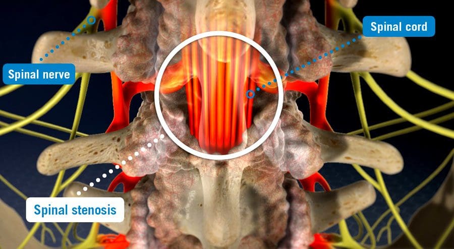
Spinal stenosis is when space somewhere along or within the spine begins to narrow, closing off the ability of normal/comfortable movement and nerve circulation. It can affect different areas, including the cervical/neck, lumbar/low back, and, less commonly, the thoracic/upper or mid-back regions causing tingling, numbness, cramping, pain, muscle weakness, or a combination in the back, leg/s, thighs, and buttocks. There can be various factors causing the stenosis; correct diagnosing is the first step, and where a spinal stenosis MRI comes in.
Spinal Stenosis MRI
Stenosis can be challenging to diagnose as it is more of a symptom/complication than a condition, often caused by herniated discs, bone spurs, a congenital condition, post-surgery, or after an infection. Magnetic resonance imaging/MRI is a common test used in diagnosis.
Diagnosis
A healthcare professional, like a chiropractor, physical therapist, spine specialist, or physician, will begin with understanding symptoms and medical history.
A physical exam will be conducted to learn more about the location, duration, positions, or activities that decrease or worsen the symptoms.
Additional tests include muscle strength, gain analysis, and balance testing to help better understand where the pain is coming from.
To confirm a diagnosis, imaging will be required to see what is going on.
An MRI uses computer-generated imaging to produce images that show bone and soft tissues, like muscles, nerves, and tendons, and if they are compressed or irritated.
A healthcare professional and MRI technician will go over the safety requirements before the imaging.
Because the machine uses powerful magnets, there can be no metal on or in the body, like implanted prostheses or devices that include:
A different imaging test may be used if an individual cannot have an MRI like a CT scan.
An MRI can range from several minutes to an hour or longer, depending on how many positions are necessary to isolate the injured area and get a clear image. The test is painless, but sometimes individuals are asked to maintain a specific position that could be uncomfortable. The technician/s will ask if there is discomfort and offer any help to make the experience as easy as possible.
Treatment
Not all cases of stenosis cause symptoms, but there are treatment options that a healthcare professional can recommend.
Conservative care is the first recommendation that includes chiropractic, decompression, traction, and physical therapy.
Treatment increases muscle strength, improves range of motion, improves posture and balance, decreases discomfort symptoms, and incorporates strategies to prevent and manage symptoms.
Prescription medications could be part of a larger treatment plan.
Surgery could become an option in more severe cases where conservative care is not working.
Spinal Stenosis
References
Database of Abstracts of Reviews of Effects (DARE): Quality-assessed Reviews [Internet]. York (UK): Centre for Reviews and Dissemination (UK); 1995-. Diagnosis of lumbar spinal stenosis: an updated systematic review of the accuracy of diagnostic tests. 2013. Available from: https://www.ncbi.nlm.nih.gov/books/NBK142906/
Ghadimi M, Sapra A. Magnetic Resonance Imaging Contraindications. [Updated 2022 May 8]. In: StatPearls [Internet]. Treasure Island (FL): StatPearls Publishing; 2022 Jan-. Available from: https://www.ncbi.nlm.nih.gov/books/NBK551669/
Gofur EM, Singh P. Anatomy, Back, Vertebral Canal Blood Supply. [Updated 2021 Jul 26]. In: StatPearls [Internet]. Treasure Island (FL): StatPearls Publishing; 2022 Jan-. Available from: https://www.ncbi.nlm.nih.gov/books/NBK541083/
Lurie, Jon, and Christy Tomkins-Lane. “Management of lumbar spinal stenosis.” BMJ (Clinical research ed.) vol. 352 h6234. 4 Jan. 2016, doi:10.1136/bmj.h6234
Stuber, Kent, et al. “Chiropractic treatment of lumbar spinal stenosis: a review of the literature.” Journal of chiropractic medicine vol. 8,2 (2009): 77-85. doi:10.1016/j.jcm.2009.02.001
Sciatica pain can radiate to the knee. Individuals with sciatica do report unique/unusual knee pain that was never there and no past or recent physical injuries. Sciatica is the culprit, as the knee muscles are powered and controlled by nerves in the lower spine. Irritation or compression of these nerves can cause symptoms that can include: random back pain, hamstring tightness, weakness in the hips or quadriceps, the development of bunions, and knee pain and/or weakness. Chiropractic treatment can release the compression, heal the sciatic nerve, and alleviate knee problems.
Sciatica Pain Can Radiate To The Knee
Spine conditions that can cause sciatica include:
Disc herniation – Where the inside of the discs leak out and compress and/or irritate surrounding nerves.
Spinal stenosis – The spinal canal begins to narrow, not allowing enough space for the nerves to rest comfortably, resulting in compressed nerves.
Spondylolisthesis – A condition that occurs when a vertebrae slips forward onto the vertebrae below it.
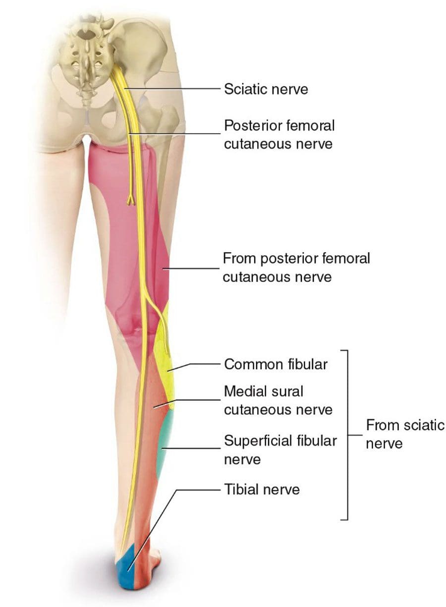
Any can cause irritation, inflammation, or compression of the sciatic nerve leading to painful sensations that extends from the lower back down through the leg.
Symptoms
Common knee symptoms that may be experienced with sciatica include:
A dull ache, warm sensation, or sharp pain around the knee.
Bunions form from weakened stabilizing muscles that affect walking, running, and standing posture.
As sciatica pain can radiate to the knee, individuals will usually also experience pain in their buttocks, thigh, calf, and/or foot. The nerve sensations and other symptoms in the knee can be felt through a branch of the sciatic nerve known as the peroneal nerve.
Duration
The knee pain will last as long as sciatica does, depending on the type of sciatica, whether it is acute or chronic.
An acute sciatic episode usually resolves after a few weeks, with possible future flare-ups.
Chronic sciatica is a long-term condition that does not resolve independently and necessitates intervention by a specialist.
Chiropractic Treatment Plan
Depending on the diagnosis, a chiropractor will develop a personalized treatment plan to address the root cause and heal the injury. The treatment plan will include therapeutic massage, posture training, and at-home self-care to help heal and prevent future sciatica.
Massage Rehabilitation
Physical therapy and therapeutic massage will loosen and relax the muscles, nerves, tendons, and ligaments.
Heat and ice, exercises, and stretches will prepare the muscles and nerves for chiropractic decompression adjustments.
Posture Training
Training will be provided to maintain the back, hips, knees, and feet in proper alignment.
Training on removing pressure from the lower back and restabilizing the rest of the body.
Training on proper body mechanics, safe lifting techniques, and injury prevention.
Self-Pain Management
Training on self-care habits that include healthy weight, core strengthening exercises and stretches for the back muscles, and proper rest for a full recovery.
An anti-inflammatory diet to reduce/eliminate inflammation and achieve a healthy weight and a nutrition plan to maintain overall health.
Surgery
Surgery is the final option when conservative treatments are not working.
Treating Severe & Complex Sciatica Syndromes
References
Dydyk AM, Khan MZ, Singh P. Radicular Back Pain. [Updated 2021 Nov 2]. In: StatPearls [Internet]. Treasure Island (FL): StatPearls Publishing; 2022 Jan-. Available from: https://www.ncbi.nlm.nih.gov/books/NBK546593/
Hirabayashi, Hiroki, et al. “Characteristics of L3 nerve root radiculopathy.” Surgical neurology vol. 72,1 (2009): 36-40; discussion 40. doi:10.1016/j.surneu.2008.08.073
Jandre Reis, Felipe Jose, and Adriana Ribeiro Macedo. “Influence of Hamstring Tightness in Pelvic, Lumbar and Trunk Range of Motion in Low Back Pain and Asymptomatic Volunteers during forwarding Bending.” Asian spine journal vol. 9,4 (2015): 535-40. doi:10.4184/asj.2015.9.4.535
Jeong, Ui-Cheol, et al. “The effects of self-mobilization techniques for the sciatic nerves on physical functions and health of low back pain patients with lower limb radiating pain.” Journal of physical therapy science vol. 28,1 (2016): 46-50. doi:10.1589/jpts.28.46
The body is designed to move. For individuals who spend a significant amount of time driving each day, whether for a living or a long commute, over time can lead to headaches, neck and back pain, sciatica, and increases the risk for serious injury. Chiropractic can retrain individuals to practice healthy driving posture. This is accomplished through decompression and massage therapy combined with recommended stretches/exercises, and an anti-inflammatory diet will bring pain relief and help prevent injury.
Healthy Driving Posture
Two main reasons driving impacts the back are poor posture and being in a fixed position for an extended period. Individuals who regularly drive for more than 4 hours a day are more at risk. An unhealthy driving posture can lead to an increased risk of discomfort/pain in the:
Over time these issues can become chronic, making the body vulnerable to various injuries.
Back Pain Symptoms
Sometimes back pain needs immediate medical attention if any of the following symptoms present:
Inflammation in the back.
Swelling on the back.
Constant pain does not go away or ease up after resting or movement.
Pain in the upper back that radiates to the chest.
A high temperature.
Unexplained weight loss.
Loss of bladder or bowel control.
Numbness or tingling around the buttocks or groin area.
Driving Recommendations
Spine Support
Slide the tailbone as close to the back of the seat as possible.
Leave a gap between the back of the knees and the front of the seat.
If the vehicle doesn’t allow for the proper position, a back support cushion can help.
Raise The Hips
If possible, adjust the area you sit on, so the thighs are supported along their entire length.
The knees should be slightly lower than the hips.
This will increase circulation to the back muscles while opening up the hips.
Sitting Too Close
An individual should be able to comfortably reach the pedals and depress them through their full range with the entire foot.
A safety study found that drivers whose chests were closer to the wheel were significantly more likely to suffer head, neck, and chest injuries in front and rear-end collisions.
Proper Height
Ensure the seat raises the eye level a few inches above the steering wheel to allow sufficient clearance between the head and roof.
Seat Angle
The angle of the back of the seat should go a little beyond 90 degrees to 100-110 degrees places minimal pressure on the back.
Leaning too far back forces the individual to raise/push their head and neck forward, which can cause neck pain, shoulder pain, and tingling in the fingers.
Headrest Height
The top of the headrest should be between the top of the ears and the top of the head.
It should slightly touch the back of the head when sitting with a healthy driving posture.
This exercise activates the abdominal and external oblique muscles.
Press the lower back into the car seat.
Inhale and tilt the pelvis forward to create an arch in the lower back.
Hold for 3 seconds, then release.
Repeat ten times.
Posture is more than just how one carries themselves. The effects of unhealthy posture can carry over into an individual’s physical, mental, and emotional health. Whether it’s caused by injury, stress, work, or sports, a professional chiropractor will help you get back to optimal health.
Driving Position
References
Cvetkovic, Marko M et al. “Assessing Post-Driving Discomfort and Its Influence on Gait Patterns.” Sensors (Basel, Switzerland) vol. 21,24 8492. 20 Dec. 2021, doi:10.3390/s21248492
Pope, Malcolm H et al. “Spine ergonomics.” Annual review of biomedical engineering vol. 4 (2002): 49-68. doi:10.1146/annurev.bioeng.4.092101.122107
Tinitali, Sarah, et al. “Sitting Posture During Occupational Driving Causes Low Back Pain; Evidence-Based Position or Dogma? A Systematic Review.” Human factors vol. 63,1 (2021): 111-123. doi:10.1177/0018720819871730
van Veen, Sigrid, and Peter Vink. “Posture variation in a car within the restrictions of the driving task.” Work (Reading, Mass.) vol. 54,4 (2016): 887-94. doi:10.3233/WOR-162359
IFM's Find A Practitioner tool is the largest referral network in Functional Medicine, created to help patients locate Functional Medicine practitioners anywhere in the world. IFM Certified Practitioners are listed first in the search results, given their extensive education in Functional Medicine