Individuals with plantar fasciitis may experience consistent flare-ups. Can knowing the causes help to find pain relief?
Plantar Fasciitis Flare-Up
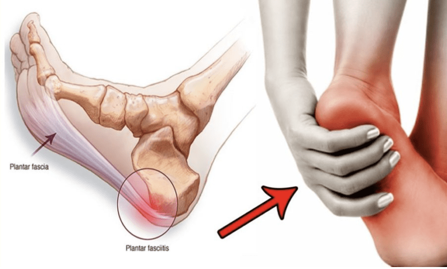
Plantar fasciitis is a common cause of heel and foot pain. The plantar fascia is a band of tissue that runs along the bottom of the foot and becomes inflamed. Certain factors can cause plantar fasciitis flare-ups, including:
Increased levels of physical activity.
Not stretching regularly.
Wearing shoes without proper support.
Weight gain.
Causes
A plantar fasciitis flare-up is often triggered by physical activity. (MedlinePlus. U.S. National Library of Medicine. 2022) It can also be brought on by underlying conditions, like increased body weight, arthritis, or the shape of the foot. (Johns Hopkins Medicine. 2023) Despite the root cause, there are activities and experiences that can contribute to and/or worsen the condition.
New Exercise Routine
Being highly physically active can exacerbate plantar fasciitis symptoms.
High heels, boots, or shoes that raise the heel above the toes.
Worn-out shoes like exercise workout shoes.
Not Stretching Properly or At All
Tight calves can increase pressure on the plantar fascia.
Stretching the calves, Achilles tendon/heel, and the bottom of the feet is highly recommended to help treat and prevent the condition. (Johns Hopkins Medicine. 2023)
Not stretching thoroughly or skipping stretches can worsen symptoms.
Individuals with plantar fasciitis are recommended to stretch before and after physical activities, exercise, before going to bed, and after waking up.
Working Through the Pain
Individuals may try to continue physical activities during a flare-up.
This is not recommended as doing so can cause more pain and worsen the condition.
When pain presents, it’s recommended to:
Stop all activities that strain the feet
Stay off the feet for at least a week.
Tearing the Plantar Fascia
The plantar fascia rarely tear completely from repeated stress known as a plantar fascia rupture.
Pascoe, S. C., & Mazzola, T. J. (2016). Acute Medial Plantar Fascia Tear. The Journal of orthopaedic and sports physical therapy, 46(6), 495. https://doi.org/10.2519/jospt.2016.0409
Individuals that sit at a desk/workstation for hours for work or school, or drive for a living, could be fostering a long-term condition known as forward head posture. Can understanding the signs and symptoms help to prevent the condition?
Forward Head Posture
Neck pain often causes or is caused by misalignment in the area between the shoulders and head. Forward head posture is a common problem that can strain the neck muscles, leading to pain and worsening neck, shoulder, and back posture. (Jung-Ho Kang, et al., 2012) For individuals who are at risk of developing or are already showing signs/symptoms, it’s important to get medical attention to prevent complications, such as chronic neck pain or compressing a nerve. Individuals can continue to do the work that they need to do but may need some postural adjustments and re-training so as not to continue straining the neck while working.
Postural Deviation
The head is in a healthy alignment with the neck when the ears line up with the gravity line.
The gravity line is an imaginary straight line that represents gravity’s downward pull.
It is used in posture assessments as a reference for noting the positions of the body and determining the presence of any postural misalignment or deviation.
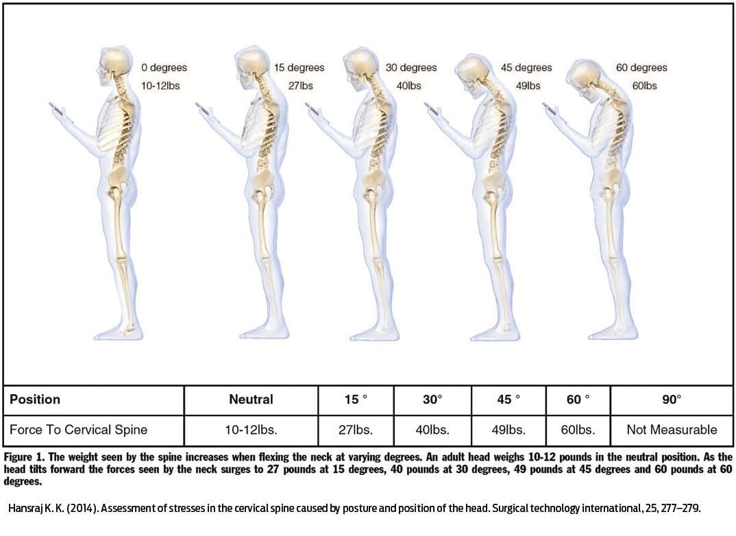
A forward head posture occurs when the head begins to position forward of the gravity line when looking at the body from the side.
Forward head posture is a postural deviation because the head varies from the reference line. (Jung-Ho Kang, et al., 2012)
Muscle Imbalances
Forward head posture often results in a strength imbalance between muscles that support and move your neck, shoulders, and head. (Dae-Hyun Kim, et al., 2018)
The muscles in the back of the neck become shortened and overactive as they flex forward, while the muscles in the front become lengthened, weaker, and strained when they relax.
Kyphosis
Kyphosis also known as hunchback is when the shoulders round forward, and the head is also brought forward. (Jung-Ho Kang, et al., 2012) After many hours sitting at a desk, computer, or driving, kyphosis can also cause and/or worsen forward head posture.
This occurs because the upper back area supports the neck and head.
When the upper back moves or changes position, the head and neck follow.
The majority of the head’s weight is in the front, and this contributes to the forward movement.
An individual with kyphosis has to lift their head to see.
Treatment
A chiropractic injury specialist team can develop a personalized treatment plan to relieve pain symptoms, provide postural retraining, realign the spine, and restore mobility and function.
Standing and sitting using a healthy posture, along with exercises to strengthen the neck muscles, can help get the spine in alignment. (Elżbieta Szczygieł, et al., 2019)
Targeted stretching can help if the neck muscles are tight.
At-home stretches may also relieve pain
Risk Factors
Pretty much everyone is at risk of developing a forward head posture. Common risk factors include:
Constantly looking down at a phone and staying in this position for a long time aka text neck.
Desk jobs and computer use can significantly round the shoulders and upper back, leading to a forward head posture. (Jung-Ho Kang, et al., 2012)
Driving for a living causes prolonged back, neck, and shoulder positioning.
Sleeping or reading with a large pillow under the head can contribute to forward head posture.
Doing work that requires dexterity and close-up positions, like a seamstress or technician can cause over-positioning of the neck.
Individuals who regularly carry a significant amount of weight in front of their body may begin to develop kyphosis.
An example is carrying a child or another load in front of the body.
Large breasts can also increase the risk of kyphosis and forward head posture.
Neck Injuries
References
Kang, J. H., Park, R. Y., Lee, S. J., Kim, J. Y., Yoon, S. R., & Jung, K. I. (2012). The effect of the forward head posture on postural balance in long time computer based worker. Annals of rehabilitation medicine, 36(1), 98–104. https://doi.org/10.5535/arm.2012.36.1.98
Kim, D. H., Kim, C. J., & Son, S. M. (2018). Neck Pain in Adults with Forward Head Posture: Effects of Craniovertebral Angle and Cervical Range of Motion. Osong public health and research perspectives, 9(6), 309–313. https://doi.org/10.24171/j.phrp.2018.9.6.04
Szczygieł, E., Sieradzki, B., Masłoń, A., Golec, J., Czechowska, D., Węglarz, K., Szczygieł, R., & Golec, E. (2019). Assessing the impact of certain exercises on the spatial head posture. International journal of occupational medicine and environmental health, 32(1), 43–51. https://doi.org/10.13075/ijomeh.1896.01293
Hansraj K. K. (2014). Assessment of stresses in the cervical spine caused by posture and position of the head. Surgical technology international, 25, 277–279.
Individuals feeling tingling or pins and needles sensations that overtake the arms or legs could be experiencing paresthesia, which occurs when a nerve has been compressed or damaged. Can knowing the symptoms and causes help in diagnosis and treatment?
Paresthesia Body Sensations
The numbness or tingling feeling when an arm, leg, or foot has fallen asleep is not so much about blood circulation but nerve function.
Paresthesia is an abnormal sensation felt in the body due to the compression or irritation of nerves.
It can be a mechanical cause like a compressed/pinched nerve.
Or it may be due to a medical condition, injury, or illness.
The symptoms typically last for 30 minutes or less.
Shaking the affected limb often relieves the sensations.
Paresthesia usually affects only one arm or leg at a time.
However, both arms and legs can be affected, depending on the cause.
Consult a healthcare provider if the symptoms last for more than 30 minutes. Treatment may be required if paresthesia body sensations are brought on by a serious underlying cause.
Causes
Sitting with incorrect and unhealthy postures can compress a nerve and generate symptoms. However, some causes are more concerning and can include:
If the symptoms don’t go away after 30 minutes or keep returning for unknown reasons, call a healthcare provider to find out what is causing the abnormal sensations. A worsening case should be monitored by a healthcare provider.
Peripheral neuropathy caused by diabetes usually begins with a feeling of paresthesia in the foot/feet and can worsen and lead to other complications.
A healthcare provider will work with the individual to understand the symptoms and perform the appropriate diagnostic tests to determine the cause. A healthcare provider will choose the tests based on a physical examination. Common diagnostic procedures include: (Merck Manual Professional Version. 2022)
Magnetic resonance imaging – MRI of the spine, brain, or extremities.
X-ray to rule out bone abnormalities, like a fracture.
Blood tests.
Electromyography – EMG studies.
Nerve conduction velocity – NCV test.
If paresthesia is accompanied by back or neck pain, a healthcare provider may suspect a compressed/pinched spinal nerve.
If the individual has a history of diabetes that is poorly controlled, they may suspect peripheral neuropathy.
Treatment
Treatment for paresthesia depends on the diagnosis. A healthcare provider can help determine the best course of action for the specific condition.
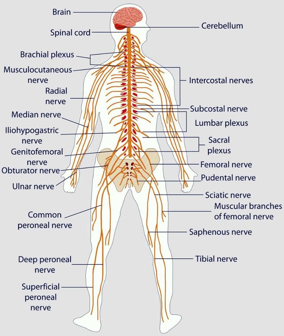
Nervous System
If symptoms are triggered by a central nervous condition like MS, individuals will work closely with their healthcare provider to get the appropriate treatment.
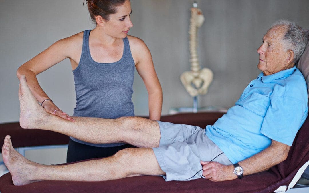
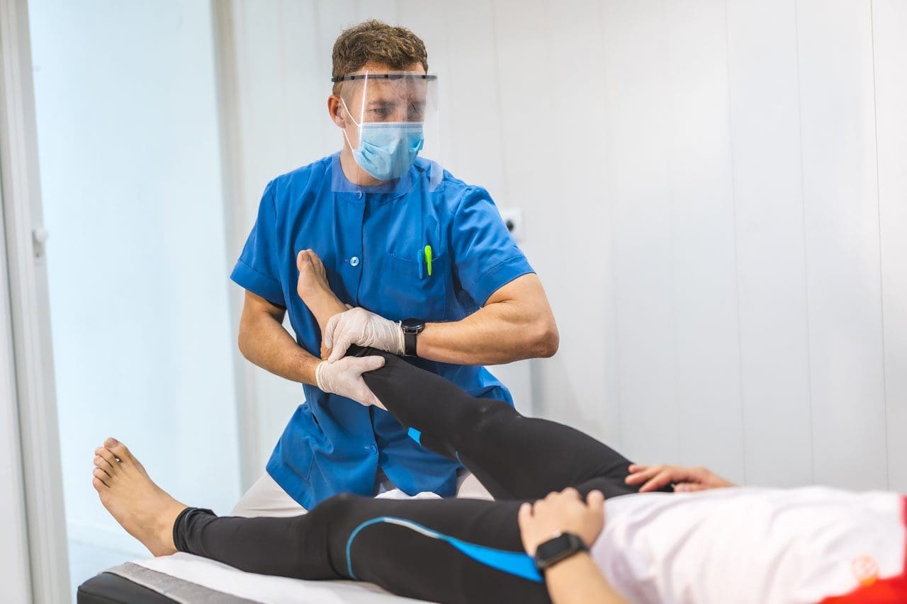
If paresthesia is caused by compression of a spinal nerve, like sciatica, individuals may be referred to a chiropractor and physical therapy team to release the nerve and pressure. (Julie M. Fritz, et al., 2021)
A physical therapist may prescribe spinal exercises to relieve compression of the nerve and restore normal sensations and motion.
Strengthening exercises to restore flexibility and mobility may be prescribed if weakness presents along with paresthesia body sensations.
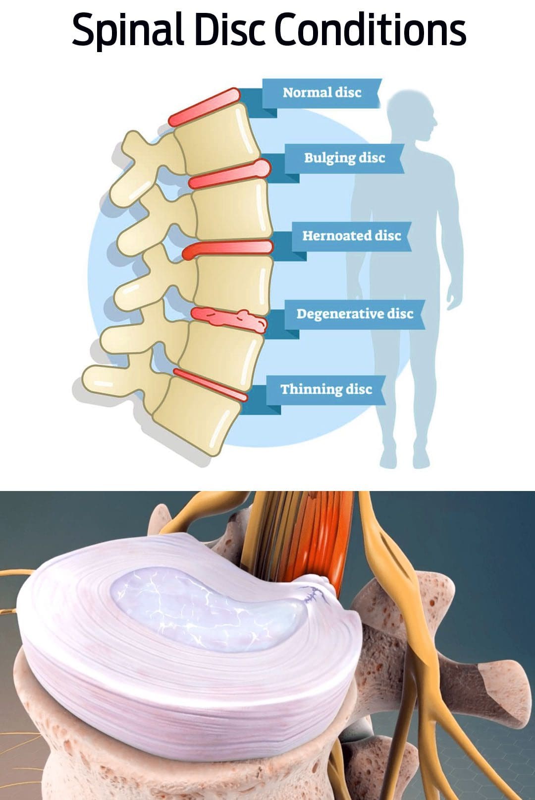
Herniated Disc
If a herniated disc is causing the abnormal sensations, and there has been no improvement with conservative measures, a healthcare provider may suggest surgery to relieve pressure on the nerve/s. (American Association of Neurological Surgeons. 2023)
In surgical procedures like a laminectomy or discectomy, the objective is to restore nerve function.
Post-surgery, individuals may be recommended to a physical therapist to help regain mobility.
National Institute of Neurological Disorders and Stroke. (2023) Paresthesia.
American Association of Neurological Surgeons. (2023) Herniated disc.
National Institute of Diabetes and Digestive and Kidney Diseases. (2018) Peripheral neuropathy.
Merck Manual Professional Version. (2022) Numbness.
Razazian, N., Yavari, Z., Farnia, V., Azizi, A., Kordavani, L., Bahmani, D. S., Holsboer-Trachsler, E., & Brand, S. (2016). Exercising Impacts on Fatigue, Depression, and Paresthesia in Female Patients with Multiple Sclerosis. Medicine and science in sports and exercise, 48(5), 796–803. https://doi.org/10.1249/MSS.0000000000000834
Fritz, J. M., Lane, E., McFadden, M., Brennan, G., Magel, J. S., Thackeray, A., Minick, K., Meier, W., & Greene, T. (2021). Physical Therapy Referral From Primary Care for Acute Back Pain With Sciatica : A Randomized Controlled Trial. Annals of internal medicine, 174(1), 8–17. https://doi.org/10.7326/M20-4187
Individuals dealing with back pain problems could be suffering from a bulging disc. Could knowing the difference between slipped and herniated disc symptoms help with treatments and finding relief?
Bulging Disc Pain
Back pain can become debilitating if not treated properly. A bulging disc is a common cause of cervical, thoracic, and lower back pain symptoms. It happens when one of the fluid-filled cushions between the vertebrae begins to shift out of place. Instead of being aligned with the edges, the disc bulges over. This begins to generate pressure on the nerves causing pain and inflammation.
Bulging discs are often caused by age, but repetitive movements and/or lifting heavy objects can contribute to the condition.
Symptoms can resolve on their own, but individuals are recommended to consult with a physical therapist and/or chiropractor to make sure the disc healed properly, otherwise, it can lead to worsening and/or further injuries.
Bulging Disc vs. Herniated Disc
Bulging and herniated discs cause pain symptoms.
They both can be linked to injuries and degenerative disc disease but are not the same condition. (Penn Medicine. 2018)
This is because the lower back is subject to all kinds of pressure and movement with daily activities, increasing the chances of pain and injuries.
The next most common place is the neck/cervical spine where there are constant movements making it prone to injury and pain symptoms.
Causes
Bulging discs are most often caused by body aging and normal wear and tear. As time goes on the intervertebral discs naturally degenerate, known as degenerative disc disease. This can cause the discs to pull downward, causing them to bulge from their placement. (Penn Medicine. 2018) Factors that can cause or worsen the condition include:
Practicing unhealthy postures.
Repetitive motions.
Lifting heavy objects
Spinal injuries.
Medical history of spinal or disc disease in the family.
Individuals with back pain that interferes with daily functions or has lasted longer than six weeks, should see a healthcare provider for a diagnosis. They will order a magnetic resonance imaging scan/MRI, which can show where a disc is protruding. (American Academy of Neurological Surgeons. 2023)
Rest
For bulging disc pain, resting the back is necessary. However,
Individuals diagnosed with peripheral neuropathy, or with small fiber neuropathy, can understanding symptoms and causes help with potential treatments?
Small Fiber Neuropathy
Small fiber neuropathy is a specific classification of neuropathy, as there are different types, which are nerve injury, damage, disease, and/or dysfunction. Symptoms can result in pain, loss of sensation, and digestive and urinary symptoms. Most cases of neuropathy like peripheral neuropathy involve small and large fibers. Common causes include long-term diabetes, nutritional deficiencies, alcohol consumption, and chemotherapy.
Small fiber neuropathy is diagnosed after diagnostic testing showing it is clear that the small nerve fibers are involved.
The small nerve fibers detect sensation, temperature, and pain and help regulate involuntary functions.
Isolated small-fiber neuropathy is rare, but research is ongoing on the type of nerve damage and potential treatments. (Stephen A. Johnson, et al., 2021)
Small fiber neuropathy is not specifically dangerous but is a sign/symptom of an underlying cause/condition that is damaging the body’s nerves.
Pain – symptoms can range from mild or moderate discomfort to severe distress and can happen at any time.
Loss of sensation.
Because the small nerve fibers help with digestion, blood pressure, and bladder control – symptoms of autonomic dysfunction can vary and can include:
Constipation, diarrhea, incontinence, urinary retention – the inability to completely drain the bladder.
If there is progressing nerve damage, the intensity of the pain can decrease, but the loss of normal sensation and autonomic symptoms can worsen. (Josef Finsterer, Fulvio A. Scorza. 2022)
Hypersensitivity to touch and pain sensations can cause pain without a trigger.
The loss of sensation can make individuals unable to accurately detect sensations of touch, temperature, and pain in affected areas, which can lead to various types of injuries.
Although more research is needed, certain disorders that were not considered neuropathies may have small fiber neuropathy components involved.
A study suggested that neurogenic rosacea, a skin condition, could have some elements of small fiber neuropathy. (Min Li, et al., 2023)
These small nerve fibers are distributed throughout the body including the tops of the fingers and toes, trunk, and internal organs.
These fibers are usually located in the superficial areas of the body, such as close to the skin’s surface. (Mohammad A. Khoshnoodi, et al., 2016)
The small nerve fibers that get damaged are involved in transmitting pain and temperature sensations.
Most nerves have a special type of insulation called myelin that protects them and increases the speed of nerve impulses.
Small nerve fibers may have a thin sheath, making them more susceptible to injury and damage at earlier stages of conditions and diseases. (Heidrun H. Krämer, et al., 2023)
Individuals At Risk
Most types of peripheral neuropathy cause damage to the small and large peripheral nerve fibers. Because of this, most neuropathies are a mix of small-fiber and large-fiber neuropathy. Common risk factors for mixed fiber neuropathy include: (Stephen A. Johnson, et al., 2021)
Diabetes
Nutritional deficiencies
Overconsumption of alcohol
Autoimmune disorders
Medication toxicity
Isolated small-fiber neuropathy is rare, but there are conditions that are known to contribute to the cause and include: (Stephen A. Johnson, et al., 2021)
Sjogren Syndrome
This autoimmune disorder causes dry eyes and mouth, dental problems, and joint pain.
It can also cause nerve damage throughout the body.
Fabry Disease
This condition causes a buildup of certain fats/lipids in the body that can lead to neurological effects.
Amyloidosis
This is a rare disorder that causes a buildup of proteins in the body.
The proteins can damage tissues like the heart or nerves.
Lewy Body Disease
This is a neurological disorder that causes dementia and impaired movement and can lead to nerve damage.
Lupus
This is an autoimmune disease that affects joints, skin, and sometimes nerve tissue.
Viral Infection
These infections typically cause a cold or gastrointestinal/GI upsetness.
Less often they can cause other effects like small fiber neuropathy.
These conditions have been seen to cause isolated small-fiber neuropathy or begin as small-fiber neuropathy before progressing to the large nerve fibers. They can also begin as a mixed neuropathy, with small and large fibers.
Progression
Often the damage progresses at a relatively moderate rate, leading to added symptoms within months or years. The fiber nerves that are affected by the underlying condition usually progressively deteriorate, regardless of where they are located. (Mohammad A. Khoshnoodi, et al., 2016) Medications can help alleviate damage to the peripheral nerves. For individuals that are diagnosed in the early stage, it is possible to stop the progression, and potentially prevent involvement of the large fibers.
Treatments
Treatment toward preventing the progression requires controlling the underlying medical condition with treatment options depending on the cause. Treatments that can help prevent the progression include:
Blood sugar control for individuals with diabetes.
Immune suppression for control of autoimmune diseases.
Plasmapheresis – blood is taken and the plasma is treated and returned or exchanged for the treatment of autoimmune diseases.
Symptom Treatment
Individuals can get treatment for the symptoms that will not reverse or cure the condition but can help with temporary relief. Symptomatic treatment can include: (Josef Finsterer, Fulvio A. Scorza. 2022)
Pain management can include medications and/or topical analgesics.
Physical therapy – stretching, massage, decompression, and adjustments to keep the body relaxed and flexible.
Rehabilitation to help improve coordination, which can be impaired by loss of sensation.
Medications to relieve GI symptoms.
Wearing specialized clothes such as neuropathy socks to help with foot pain symptoms.
Treatment and medical management of neuropathies usually involve a neurologist. A neurologist may prescribe medication to help alleviate pain symptoms and provide medical interventions like immunotherapy if there is concern that an autoimmune process could be the cause. Additionally, treatment could include the care of a physical medicine and rehabilitation physician or a physical therapy team to provide stretches and exercises to help strengthen the body and maintain mobility and flexibility.
Peripheral Neuropathy Myths & Facts
References
Johnson, S. A., Shouman, K., Shelly, S., Sandroni, P., Berini, S. E., Dyck, P. J. B., Hoffman, E. M., Mandrekar, J., Niu, Z., Lamb, C. J., Low, P. A., Singer, W., Mauermann, M. L., Mills, J., Dubey, D., Staff, N. P., & Klein, C. J. (2021). Small Fiber Neuropathy Incidence, Prevalence, Longitudinal Impairments, and Disability. Neurology, 97(22), e2236–e2247. https://doi.org/10.1212/WNL.0000000000012894
Finsterer, J., & Scorza, F. A. (2022). Small fiber neuropathy. Acta neurologica Scandinavica, 145(5), 493–503. https://doi.org/10.1111/ane.13591
Krämer, H. H., Bücker, P., Jeibmann, A., Richter, H., Rosenbohm, A., Jeske, J., Baka, P., Geber, C., Wassenberg, M., Fangerau, T., Karst, U., Schänzer, A., & van Thriel, C. (2023). Gadolinium contrast agents: dermal deposits and potential effects on epidermal small nerve fibers. Journal of neurology, 270(8), 3981–3991. https://doi.org/10.1007/s00415-023-11740-z
Li, M., Tao, M., Zhang, Y., Pan, R., Gu, D., & Xu, Y. (2023). Neurogenic rosacea could be a small fiber neuropathy. Frontiers in pain research (Lausanne, Switzerland), 4, 1122134. https://doi.org/10.3389/fpain.2023.1122134
Khoshnoodi, M. A., Truelove, S., Burakgazi, A., Hoke, A., Mammen, A. L., & Polydefkis, M. (2016). Longitudinal Assessment of Small Fiber Neuropathy: Evidence of a Non-Length-Dependent Distal Axonopathy. JAMA neurology, 73(6), 684–690. https://doi.org/10.1001/jamaneurol.2016.0057
Can individuals with low back pain find relief with spinal decompression combined with chiropractic care to reduce muscle pain?
Introduction
Everyone worldwide has dealt with low back pain, a multifactorial common problem that can mask other musculoskeletal disorders. Low back pain can develop naturally through spinal degeneration, normal environmental factors people put their bodies through, or traumatic factors that can gradually affect the lumbar region over time. The lower back is part of the lumbar spine region as it takes most of the upper body’s weight and stabilizes the lower body when in motion. The lumbar region is thick to support the upper body’s weight and is protected by the surrounding ligaments, muscles, and tissues; however, it is the most susceptible to injury. Many people don’t realize they are experiencing low back pain until an accident or repetitive motions begin to affect the surrounding ligaments or the lumbar intervertebral discs are severely compressed. To that point, many individual are experiencing radiating pain in their lower extremities. However, all is not lost, as there are numerous ways to alleviate low back pain and provide relief to many individuals. Today’s article focuses on the factors that lead to the development of low back pain and how treatments like chiropractic care and spinal decompression can help reduce low back pain and its symptoms. We speak with certified medical providers who incorporate our patients’ information to provide numerous treatment plans to ease low back pain and its associated symptoms. We also inform our patients that there are non-surgical options like chiropractic care and spinal decompression to minimize the radiating pain associated with low back pain. We encourage our patients to ask intricated and educational questions to our associated medical providers about the pain-like symptoms they are experiencing correlating with the lower back. Dr. Alex Jimenez, D.C., utilizes this information as an academic service. Disclaimer
Factors That Lead To The Development Of Low Back Pain
Do you feel muscle aches and pains in your lower back when stretching? Does the pain cause issues in your mobility when going to run errands? Or do you feel sudden or gradual pain after a long workday of carrying heavy objects or excessively sitting at your desk? When many individuals are experiencing pain in these various scenarios, it can lead to the development of a lower back that correlates with these environmental factors. Low back pain is a common nuisance that many working and non-working individuals experience occasionally. When various risk factors are associated with low back pain, it can cause many individuals to deal with the physical and psychological demands that cause them to do multiple things like heavy lifting, awkward positions, and excessive bending, which can develop pain-like symptoms in the lumbar region. (To et al., 2021) At the same time, compared with other health conditions, low back pain is the highest global burden, causing many individuals to miss out on important events like work and daily activities. (Petrozzi et al., 2020) This causes them to live a life of disability and causes them to feel miserable. Low back pain can affect a person’s socioeconomic demographic and the functionality that they seek the treatment they deserve.
Many individuals who experience low back pain will often lead a life of disability and socioeconomic burden correlated with these factors. (Wong et al., 2022) Many people often think that low back pain only happens to older adults, but low back pain can happen to anybody at any age. Many factors that correlate to the development of low back pain include:
Improper heavy lifting
Walking incorrectly
Being in a slouched or hunched position
An auto accident
Obesity
Gastrointestinal issues
Referred musculoskeletal disorders
Physical inactivity
Many of these environmental factors are associated with low back pain that many people are unaware of when doing their routine. This is due to the surrounding tissues, ligaments, and muscles being overused and affected and the intervertebral disc being constantly compressed from repetitive motions. When this happens, many individuals will often seek treatment to reduce the painful effects of low back pain.
How Chiropractic Care Can Transform Pain Into Relief- Video
When it comes to reducing low back pain, many individuals will try home remedies to mitigate muscle pain and reduce inflammation associated with low back pain. This provides temporary relief to acute low back pain, but many will seek non-surgical treatment when it comes to chronic low back pain. Non-surgical treatments can help reduce low back pain and its associated symptoms through mechanical and manual techniques. Non-surgical treatments, which include chiropractic care and spinal decompression, could help alleviate low back pain. Since low back pain is associated with altered motor control within the lumbar region, it can hinder lumbar stability, cause impaired detection of passive motion, and affect postural stability. (Fagundes Loss et al., 2020) With non-surgical treatments, many pain specialists can incorporate vertebral mobilizations and manipulation procedures to improve spinal mobility and reduce pain in the lumbar region. The video above explains how chiropractic care and spinal decompression can alleviate lower back pain and help restore functionality in the body.
Chiropractic Care & Low Back Pain
When it comes to reducing low back pain associated with environmental factors, many pain specialists can look at the physical distress cognitive, and functional patterns that are correlated with faulty lumbar movement. (Khodadad et al., 2020) This allows the pain specialist to create a personalized treatment plan for low back pain. Non-surgical treatments like chiropractic care are excellent for reducing low back pain. Chiropractic care incorporates spinal manipulation techniques to realign the spine and help stretch the affected surrounding muscles. Many individuals who include chiropractic care in their routine find significant pain reduction and reduced disability after a few consecutive treatments. (Gevers-Montoro et al., 2021) Chiropractic care can be combined with physical and massage therapy to stretch and strengthen the affected muscles. This, in turn, helps the individual regain spinal mobility and reduce pain.
Spinal Decompression & Low Back Pain
Spinal decompression is another non-surgical treatment that can help alleviate low back pain, like chiropractic care. Spinal decompression utilizes gentle traction on the lumbar spine to be gently pulled to relieve the affected muscles causing lower back pain. Spinal decompression can also ease leg pain associated with low back pain and reduce referred pain affecting the lower extremities. (Wang et al., 2022) Spinal decompression can also improve spinal disc height and help improve muscle strength and trunk endurance. (Kang et al., 2016) The combination of spinal decompression and chiropractic care to reduce lower back pain is effective as these two forms of non-surgical treatments can effectively reduce many people’s musculoskeletal pain and make them aware of the environmental factors that are the root causes of their lower back pain and prevent it from returning.
References
Fagundes Loss, J., de Souza da Silva, L., Ferreira Miranda, I., Groisman, S., Santiago Wagner Neto, E., Souza, C., & Tarrago Candotti, C. (2020). Immediate effects of a lumbar spine manipulation on pain sensitivity and postural control in individuals with nonspecific low back pain: a randomized controlled trial. Chiropr Man Therap, 28(1), 25. https://doi.org/10.1186/s12998-020-00316-7
Gevers-Montoro, C., Provencher, B., Descarreaux, M., Ortega de Mues, A., & Piche, M. (2021). Clinical Effectiveness and Efficacy of Chiropractic Spinal Manipulation for Spine Pain. Front Pain Res (Lausanne), 2, 765921. https://doi.org/10.3389/fpain.2021.765921
Kang, J.-I., Jeong, D.-K., & Choi, H. (2016). Effect of spinal decompression on the lumbar muscle activity and disk height in patients with herniated intervertebral disk. Journal of Physical Therapy Science, 28(11), 3125-3130. https://doi.org/10.1589/jpts.28.3125
Khodadad, B., Letafatkar, A., Hadadnezhad, M., & Shojaedin, S. (2020). Comparing the Effectiveness of Cognitive Functional Treatment and Lumbar Stabilization Treatment on Pain and Movement Control in Patients With Low Back Pain. Sports Health, 12(3), 289-295. https://doi.org/10.1177/1941738119886854
Petrozzi, M. J., Rubinstein, S. M., Ferreira, P. H., Leaver, A., & Mackey, M. G. (2020). Predictors of low back disability in chiropractic and physical therapy settings. Chiropr Man Therap, 28(1), 41. https://doi.org/10.1186/s12998-020-00328-3
To, D., Rezai, M., Murnaghan, K., & Cancelliere, C. (2021). Risk factors for low back pain in active military personnel: a systematic review. Chiropr Man Therap, 29(1), 52. https://doi.org/10.1186/s12998-021-00409-x
Wang, W., Long, F., Wu, X., Li, S., & Lin, J. (2022). Clinical Efficacy of Mechanical Traction as Physical Therapy for Lumbar Disc Herniation: A Meta-Analysis. Comput Math Methods Med, 2022, 5670303. https://doi.org/10.1155/2022/5670303
Wong, C. K., Mak, R. Y., Kwok, T. S., Tsang, J. S., Leung, M. Y., Funabashi, M., Macedo, L. G., Dennett, L., & Wong, A. Y. (2022). Prevalence, Incidence, and Factors Associated With Non-Specific Chronic Low Back Pain in Community-Dwelling Older Adults Aged 60 Years and Older: A Systematic Review and Meta-Analysis. J Pain, 23(4), 509-534. https://doi.org/10.1016/j.jpain.2021.07.012
As research increases in regenerative medicine with the potential of being able to regrow thyroid tissue, could regeneration therapy eliminate the need for patients to take thyroid replacement hormones?
Thyroid Regenerative Therapy
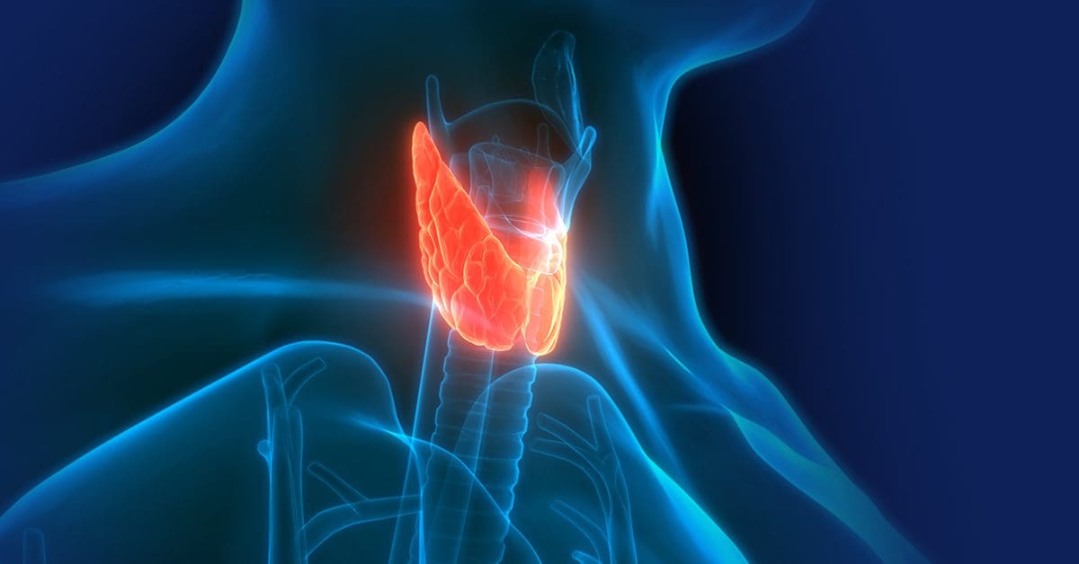
A great hope for regenerative therapy is the ability to grow healthy organs. One of the organs being looked at is the thyroid gland. The goal is to re-grow thyroid tissue in:
Individuals who had to have the gland removed because of thyroid cancer.
Individuals who were born without a fully developed gland.
As science advances and research has expanded from laboratory and animal experiments to test tube human thyroid cell studies, the use of stem cell therapy for this purpose is not there yet, as more extensive research is needed for human consideration.
Human Research
Research on the use of thyroid regenerative therapy for thyroid disease has not published studies in which stem cell therapy has been attempted in human thyroid patients.
The studies that have been done were conducted in mice, and any findings of this research cannot automatically be applied to humans. (H. P. Gaide Chevronnay, et al., 2016)
In human thyroid tissue in test tube studies, the stimulation of cells was achieved in a way that raised the question of making cancerous transformations more likely if it were to be attempted in humans. (Davies T.F., et al., 2011)
Recent Studies
Current research involves advances in embryonic stem cell – ESC and induced pluripotent stem cell – iPSC. (Will Sewell, Reigh-Yi Lin. 2014)
ESCs, also known as pluripotent stem cells, can increase any type of cell in the body.
They are harvested from embryos that were produced, but not implanted, during IVF procedures.
iPSCs are pluripotent cells that have been developed using a reprogramming process of adult cells.
Follicular cells are thyroid cells that make thyroid hormones – T4 and T3 and have been produced from the embryonic stem cells of mice.
In a study published in the journal Cell Stem Cell in 2015, these cells had the ability to grow and were also able to start making thyroid hormone within two weeks. (Anita A. Kurmann, et al., 2015)
After eight weeks, cells that were transplanted into mice that did not have thyroid glands had normal amounts of thyroid hormone.
New Thyroid Gland
Investigators at Mount Sinai Hospital induced human embryonic stem cells into thyroid cells.
They were looking at the possibility of creating a new-like thyroid gland in individuals who have had their thyroid surgically removed.
The future looks promising for the ability to regrow thyroid tissue and eliminate thyroid replacement hormone. However, far more research is needed for this to even be considered a possibility.
Cracking the Low Thyroid Code Assessment Guide
References
Gaide Chevronnay, H. P., Janssens, V., Van Der Smissen, P., Rocca, C. J., Liao, X. H., Refetoff, S., Pierreux, C. E., Cherqui, S., & Courtoy, P. J. (2016). Hematopoietic Stem Cells Transplantation Can Normalize Thyroid Function in a Cystinosis Mouse Model. Endocrinology, 157(4), 1363–1371. https://doi.org/10.1210/en.2015-1762
Davies, T. F., Latif, R., Minsky, N. C., & Ma, R. (2011). Clinical review: The emerging cell biology of thyroid stem cells. The Journal of clinical endocrinology and metabolism, 96(9), 2692–2702. https://doi.org/10.1210/jc.2011-1047
Sewell, W., & Lin, R. Y. (2014). Generation of thyroid follicular cells from pluripotent stem cells: potential for regenerative medicine. Frontiers in endocrinology, 5, 96. https://doi.org/10.3389/fendo.2014.00096
Kurmann, A. A., Serra, M., Hawkins, F., Rankin, S. A., Mori, M., Astapova, I., Ullas, S., Lin, S., Bilodeau, M., Rossant, J., Jean, J. C., Ikonomou, L., Deterding, R. R., Shannon, J. M., Zorn, A. M., Hollenberg, A. N., & Kotton, D. N. (2015). Regeneration of Thyroid Function by Transplantation of Differentiated Pluripotent Stem Cells. Cell stem cell, 17(5), 527–542. https://doi.org/10.1016/j.stem.2015.09.004
Tuttle, R. M., & Wondisford, F. E. (2014). Welcome to the 84th annual meeting of the American Thyroid Association. Thyroid : official journal of the American Thyroid Association, 24(10), 1439–1440. https://doi.org/10.1089/thy.2014.0429
IFM's Find A Practitioner tool is the largest referral network in Functional Medicine, created to help patients locate Functional Medicine practitioners anywhere in the world. IFM Certified Practitioners are listed first in the search results, given their extensive education in Functional Medicine