Hamstring muscle injuries are common, especially in athletes and individuals with physically demanding jobs. Is there a better chance of full recovery with surgical repair and post-op rehabilitation?
Hamstring Muscle Tear
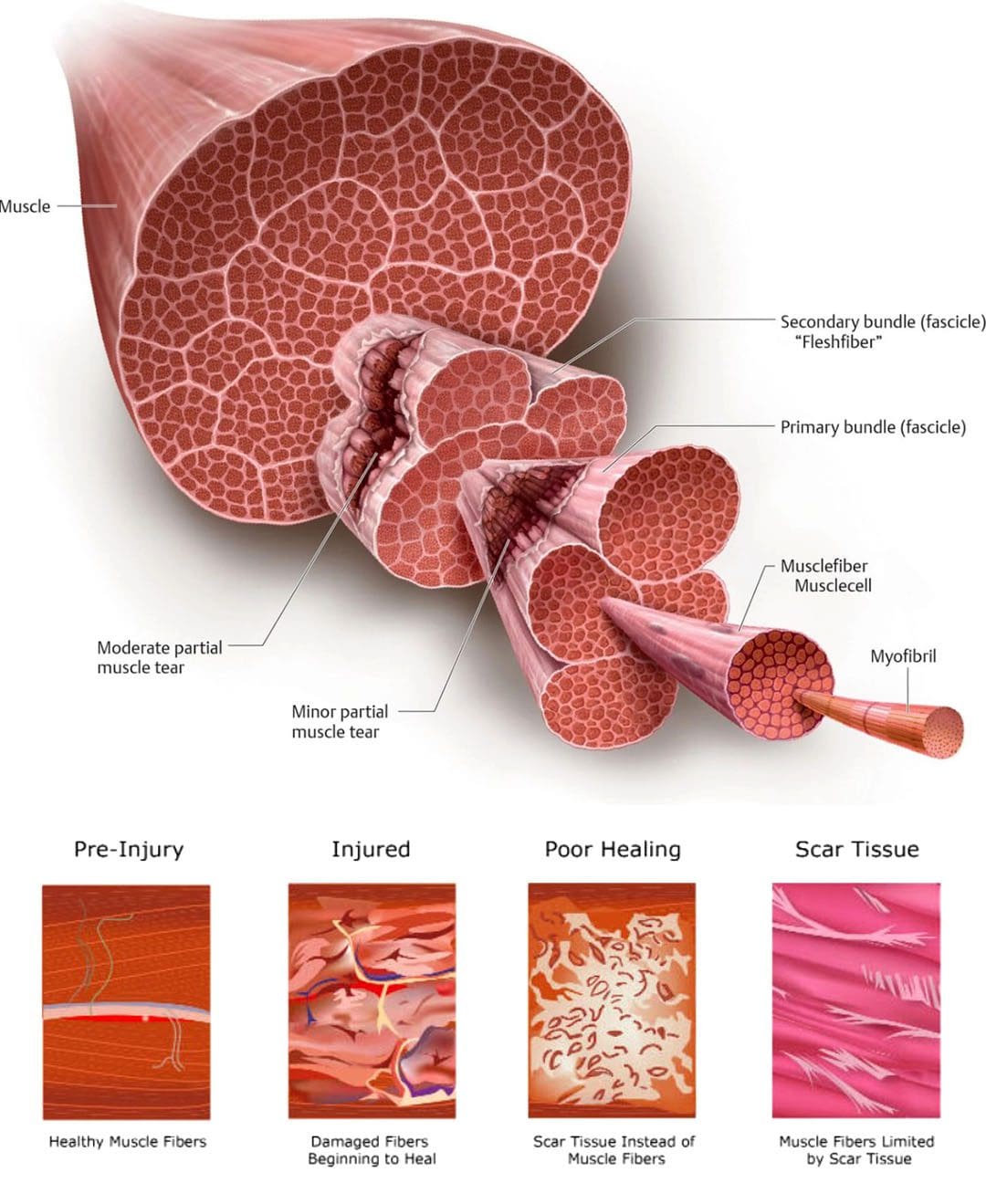
Most often, hamstring muscle injuries are partial tears of the muscle. These types of injuries are muscle strains that occur when the muscle fibers are stretched beyond their normal limits. Complete tears of the hamstring muscle are unusual, but they do occur in both athletes and non-athletes. Determining the optimal treatment plan depends on:
The severity of the tendon tear
The expectations of the injured individual.
Incomplete tears are when the hamstring muscle is stretched too far, but not completely detached.
Complete tears usually occur at the top of the muscle where the tendon tears away from the pelvis.
A complete tear usually occurs when there is a sudden flexion of the hip and extension of the knee joint – when the muscle contracts in this position, it gets stretched beyond its limits.
Basic hamstring strains can be treated with simple steps – rest, ice, anti-inflammatory medications, and conservative therapies.
Symptoms
Symptoms of a hamstring muscle strain can include pain, bruising, swelling, and movement difficulty. (American Academy of Orthopaedic Surgeons. 2021) Individuals who sustain this injury typically experience sudden sharp pain. Signs of a tear can include:
Sharp pain where the buttock and thigh meet.
Difficulty walking.
Sitting can be difficult as the edge of a chair can place pressure directly on the injury.
Spasms and cramping sensations in the back of the thigh.
Weakness in the leg, specifically when bending the knee or lifting the leg behind the body.
Numbness or burning sensations as a result of sciatic nerve irritation.
Swelling and bruising in the back of the thigh – over time it can travel down to the back of the knee and calf and possibly into the foot.
With a complete hamstring tear, there is usually significant swelling and bruising that develops in the back of the thigh.
Diagnosis
The symptoms can be difficult to spot in the early stages which is why X-rays of the hip or thigh are usually obtained.
In some situations, a fragment of bone can get pulled off the pelvis along with the hamstring muscle attachment. MRI testing can be performed to evaluate the attachment and can define critical features of a complete hamstring muscle tear, including: (American Academy of Orthopaedic Surgeons. 2021)
The number of tendons involved.
Complete versus incomplete tearing.
The amount of retraction – the amount the tendons have pulled back.
This will guide the development of treatment.
Treatment
The treatment of a complete tear will depend on different factors. The other variable is the patient and their expectations.
Treatment is more aggressive in younger individuals like high-level athletes.
Treatment is less aggressive in middle-aged individuals.
Often a single tendon tear can be treated non-surgically.
When one tendon is involved, it is typically not pulled very far from its normal attachment and will develop scar tissue in a positive position.
Conversely, when three tendons have been torn, they usually pull more than a few centimeters away from the bone. These cases have better results with surgical repair. (UW Health. 2017)
Surgeons will use patient characteristics – high-level athletes or less physically active individuals – to guide treatment recommendations.
Rehabilitation
Rehabilitation following surgery can take 3-6 months or longer.
The first six weeks limit weight-bearing with the use of crutches.
Patients may be recommended to wear a brace to reduce tension on the repaired hamstring tendons.
Strengthening does not begin until three months post-op, and even light activities are usually delayed. (UW Health. 2017)
Because this injury can have a long recovery time, some individuals may choose nonsurgical treatment.
Sometimes these individuals experience symptoms of discomfort from sitting and may exhibit long-term weakness of the hamstring muscle.
Full recovery from a complete hamstring muscle injury takes time. Studies have shown high-level athletes are able to resume competitive sports after the repair and rehabilitation of an acute hamstring muscle injury. (Samuel K. Chu, Monica E. Rho. 2016)
Delaying surgical treatment may not always lead to optimal results.
When the tendon is torn away from its normal attachment, it begins to scar around the surrounding soft tissues.
When there is a delay of more than a few weeks following the initial injury, regaining the full length of the tendon and muscle can be challenging.
This could delay the rehabilitation process and may limit the potential for full recovery. (Ho Yoon Kwak, et al., 2011)
With severe injuries, there is a better chance of full recovery with surgical repair but could involve a long recovery and commitment to a post-op rehabilitation plan.
Chu, S. K., & Rho, M. E. (2016). Hamstring Injuries in the Athlete: Diagnosis, Treatment, and Return to Play. Current sports medicine reports, 15(3), 184–190. https://doi.org/10.1249/JSR.0000000000000264
Kwak, H. Y., Bae, S. W., Choi, Y. S., & Jang, M. S. (2011). Early surgical repair of acute complete rupture of the proximal hamstring tendons. Clinics in orthopedic surgery, 3(3), 249–253. https://doi.org/10.4055/cios.2011.3.3.249
When individuals experience a neuromusculoskeletal injury strain, can following basic pulled muscle treatment protocols help in healing and a full recovery?
Pulled Muscle Treatment
A pulled muscle or muscle strain occurs when a muscle is stretched beyond its ability resulting in discomfort symptoms and mobility issues. Microscopic tears can occur within the muscle fibers potentially worsening the injury. This type of injury usually causes mild to severe pain, bruising, and immobility, and nerve injuries can develop as well. Common muscle strains include:
Pulled hamstrings
Groin strains
Pulled abdominal muscles
Calf strains
Pulled muscle treatment requires patience to promote proper healing and restoration of optimal function.
Individuals need to focus on the different stages of healing.
Gradually increase activity levels as the body allows to prevent stiffness and atrophy which can cause complications.
Symptoms
The usual symptoms of this type of injury include:
Pain
Limited mobility
Muscle spasms
Swelling
Bruising
Often individuals will feel a sudden grabbing or tearing sensation and are then unable to continue the activity.
Can limit the ability to perform certain activities.
May have moderate swelling and bruising.
Grade III
Severe injury that can cause significant pain.
Muscle spasms.
Swelling.
Significant bruising.
Basic Treatment Protocols
Most pulled muscle strain injuries heal with simple treatment. Following the right steps can ensure an expedited recovery. In the early stages after the injury, there is a balance between doing too much or not enough. The amount of activity an individual will be able to do, and the time required for recovery depends on the severity of the injury. Here are some guidelines in the right direction.
Rest
Rest is recommended for the early recovery stage.
Depending on the severity of the injury this could last from one to five days.
Immobilization is usually not necessary, and not moving at all can lead to muscle and joint stiffness.
To avoid injuries make sure the muscles are not over-exerted.
Gradually increase activity levels when starting an exercise program to build endurance.
Properly Warming Up
Warming up before taking on physical activities will help loosen the muscles and prevent injuries.
Beginning work or exercise with stiff muscles can lead to an increased chance of strain.
Studies have shown that temperature can influence the stiffness of a muscle. (K. W. Ranatunga. 2018)
Maintaining body and muscle warmth helps prevent injury and re-injury.
Injuries and Chiropractic: The Road To Recovery
References
Hospital for Special Surgery, Muscle Strain: What You Need to Know About Pulled Muscles.
Kary J. M. (2010). Diagnosis and management of quadriceps strains and contusions. Current reviews in musculoskeletal medicine, 3(1-4), 26–31. https://doi.org/10.1007/s12178-010-9064-5
Malanga, G. A., Yan, N., & Stark, J. (2015). Mechanisms and efficacy of heat and cold therapies for musculoskeletal injury. Postgraduate medicine, 127(1), 57–65. https://doi.org/10.1080/00325481.2015.992719
Mair, S. D., Seaber, A. V., Glisson, R. R., & Garrett, W. E., Jr (1996). The role of fatigue in susceptibility to acute muscle strain injury. The American journal of sports medicine, 24(2), 137–143. https://doi.org/10.1177/036354659602400203
Ranatunga K. W. (2018). Temperature Effects on Force and Actin⁻Myosin Interaction in Muscle: A Look Back on Some Experimental Findings. International journal of molecular sciences, 19(5), 1538. https://doi.org/10.3390/ijms19051538
As the body ages, individuals want to stay active and maintain a healthy pain free lifestyle. Can regenerative cells for arthritis and cartilage damage be the future of neuromusculoskeletal medicine and joint healing?
Regenerative Cells For Arthritis and Cartilage Damage
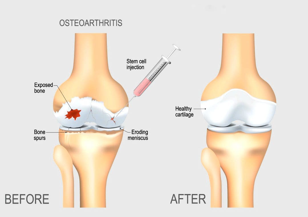
Individuals want to continue to do the physical activities they love, which require healthy joints. Scientists are learning how to harness the abilities of regenerative cells to repair and regrow damaged and deteriorated cartilage. Current stem cell treatment of cartilage problems has not been shown to reverse the effects of arthritis and while studies show clinical improvement, further research is necessary. (Bryan M. Saltzman, et al., 2016)
Cartilage and How It Gets Damaged
Cartilage is a type of connective tissue. In the joints, there are a few types of cartilage. The most commonly referred to is the smooth lining known as articular or hyaline cartilage. This type forms a smooth layer of cushion on the end of a bone at the joint. (Rocky S. Tuan, et al., 2013)
The tissue is very strong and has the ability to compress and absorb energy.
It is very smooth allowing a joint to glide effortlessly through a limb’s range of motion.
When joint cartilage is damaged, the cushioning can wear down.
In traumatic injuries, a sudden force can cause the cartilage to break off and/or suffer damage, that exposes the underlying bone.
In osteoarthritis – degenerative or wear-and-tear arthritis, the smooth layer can wear down thin and unevenly.
Eventually, the cushion wears away, the joints become inflamed and swollen and movements become stiff and painful.
There are treatments for arthritis and cartilage damage, but these treatments are usually focused on relieving symptoms by smoothing down the damaged cartilage or replacing the joint surface with an artificial implant, like knee replacement or hip replacement surgeries. (Robert F. LaPrade, et al., 2016)
Regenerative Cells
Regenerative stem cells are special cells that have the ability to multiply and develop into different types of tissue. In an orthopedic surgery setting for joint problems, stem cells are obtained from adult stem cell primary sources which are bone marrow and fatty tissue. These cells have the ability to develop into cartilage cells, called chondrocytes. (Rocky S. Tuan, et al., 2013)
They also help by stimulating the body to reduce inflammation, stimulate cell repair, and improve blood circulation.
This process is caused by cellular signals and growth factors to stimulate the body to activate the healing processes.
Once stem cells have been obtained, they need to be delivered to the area of cartilage damage.
Cartilage is a complex tissue that is described as a scaffold structure that is composed of collagen, proteoglycans, water, and cells. (Rocky S. Tuan, et al., 2013)
To regenerate cartilage, the complex tissues must also be reconstructed.
There are studies on types of tissue scaffolds engineered to recreate a similar type of cartilage structure.
The stem cells can then be injected into the scaffold, in hopes of restoring a normal type of cartilage.
Non-Surgical Arthritis Treatments
Standard treatments such as cortisone shots or physical therapies work as well and provide benefits that could be utilized in combination with regenerative cells for arthritis and cartilage damage in the near future. Data takes time and therefore how this impacts the long-term health of a joint needs continued research in terms of tissue engineering and cell delivery to determine the best approach to help individuals.
Arthritis
References
LaPrade, R. F., Dragoo, J. L., Koh, J. L., Murray, I. R., Geeslin, A. G., & Chu, C. R. (2016). AAOS Research Symposium Updates and Consensus: Biologic Treatment of Orthopaedic Injuries. The Journal of the American Academy of Orthopaedic Surgeons, 24(7), e62–e78. https://doi.org/10.5435/JAAOS-D-16-00086
Saltzman, B. M., Kuhns, B. D., Weber, A. E., Yanke, A., & Nho, S. J. (2016). Stem Cells in Orthopedics: A Comprehensive Guide for the General Orthopedist. American journal of orthopedics (Belle Mead, N.J.), 45(5), 280–326.
Tuan, R. S., Chen, A. F., & Klatt, B. A. (2013). Cartilage regeneration. The Journal of the American Academy of Orthopaedic Surgeons, 21(5), 303–311. https://doi.org/10.5435/JAAOS-21-05-303
Back problems and discomfort symptoms are a common ailment that many types of healthcare providers diagnose and treat it. Can knowing a little about what each back pain specialist does and what they have to offer help in choosing a provider?
Back Pain Specialists
Nowadays individuals have options for treating back pain. Primary healthcare providers, general practitioners, pediatricians, and emergency room workers are usually the first to examine the injury or problem. If they are unable to properly diagnose or treat the injury, they will refer the individual to a specialist. Specialists include:
Osteopaths
Chiropractors
Orthopedists
Rheumatologists
Neurologists
Neurosurgeons.
They specialize in chronic conditions and diseases like arthritis or problems like herniated discs. Complementary and alternative providers treat individuals solely or with help from a care team. They look at the whole body and focus on improving overall function.
Family and General Practitioners
When neck or back pain begins a regular doctor that’s usually a family or general practitioner/GP or primary care provider PCP is a recommended place to start. They will:
Order diagnostic tests.
Recommend exercises and stretches.
Prescribe medication.
Refer the patient to a physical therapist or other back pain specialist.
However, studies suggest general providers can be uninformed and slow to adopt new back treatments. (Paul B. Bishop, Peter C. Wing. 2006) It is recommended to research possible treatment options, ask a lot of questions during an appointment, and ask or request a referral to a specialist.
Pediatricians
Pediatricians diagnose and treat children. They cover a wide range of issues including back problems and injuries. As with a general practitioner or primary care provider, a child’s pediatrician is the place to start. Depending on the child’s needs, they’ll refer them to the proper specialist.
Emergency Healthcare Providers
When there is serious neck or back trauma, individuals need to go to the emergency room. Trauma can include automobile collisions, sports accidents, work accidents, and/or personal home accidents. Someone with a possible spinal injury should not be moved. (W Yisheng, et al., 2007) Go to the ER if there is back pain with loss of bowel or bladder control, or the legs become progressively weaker. These are symptoms of an emergency condition known as cauda equina syndrome. (American Association of Neurological Surgeons. 2023)
Orthopedists
Orthopedists and orthopedic surgeons treat the musculoskeletal system, which includes:
Muscles
Bones
Joints
Connective tissues
Cartilage
Common orthopedic issues include:
Repetitive stress injuries
Sports injuries
Bursitis
Tendonitis
Ruptured discs
Nerve impingement
Scoliosis
Osteoporosis
Osteoarthritis
Orthopedics can overlap with other specialties. As orthopedists and rheumatologists both treat arthritis and orthopedic surgeons and neurosurgeons do some of the same procedures that include spinal fusions and discectomies.
Rheumatologists
A rheumatologist treats autoimmune, inflammatory, and musculoskeletal conditions that can include different types of arthritis, lupus, and Sjogren’s syndrome. A primary care provider may refer a patient to a rheumatologist if they have symptoms that include:
Sacroiliitis – inflammation of the sacroiliac joint at the base of the spine.
Axial spondylosis – a form of spinal arthritis.
Axial spondylosis – spinal arthritis that causes the bones to fuse together.
Rheumatologists can also treat spinal stenosis or advanced osteoarthritis as they overlap with orthopedists.
Neurologists
A neurologist specializes in the function of the nervous system. They treat disorders of the brain, spinal cord, and nerves that include:
Parkinson’s disease
Multiple sclerosis
Alzheimer’s disease
Chronic back or neck pain
They are experts in the origins of pain. (David Borsook. 2012) However, a neurologist does not perform spine surgery.
Neurosurgeons
A neurosurgeon specializes in nervous system surgical procedures that include the brain, spine, and spinal cord. However, neurosurgeons do not provide overall treatment for back pain because they are usually the last to be seen after exhausting all other treatment options.
Osteopaths
An osteopath is a licensed physician who practices medicine using conventional treatments and osteopathic manipulative medicine. They have the same education as an MD plus 500 hours of musculoskeletal system studies. (National Library of Medicine. 2022) They take the same exams and are licensed as an MD. Many osteopaths are primary care providers. For back pain, they will focus on:
Posture rehabilitation and training.
Stretching
Therapeutic massage
Spinal manipulation
The goal is to relieve pain and muscle tension, increase mobility, and improve musculoskeletal function.
Physiatrists
Physiatrists are holistic providers that focus on physical function. They can be thought of as a primary care provider plus a physical therapist. These back pain specialists provide rehabilitation for various types of conditions and injuries including:
Back pain
Sports injuries
Stroke
Often they will coordinate a team of specialists to develop a targeted treatment plan.
Chiropractor
Chiropractic is a hands-on alternative medicine. The goal is to restore neuromusculoskeletal function by realigning the spine to its proper form. They do this with spinal manipulations, non-surgical mechanical decompression, traction, and massage techniques. (Michael Schneider, et al., 2016)
The purpose of most chiropractic adjustments is to relax and retrain tight muscles and increase flexibility.
Chiropractic helps to relieve stiff muscles and restore range of motion.
Individuals may not be referred to a chiropractor if they:
Have loose-joints
Have connective tissue problems or conditions.
Have osteoporosis/thinning bones
All types of back pain specialists provide various forms of therapy that may be able to help.
Spinal Decompression in Depth
References
Bishop, P. B., & Wing, P. C. (2006). Knowledge transfer in family physicians managing patients with acute low back pain: a prospective randomized control trial. The spine journal : official journal of the North American Spine Society, 6(3), 282–288. https://doi.org/10.1016/j.spinee.2005.10.008
Yisheng, W., Fuying, Z., Limin, W., Junwei, L., Guofu, P., & Weidong, W. (2007). First aid and treatment for cervical spinal cord injury with fracture and dislocation. Indian journal of orthopaedics, 41(4), 300–304. https://doi.org/10.4103/0019-5413.36991
Schneider, M., Murphy, D., & Hartvigsen, J. (2016). Spine Care as a Framework for the Chiropractic Identity. Journal of chiropractic humanities, 23(1), 14–21. https://doi.org/10.1016/j.echu.2016.09.004
Individuals that experience nerve pain in the foot could be caused by a number of different conditions, can recognizing the most common causes help in developing an effective treatment plan?
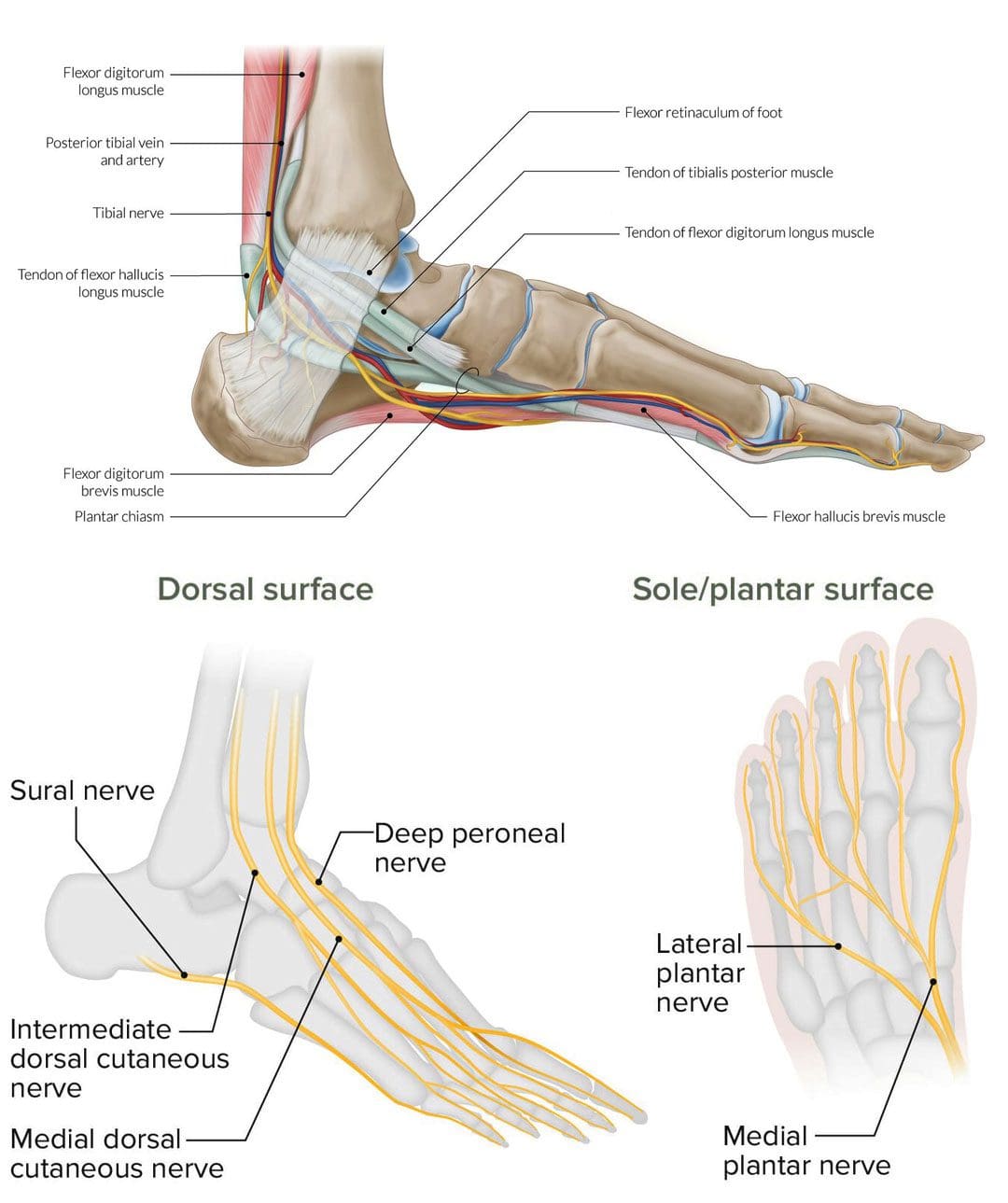
Nerve Pain In The Foot
These sensations can feel like a burning, shooting, electrical, or stabbing pain and can happen while in motion or at rest. It can occur on the top of the foot or through the arch. The area closest to the nerve may be sensitive to the touch. A number of different conditions can cause nerve pain in the foot, including:
Morton’s neuroma
Pinched nerve
Tarsal tunnel syndrome
Diabetic peripheral neuropathy
Herniated disc
Morton’s Neuroma
Morton’s neuroma involves the nerve that runs between the third and fourth toes, but can sometimes occur between the second and third toes becoming thicker. Typical symptoms include a burning or shooting pain in the area, usually while walking. (Nikolaos Gougoulias, et al., 2019) Another common symptom is the sensation of pressure beneath the toes like the sock is bunched up underneath. Treatments can include:
Arch supports
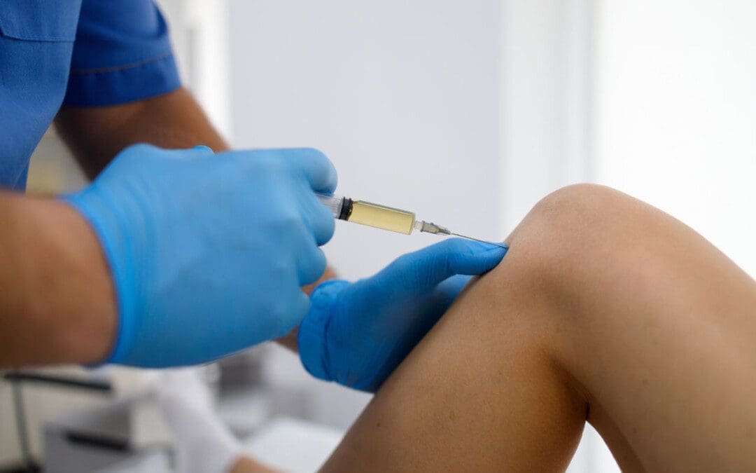
Cortisone injections to decrease swelling
Footwear modifications – can include lifts, orthotics combined with metatarsal pads, and rocker soles, to provide cushion where needed.
Things that increase the risk of developing the condition include:
Regularly wearing high-heels – the condition occurs more frequently in women.
Shoes that are too tight.
Participating in high-impact sports like running.
Having flat feet, high arches, bunions, or hammertoes.
Pinched Nerve
A pinched nerve can feel like shooting or burning pain. Nerve entrapment can occur in various regions of the foot or the area on top of the foot may feel sensitive. Causes can be caused by: (Basavaraj Chari, Eugene McNally. 2018)
Trauma that causes swelling.
Blunt impact.
Tight shoes.
Treatment can include:
Massage
Physical therapy
Rest
Footwear modifications
Anti-inflammatories.
Things that increase the risk of developing a pinched nerve in the foot include:
Poor-fitting footwear.
Repetitive stress injury.
Trauma to the foot.
Obesity.
Rheumatoid arthritis.
Tarsal Tunnel Syndrome
Another type of nerve entrapment is tarsal tunnel syndrome. Tarsal tunnel syndrome is “anything that produces compression on the posterior tibial nerve.” (American College of Foot and Ankle Surgeons. 2019) The tibial nerve is located near the heel. Symptoms include numbness and foot cramps, burning, tingling, or shooting sensations that often radiate from the instep/arch. Both can worsen while the foot is at rest, like when sitting or sleeping. Treatment can consist of:
Placing padding in the shoe where the foot is being compressed to relieve the pain.
Custom foot orthotics.
Cortisone shots or other anti-inflammatory treatments.
Surgery may be necessary to release the nerve.
Conditions that compress the tibial nerve and can lead to tarsal tunnel syndrome include:
Flat feet
Fallen arches
Ankle sprain
Diabetes
Arthritis
Varicose veins
Bone spurs
Diabetic Peripheral Neuropathy
Long-term high blood sugar/glucose associated with diabetes can lead to a form of nerve damage known as peripheral neuropathy. (Centers for Disease Control and Prevention. 2022) Neuropathy pain feels like burning or shooting pain, or the sensation of walking on bubble wrap that usually shows up overnight. The pain can come and go as well as a gradual loss of feeling in the feet that begins in the toes and moves up the foot. It’s estimated that around half of individuals with diabetes will eventually develop neuropathy. (Eva L. Feldman, et al., 2019) Treatments can include:
Physical therapy massage to increase circulation.
Topical treatments with capsaicin.
Vitamin B.
Blood sugar management.
Alpha lipoic acid.
Medication.
Individuals with diabetes have an increased risk of developing peripheral neuropathy if:
Blood sugar is not well-controlled.
Diabetes has been present for many years.
Kidney disease.
Smoke.
Overweight or obese.
Herniated Disc
Nerve pain in the foot can be caused by spinal issues. A herniated disc in the lower back can irritate and compress the nerves, causing pain that radiates down the leg and foot. Additional symptoms usually include muscle weakness in the legs and/or numbness and tingling. Most herniated discs don’t require surgery and get better with conservative treatment. (Wai Weng Yoon, Jonathan Koch. 2021) If symptoms don’t improve or worsen, a healthcare provider may recommend surgery. Herniated discs are most common in young and middle-aged adults. Increased chances of developing a herniated disc can come from:
Degenerative changes in the spine from normal age wear and tear.
Physically demanding job.
Lifting incorrectly.
Overweight or obese.
Genetic predisposition – family history of herniated discs.
Spinal Stenosis
Spinal stenosis occurs when the spaces in the spine begin to narrow, creating pressure on the spinal cord and nerve roots. It is usually caused by wear and tear on the spine as the body ages. Stenosis in the lower back can cause burning pain in the buttocks and leg. As it progresses pain can radiate into the feet along with numbness and tingling. Conservative treatment consists of physical therapy exercises and non-steroidal anti-inflammatory medications/NSAIDs. (Jon Lurie, Christy Tomkins-Lane. 2016) Cortisone injections can be beneficial and if the condition worsens, surgery may be an option. Risk factors include:
Physical trauma – after surgery or an automobile or sports accident.
Certain cancer, antiviral medications, or antibiotics.
Complex regional pain syndrome.
Tumors that irritate and/or compress a nerve.
Liver or kidney disease.
Infectious diseases – Lyme disease complications or viral infections.
Nerve pain in the foot is definitely a reason to see a healthcare provider. Early diagnosis can help prevent symptom progression and future problems. Once the cause of the pain has been identified, the healthcare team can work together to develop a personalized treatment plan to release compressed nerves and restore mobility and function. See a healthcare provider right away if the pain and symptoms worsen, or if there are difficulties standing or walking.
Chiropractic After Accidents and Injuries
References
Gougoulias, N., Lampridis, V., & Sakellariou, A. (2019). Morton’s interdigital neuroma: instructional review. EFORT open reviews, 4(1), 14–24. https://doi.org/10.1302/2058-5241.4.180025
Chari, B., & McNally, E. (2018). Nerve Entrapment in Ankle and Foot: Ultrasound Imaging. Seminars in musculoskeletal radiology, 22(3), 354–363. https://doi.org/10.1055/s-0038-1648252
Feldman, E. L., Callaghan, B. C., Pop-Busui, R., Zochodne, D. W., Wright, D. E., Bennett, D. L., Bril, V., Russell, J. W., & Viswanathan, V. (2019). Diabetic neuropathy. Nature reviews. Disease primers, 5(1), 42. https://doi.org/10.1038/s41572-019-0097-9
Yoon, W. W., & Koch, J. (2021). Herniated discs: when is surgery necessary?. EFORT open reviews, 6(6), 526–530. https://doi.org/10.1302/2058-5241.6.210020
Lurie, J., & Tomkins-Lane, C. (2016). Management of lumbar spinal stenosis. BMJ (Clinical research ed.), 352, h6234. https://doi.org/10.1136/bmj.h6234
Staff, N. P., & Windebank, A. J. (2014). Peripheral neuropathy due to vitamin deficiency, toxins, and medications. Continuum (Minneapolis, Minn.), 20(5 Peripheral Nervous System Disorders), 1293–1306. https://doi.org/10.1212/01.CON.0000455880.06675.5a
Can combining chiropractic treatment with the common therapies of medication, exercise, and/or physical therapy help relieve sciatic endometriosis pain symptoms?
Sciatic Endometriosis
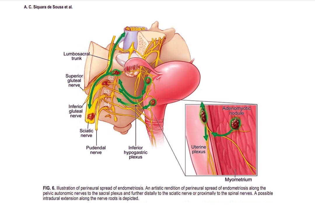
Sciatic endometriosis is a condition in which endometrial cells (tissue that resembles the lining of the uterus) grow outside of the uterine lining and compress the sciatic nerve. This places stress and pressure on the nerve causing back, pelvic, hip, and leg pain, especially before and during the menstrual cycle. It can also cause pain, irregular periods, and infertility. (The American College of Obstetricians and Gynecologists. 2021)
These areas of endometrial tissue growth are also known as lesions or implants.
Women with sciatic endometriosis often experience leg pain and weakness around the time of their menstrual cycle. (Lena Marie Seegers, et al., 2023)
Sciatic endometriosis can also cause pain when urinating, during a bowel movement, during sex, and fatigue, and irregular vaginal bleeding.
The abnormal growth may be caused by higher-than-normal levels of estrogen.
Researchers believe that endometriosis is related to retrograde menstruation, which causes menstrual blood to flow back into the pelvis instead of out through the vagina. (World Health Organization. 2023)
Sometimes, the cells grow in the area of the pelvis right above the sciatic nerve. (Adaiah Yahaya, et al., 2021)
The sciatic nerve is the longest nerve in the body and travels down the back of each leg. (Johns Hopkins Medicine. 2023)
When endometrial lesions place pressure on the sciatic nerve, they can cause irritation and inflammation leading to severe pelvic pain, which makes it harder to conceive. (Liang Yanchun, et al., 2019)
Symptoms
Some women with endometriosis experience no symptoms or misinterpret the symptoms as typical premenstrual syndrome/PMS signs. The most common signs and symptoms of sciatic endometriosis include:
Difficulty walking or standing.
Loss of sensation, muscle weakness, and reflex alteration.
Limping.
Balance problems.
Bloating and nausea.
Constipation or diarrhea before or after a period.
Painful, heavy, and/or irregular periods.
Bleeding between periods.
Pain during sex, urination, and bowel movements.
Pain in the stomach, pelvis, lower back, hips, and buttocks. (MedlinePlus. 2022)
Weakness, numbness, tingling, burning, or dull aching sensations in the back of one or both legs.
Endometriosis, including sciatic endometriosis, typically cannot be diagnosed with a pelvic examination or ultrasound by themselves. A healthcare provider may need to perform a biopsy using laparoscopy and discuss menstrual cycles, symptoms, and medical history.
The laparoscopy procedure involves making tiny incisions and taking a tissue sample with tools attached to a thin tube with a camera. (MedlinePlus. 2022)
Imaging tests, like magnetic resonance imaging/MRI, and computed tomography/CT scans, can help provide essential information about the location and size of any endometrial lesions. (The American College of Obstetricians and Gynecologists. 2021)
Treatment
Symptoms can sometimes be temporarily relieved with over-the-counter/OTC pain relievers. Depending on the condition and severity a healthcare provider may prescribe hormonal treatment to prevent new endometrial implants from growing. These can include:
Hormonal birth control.
Progestin – a synthetic form of progesterone.
Gonadotropin-releasing hormone – GnRH agonists.
If pain persists or worsens, individuals may need to undergo surgery to remove the tissue.
The American College of Obstetricians and Gynecologists. Endometriosis.
Seegers, L. M., DeFaria Yeh, D., Yonetsu, T., Sugiyama, T., Minami, Y., Soeda, T., Araki, M., Nakajima, A., Yuki, H., Kinoshita, D., Suzuki, K., Niida, T., Lee, H., McNulty, I., Nakamura, S., Kakuta, T., Fuster, V., & Jang, I. K. (2023). Sex Differences in Coronary Atherosclerotic Phenotype and Healing Pattern on Optical Coherence Tomography Imaging. Circulation. Cardiovascular imaging, 16(8), e015227. https://doi.org/10.1161/CIRCIMAGING.123.015227
World Health Organization. Endometriosis.
Yahaya, A., Chauhan, G., Idowu, A., Sumathi, V., Botchu, R., & Evans, S. (2021). Carcinoma arising within sciatic nerve endometriosis: a case report. Journal of surgical case reports, 2021(12), rjab512. https://doi.org/10.1093/jscr/rjab512
Johns Hopkins Medicine. Sciatica.
Yanchun, L., Yunhe, Z., Meng, X., Shuqin, C., Qingtang, Z., & Shuzhong, Y. (2019). Removal of an endometrioma passing through the left greater sciatic foramen using a concomitant laparoscopic and transgluteal approach: case report. BMC women’s health, 19(1), 95. https://doi.org/10.1186/s12905-019-0796-0
MedlinePlus. Endometriosis.
Center for Endometriosis Care. Sciatic endometriosis.
Chen, S., Xie, W., Strong, J. A., Jiang, J., & Zhang, J. M. (2016). Sciatic endometriosis induces mechanical hypersensitivity, segmental nerve damage, and robust local inflammation in rats. European journal of pain (London, England), 20(7), 1044–1057. https://doi.org/10.1002/ejp.827
Siquara de Sousa, A. C., Capek, S., Howe, B. M., Jentoft, M. E., Amrami, K. K., & Spinner, R. J. (2015). Magnetic resonance imaging evidence for perineural spread of endometriosis to the lumbosacral plexus: report of 2 cases. Neurosurgical focus, 39(3), E15. https://doi.org/10.3171/2015.6.FOCUS15208
Individuals who have suffered cervical acceleration-deceleration/CAD more commonly known as whiplash, can experience headaches, and other symptoms like neck stiffness, pain, fatigue, and shoulder/neck/back discomfort. Can non-surgical and conservative treatments help alleviate the symptoms?
Cervical Acceleration – Deceleration or CAD
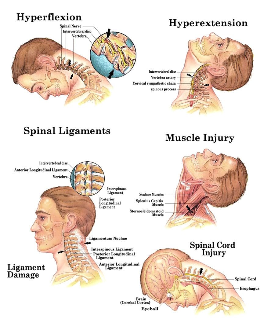
Cervical acceleration-deceleration is the mechanism of a neck injury caused by a forceful back-and-forth neck motion. It happens most commonly in rear-end vehicle collisions when the head and neck whip forward and backward with intense acceleration and/or deceleration causing the neck to flex and/or extend rapidly, more than normally, straining and possibly tearing muscle tissues and nerves, ligaments, dislocation of spinal discs and herniations, and cervical bone fractures.
For symptoms that do not improve or worsen after 2 to 3 weeks, see a healthcare provider or chiropractor for further evaluation and treatment.
Whiplash injuries strain or sprain the neck muscles and/or ligaments, but can also affect the vertebrae/bones, disc cushions between the vertebrae, and/or the nerves.
Whiplash symptoms can present right away, or after several hours to a few days after the incident, and tend to worsen in the days after the injury. Symptoms can last a few weeks to a few months, and can severely limit activity and range of motion. Symptoms can include: (National Institute of Neurological Disorders and Stroke. 2023)
Pain that extends into the shoulders and back.
Neck stiffness
Limited neck motion
Muscle spasms
Numbness and tingling sensations – paresthesias or pins and needles in the fingers, hands, or arms.
Headaches – A whiplash headache usually starts at the base of the skull and can vary in intensity. Most individuals experience pain on one side of the head and toward the back, though some can experience symptoms all over their head, and a small number experience headaches on the forehead or behind the eyes. (Monica Drottning. 2003)
The headaches can become aggravated by moving the neck around, especially when looking up.
Headaches are often associated with shoulder pain along with sensitive neck and shoulder muscles that when touched can increase pain levels.
Whiplash headaches can lead to chronic neck-related headaches known as cervicogenic headaches. (Phil Page. 2011)
It is also important to rest your neck area following the injury.
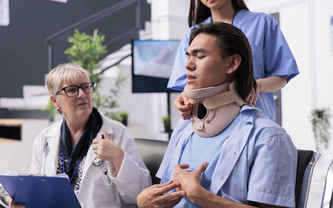
A cervical collar can be utilized temporarily to stabilize the neck, but for long-term recovery, it is recommended to keep the area mobile.
Physical activity reduction until the individual can look over both shoulders, and tilt their head all the way forward, all the way back, and from side to side without pain or stiffness.
Non-steroidal anti-inflammatories – NSAIDs – Ibuprofen or Naproxen.
Muscle relaxers
If symptoms do not improve, a healthcare provider may recommend physical therapy and/or stronger pain medications. For whiplash headaches that last for several months, acupuncture, or spinal injections may be recommended.
Neck Injuries
References
National Institute of Neurological Disorders and Stroke. Whiplash Information Page.
Drottning M. (2003). Cervicogenic headache after a whiplash injury. Current pain and headache reports, 7(5), 384–386. https://doi.org/10.1007/s11916-003-0038-9
Page P. (2011). Cervicogenic headaches: an evidence-led approach to clinical management. International journal of sports physical therapy, 6(3), 254–266.
IFM's Find A Practitioner tool is the largest referral network in Functional Medicine, created to help patients locate Functional Medicine practitioners anywhere in the world. IFM Certified Practitioners are listed first in the search results, given their extensive education in Functional Medicine