For individuals considering acupuncture for sciatica relief and management, can knowing how it works and what to expect during a session help in making the decision?
Acupuncture Sciatica Treatment Session
Acupuncture for sciatica is a safe and effective medical treatment to relieve and manage pain symptoms. Studies suggest it is as effective as other treatment strategies and causes fewer side effects. (Zhihui Zhang et al., 2023) The frequency of acupuncture to relieve sciatica pain depends on the severity of the condition and injury, but many report improvement within two to three weeks. (Fang-Ting Yu et al., 2022)
Needle Placement
Circulation problems can cause the body’s energy to stagnate in one or more meridians/channels, leading to pain in and around the surrounding area. (Wei-Bo Zhang et al., 2018)
The objective of acupuncture is to restore optimal circulation by stimulating specific points in the body called acupoints.
Thin, sterile needles stimulate the acupoints to activate the body’s natural healing abilities and relieve pain. (Heming Zhu 2014)
Some practitioners use electroacupuncture – a gentle, mild electrical current is applied to the needles and passes through the tissues to activate the nervous system. (Ruixin Zhang et al., 2014)
Acupoints
Acupuncture sciatica treatment involves specific acupoints along the bladder and gallbladder meridians.
Bladder Meridian – BL
The bladder meridian/BL runs down the back along the spine, hips, and legs. The acupoints within the meridian for sciatica include: (Fang-Ting Yu et al., 2022)
BL 23 -Shenshu – Location on the lower back, near the kidney.
BL 25 – Dachangshu – Location on the lower back.
BL 36 – Chengfu – Location on the back of the thigh, just below the buttocks.
GB 30 – Huantiao – Location on the back, where the buttocks meet the hips.
GB 34 – Yanglingquan – Location on the outside of the leg, below the knee.
GB 33 – Xiyangguan – Location lateral to the knee, on the side.
Stimulating acupoints in these meridians increases blood flow to the area, reduces inflammation, and releases endorphins and other pain-relieving neurochemicals to relieve symptoms. (Ningcen Li et al., 2021) The specific acupoints vary depending on symptoms and the root cause. (Tiaw-Kee Lim et al., 2018)
Example Patient
An example of acupuncture sciatica treatment session: A patient with persistent shooting pain extending down the back and side of the leg. A standard treatment consists of the following:
The acupuncturist thoroughly goes over the patient’s medical history and symptoms and has the patient point to where the pain is located.
Then, they palpate on and around the area to find where the pain worsens and lessens, communicating with the patient as they go along.
Depending on the site and severity, they may start placing needles at the lower back, focusing on the site of the injury.
Sometimes, the sacrum is involved, so the acupuncturist will place needles on those acupoints.
They then move to the back of the leg and insert needles.
The needles are retained for 20-30 minutes.
The acupuncturist leaves the room or treatment area but regularly checks in.
The patient may feel a warmth, tingling, or mild heaviness, which is a normal response. This is where patients report a calming effect. (Shilpadevi Patil et al., 2016)
The needles are carefully removed.
The patient may feel deeply relaxed and will be advised to get up slowly to avoid dizziness.
There may be soreness, redness, or bruising at the needle insertion site, which is normal and should resolve quickly.
The patient will be given recommendations as to avoiding strenuous activity, properly hydrating, and performing gentle stretches.
Acupuncture Benefits
Acupuncture has been shown to be a complementary therapy for pain relief and management. The benefits of acupuncture:
Improves Circulation
Acupuncture stimulates blood circulation, which nourishes damaged or irritated nerves and promotes healing.
This helps relieve sciatica symptoms, like numbness, tingling, and pain. (Song-Yi Kim et al., 2016)
Releases Endorphins
Acupuncture triggers the release of endorphins and other natural pain-relieving chemicals, which help relieve pain. (Shilpadevi Patil et al., 2016)
Regulates the Nervous System
Acupuncture rebalances the sympathetic and parasympathetic responses, which reduces stress, tension, and pain. (Xin Ma et al., 2022)
Relaxes the Muscles
Nerve pain often accompanies muscle tension and spasms.
Acupuncture relaxes tight muscles, reducing pressure and providing relief. (Zhihui Zhang et al., 2023)
From Symptoms to Solutions
References
Zhang, Z., Hu, T., Huang, P., Yang, M., Huang, Z., Xia, Y., Zhang, X., Zhang, X., & Ni, G. (2023). The efficacy and safety of acupuncture therapy for sciatica: A systematic review and meta-analysis of randomized controlled trails. Frontiers in neuroscience, 17, 1097830. https://doi.org/10.3389/fnins.2023.1097830
Yu, F. T., Liu, C. Z., Ni, G. X., Cai, G. W., Liu, Z. S., Zhou, X. Q., Ma, C. Y., Meng, X. L., Tu, J. F., Li, H. W., Yang, J. W., Yan, S. Y., Fu, H. Y., Xu, W. T., Li, J., Xiang, H. C., Sun, T. H., Zhang, B., Li, M. H., Wan, W. J., … Wang, L. Q. (2022). Acupuncture for chronic sciatica: protocol for a multicenter randomised controlled trial. BMJ open, 12(5), e054566. https://doi.org/10.1136/bmjopen-2021-054566
Zhang, W. B., Jia, D. X., Li, H. Y., Wei, Y. L., Yan, H., Zhao, P. N., Gu, F. F., Wang, G. J., & Wang, Y. P. (2018). Understanding Qi Running in the Meridians as Interstitial Fluid Flowing via Interstitial Space of Low Hydraulic Resistance. Chinese journal of integrative medicine, 24(4), 304–307. https://doi.org/10.1007/s11655-017-2791-3
Zhu H. (2014). Acupoints Initiate the Healing Process. Medical acupuncture, 26(5), 264–270. https://doi.org/10.1089/acu.2014.1057
Zhang, R., Lao, L., Ren, K., & Berman, B. M. (2014). Mechanisms of acupuncture-electroacupuncture on persistent pain. Anesthesiology, 120(2), 482–503. https://doi.org/10.1097/ALN.0000000000000101
Perreault, T., Fernández-de-Las-Peñas, C., Cummings, M., & Gendron, B. C. (2021). Needling Interventions for Sciatica: Choosing Methods Based on Neuropathic Pain Mechanisms-A Scoping Review. Journal of clinical medicine, 10(10), 2189. https://doi.org/10.3390/jcm10102189
Li, N., Guo, Y., Gong, Y., Zhang, Y., Fan, W., Yao, K., Chen, Z., Dou, B., Lin, X., Chen, B., Chen, Z., Xu, Z., & Lyu, Z. (2021). The Anti-Inflammatory Actions and Mechanisms of Acupuncture from Acupoint to Target Organs via Neuro-Immune Regulation. Journal of inflammation research, 14, 7191–7224. https://doi.org/10.2147/JIR.S341581
Lim, T. K., Ma, Y., Berger, F., & Litscher, G. (2018). Acupuncture and Neural Mechanism in the Management of Low Back Pain-An Update. Medicines (Basel, Switzerland), 5(3), 63. https://doi.org/10.3390/medicines5030063
Kim, S. Y., Min, S., Lee, H., Cheon, S., Zhang, X., Park, J. Y., Song, T. J., & Park, H. J. (2016). Changes of Local Blood Flow in Response to Acupuncture Stimulation: A Systematic Review. Evidence-based complementary and alternative medicine : eCAM, 2016, 9874207. https://doi.org/10.1155/2016/9874207
Patil, S., Sen, S., Bral, M., Reddy, S., Bradley, K. K., Cornett, E. M., Fox, C. J., & Kaye, A. D. (2016). The Role of Acupuncture in Pain Management. Current pain and headache reports, 20(4), 22. https://doi.org/10.1007/s11916-016-0552-1
Ma, X., Chen, W., Yang, N. N., Wang, L., Hao, X. W., Tan, C. X., Li, H. P., & Liu, C. Z. (2022). Potential mechanisms of acupuncture for neuropathic pain based on somatosensory system. Frontiers in neuroscience, 16, 940343. https://doi.org/10.3389/fnins.2022.940343
For individuals looking to improve their spinal health, can understanding the anatomy of the intervertebral foramen help in injury rehabilitation and prevention?
Intervertebral Foramen
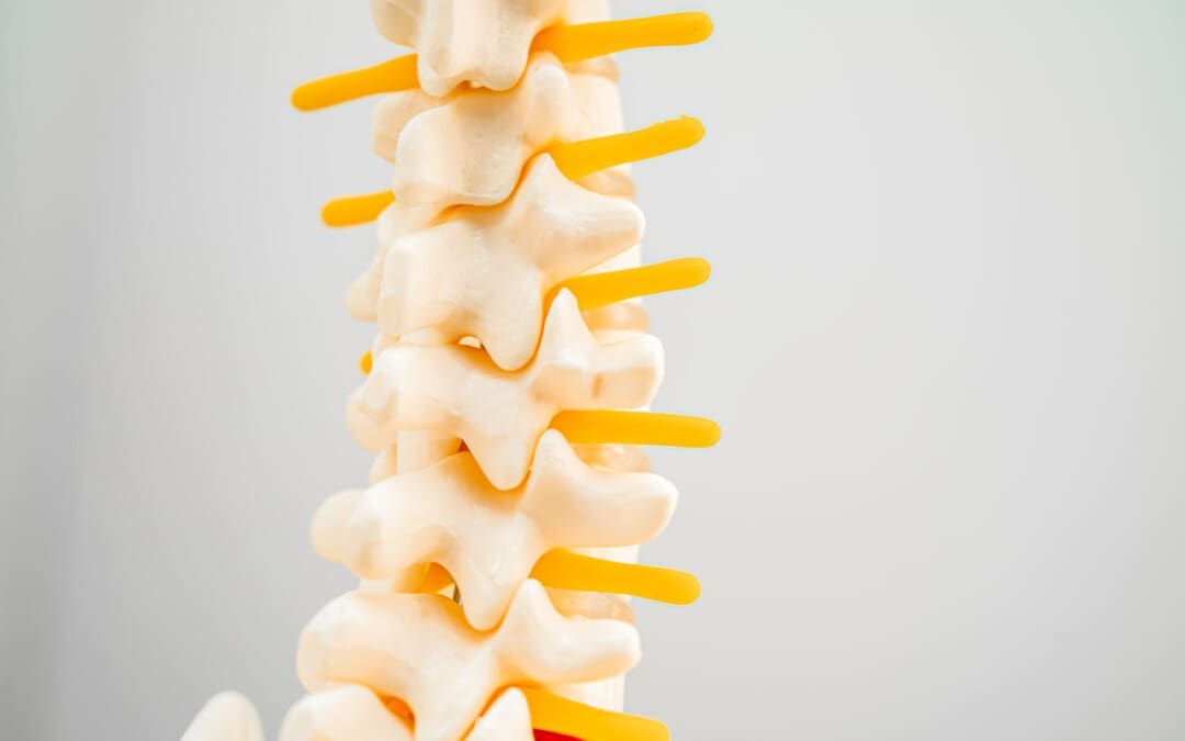
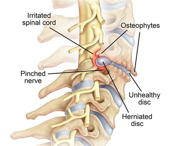
The intervertebral foramen, aka neural foramen, is the opening between the vertebrae through which spinal nerve roots connect and exit to other body areas. If the foramina narrows, it can place added pressure on the nerve roots near and around them, causing pain symptoms and sensations. This is known as neuroforaminal stenosis. (Sumihisa Orita et al., 2016)
Anatomy
The vertebrae comprise the spinal column.
They protect and support the spinal cord and most of the weight placed on the spine.
Foramen is the singular form, and foramina is the plural form.
Structure
The body is the large, round part of the bone that makes up each vertebra.
The body of each vertebra is attached to a bony ring.
Stenosis can occur in the spinal canal, known as central canal stenosis, and the foramina.
Pain brought on by neuroforaminal spinal stenosis and arthritis-related bone growth/bone spurs/osteophytes that are present in one or more foramen rub against the nerve root that passes through the space, causing radicular pain.
Pain accompanied by other sensations, like tingling or numbness, is known as radiculopathy. (Young Kook Choi, 2019)
The main symptom is pain.
Numbness and/or tingling can present depending on the injury.
Neurogenic claudication occurs as a result of ischemia or a lack of blood circulation to the nerves and typically presents with a heaviness in the legs.
It is typically associated with central stenosis rather than foraminal stenosis.
Most individuals with spinal stenosis feel better when flexing or bending forward and worse when arching their backs.
Stenosis treatment aims to relieve pain and prevent nerve symptoms from occurring or worsening. Conservative treatments are recommended and can be highly effective.
These include:
Myelopathy in the neck and/or upper or mid-back (myelopathy symptoms are spinal cord related and occur in central canal stenosis) (Cleveland Clinic. 2021)
Intense incapacitating pain
Different surgical techniques include:
Decompression laminectomy – entails removing the buildup of bone in the spinal canal.
Spinal fusion – when there is instability of the spine or severe foraminal stenosis.
Orita, S., Inage, K., Eguchi, Y., Kubota, G., Aoki, Y., Nakamura, J., Matsuura, Y., Furuya, T., Koda, M., & Ohtori, S. (2016). Lumbar foraminal stenosis, the hidden stenosis including at L5/S1. European journal of orthopaedic surgery & traumatology : orthopedie traumatologie, 26(7), 685–693. https://doi.org/10.1007/s00590-016-1806-7
American Academy of Orthopaedic Surgeons. (2020). Spine Basics (OrthoInfo, Issue. https://orthoinfo.aaos.org/en/diseases–conditions/spine-basics/
American Academy of Orthopaedic Surgeons. (2021). Lumbar spinal stenosis (OrthoInfo, Issue. https://orthoinfo.aaos.org/en/diseases–conditions/lumbar-spinal-stenosis/
Choi Y. K. (2019). Lumbar foraminal neuropathy: an update on non-surgical management. The Korean journal of pain, 32(3), 147–159. https://doi.org/10.3344/kjp.2019.32.3.147
Lee, S. Y., Kim, T. H., Oh, J. K., Lee, S. J., & Park, M. S. (2015). Lumbar Stenosis: A Recent Update by Review of Literature. Asian spine journal, 9(5), 818–828. https://doi.org/10.4184/asj.2015.9.5.818
Lurie, J., & Tomkins-Lane, C. (2016). Management of lumbar spinal stenosis. BMJ (Clinical research ed.), 352, h6234. https://doi.org/10.1136/bmj.h6234
For individuals experiencing musculoskeletal issues and pain symptoms, can learning about biomechanics and how it applies to movement, physical training, and performance, help in injury treatment and prevention?
Biomechanics
Biomechanics studies all life forms and their mechanical workings. Many think of biomechanics in sports and athletic performance, but biomechanics helps create and improve technologies, equipment, and injury rehabilitation techniques. (Tung-Wu Lu, Chu-Fen Chang 2012) Scientists, sports medicine doctors, physiotherapists, chiropractors, and conditioning specialists utilize biomechanics to help develop training protocols and techniques to improve therapy outcomes.
Body Movement
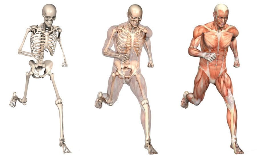
Biomechanics studies the movement of the body, including how muscles, bones, tendons, and ligaments work together, especially when movement is not optimal or correct. It is part of the larger field of kinesiology, specifically focusing on motion mechanics and analysis of how all the individual parts of the body work together to make up athletic and normal movements. (José M Vilar et al., 2013) Biomechanics includes:
Structure of bones and muscles.
Movement ability.
Mechanics of blood circulation, renal function, and other functions.
The study of forces and the effects of these forces on the tissues, fluid, or materials used for diagnosis, treatment, or research. (Jose I. Priego-Quesada 2021)
Sports
Sports biomechanics studies motion in exercising, training, and sports, which incorporates physics and the laws of mechanics. For example, the biomechanics of a specific exercise looks at:
Body position.
Movement of the feet, hips, knees, back, shoulders, and arms.
Knowing the correct movement patterns helps make the most of the exercise while preventing injuries, correcting form mistakes, informing training protocols, and increasing positive results. Understanding how the body moves and why it moves the way it does helps medical professionals prevent and treat injuries, alleviate pain symptoms, and improve performance.
Equipment
Biomechanics is used in the development of physical and sports equipment to improve performance. For example, a shoe can be designed for optimal performance for a skateboarder, long-distance runner, or soccer player. Playing surfaces are also studied for this purpose, such as how the surface stiffness of artificial turf affects athletic performance. (Jose I. Priego-Quesada 2021)
Individuals
Biomechanics can analyze an individual’s movements for more effective movement during training and games.
For example, an individual’s running gait or swing can be filmed with recommendations on what to change to improve.
Injuries
The science studies the causes, treatment, and prevention of neuromusculoskeletal injuries.
The research can analyze the forces that cause injuries and provide information for medical professionals on how to reduce the risk of injury.
Training
Biomechanics studies sports techniques and training systems to develop ways to improve efficiency.
This can include research on positioning, release, follow-through, etc.
It can analyze and help design new training techniques based on the mechanical demands of the sport, aimed at resulting in better performance.
For example, muscle activation is measured in cycling using electromyography and kinematics, which helps researchers analyze factors like posture, components, or exercise intensity that affect activation. (Jose I. Priego-Quesada 2021)
Motions
In biomechanics, the body’s motions are referred to from anatomical positioning:
Standing upright, with the gaze straight ahead
Arms at the sides
Palms facing forward
Feet spaced slightly apart, toes forward.
The three anatomical planes include:
Sagittal – median – Dividing the body into right and left halves is the sagittal/median plane. Flexion and extension occur in the sagittal plane.
Frontal – The frontal plane divides the body into front and back sides but also includes abduction, or moving a limb away from the center, and adduction, or moving a limb towards the center in the frontal plane.
Transverse – horizontal. – The upper and lower parts of the body are divided by the transverse/horizontal plane. Rotating movements occur here. (American Council on Exercise 2017)
Moving the body in all three planes occurs with daily activity. This is why performing exercises in each plane of motion to build strength, function, and stability is recommended.
Tools
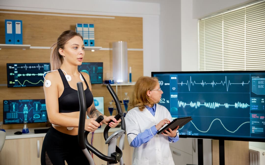
Various tools are used to study biomechanics. Studies are usually performed using a device known as electromyography or EMG sensors. Sensors are placed on the skin and measure the amount and degree of muscle fiber activation in certain muscles during test exercises. EMGs can help:
Researchers understand which exercises are more effective than others.
Therapists know whether patients’ muscles are properly operating and functioning.
Dynamometers are another tool that helps measure muscle strength.
They measure the force output generated during muscle contractions to see if the muscles are sufficiently strong.
They are used to measure grip strength, which can be an indicator of overall strength, health, and longevity. (Li Huang et al., 2022)
Beyond Adjustments: Chiropractic and Integrative Healthcare
References
Lu, T. W., & Chang, C. F. (2012). Biomechanics of human movement and its clinical applications. The Kaohsiung journal of medical sciences, 28(2 Suppl), S13–S25. https://doi.org/10.1016/j.kjms.2011.08.004
Vilar, J. M., Miró, F., Rivero, M. A., & Spinella, G. (2013). Biomechanics. BioMed research international, 2013, 271543. https://doi.org/10.1155/2013/271543
Priego-Quesada J. I. (2021). Exercise Biomechanics and Physiology. Life (Basel, Switzerland), 11(2), 159. https://doi.org/10.3390/life11020159
American Council on Exercise. Makeba Edwards. (2017). Planes of Motion Explained (Exercise Science, Issue. https://www.acefitness.org/fitness-certifications/ace-answers/exam-preparation-blog/2863/the-planes-of-motion-explained/
Huang, L., Liu, Y., Lin, T., Hou, L., Song, Q., Ge, N., & Yue, J. (2022). Reliability and validity of two hand dynamometers when used by community-dwelling adults aged over 50 years. BMC geriatrics, 22(1), 580. https://doi.org/10.1186/s12877-022-03270-6
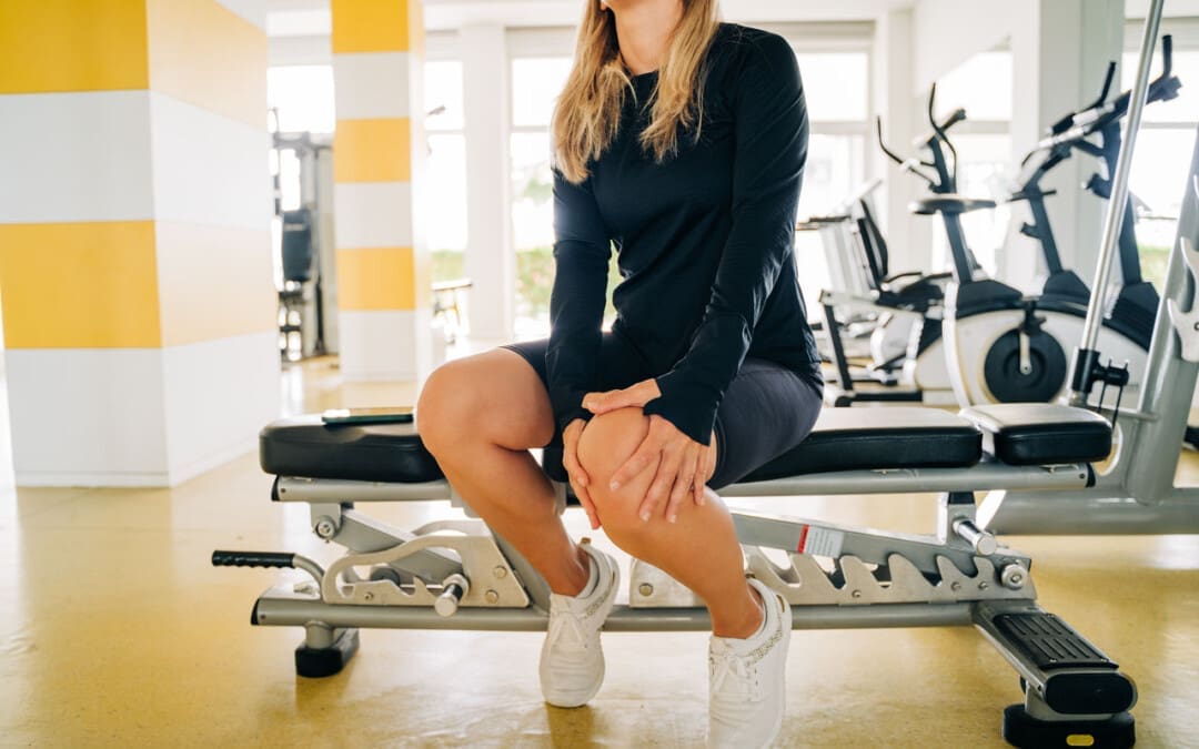
Knee injuries can present in physically active individuals that lift weights. Can understanding the types of weightlifting knee injuries help in prevention?
Weightlifting Knee Injuries
Weight training is very safe for the knees as regular weight training can improve knee strength and prevent injury as long as the correct form is followed. For Individuals with knee injuries from other activities, incorrect weight-training exercises could worsen the injury. (Ulrika Aasa et al., 2017) As well as, sudden twisting movements, poor alignment, and pre-existing injuries can increase the risk of worsening or creating further injuries. (Hagen Hartmann et al, 2013) The body and the knees are designed to support vertical forces on the joints.
Common Injuries
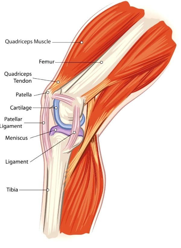
Weightlifting knee injuries occur as the knee joints endure a wide range of stresses and strains. In weight training, the ligaments that attach to the complex bone system of the knee joint can be damaged by incorrect movements, overloading the weight, and increasing the weight too soon. These injuries can result in pain, swelling, and immobility that can range from minor to severe, from a sprain or a slight tear to a complete tear in serious cases.
Anterior Cruciate Ligament – ACL – Injury
This ligament attaches the thigh’s femur bone to the lower leg’s shin bone/tibia and controls excessive rotation or extension of the knee joint. (American Academy of Family Physicians. 2024)
Anterior means front.
ACL injuries are seen mostly in athletes but can happen to anybody.
Severe damage to the ACL usually means surgical reconstruction and up to 12 months of rehabilitation.
When weightlifting, try to avoid twisting knee movements, intentionally or accidentally, under excessive load.
Posterior Cruciate Ligament – PCL – Injury
The PCL connects the femur and tibia at different points to the ACL.
It controls any backward motion of the tibia at the joint.
Injuries occur most with high-impact forces as a result of accidents and sometimes in activities where forceful trauma to the knee occurs.
Medial Collateral Ligament – MCL – Injury
This ligament maintains the knee from bending too far to the inside/medially.
Injuries mostly occur from impact to the outside of the knee or from accidental bodyweight force on the leg that bends at an unusual angle.
Lateral Collateral Ligament – LCL – Injury
This ligament connects the smaller bone of the lower leg/fibula to the femur.
It is opposite to the MCL.
It maintains excessive outward movement.
LCL injuries occur when a force pushes the knee out.
Cartilage Injury
Cartilage prevents bones from rubbing together and cushions impact forces.
Knee menisci are cartilage that cushions the knee joints inside and outside.
Other types of cartilage protect the thigh and shin bones.
When cartilage gets torn or damaged, surgery may be required.
Tendonitis
Aggravated and overused knee tendons can lead to weightlifting knee injuries.
A related injury known as iliotibial band syndrome/ITB causes pain to the outside of the knee, usually in runners, but it can occur from overuse.
Rest, stretching, physical therapy, and anti-inflammatory medication are a common treatment plan.
The condition causes the cartilage to deteriorate and bones to rub together, resulting in pain and stiffness.
Prevention
Individuals can minimize their risk of weightlifting knee injuries and pain by following their doctor’s and personal trainers’ recommendations.
Individuals with an existing knee injury should follow their doctor’s or physical therapist’s recommendations.
A knee sleeve can keep the muscles and joints secure, providing protection and support.
Stretching the leg and knee muscles can maintain joint flexibility.
Avoid sudden lateral movements.
Possible recommendations can include:
Avoiding Certain Exercises
Isolation exercises like leg curls, standing, or on a bench, as well as using the leg extension machine, can stress the knee.
Deep Squat Training
Research shows that the deep squat can protect against lower leg injury if the knee is healthy. However, this is when done with proper technique, under expert supervision, and with a gradual progressive load. (Hagen Hartmann et al, 2013)
Individuals should talk to their doctor before beginning a new exercise routine. A personal trainer can provide training in learning the proper technique and weightlifting form.
How I Tore my ACL Part 2
References
Aasa, U., Svartholm, I., Andersson, F., & Berglund, L. (2017). Injuries among weightlifters and powerlifters: a systematic review. British journal of sports medicine, 51(4), 211–219. https://doi.org/10.1136/bjsports-2016-096037
Hartmann, H., Wirth, K., & Klusemann, M. (2013). Analysis of the load on the knee joint and vertebral column with changes in squatting depth and weight load. Sports medicine (Auckland, N.Z.), 43(10), 993–1008. https://doi.org/10.1007/s40279-013-0073-6
American Academy of Family Physicians. ACL injury. (2024). ACL injury (Diseases and Conditions, Issue. https://familydoctor.org/condition/acl-injuries/
Mellinger, S., & Neurohr, G. A. (2019). Evidence based treatment options for common knee injuries in runners. Annals of translational medicine, 7(Suppl 7), S249. https://doi.org/10.21037/atm.2019.04.08
Driban, J. B., Hootman, J. M., Sitler, M. R., Harris, K. P., & Cattano, N. M. (2017). Is Participation in Certain Sports Associated With Knee Osteoarthritis? A Systematic Review. Journal of athletic training, 52(6), 497–506. https://doi.org/10.4085/1062-6050-50.2.08
For individuals starting to lift weights, motor units are important for muscle movement. Can building more motor units help build strength and maintain muscle mass?
Motor Units
Motor units control the skeletal muscles and are the force behind every body movement. (C J. Heckman, Roger M. Enoka 2012)
This includes voluntary movements like lifting weights and involuntary movements like breathing. When lifting objects and weights, the body adapts to motor unit needs, meaning that individuals must consistently increase the weight to progress.
Lifting weights regularly trains the body to generate more motor units and force.
General guidelines recommend lifting weights for all muscle groups two to three non-consecutive days a week.
Consistency helps maintain muscle mass.
Regular progression increases the risk of plateauing.
What They Are
Exercise increases the body’s muscle strength, while sedentariness and inactivity weaken them. A motor unit is a single nerve cell/neuron that supplies the nerves to innervate a group of skeletal muscles. The neuron receives signals from the brain that stimulate all the muscle fibers in that particular motor unit to generate movement.
Muscles comprise different fiber types.
They are attached to the bones with connective tissue, which is stronger than the muscle.
Multiple motor units are dispersed throughout the muscle.
The motor units help ensure muscle contraction force is evenly spread throughout the muscle.
Motor units are different sizes and operate differently depending on where and what they do.
Small motor units might only innervate five or ten fibers. For example, to blink or sniff.
Large motor units can comprise hundreds of muscle fibers for swinging or jumping movements.
How They Work
The number of units activated depends on the task. Stronger muscle contractions require more. However, fewer units are needed to accomplish the movement for individuals expending less effort.
Contraction
Once a unit receives a signal from the brain, the muscle fibers contract simultaneously.
The force generated depends on how many units are required to accomplish the task. (Purves D. et al., 2001)
For example, picking up small objects like a pen and paper requires only a few units to generate the force needed.
If picking up a heavy barbell, the body needs more units because more force is needed to lift the heavier load.
The body can generate more force with stronger muscles.
This happens when lifting weights on a regular basis and overloading the muscles with more weight than they can handle.
This process is known as adaptation.
Adaptation
The purpose of lifting weights is to challenge the muscles so they adapt to the new challenge and grow in strength and mass. Motor units are a major part of the adaptation process. (Dr. Erin Nitschke. American Council on Exercise. 2017)
As individuals continue working out, their ability to generate more force increases and units activate more rapidly.
This makes movements more efficient.
Individuals can increase motor unit recruitment by consistently increasing the weight challenge to their muscles.
The development creates movement memory.
A relationship between the brain, muscles, and motor units is established even if the individual stops working out. The pathways are still there no matter how long the individual takes off.
When returning to training, the body will remember how to ride a bike, do a bicep curl, or squat.
However, the muscles will not have the same strength as the strength needs to be built back along with endurance that may have been lost.
It is the movement memory that remains.
Military Training and Chiropractic Care: Maximizing Performance
References
Heckman, C. J., & Enoka, R. M. (2012). Motor unit. Comprehensive Physiology, 2(4), 2629–2682. https://doi.org/10.1002/cphy.c100087
Purves D, Augustine GJ, Fitzpatrick D, et al., editors. (2001). Neuroscience. 2nd edition. Sunderland (MA): Sinauer Associates; 2001. The Motor Unit. Available from: https://www.ncbi.nlm.nih.gov/books/NBK10874/
Dr. Erin Nitschke. American Council on Exercise. (2017). How Muscle Grows (Exercise Science, Issue. https://www.acefitness.org/resources/everyone/blog/6538/how-muscle-grows/
Pete McCall. American Council on Exercise. (2015). 10 Things to Know About Muscle Fibers (Exercise Science, Issue. https://www.acefitness.org/resources/pros/expert-articles/5411/10-things-to-know-about-muscle-fibers/
For individuals suffering from back pain, can knowing basic chiropractic terminology help in understanding diagnosis and treatment plan development?
Chiropractic Terminology
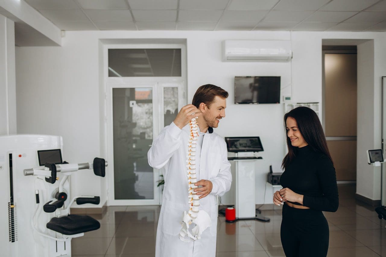
The chiropractic principle is that a properly aligned spine positively affects an individual’s overall health. One of the main aspects of chiropractic care is applying calculated force to the spinal joints to restore correct spinal alignment. Chiropractic terminology describes specific types of techniques and care.
General Subluxation
A subluxation can mean different things for various doctors. In general, a subluxation is a significant structural displacement or an incomplete or partial dislocation of a joint or organ.
To medical doctors, a subluxation refers to a partial dislocation of a vertebrae.
This is a serious condition, usually brought on by trauma, that can result in a spinal cord injury, paralysis, and/or death.
X-rays show a conventional subluxation as an obvious disconnect between the vertebrae.
Chiropractic Subluxation
The chiropractic interpretation is more subtle and refers to the misalignment of adjacent spinal vertebrae.
Subluxation in this context refers to position changes in the joints and soft tissues of the spine.
Vertebral misalignment is believed to lead to pain and abnormal intervertebral joint motion.
This difference between the serious subluxation medical condition and the chiropractic version may cause individuals to dismiss seeking back pain treatments.
Motion Segment
Chiropractors and surgeons use it as a technical term.
Motion segment refers to two adjacent vertebrae and the intervertebral disc between them.
This is the area chiropractors assess and adjust.
Adjustment
The chiropractor performs a spinal manual adjustment to realign joint subluxations.
Adjustments involve applying force to motion segments to bring them back into a centered alignment.
The goal for adjustments and realigning the vertebrae includes:
Spinal manipulation is a technique used by chiropractors to provide relief for musculoskeletal pain related to the back and neck. Manipulation provides mild to moderate relief and works as well as some conventional treatments like pain-relieving medications. (Sidney M. Rubinstein et al., 2012)
Spinal manipulation is divided into grades of mobilization.
Depending on their training, practitioners of various medical disciplines may be licensed to perform grade 1 to grade 4 mobilizations.
Only physical therapists, osteopathic physicians, and chiropractors are licensed to perform grade 5 mobilizations, which are high-velocity thrust techniques.
Most massage therapists, athletic trainers, and personal trainers are not licensed to perform spinal manipulations.
Based on a systematic review, the effectiveness of these treatments found that there is quality evidence that manipulation and mobilization can help reduce pain and improve function for individuals with chronic low back pain, with manipulation appearing to produce a more profound effect than mobilization. Both therapies are safe, with multimodal treatments potentially being an effective option. (Ian D. Coulter et al., 2018)
As with any treatment, results vary from person to person and with different chiropractors. There are also potential risks with spinal manipulation. Though rare, cervical, carotid, and vertebral artery dissections have occurred with cervical/neck manipulation. (Kelly A. Kennell et al., 2017) Individuals with osteoporosis may be advised to avoid chiropractic adjustments or manipulation because of the increased risk of injury. (James M. Whedon et al., 2015)
Many individuals choose chiropractic treatment for a variety of conditions. Understanding chiropractic terminology and reasoning allows individuals to ask questions as they discuss their symptoms to develop a personalized treatment plan and restore function and wellness.
What Causes Disc Herniation?
References
Henderson C. N. (2012). The basis for spinal manipulation: chiropractic perspective of indications and theory. Journal of electromyography and kinesiology : official journal of the International Society of Electrophysiological Kinesiology, 22(5), 632–642. https://doi.org/10.1016/j.jelekin.2012.03.008
Blanchette, M. A., Stochkendahl, M. J., Borges Da Silva, R., Boruff, J., Harrison, P., & Bussières, A. (2016). Effectiveness and Economic Evaluation of Chiropractic Care for the Treatment of Low Back Pain: A Systematic Review of Pragmatic Studies. PloS one, 11(8), e0160037. https://doi.org/10.1371/journal.pone.0160037
Rubinstein, S. M., Terwee, C. B., Assendelft, W. J., de Boer, M. R., & van Tulder, M. W. (2012). Spinal manipulative therapy for acute low-back pain. The Cochrane database of systematic reviews, 2012(9), CD008880. https://doi.org/10.1002/14651858.CD008880.pub2
Coulter, I. D., Crawford, C., Hurwitz, E. L., Vernon, H., Khorsan, R., Suttorp Booth, M., & Herman, P. M. (2018). Manipulation and mobilization for treating chronic low back pain: a systematic review and meta-analysis. The spine journal : official journal of the North American Spine Society, 18(5), 866–879. https://doi.org/10.1016/j.spinee.2018.01.013
Kennell, K. A., Daghfal, M. M., Patel, S. G., DeSanto, J. R., Waterman, G. S., & Bertino, R. E. (2017). Cervical artery dissection related to chiropractic manipulation: One institution’s experience. The Journal of family practice, 66(9), 556–562.
Whedon, J. M., Mackenzie, T. A., Phillips, R. B., & Lurie, J. D. (2015). Risk of traumatic injury associated with chiropractic spinal manipulation in Medicare Part B beneficiaries aged 66 to 99 years. Spine, 40(4), 264–270. https://doi.org/10.1097/BRS.0000000000000725
For individuals dealing with lower back pain, it could be quadricep muscle tightness causing the symptoms and posture problems. Can knowing the signs of quadricep tightness help prevent pain and avoid injury?
Quadriceps Tightness
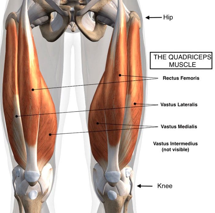
Quadriceps muscles are in the front of the thigh. Forces that could be creating chronic pain and posture problems could be happening at the same time are:
Quadricep tightness causes lower back pain as the pelvis gets pulled down.
Tight quadriceps lead to weakened hamstring muscles.
These are the opposing muscles behind the thigh.
Stress and pressure on the hamstrings can cause back pain and problems.
The rectus femoris attaches to the pelvis at the anterior superior iliac spine, which is the front part of the hip bone.
The rectus femoris is the only muscle in the group that crosses over the hip joint, which also affects movement.
When the quadriceps, especially the rectus femoris, become tight, they pull down on the hips.
The pelvis tilts downward or forward, technically referred to as the anterior tilt of the pelvis. (Anita Król et al., 2017)
The spine is between the pelvis, and if the pelvis tilts forward, the lumbar spine compensates by arching.
A larger arch in the lower back is referred to as excessive lordosis and often causes tightness and pain in the back muscles. (Sean G. Sadler et al., 2017)
Hamstring Compensation
When the quadriceps tighten and the pelvis gets pulled down, the back has an abnormal lift. This puts the hamstring on a consistent stretch that can cause pain symptoms.
Healthy posture and hamstring muscle tone help maintain correct pelvic positioning in the back.
This is correct because it helps maintain a comfortable position.
Quadricep tightness can set off a reaction as the pelvis tilts down in front and up in the back while overly stretching the hamstrings.
Pain and soreness are the usual result
Lack of hamstring strength and quadriceps stretching can cause the hamstrings to lose their ability to support correct pelvic and spinal positions. (American Council on Exercise. 2015)
Knowing When Quads Are Tightening
Individuals often don’t realize their quadriceps are tight, especially those who spend most of the day sitting.
The more time spent in a chair can cause the quadriceps and lower back muscles to tighten steadily.
Individuals can try a few tests at home:
Standing Up
Push the hips forward.
Push from the sitting bones so you’re at the correct level.
How far forward do the hips go?
What is felt?
Pain could indicate tight quadriceps.
In A Lunge Position
With one leg forward and bent in front of the other.
The back leg is straight.
How far forward does the leg go?
What is felt?
How does the front of the hip on the back leg feel?
Standing Bent Leg
Stand with the front leg bent and the back leg straight.
Discomfort in the back leg could mean tight quadriceps.
In A Kneeling Position
Arch the back
Grab the ankles
Modify the position to adjust for any pain or joint issues.
If you have to prop yourself up or modify the pose to reduce pain, it could be tight quadriceps.
Helping to understand the condition can help in communication with a healthcare provider.
A healthcare provider and/or physical therapist can conduct a posture evaluation examination to test the quadriceps.
Understanding Academic Low Back Pain: Impact and Chiropractic Solutions
References
Kripa, S., Kaur, H. (2021). Identifying relations between posture and pain in lower back pain patients: a narrative review. Bulletin of Faculty of Physical Therapy, 26(34). https://doi.org/doi: 10.1186/s43161-021-00052-w
Król, A., Polak, M., Szczygieł, E., Wójcik, P., & Gleb, K. (2017). Relationship between mechanical factors and pelvic tilt in adults with and without low back pain. Journal of back and musculoskeletal rehabilitation, 30(4), 699–705. https://doi.org/10.3233/BMR-140177
Sadler, S. G., Spink, M. J., Ho, A., De Jonge, X. J., & Chuter, V. H. (2017). Restriction in lateral bending range of motion, lumbar lordosis, and hamstring flexibility predicts the development of low back pain: a systematic review of prospective cohort studies. BMC musculoskeletal disorders, 18(1), 179. https://doi.org/10.1186/s12891-017-1534-0
American Council on Exercise. (2015). 3 Stretches for Opening Up Tight Hips (Fitness, Issue. https://www.acefitness.org/resources/everyone/blog/5681/3-stretches-for-opening-up-tight-hips/
IFM's Find A Practitioner tool is the largest referral network in Functional Medicine, created to help patients locate Functional Medicine practitioners anywhere in the world. IFM Certified Practitioners are listed first in the search results, given their extensive education in Functional Medicine