by Dr Alex Jimenez DC, APRN, FNP-BC, CFMP, IFMCP | Chiropractic, Integrative Functional Wellness, Integrative Medicine, Wellness
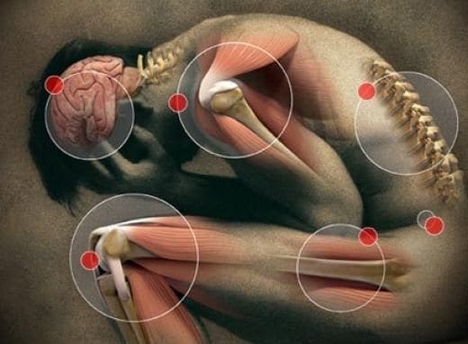
El Paso TX. Chiropractor Dr. Alex Jimenez takes a look at medication for joint pain and how they can make the pain worse.
Non-steroidal anti-inflammatory drugs (NSAIDs) are as common as candy, a staple of every home medicine cabinet and tossed casually in desk drawers, purses, and briefcases. Many people take these drugs, which include ibuprofen (sold as Motrin and Advil), naproxen (Aleve), and aspirin, at the first sign of a�headache or muscle cramps � and they are a daily ritual for many people living with arthritis.
But few people realize that NSAIDs carry a black-box warning, the strictest warning issued by the Food and Drug Administration. �Most people think that the government or FDA would not allow something dangerous on the market, especially since most of them are over-the-counter and [used] without a prescription,� says integrative medicine expert Sunil Pai, MD, author of An Inflammation Nation. �A black-box warning is the FDA�s attempt to let you know that you can end up in a casket if you are unlucky enough to suffer one of a medication�s serious reactions.�
Not only have NSAIDs been linked to a slew of serious side effects, including ulcers, hearing loss, allergic reactions and miscarriages, but they can actually worsen some of the conditions, such as arthritis, they are supposed to help.
�The scientific literature makes it abundantly clear that NSAIDs�have a significant negative effect on cartilage,� which accelerates the deterioration of arthritic joints, says Pai. �NSAIDs have no beneficial effect on [cartilage] and speed up the very disease for which they are most used and prescribed.�
Even worse, NSAIDs do not address the underlying conditions that cause pain and inflammation, such as a leaky gut, and can even exacerbate them. Stress, infections, alcohol, and a poor diet can all irritate the gut lining and lead to a leaky gut, but so can NSAIDs.
�If you use a full therapeutic dose of NSAIDs for two weeks, there is a 75 percent chance you will develop a leaky gut that doesn�t go away when you stop taking the drug, Leo Galland, MD, tellsExperience Life magazine.
6 Simple Dietary Interventions To Fight &�Heal A Leaky Gut

So, how can people with acute or chronic inflammatory conditions fight pain naturally? Some simple dietary interventions go a long way towards fighting inflammation and healing a leaky gut.
1. Try an Elimination Diet
Removing common foods that can irritate the gut, including gluten, sugar, dairy, processed foods and soy, can jumpstart the healing process. Sugar (and refined grains, which turn to sugar in the body), for example, is one of the single biggest drivers of inflammation and its downstream consequences.
When sugar cravings strike, try roasting root vegetables or sweet potatoes. Roasting concentrates the natural sweetness of the plant, but the fiber slows down sugar absorption in the bloodstream.
2. Eat Whole Foods
Michael Pollen�s recommendation � �Eat food. Not too much. Mostly plants.� � Is great advice when it comes to naturally fighting inflammation. Eating a Standard American Diet (SAD) � high in processed foods, unhealthy fats, and sugars � is like pouring kerosene on inflammation�s fire. Eating whole foods, rich in phytonutrients, helps put out that fire.
One fun way to eat more plants? Strive to �eat the rainbow,� or get at least one whole food from all the different colors of the rainbow each day:
- Red (pomegranates, strawberries, tomatoes)
- Orange (sweet potatoes, carrots)
- Yellow (lemon, squash)
- Green (avocado, Brussels sprouts, green tea)
- Blue/purple (berries, olives)
- White/tan/brown (garlic, onion, mushrooms).
Animal protein doesn�t need to be avoided if it�s grass-fed and pastured. Instead, try to reverse the ratio on your dinner plate: Make meat the side dish and vegetables the main course.
3. Supplement with Glutamine
Glutamine helps heal your gut by fueling the cells in your gut lining. You could think of it as a leaky gut superhero. �Glutamine heals the intestinal lining more than any other nutrient,� Liz Lipski, Ph.D., CCN, author of Digestive Wellness, tells Experience Life.
4. Get Your Omega-3s
Omega-3 fatty acids are natural inflammation fighters. Good whole food sources of omega-3s include wild-caught fish, grass-fed meat, pastured eggs, algae, and seeds such as hemp, chia, and flax. A high-quality omega-3 supplement is also worth considering. Even on a largely whole-foods-based, it can be hard to get the recommended daily amount of omega-3s.
5. Drink Bone Broth
Bone broth is one of the best natural sources of collagen, a protein found in abundance in our ligaments, tendons, bones, and skin. The collagen in broth is easily absorbed by our tissues and can not only help promote healthier connective tissue and ease joint pain, but it can also help heal a leaky gut. The best bone broth is homemade�but increasingly high-quality bone broth is available for purchase at cooperatives and health food stores.
6. Consider Botanical First Aid
Many plants are powerful inflammation fighters. Turmeric may be the best known and most studied.�Recent research suggests that the active ingredient in turmeric (called curcumin) has anti-inflammatory, antioxidant, antiviral, antibacterial, antifungal, and anticancer activities on par with commonly prescribed arthritis drugs like Enbrel and Humira.
A lot of other plants and plant compounds show similar activity in the body, including ginger, bromelain (an enzyme found in pineapple), capsaicin (the active ingredient in hot peppers), and ginger. Consult your healthcare practitioner before taking botanical supplements.

Call Today!
Source:
http://www.drfranklipman.com/problem-nsaids-yes-mean-advil/

by Dr Alex Jimenez DC, APRN, FNP-BC, CFMP, IFMCP | Chiropractic, Chronic Back Pain, Migraines, Neck Pain
El Paso TX. Chiropractor, Dr. Alex Jimenez discusses ways�to assist you to get the care you’ll need at your next appointment.
Dr. Michael J. Cooney, D.C�I�ve been treating patients with severe and chronic pain from around the corner in Rutherford, New Jersey to as far away as Australia and South Africa.
From our patient�s first reference to the past treatment office visit, the success of any pain treatment we prescribe is contingent upon us (the health care provider) correctly treating the root cause of your pain.
As the patient, just describing your intense pain or neuropathic pain is a high stakes� conversation that is �. I am able to read your medical history, attributing lab results and physician reports, however this really is secondary to understanding each patient�s pain mechanics. It is absolutely vital this is communicated to your pain management provider as correctly as possible.
For those fighting �invisible pain” such as fibromyalgia, CRPS (complex areas pain syndrome), RSD (reflex sympathetic dystrophy), diabetic neuropathy or long-term pain after cancer treatment, correctly communicating the place, frequency and depth of the discomfort can be especially demanding and emotionally taxing.
You might wish to bring this short article for your next doctor visit and go over each of the key pain description points I�ve outlined below.
I really hope your doctor will ask you these questions, but if not, you are able to behave as your own pain promoter and offer this information.
�Tell Me About Your Pain�
Based upon your medical records, we already know the reason behind your pain (injury or ailment). In order to restart your highest quality of life possible, our goal would be to remove or minimize this symptom.
Pain symptoms are private, subjective –and unique. (What Joe describes as �unbearable pain� could be considered �fairly disagreeable pain� to Mike). Through the years, I developed my own �pain diagnostic� conversation with patients to assist my team and I understand what, where, when and just how much pain patients are feeling.
I�ve outlined key points below:
Time Matters
This is key to a proper analysis. Don�t presume we know you�ve combated with this pain to get a month a year or a decade.
1.I�ve had this pain for _________________.
2.How frequently and how long does it last?
3.What ignites (flare) or lessens your pain and for how long?
Location, Location, Location
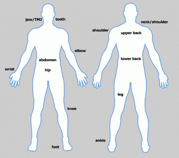
Graphic of a human body with a rear & front view (see above)
Doctors may instruct you to indicate the area/s where your pain is concentrated. They may also request that you notice a difference between pain which is on pain and the surface that’s below the surface.
The front and back of the unisex individual figure are the most identifiable, although this tool comes from the McGill Pain Questionnaire including other measurements.
Most referring physicians, regardless of their medical specialty, utilize 1 to 10 point pain scale that is simple, so�I keep everyone on the same page.
This tool comes from the McGill Pain Questionnaire including other measurements, but the front and back of the unisex person body are the most identifiable.
How Bad Is Your Pain – Measurement Tool
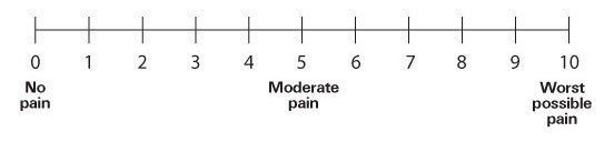
Simply said, take into consideration where your pain level falls the majority of the time�unless you experience extreme pain changes.
No Pain
0 � Painfree
Manageable Pain
1 � Pain is quite mild, barely noticeable. You don’t think about it.
2 � Small pain. Annoying and may have occasional twinges that are stronger.
3 � Pain distracting and is noticeable, you may get used to it and adapt.
Moderate�Pain�Disrupts Regular Day-To-Day Living Tasks
4 � Moderate pain. If you should be deeply in an action, it may be blown off to get a time frame, but is diverting.
5 � Moderately strong pain. It can’t be dismissed for more than a few minutes, but you still can manage to work or participate in some social activities.
6 � Rather strong pain that interferes with normal daily activities. Difficulty focusing.
Severe�Pain�Disabling; Debilitating, Reduces Daily Quality Of Life, Cannot Live Independently
7� Severe pain that dominates your senses and significantly restricts your capability to perform ordinary daily tasks or maintain social relationships. Interferes with sleep.
8� Intense pain. Physical action is seriously limited. Conversing requires great exertion.
9. Not able to converse. Weeping outside or moaning uncontrollably.
10� Unspeakable pain. Perhaps and bedridden delirious. Mobility may be undermined.
�My Pain Feels Like��
Most of the time, patients experience one or two consistent pain �feelings� but some can experience a variety of sensations.
The most common pain kinds are:
- Sharp stabbing pain
- Extreme heat or burning sensation
- Extreme cold
- Throbbing, inflamed tissue
- Susceptibility to contact / touching
- Itching
- Numbness, tingling, pins & needles
Create A Pain Journal

I motivate patients or their family members to document a weeklong pain cycle till they meet with chiropractic, their pain management or alternative medicine team.
Additionally, jot down any treatments or activities�that lessen or increase your discomfort.
As an example, maybe you have discovered that hot showers or cold weather allows you to feel worse, but exercise or Epsom salt baths makes the pain more manageable.
If you come prepared with all this information, your time with all the physician can be spent focusing on next steps and also a treatment plan, rather than a lengthy Q & A review of the information supplied here.
More importantly, addressing these issues beforehand will ensure your physician receives up-to-date, higher quality information.
Consequently, your case could be assessed more quickly and a pain management plan can be placed into action to begin removing or reducing your suffering as rapidly and efficiently as possible.
Call Today!

by Dr Alex Jimenez DC, APRN, FNP-BC, CFMP, IFMCP | Chiropractic, Spine Care, Wellness
The understanding the spine is somehow involved in health and wellness, in addition to the practice of utilizing manual manipulation as a way to obtain healing, dates back to the time of the ancient Greek philosophers. In fact, Hippocrates once said, “Get understanding of the back, for this is the requisite for several ailments.”
The First�Chiropractic Adjustment
Modern chiropractic, however, indicates its beginnings in the late 1800s, �when a Canadian living in the US, Daniel David Palmer, a self educated teacher and healer, performed the very first spinal manipulation on a patient.
That patient was Harvey Lillard, a janitor who worked in Palmer’s building. Lillard was almost totally deaf and mentioned to Palmer that he lost his hearing many years before when he was bending over and felt a “pop” in his upper back.
Palmer, who had been a practitioner of magnet therapy (a common therapy of the time) was fairly learned in physiology and incredibly interested in how a spine interacts with all the remainder of the entire body’s systems.
He found a difficulty with one of his vertebra and examined Lillard’s back. Palmer manipulated Lillard’s vertebra and an amazing event occurred�Lillard’s hearing was restored. Today, this process is referred to as a chiropractic adjustment.
Palmer soon found that alterations could alleviate patients’ pain as well as other symptoms. These problems with vertebrae are called chiropractic subluxations.
He started to use these “hand treatments” to treat many different ailments, including sciatica, migraine headaches, stomach complaints, epilepsy, and heart trouble. In 1898, he started the Palmer School & Infirmary in Davenport, Iowa, and began teaching his chiropractic techniques.

Initial Resistance In The Medical Community
The medical community failed to immediately embrace techniques and Palmer’s chiropractic theories. The called him a “quack” and refused to recognize his achievements. At one point, Palmer spent time in jail because of his violation and was indicted for practicing medicine with no license.
Research has shown that Palmer was not the fish monger that was unlearned that some in the health care profession claim. An investigation of this library, which he quoted in his letters, showed that he was up to date in his knowledge at the turn of the 20th century. Obviously, his theories, in the light of 21st century knowledge, seem uneducated.
Chiropractic Today
Today, chiropractors are licensed in most European countries, Canadian provinces, all the US states, Australia and New Zealand. There tend to be more than 50,000 practicing chiropractors in the US alone . Despite its North American roots, there are more chiropractic educational programs beyond North America.
Call Today!

by Dr Alex Jimenez DC, APRN, FNP-BC, CFMP, IFMCP | Athletes, Complex Injuries
The majority of clients that present to the clinic with anterior knee pain over the coming year will more often than not have a patellofemoral (PF) problem.
It may be a slight bit of biomechanical mal-alignment that has stirred up the knee cap – this is the good, or they may have started to wear the cartilage behind the knee cap and as a result it has softened – chondromalacia � this is the bad. They may even have worn a hole into the knee cap cartilage and they now have a chondral defect, or worse still an osteochondral defect – the downright ugly.
These problems affect runners, cross fitters, group exercise enthusiasts (PUMP classes) and simple recreational walkers who spend a lot of time on hills and stairs.
How These Extremes Are Managed Will Differ
The biomechanical irritations and the chondromalacia versions can be managed conservatively with a combination of local treatment modalities and correcting the biomechanical faults. The more serious chondral/osteochondral defects often need some surgical intervention as often the pathology is too advanced to respond to conservative treatment alone.
Understanding the exact mechanical contributions of the knee cap in relation to the femur is critical for the therapist to effectively manage these problems.
At the local PF level, the fault is usually a malposition of the patella in the femoral trochlear groove. Often the knee cap is being pulled too far laterally and superiorly in the groove, creating an uneven contact situation between the knee cap and the femur. The PF compression force during loaded knee flexion (squats, lunges etc.) is no longer optimal and usually a smaller portion of the patella cartilage is taking all the load. This wears the cartilage down and creates pain and pathology. This is most noticeable as the knee flexes to 30 degrees and onwards as it is this knee flexion angle where the knee cap enters the femoral trochlear groove.

The more distant (but often dominant) faults lie at the hip/pelvis and at the feet. Below is a breakdown of common biomechanical faults that may contribute to PF pain syndromes.
1. Overpronation
If the foot pronates (rolls in) for too long or too much, the pronated midfoot forces the tibia to remain internally rotated. The femur follows the tibia and also internally rotates. This creates a mal-alignment at the knee whereby the PF arrangement is altered and the knee cap shifts laterally. We are all familiar with the Q angle of the knee and how this affects the PF alignment.
Common causes of overpronation may be structural flatfoot problems that can be corrected with orthotics and shoe selection. However, tight soleus (that limits dorsiflexion) or a tight and overactive peroneal system that everts the foot and flattens the foot can also be a cause.
Stretching and loosening the soleus and peroneals along with strengthening the anti-pronation muscles such as tibialis posterior, flexor hallucis longus and flexor digitorum longus may help fix this problem.
http://secure.newsletters.co.uk/sportsinjurybulletin/image/overpronation…
2. Hip Joint FADDIR
FADDIR represents a flexed, adducted and internally rotated hip joint at foot strike. This is often caused by tight and overactive hip flexors such as TFL and the adductors and weakness in the abductors (gluteus medius) and external hip rotators (gemellus, obturator muscles). This hip posture forces the femur to roll inwards and as a result the knee is deviated medially and away from the vertical line drawn up from the foot. This also increases the Q angle and PF misalignment results and perpetuates the local knee imbalance of tight and overactive lateral quadriceps and lateral hamstrings along with ITB tightness. As a result the VMO weakens.
Loosening the overactive TFL, adductors, lateral quad, ITB and lateral hamstring whilst strengthening the gluteus medius, hip external rotators and VMO may help this biomechanical mal-alignment.
3. Pelvic Trendelenburg
Defined as lateral pelvic shift whereby at stance phase the opposite side of the pelvis drops down below the height of the pelvis on the stance side. This is usually caused by a weak gluteus medius complex that is unable to hold the pelvis stable during stance phase. The implications again are that this causes the knee to roll in and increase the Q angle. The solution is to muscle up the gluteus medius.
http://secure.newsletters.co.uk/sportsinjurybulletin/image/pelvictrendel…
4. Hip Flexor To Extensor Imbalance
This often forgotten about imbalance creates a situation whereby the individual finds it difficult to attain hip extension at the end of stance phase. The hip remains locked in a degree of flexion.
The knock on effect is that the knee also stays locked in some flexion. With the knee in flexion, the knee cap is now compressed against the femur, compression on the underside of the kneecap may result. To fix this the therapist needs to stretch/loosen the hip flexors and strengthen the gluteus maximus to promote more hip extension.

Call Today!
by Dr Alex Jimenez DC, APRN, FNP-BC, CFMP, IFMCP | Chiropractic, Chronic Back Pain, Lower Back Pain
Chiropractors think that good health is decided by way of a healthy nervous system, particularly a healthy spinal column. Sometimes, vertebrae become misaligned and put pressure on the nerves exiting the spinal cord. The misalignment of a vertebra is called a chiropractic subluxation.
Chiropractors use specific methods to return the vertebrae in their proper locations or muster them to allow them to go freely when subluxations happen. These techniques are called spinal manipulations or adjustments. During an adjustment, the vertebra is freed in the misaligned location and returned to the right place in the spinal column. The adjustment permits the entire body to cure and preserve homeostasis once performed.
Chiropractors Are Trained In Many Different Adjustment Techniques
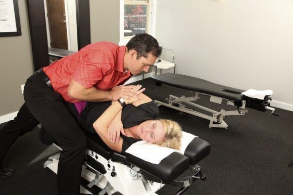
Some are done by hand; some necessitate using specialized instruments. Since each patient is different, your chiropractor will pick the best technique for the state. Nevertheless, don’t hesitate to ask the chiropractor which technique she or he will be doing and the way that it will be achieved.
Common Adjustment Techniques Employed By Chiropractors Are The Following:
- Toggle Drop – this is when the chiropractor presses down firmly on a specific part of the back. Subsequently, using a drive that is precise and rapid, the chiropractor aligns the spine. This really is accomplished to enhance mobility in the vertebral joints.
- Motion Palpation – this hand-on procedure is performed to determine in case your vertebrae are moving freely inside their normal planes of motion.
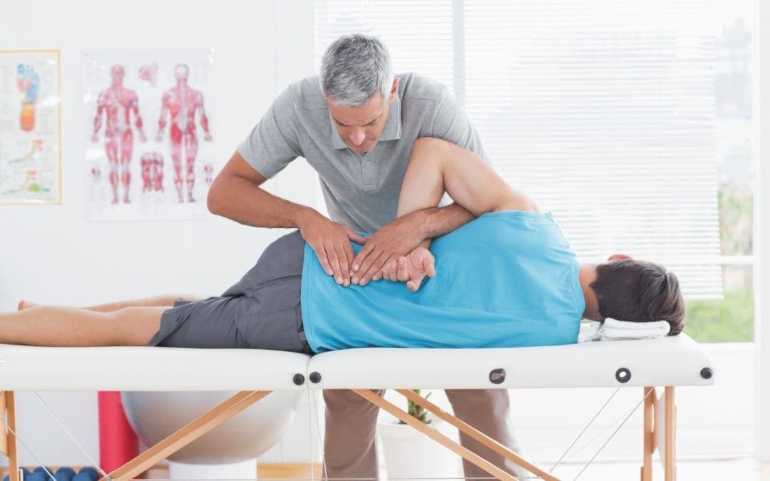
- Lumbar Spin -the chiropractor positions the patient on her or his side, then implements a thrust that is quick and precise returning it to its proper place.
- Release Work – the chiropractor applies gentle pressure using her or his fingertips to separate the vertebrae.
The chiropractor applies a quick thrust at once the table drops. The dropping of the table allows for a lighter adjustment without the twisting postures that can accompany the manual adaptation.
- Instrument adjustments – of correcting the spinal column frequently the gentlest ways. The patient lies on the table while a string is used by the chiropractor face down – filled activator instrument to do the adjustment. This technique is frequently used to perform adjustments on creatures too.
- Manipulation done under anesthesia (or twilight sedation) – this is performed by a chiropractor certified in this technique in a hospital outpatient setting when you’re unresponsive to traditional adjustments
Keep in mind that before you experience complete relief out of your symptoms you may really need to go back to the chiropractor’s office for additional adjustments.
Call Today!

by Dr Alex Jimenez DC, APRN, FNP-BC, CFMP, IFMCP | Chiropractic, Mobility & Flexibility, Power & Strength
I have been travelling through Athens and now Istanbul. My 11 year old is a Percy Jackson nut and has been filling me in with the who’s who of Greek mythology and I am learning Latin words every day. Quite an education!
I looked up the word syndesmosis and the Latin translation is “(New Latin, from Greek sundesmos) bond, ligament, from sundein, meaning to bind together”. As sports injury professionals, we know syndesmosis to be the joint articulation between the tibia and the fibula bones around the ankle. These two bones are �bound’ together with very firm and strong ligaments.
Syndesmosis comes to mind after I saw a girl sprain a syndesmosis at the Archaeological Museum in Istanbul today. This poor girl was preoccupied by the hundreds of cats and kittens running all over the place and did not see the uneven cobblestones on which she placed her foot. At the same time, she turned to change direction. This is a common mechanism of injury for a syndesmosis – a forced dorsiflexion and rotation on a fixed foot.
Rehab Masterclass Issue 140 Of Sports Injury Bulletin
Of all the ankle injuries, injury to the syndesmosis is the biggest pest to sports physios and the like. And unlike simple garden variety ankle sprains that heal quickly, the syndesmosis takes a LONG time to heal properly. If you deal with athletes that are susceptible to syndesmosis sprains, I’m sure you will agree that these are harder injuries to manage because of the severe consequences if done badly.

I go into a fair bit of detail in my Sports Injury Bulletin piece about syndesmosis injuries, detailing how they happen, how to identify them and then manage them. What I would like to highlight here are the implications of mismanaging a syndesmosis sprain.
In the current issue of The Journal of Sports and Physical Therapy, a group of Japanese researchers discovered that individuals who had chronic ankle instability (CAI) had a distal fibula that was positioned more lateral compared with healthy individuals with no CAI. In effect, those who had suffered serious syndesmosis injuries in the past and ended up with a wider distance between the fibula and the tibia, suffered more ongoing ankle pain than those without a tibfib separation.
Research shows that even a 1mm displacement of the talus within the mortise (due to a wider placed fibula) can reduce the contact area in the talocrural joint by 42% (Ramsey and Hamilton 1976). Mismanaged syndesmosis injuries, resulting in an excessive amount of opening, can lead to early onset arthritic changes and chronic ankle instability. The talus bone bounces around in the now wider tibfib articulation.
A Widening Of The Fibula Is Due To One Of The Following:
Poor initial management, whereby the athlete is allowed to weight bear too early and this weight bearing forces the fibula away from the tibia as the syndesmosis ligaments are trying to heal.
The degree of damage is so severe that proper tightening of these ligaments is not possible without surgical intervention such as a screw or similar being placed between the two bones to �force’ them together.
The key for a sports injury practitioner, is to properly identify a regular ankle sprain from a more serious syndesmosis injury. If you get this part wrong and allow the athlete to get back to weight bearing too early, then expect some complaints about a chronically painful ankle some time down the track.

Call Today!
Kobayashi et al (2014). �Fibular malalignment in individuals with chronic ankle instability.’ JOPST. 44(11); pp 841-910.
Ramsey and Hamilton (1976). J Bone and J Surgery Am. 58(3); 356-357.

by Dr Alex Jimenez DC, APRN, FNP-BC, CFMP, IFMCP | Chiropractic, Chronic Back Pain, Migraines, Neck Pain
About Pain Management (Medicine) Specialists
A pain medicine specialist is a medical or osteopathic doctor who treats pain due to disease, ailment, or injury. Many of these doctors are physiatrists or anesthesiologists although called interventional pain management specialists or pain medicine. Pain medicine is a mutlidisciplinary team effort generally affecting specialists in other disciplines, complimentary alternative medicine, along with radiology, psychiatry, psychology, oncology, nursing, physical therapy, and the patient’s primary care physician or other treating doctor.

Education & Training
After graduating medical school and completing a one-year internship, the physician enters a residency program normally in physical or anesthesiology medicine but sometimes from other fields like psychiatry and neurology. Upon conclusion of a residency program (typically 3 years long), the physician completes a one-year fellowship for advanced training in pain medicine.
Many pain medicine specialists are board certified. The organizations that board certify physiatrists, anesthesiologists, neurologists, and psychiatrists all collaborate to provide the board examination for the subspecialty of Pain Medicine. You can find numerous opportunities for pain management specialists to remain current with medical and technical improvements in pain medicine, such as scientific journals and society meetings.
Targets of Pain Management
The goal of pain medicine is to handle severe or long-term pain by reducing intensity and pain frequency. Besides addressing pain problems, a multidisciplinary pain management program may address your functional goals for activities of day-to-day living. Overall, a pain medicine plan aims to give you a feeling of well-being, increase your level of action (including return to work), and reduce or eliminate your reliance on drugs.
Many Kinds of Pain Treated
Pain medicine specialists treat all sorts of pain. Severe pain is described sharp or as acute and may indicate something is wrong. The pain experienced during dental work is an instance of intense pain. Pain lasting 6 months or longer is defined as chronic. This type of pain varies from mild to serious and is consistent. Spinal arthritis (spondylosis) pain is frequently chronic. A good consequence is produced by uniting different treatments regularly although chronic pain is difficult to handle.
- Degenerative disc disease
- Facet joint pain
- Sciatica
- Cervical and lumbar spinal stenosis
- Spondylolisthesis
- Whiplash
What to Anticipate During an Appointment
Your appointment with a pain or interventional pain management practitioner is much like other doctor visits. Although there are many similarities, the focus is fast managing it, and on your pain, the cause or contributing factors.
Pain medicine physicians execute a physical and neurological examination, and review your medical history paying particular focus on pain history. You may be asked many questions about your pain
- On a scale from zero to 10, with 10 being the worse pain imaginable, speed your pain.
- When did pain start? When pain started, what were you doing?
- Does pain disperse into other regions of the body?
- Is its intensity persistent, or is it worse at different times of night or the day?
- What really helps to alleviate the pain? Why is pain worse?
- What treatments have you ever attempted? What worked? What failed?
- Would you take over the counter drugs, vitamins, or herbal nutritional supplements?
- Does one take prescription medication? If so, what, how much, and how frequently?
Most pain medicine specialists utilize a standardized drawing of the front/back of the body to let you mark where pain is sensed, as well as indicate pain spread and type (eg, light, sharp). You may be asked to complete the form each time you see with the pain physician. The finished drawing helps you to evaluate your treatment progress.
Accurate Analysis Key to Treatment
Pain medicine includes diagnosing origin or the cause of pain. Making the proper identification may include getting an X ray, CT scan, or MRI study to verify the reason for your neck pr back pain. When treating spine-associated pain (which may include arm or leg symptoms), other tests, like discography, bone scans, nerve studies (electromyography, nerve conduction study), and myelography could possibly be performed. The proper analysis is crucial to some favorable treatment plan.
Some spinal ailments and pain treatment requires involvement of other specialists, such as your primary care physician, neurosurgeon, orthopaedic surgeon, and practitioners in radiology, psychiatry, psychology, oncology, nursing, physical therapy, and complimentary alternative medicine. The pain medicine specialist may consult with and/or refer you to a neurosurgeon or orthopaedic spine surgeon to determine if your pain difficulty necessitates back operation.
Call Today!












