Neck pain that is not caused by whiplash or other trauma has a postural component as part of the underlying problem. Sitting atop the body, the health of the neck is subject to the �curvature of the spine below and the position of the head above.
See Chronic Neck Pain: What Condition Is Causing My Neck Pain?
The neck muscle pain can be caused by the following neck muscles becoming tight:Scalene muscles (three pairs of muscles that help rotate the neck)
Suboccipital muscles (four pairs of muscles used to rotate the head)
Pectoralis minor muscles (a pair of thin triangular muscles at the upper part of the chest)
Subscapularis muscles (a pair of large triangular muscles near each shoulder joint)
Levator scapulae muscles (a pair of muscles located at the back and side of the neck).
If the alignment of the head and spine is not optimal, the neck can be predisposed to injury and/or the degenerative effects of wear and tear over time.
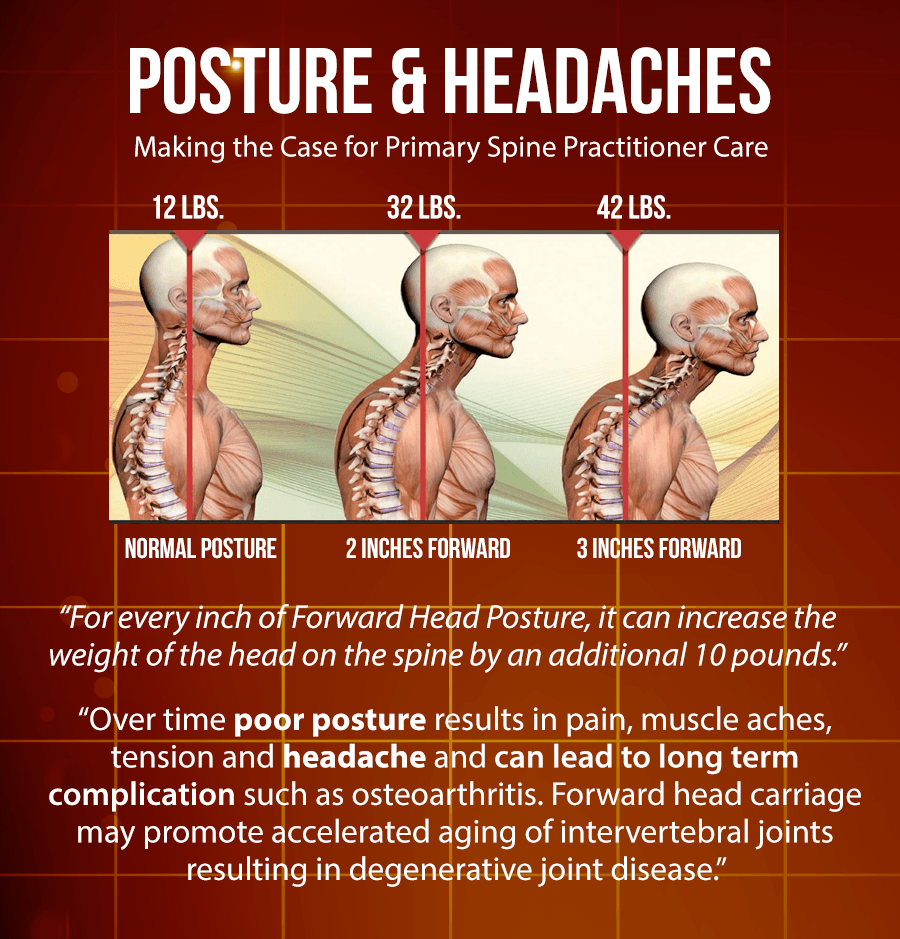
The most common condition that contributes to neck pain is forward head and shoulder posture.
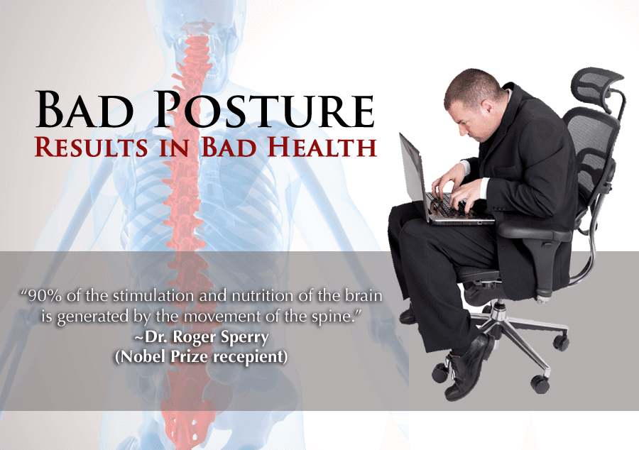
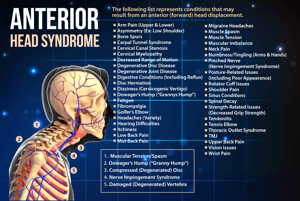
Forward head posture is when the neck slants forward placing the head in front of the shoulders.
This head position leads to several problems:
The forward pull of the weight of the head puts undue stress on the vertebrae of the lower �neck, contributing to degenerative disc disease and other degenerative neck problems.
Similarly, this posture causes the muscles of the upper back to continually overwork to counterbalance the pull of gravity on the forward head.
This position is often accompanied by forward shoulders and a rounded upper back, which not only feeds into the neck problem but can also cause shoulder pain.
The more time spent with a forward head posture, �the more likely it is that one will develop neck and shoulder problems.
Poor Posture Effects On the Lower Cervical Vertebrae
The part of the neck that is particularly vulnerable to forward head posture is the lower part of the neck, just above the shoulders.
See Cervical Spine Anatomy and Neck Pain
The lower cervical vertebrae (C5 and C6) may slightly slide or shear forward relative to one another as a result of the persistent pull of gravity on a forward head. This shear force can be a problem for patients with jobs that require them to look down or forward all day, such as pharmacists who spend many hours counting pills or data entry workers who look at a computer screen.
Prolonged shearing of the vertebrae from forward head posture eventually irritates the small facet joints in the neck as well as the ligaments and soft tissues.
This irritation can result in neck pain that radiates down to the shoulder blades and upper back, potentially causing a variety of conditions, including:
Trigger points in the muscles, which are points of exquisite tenderness that are painful to touch, along with limited range of motion
Disc degeneration problems, which may potentially lead to cervical degenerative disc disease, cervical osteoarthritis, or a cervical herniated disc.
BRAIN BASED POSTURAL CORRECTION
The neurologic system controls and coordinates all other systems of the body? What system provides the physical framework for all other systems of the body to resist gravity and function within our environment? The Posture System. These intricate systems work together to control and coordinate all experiences and actions throughout our lives.
Postural Neurology is defined as the network of neural impulses to support proper functional alignment of the Posture System. The purpose of Postural Neurology is to develop plasticity of proper structural alignment through brain based treatment protocols.
Why implement Postural Neurology into your practice? Quite simply, every musculoskeletal problem is at some level a neurologic problem. Neurology dictates movement, structure, and the function of your Posture System, overseeing and refining every single movement that you do on a daily basis.
It is an outdated system of healthcare to look at one part of the body, the site of injury or pain, and make assumptions about that person�s health and ability to function.
Identifying the area of injured tissue, then creating a rehabilitation plan around that injuryis less than adequate. The new system is all about function and whole posture patterns.
Segmental Rehabilitation & Brain Based Postural Correction
If the patient says �I hurt here,� or you test a weak muscle �here.� It does not mean that the problem lives �here.� This is simply information telling us that there is pain or dysfunction that is being expressed in that end organ. There is absolutely no indication that the problem is in that tissue. The noxious stimulus being interpreted by the brain is nothing more than a sensory input. The weak muscular output is nothing more than an insufficient motor plan.
This is the difference between segmental and brain based thinking. Segmental healthcare professionals do not see beyond the �hurt� or �weak� segment, oblivious to compensation patterns that are dampening function in the rest of the body.
Compartmentalized thinking is not adequate for a system as dynamically developed and as functionally managed as the human body. It simply won�t cut it.
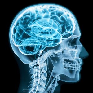
This guide introduces you to the Neurology of the Posture System, so you can understand the functional output of the brain and how it contributes to upright postural design.
Motor Cortex:�
The primary motor cortex is located in the frontal lobe of the brain.
The frontal lobe is the center of human development including executive decision-making, language, and problem-solving.
Located within the Motor cortex is the Motor Homonculus. The Motor Homonculus is a map of motor output for each part of your human anatomy. Within this map, the amount of cortex devoted to any given body region is proportional to how richly innervated that region is, not to the body region�s physical size. Areas of the body with greater or more complex sensory or motor connections
are represented as larger in the homunculus such as the hands, lips, and the face.
The primary motor cortex receives signals from the pre-motor cortex to make a movement.
The primary motor cortex then sends the information to make that movement from the cortex down the spinal cord to the skeletal muscle of that body region.
Motor movement occurs contralaterally, meaning that your left motor cortex controls movement on the right side of your body. Your right motor cortex controls movement on the left side of your body. Every action occurs in this fashion, in response to a descending signal from the brain to the body.
Is the Motor Cortex important to Posture?
The motor cortex controls motor function. The Posture System is never actually �static,� we are dynamic beings who are always in motion. The motor cortex is constantly making small movements (even minute movements) necessary to prevent falling over, and to interact within our environment. Optimal dynamic posture begins in the motor cortex and is regulated by the cerebellum.
Can I check for weakness of the Motor Cortex?�
You can check for weakness�of motor output by performing muscle tests. If a muscle on the left side is weak,
this is information to do further muscle tests to confirm for weakness of the right motor cortex.
�Sensory Cortex:
The somatosensory cortex of the parietal lobe is the center for
sensation. This is where you perceive your world and how you process all of the tactile and
proprioceptive information received from your external environment.
Just as the motor cortex is a map whose arrangement gives preference to the highly innervated parts of the body, so does the sensory homunculus. The sensory homunculus is the primary mechanism of cortical processing for sensoryinformation originating at body surfaces and other tissues.
Is the Sensory Cortex important to Posture?
The sensory cortex is very important for posture because the sensory cortex lights up with activation from novel stimuli such as proprioceptive signals and mechanoreceptor feedback
from complex movements. Lack of movement is the worst thing for your sensory cortex. In fact, with lack of stimulation to an area of the sensory cortex, the sensory map �blurs� together with less representation of that body part in the brain.
For example, when patients present an injured knee and they have worn a brace that limits their range of motion. This limited motor output of the knee results in less tactile and proprioceptive
feedback from the knee, the brain then thinks that the knee is �less important� and blurring of the cortical representation of the hip and ankle will begin to occur over the knee.
Less awareness of our body leads to poor activation and neglect of that area. To keep your patients healthy, keep them moving and activating all joints of the body.
Can I check for weakness of the Sensory Cortex?
You can check for dysfunction of the sensory cortex by performing sensory tests such as light touch,
vibration, temperature, painful stimuli, and joint position. If the patient presents with decreased sensory recognition, the contralateral sensory cortex is weak.
Meaning, that if the left side of the body cannot detect tactile sensation, this is a weakness of the right sensory cortex.
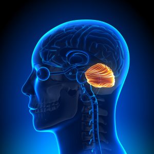
Cerebellum:
The cerebellum works in conjunction with the contralateral motor
cortex to coordinate fine movements. The cerebellum is an �inhibitor,� it reduces any extra or unnecessary motor movements to perform the desired action as accurately as possible.
The cerebellum receives input from sensory systems of the spinal cord and from other parts of the brain, and integrates these inputs to fine-tune motor activity, providing feedback to the motor cortex of how movements can be smoother and more precise. When patients have deficits of the cerebellum
they have excess movement, such as a tremor or a wide stance because they can�t balance with their feet together. Their equilibrium and ability to perform controlled motor movements is compromised.
Is the Cerebellum important to Posture?�
The cerebellum coordinates all movement, to more
precisely regulate fine movements. Proper cerebellar output means that the patient can perform their intended movement without recruiting other muscles. They are on target and on point with coordinated functions of the Posture System. Cerebellar deficits on the other hand affect the patient�s posture, they have compromised postural stability and uncoordinated dynamic postures.
Can I check for weakness of the Cerebellum?
You can check for cerebellar weakness by performing a Romberg�s test. Have the patient stand with their feet together, close their eyes, and see if they sway to one side. The patient will sway toward the side of cerebellar dysfunction.
Brainstem:
The brainstem is the center of postural control. The brainstem is made up of the midbrain, the pons, and the medulla and is the house of the nuclei of 10 of 12 of Cranial Nerves.
Each of the cranial nerves provides important sensory and motor functions for the body. Of particular importance to the Posture System are the visual and vestibular nuclei that reside in the brainstem.
The visual system controls your orientation in space, literally how you see the world. Visual fibers descend to the cervical spine controlling head posture. Visual deficits result in forward head posture, lateral head tilt, and head rotation.
The vestibular system controls balance and extension. Flexor dominant posture is weak posture; upright extended posture is healthy and optimal for better function. This system is of utmost importance for upright postural stabilization and balance.
The brainstem also controls involuntary systems of your autonomicnervous system that regulate life-sustaining processes such as breathing, heart rate, sexual function, and digestion. Plus, the brainstem modulates postural tone, and inhibits flexion toward gravity.
Is the Brainstem important to Posture?
This is the primary control center of postural stabilization! The brainstem inhibits flexion, and in conjunction with the vestibular system activates extension. Descending fibers from the cranial nerve nuclei of the eyes control your head posture, and the ability to keep your eyes parallel to the horizon during dynamic movement.
Understanding the brainstem is an important connection for all posture professionals. Your posture depends upon the functional output of the brainstem.
Can I check for weakness of the Brainstem?
Checking posture provides valuable insight into the function of the brainstem. Noted flexion of the Posture System or head posture distortion patterns indicates that there is weakness of the brainstem.
The Cranial Nerves also provide valuable insight of the function of the brainstem. Dysfunction of the cranial nerves means that there is ipsilateral weakness of the brainstem.
Spinal Pathways:�
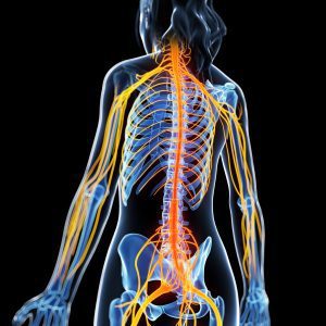
The spinal chord and its pathways are the communication system to and from the brain and the body. The descending motor pathways stimulate movement and postural control. The ascending sensory pathways carry information of sensation to be processed and �understood� in the brain.
The brain and the body are in constant communication to perform efficient movements, to stabilize without falling, and to detect signals from the environment that require a response. Sensorimotor integration happens in your spine, this is the communication highway of the nervous system.
Are the Spinal Pathways important to Posture?
The pathways are invaluable to your postural design. Pathways from the motor cortex send information to create dynamic movements,�and pathways from the brainstem descend to stabilize your postural tone. Ascending pathways from the body to the brain ignite the sensory homunculus for perception, and carry information from the primary sensors of your body for proprioception, vision, hearing, touch, smell, taste, etc.
Can I check for weakness of the Spinal Pathways?
There are many different pathways. When you understand the function of that pathway you can design a test for it. For example, the corticospinal tract descends from the motor cortex to the spine
to create motor output. Dysfunction of this pathway would present as weakness on muscle tests.
The spinothalamic tract is an ascending tract from the spine to the thalamus that recognizes stimuli associated with pain and temperature. To test this tract you would test sensory integration of hot, cold, and painful stimuli.
Neck pain that is not caused by whiplash or other trauma has a postural component as part of the underlying problem. Sitting atop the body, the health of the neck is subject to the �curvature of the spine below and the position of the head above.�For Answers to any questions you may have please call Dr. Jimenez at�915-850-0900
Injuries to the muscles and ligaments around the hip affect both competitive and recreational athletes. These injuries can interfere significantly with sports enjoyment and performance levels, and they occasionally will end participation completely. Excessive pronation and poor shock absorption have been found to be an underlying cause or a contributing factor for many leg injuries. Functional orthotics which have been custom-fitted to improve the biomechanics of the feet and reduce the extent of pronation can help to prevent many sport-related leg injuries.
Lower Extremity Problems in Athletes
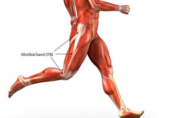
One study looked at the foot biomechanics of athletes who reported a recent foot or leg injury and compared them to an uninjured control group. The researchers determined that those athletes with more foot pronation had a much greater statistical probability of sustaining one of five leg injuries, including iliotibial band syndrome (which is due to excessive tightness of the hip abductor muscles).
This study helps us understand how providing appropriate functional foot orthotic support to patients who are involved in sports or recreational activities lowers their likelihood of developing both traumatic and overuse hip injuries.
In this paper, sixty-six injured athletes who ran at least once a week, and who had no history of traumatic or metabolic factors, were the study group. Another control group of 216 athletes were matched who did not have any symptoms of lower extremity injuries. The amount of pronation during standing and while running at �regular speed� was determined by measuring the angles of their footprints. The investigators found a significant correlation: Those athletes with more pronation had a much greater likelihood of having sustained one of the overuse athletic injuries.
Hip and Thigh Injuries
Many injuries experienced at the hip develop from poor biomechanics and gait asymmetry, especially when running. Smooth coordination of the muscles that provide balance and support for the pelvis is needed for optimum bipedal sports performance. This includes the hamstring muscles and the hip abductor muscles, especially the tensor fascia lata (the iliotibial band). When there is a biomechanical deficit from the feet and ankles, abnormal motions (such as excessive internal rotation of the entire leg) will predispose to pulls and strains of these important support muscles. The hamstrings (comprised of the biceps femoris, semimembranosus, and semitendinosus muscles) are a good example.
During running, the hamstrings are most active during the last 25% of the swing phase, and the first 50% of the stance phase. This initial 50% of stance phase consists of heel strike and maximum pronation. The hamstring muscles function to control the knee and ankle at heel strike and to help absorb some of the impact. A recent study has shown a significant decrease in electromyographic activity in the hamstrings when wearing orthotics. In fact, these investigators found that the biceps femoris (which is the most frequently injured of the three hamstring muscles) had the greatest decrease in activity of all muscles tested, including the tibialis anterior, the medial gastrocnemius, and the medial and lateral vastus muscles. The scientists in this study theorized that the additional support from the orthotics helped the hamstrings to control the position of the calcaneus and knee, so there was much less stress into the hip joint and pelvis.
Excessive Pronation and Hip Injuries
Using functional orthotics to correct excessive pronation and to treat hip problems requires an awareness of the various problems that can develop. The following is a list of the pathologies that are seen in the hip and pelvis secondary to pronation and foot hypermobility:
Iliotibial band syndrome����������������� Tensor fascia lata strain
Trochanteric bursitis����������������������� Hip flexor muscle strain
Piriformis muscle strain������������������ Hip adductor muscle strain
Hip joint capsulitis��������������������������� Anterior pelvic tilt
These conditions will develop much more easily in athletes, who push their musculoskeletal systems, and who seek more efficient and effective functional performances.
In 2002, researchers at Logan College of Chiropractic recruited a total of 40 male subjects that demonstrated bilateral pes planus or hyperpronation syndrome. Subjects were cast for custom made orthotics; their right and left Q-angles were measured with and without the orthotic in place. Thirty-nine of 40 test subjects showed reduced Q-angle, which was in the direction of correction, suggesting that wearing orthotics can improve stability and levelness of the pelvis, thus protecting the body to some degree from hip injury.
Conclusion
Excessive pronation and/or poor shock absorption have been shown to be an associated or causative factor in many leg injuries � from the foot itself, up the lower leg to the knee, thigh, and into the hip joint. The good news is that many of these conditions can be prevented with custom-fitted functional orthotics. Evaluation of foot biomechanics is a good idea in all patients, but is especially necessary for those who are recreationally active, or for anyone who has experienced hip problems.
To avoid potentially disabling hip injuries, competitive athletes must have regular evaluations of the alignment and function of their feet. Additional preventive measures include wearing well-designed and solidly-constructed shoes. When athletes are provided with custom-fitted functional orthotics, it can help prevent arch breakdown and biomechanical foot problems, and also treat numerous injuries of the lower extremities, including the hip joints.
Many injuries experienced at the hip develop from poor biomechanics and gait asymmetry, especially when running. Smooth coordination of the muscles that provide balance and support for the pelvis is needed for optimum bipedal sports performance. For Answers to any questions you may have please call Dr. Jimenez at 915-850-0900
How a chiropractic adjustment works: A displaced or luxated vertebra is when the joint surfaces are completely separated. Subluxation is a partial or incomplete separation: one in which the articulating surfaces remain in partial contact. This latter condition is so often referred to and known by chiropractors as sub-luxation.
The relationship existing between bones and nerves are so nicely adjusted that anyone of the 200 bones, more especially those of the vertebral column, cannot be displaced ever so little without impinging upon adjacent nerves. Pressure on nerves excites, agitates, creates an excess of molecular vibration, whose effects, when local, are known as inflammation, when general, as fever.
A subluxation does not restrain or liberate vital energy. Vital energy is expressed in functional activity. A subluxation may impinge against nerves, the transmitting channel may increase or decrease the momentum of impulses, not energy.
�According to BJ Palmer, a subluxation represents a displaced bone that’s impinged on a nerve, which interferes with the transmission of vital nerve energy (or, more specifically, �mental impulses.�)�
For over hundred years, chiropractors have explained chiropractic medicine by teaching patients and the medical community that bones compress which irritate spinal nerves. The ensuing nervous system dysfunctions have negative effects on the function of peripheral nervous systems, central nervous systems and patients� overall ability to maintain homeostasis.
Essentially, they go into states of dis-ease.� These discussions were in large part due to the teachings of D.D. Palmer and B.J. Palmer as previously cited. Based on the results rendered in chiropractic offices across the country and in a patient-driven model of success, the general consensus in both private practice and chiropractic academia had been to maintain status quo and simply teach what has worked in the absence of conclusive evidence, particularly in light of a lack of serious governmental funding and support for chiropractic research.� In addition, dogma has also created blinders for many, as evidence evolves to further chiropractic and its understanding, application and expansion.
Research has been published and has begun to verify D.D. and B.J. Palmer�s hypotheses were correct. While clarifying the specific physiological mechanisms related to chiropractic�s ability to alleviate pain.� As a result of initially studying pain mechanisms, contemporary research has also begun to set the foundation for understanding why chiropractic works with systemic and autonomic dysfunction and potential disease treatment through the adjustment � central nervous system connection.
It is the understanding of that connection with pain that is helping people to begin to understand the full impact of the chiropractic spinal adjustment and render the evidence to help more get well.
CENTRAL NERVOUS SYSTEM & PAIN REDUCTION
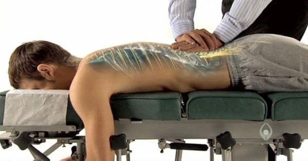
Reductions in pain sensitivity, or hypoalgesia, after spinal manipulative therapy or a chiropractic adjustment may point to a mechanism related to the central nervous system processing of pain.�These findings indicate that a chiropractic spinal adjustment affects the dorsal horns at the root levels which are located in the central nervous system.
This is the beginning of the �big picture� since once we identify the mechanism by which we can positively influence the central nervous system, we can then study that process and its effects in much more depth.
One question asked, does a chiropractic adjustment cause a general response of pain sensitivity or if the response is specific to the area where adjustment is applied? For example, changes in pain sensitivity over the cervical facets following a cervical spine SMT would indicate a local and specific effect while changes in pain sensitivity in the lumbar facets following a cervical spine SMT would suggest a general effect.�
We observed a favorable change for increased PPT [pressure pain threshold] when measured at remote anatomical sites and a similar, but non-significant change at local anatomical sites. These findings lend support to a possible general effect of SMT beyond the effect expected at the local region of SMT application.
The adjustment mechanism is theorized to result from both spinal cord mediated mechanisms and the brain. A recent model of the mechanisms of manual therapy suggests changes in pain related to SMT result from an interaction of neurophysiological responses related to the peripheral nervous system and the central nervous system at the spinal and supraspinal level.
This demonstrates that�chiropractic adjustment influences the peripheral nervous system and the central nervous system.
ACTIVATION OF BRAIN & DESCENDING NERVE PATHWAYS BEYOND AREAS TREATED
CHIROPRACTIC ADJUSTMENT VS. SPINAL MOBILIZATION
Manual therapy has clinically shown to boost pressure pain thresholds (i.e., decrease sensitivity) in both symptomatic and asymptomatic subjects. Cervical spinal manipulation has been shown to result in unilateral as well as bilateral mechanical hypoalgesia.
Compared with no manual therapy, oscillatory spinal manual therapy at T12 and L4 produced significantly higher paraspinal pain thresholds at T6, L1, and L3 in individuals with rheumatoid arthritis.
The immediate and widespread hypoalgesia associated with manual therapy treatments has been attributed to alterations in peripheral and/or central pain processing including activation of descending pain inhibitory systems.
Manual therapy triggers the central nervous system which affects areas around those being treated.�Clinical studies show spinal manipulation or an adjustment alters the central processing of mechanical stimuli. This is proven by an increased pressure pain threshold and decreased pain sensitivity in asymptomatic and symptomatic subjects following manipulation.
The thalamus also regulates states of sleep and wakefulness which plays a major role in regulating arousal, the level of awareness, and activity (Thalamus�http://en.wikipedia.org/wiki/Thalamus).
Therefore, a�chiropractic adjustment reduces pain through the thalamus. �In addition, with current knowledge of chiropractic adjustment effecting the thalamus, we can begin to offer an explanation of how the first historically reported chiropractic adjustment by D.D. Palmer helped Harvey Lilard regain his hearing.
CHIROPRACTIC REDUCES PAIN IN MULTIPLE REGIONS DUE TO LOCAL AND CNS STIMULATION
Significant changes were observed on sensory parameters, supporting the hypothesis of centrally mediated effects of a single adjustment. This suggests that the�chiropractic spinal adjustment affects the nociceptors and the mechanoreceptors at the joint level causing central modulation of an effect at the cord and/or brain level(s) and pain reductions in multiple areas as a result.
AN ADJUSTMENT WILL CREATE HIGHER FUNCTION IN CORTICAL AREAS
MT or manual therapy/chiropractic adjustment reduce pain intensity and pain sensitivity. it is reasonable to assume that the underlying therapeutic effect of MT is likely to include a higher cortical component.
In other words, thoracic adjustments produced direct and measureable effects on the central nervous system across multiple regions, which in the case of the responsible for the processing of emotion (cingulate cortex, aka limbic cortex) are regarding the insular cortex which also responsible for regulating emotion as well has homeostasis.
The motor cortex is involved in the planning and execution of voluntary movements, the amygdala�s primary function is memory and decision making (also part of the limbic system), the somatosensory cortex is involved in processing the sense of touch (remember the homunculus) and, finally, the periaqueductal gray is responsible for descending pain modulation (the brain regulating the processing of painful stimuli).
Periaqueductal Gray � � � Ascending and descending spinothalamtic tracts carrying pain and temperature fibers
The global effects of a chiropractic adjustment simply put, the processing of pain may be modulated or regulated from an external force without the use of pharmacy or surgery? Pain is deeply tied to the most primitive regions of the central nervous system and it appears (as chiropractors have observed clinically for 116 years) that therapeutically speaking, we can have an influence on these higher centers with little or no side-effects.
Chiropractic spinal adjustments create functional changes in multiple regions of the brain. In addition, this has far reaching effects in setting the foundation for understanding how the adjustment works in systemic and possibly autonomic changes by being able to measure and reproduce functional changes within the brain as direct sequellae.
MUSCLE IMPAIRMENT CREATES CNS ALTERATIONS & THE�NECESSITY FOR BOTH SHORT-TERM & LONG-TERM�CHIROPRACTIC CARE
ADJUSTMENTS WORK � SPINAL MOBILIZATION DOES NOT
What this means is that there are observable changes in the function of the central nervous system seen in patients with musculoskeletal conditions.� That is something that chiropractors have observed clinically and shows the medical necessity for chiropractic care for both short and long term management as well as in the prevention of pain syndromes.
Evidence suggests that muscle damage occurs early in spinal complaints, and that such muscle damage does not automatically resolve even when pain symptoms improve. This has led some authors to suggest that the deficits in proprioception and motor control, rather than the pain itself, may be the main factors defining the clinical picture and chronicity of various chronic pain conditions.
This evidence suggests that chiropractic manipulation may have a positive neuromodulatory effect on the central nervous system, and this may play a role in the effect it has in the treatment of neck pain.
When the input signal is no longer encoded as a result of joint dysfunction and altered afferent input, the cerebellum must adjust to new encodings that dictate the body schema and affect proper execution of the motor task.
The significance is that it suggests that chiropractic adjustments improve neck dysfunction, and through plasty changes in the cerebellum, there is resultant motor learning and increased function.
CONCLUSION
Chiropractic adjustments influence the peripheral nervous system and the central nervous system. In the central nervous system, chiropractic spinal adjustments reduce pain by effecting the thalamus and descending central pain pathways.
The areas of the brain affected by chiropractic adjustments effect the following functions: emotions, learning, motivation, memory, consciousness, homeostasis, perception, motor control, self-awareness, cognitive function, voluntary movements, decision making, touch, temperature, pain of the skin- epithelial tissue-skeletal muscles-bones-internal organs and cardiovascular system.
For over hundred years, chiropractors have explained chiropractic medicine by teaching patients and the medical community that bones compress which irritate spinal nerves. People have begun�to understand the full impact of the chiropractic spinal adjustment and render the evidence to help more people get well.
The natural curve of the spine is reinforced by correct posture. Posture not only influences how you look but helps you breathe, improves concentration, supports vital organ function and stimulates overall well-being.
Ignoring harmful postural habits leads to illness, discomfort and pain, and increases the risk for pathogenesis of disease in the body. �You can improve your posture and nervous system function and improve your quality of life significantly.
At the bottom of this article, you will discover a video and narrative for�5 exercises that will help you correct defects and optimize your posture in minutes each day. �Much like we brush and floss our teeth for dental health, we need to work on our posture each and every day!
Spinal Curve Dictates Health
The body was built with a curve in the spine to provide support and balance to the musculoskeletal system. It is essential for preventing deformation to bones, joints, muscles and tendons. Correct posture protects against disc degeneration that can lead to inflammatory conditions and disease. It is also critical for protecting the central nervous system.
Postural experts�understand that poor posture weakens the body�s ability to defend itself against infectious and chronic diseases. �They also view poor posture as a marker for the presence of chronic illness. �Today, many posture focused doctors such as chiropractors advocate for health measures in schools to educate students about correct posture techniques to reduce the current posture epidemic brought on by modern technologies such as cell phones that force us into a forward head shift.
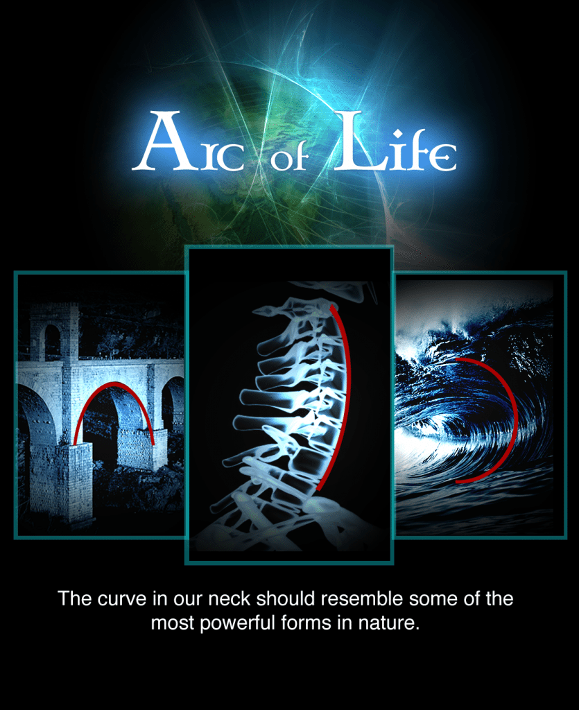
Doctors refer to the natural curve of the neck as the �arch of life.� This arch should have a 40-45 degree curve. �As the arch reduces it becomes unstable and results in a forward head shift, which depending upon the severity can add up to 30 pounds of additional weight on the spinal discs, ligaments and tendons.
This postural issue causes major stress on the�musculoskeletal system. Loss of the spine�s natural curve inhibits normal physiological and nervous system functions.� The ability to protect the brain stem and support the communication of nerve impulses throughout the body becomes suppressed.
Effects of Subluxation
Unfortunately, we are a modern age characterized by reoccurring activities that creates stress on the supporting spinal column. Slouching, crossing legs, cell phone use, and incorrect ergonomic practices at home, school and work causes poor posture. This leads to the abnormal curvature of the spine�and abnormal stress on the nervous system, which is known as subluxation.
Subluxation interferes with nerve impulses and can manifest in numerous physical symptoms.
Examples include:
Neuropathy
Back pain or neck pain
Chronic pain common in the hips, joints, lower back, pelvis and knees
Irritation of a specific area such as arm pain
Weakened immune system
Organ dysfunction
Inability to move or exercise normally
Dizziness and loss of balance
Loss of bladder or bowel control
Autoimmune conditions
Headaches�and migraines
Fatigue
Although our very own chronic habits lead to forward head posture, the spine is also susceptible to trauma from birth, regular physical activities from exercise and sports, as well as accidental injury from car accidents and falls.
Depending on the type of injury and which nerve pathways of the spine become disrupted, spinal subluxations�can increase the risk for weakened immunity and lowered quality of life.
Dangers of Forward Head Posture
Vanity is of least concern when it comes to the problems associated with forward head posture. As the head shifts forward, the thoracic spine moves and the weakening of the shoulder blades causes slouching and the appearance of hunchback. These harmful effects trigger the descent downward of vital organs in the chest. There is a decrease in lung capacity, a reduced flow of oxygen into the diaphragm and a lower�rate of oxygen reaching the cells.
A reduction in circulating oxygen in the body poses serious health threats. Oxygen is essential for survival because it maintains homeostatic functions including hormone balance, supports blood flow, protects the body from chronic disease and cancer, fuels nutrient absorption and protects the health and healing of cells, tissues and organs.
Studies have shown that forward head posture is an indication of poor health and is reported to cause the impairment of simple activities including walking or sitting comfortably. It is likely that physical limitations of the body marked by poor posture accelerate the aging process and increases inflammation. This is a result of the body�s inability to manage stress and tissue trauma appropriately.
Other evidence supports that the correction of forward head posture:
Alleviates symptoms of respiratory complications such as asthma
Increases breathing and lung oxygenation essential for physical fitness
Strengthens abdominal muscles
Restores enlarged tonsils
Improves glandular function in the head and neck
Improvements from Chiropractic Care:
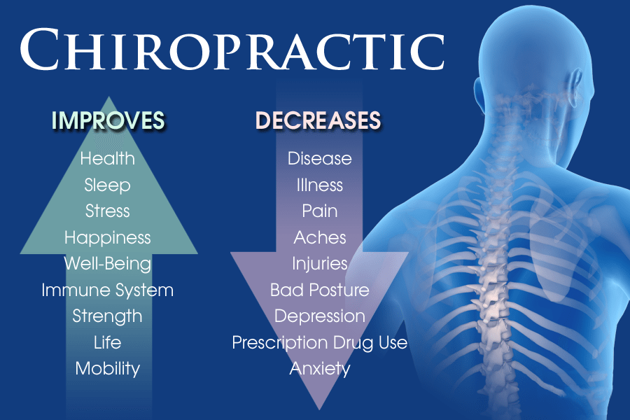
Chiropractic adjustments�can help compensate for postural abnormalities leading to an improvement in the health of the spine and the whole body. Research performed by Dr. Morningstar and Dr. Jockers found that chiropractic adjustments and rehabilitation exercises lead to the correction of forward head posture and cervical lordosis and restored pulmonary function.
Findings of the study also suggests that chiropractic care can improve the function of the autonomic nervous system by relieving tension that hinders the vital communication between the brain and body.
Corrective care chiropractors are able to identify abnormal features of the spine using x-rays, postural pictures, nerve scans and various technologies. Doctors then develop a specific care plan used to treat each individual�s concern focusing on chiropractic adjustments and rehabilitative techniques like the 5 exercises that follow.
5 Exercises:
In 30 to 90 days you can improve posture, breathe better, boost oxygenation throughout your body and alleviate tension and pain. Performing each of the following exercises for 1 minute twice a day will boost your body�s total healing potential, reduce your risk of developing degenerative disease and help you to thrive in life.
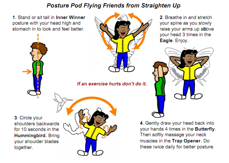
1) �Hummingbird:
Remove the slouch from poor posture and realign the head with the spine by practicing the hummingbird. This exercise strengthens the muscles between the shoulder blades, improves muscle fibers around the thoracic spinal column, opens up pectoral muscles and lifts the rib cage.
Instructions for Exercise:
Lift arms so that they are parallel to the floor.
Bend elbows and face palms forward to form a 90 degree angle between the bicep and forearm.
Rotate arms backwards in a circular movement while squeezing shoulder blades together.
Repeat for 1 minute.
2) �The Eagle:
With this exercise imagine opening up your arms just like an eagle spreads its wings. Stretching your arms overhead will open up the lungs. This boosts oxygen intake to stimulate tissue regeneration in the body and increase blood flow.
Instructions for Exercise:
Stand with feet shoulder-width apart.
Start with arms lowered and adjacent to sides.
Simultaneously lift arms above the head,
Pause for a moment; and
Lower arms back down to sides in a slow and controlled movement.
Repeat for 1 minute.
3) �Butterfly:
The butterfly is an extraordinary exercise to correct forward head posture. Performing this exercise regularly targets muscles in the neck and shoulders that give rise to chronic neck pain.
Instructions for Exercise:
Focus on lifting chest toward the ceiling.
Bring hands back against head so that thumbs point down. *Optional: If reduced flexibility hinders your ability to lift arms and hands behind head, perform exercise standing flat against a wall. You can also align back of head against car seat.
Use about 10% of strength to push head backward while keeping head straight.
Pause for approximately 10 seconds.
Relax and repeat for 1 minute.
4) �Chin Tuck:
An opposing exercise to the butterfly is the chin tuck. This exercise provides balance to the opposite core muscles in the neck by strengthening the deep neck flexors. Performing the chin tuck helps balance the head and neck reducing the occurrence of forward head shift.
Instructions for Exercise:
Focus on lifting chest toward the ceiling.
Maintain head position with ears over the neck.
Place 1 hand on forehead; and
Gently push forward with about 10% of strength (muscles in neck should contract and head should appear immobile).
Repeat for 1 minute.
5) �Trap Opener:
The trapezius muscles stabilize the shoulder blades providing support for head and neck movement. Habitual forward head posture pulls and weakens the shoulder blades which consequently also relocates the ribcage.
Unlike the previous four exercise, the trap opener is a favorite amongst patients and is not intended to stretch or strengthen the trap muscles. Rather, perform this exercise to release stress from the trapezius muscles and get rid of the so called �monkey� on your back.
Instructions for Exercise:
Relax shoulders.
Drop the chin towards the chest.
Roll head slightly to the right side.
Use right hand to massage trapezius muscles on the upper left side of the back.
Repeat exercise for opposite side.
Perform for 1 minute.
Summary:
Don�t ignore the signs of poor posture that can impact your ability to sleep restfully at night or concentrate clearly during the day. Poor posture has devastating effects on the body that can only be treated with mindful practices. Utilize these 5 exercises to improve your posture and reap its benefits reflected in overall health and well being.
Incorrect�postural habits lead to illness, discomfort, pain, and increases the risk for pathogenesis of disease in the body. �You can improve your posture, nervous system function and improve your quality of life significantly. Correct Posture not only influences how you look but helps you breathe, improves concentration, supports vital organ function and stimulates overall well-being. For Answers to any questions you may have please call Dr. Jimenez at 915-850-0900
Chiropractor, Dr. Alexander Jimenez looks at how sciatica can affect what you do and where you go.
Popular songstress hears her “Body Talk” and finds “Let’s Get Physical” challenging due to low back and leg pain.
In the event you suffer from sciatica, you may have to take time off work due to debilitating leg pain and low back. That�s just what occurred to Olivia Newton-John, who postponed her May 2017 performances because of sciatica flare up.
A statement posted on the star�s official Facebook page said, �We are so sorry to announce the Olivia concerts scheduled for May will be postponed due to a issue that is negative with Olivia’s sciatica.�
In a Facebook post that was separate, Newton John expressed disappointment at having to postpone the shows, but that the symptoms wouldn’t allow her to perform. �I have had a long running issue with sciatica, � she wrote. �I need to take this time with this very debilitating condition.�
Sciatica�s hallmark symptoms of intense low back and leg pain make it among the most well known spinal issues. Individuals who have sciatica frequently describe their pain as electrical-like as it runs in the buttock down and down the trunk of the thigh to the foot. Moving your legs often makes the pain worse performing all night as Newton-John her pain would have during her concerts may have greatly intensified.
Newton-John�s choice to take time to handle her sciatica is an excellent example for anyone who lives with acute low back and leg pain. Avoiding activities that aggravate your sciatica and focusing on mild activities and other treatments go a long way to lower your pain.
Keep Sciatica Pain From Increasing
By Staying Informed
Does one have problems with sciatica? SpineUniverse has many resources that will help you understand this sciatic nerve-associated pain and also the treatments that may assist you to will find relief:
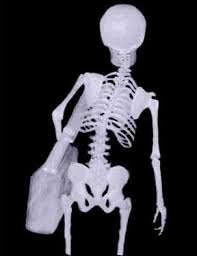
Soldiers routinely carry between 50 and 100 pounds of gear i.e. body armor, weapons, flack vests, rucksacks, and ammo.�This weight is directly affecting their spine and increasing spine related problems for military men and women. Carrying gear weighing over 10% of a persons body weight is known to cause damage to the spine and vertebral discs. It is no surprise that the main complaint from soldiers returning home would be neck & back pain, followed by hip and knee problems. The VA has seen over 396,000 veterans already for these musculoskeletal problems, which is roughly 31% of the returning soldiers. It is assumed many more have not sought medical care or have gone to private medical center.
The Spine Maximum Load Restriction
The spine is made to hold a human being up right, and allow freedom of movement. Like any structure there are �maximum load restrictions�. When we increase the weight placed on the spine by more than 10% of our normal body weight it begins to stress and degenerate the vertebral discs that act like the shock absorbers between the spinal vertebrae. If these discs become worn and degenerated the spine can no longer function correctly. Compensation and adaptation sets into the spine and other structures such as the hips, knees, and muscles of the back.
Posture Misalingment Will Degenerate The Spine & Cause Back Pain.
An excess amount of weight on the spine puts the body into a compromised postural position to balance the weight. This causes the postural distortions patterns, which are known to degenerate the spine and cause back pain. Soldiers are trained to handle extreme situations. They are �mentally tough� and physically fit, however the spine can�t be trained to withstand these extreme loads. Forcing ones body to maintain extreme stress and weight bearing begins to slowly deteriorate the spine.
Anybody Overloaded With Weight Will Damage The Spine
A soldier may be tough and made to deal with the pain and stress, however, as they age, these traumas to the spine weaken the spine reducing them to canes, wheel chairs, and in some cases in ability to even get out of bed. The pain is traumatic and the damage is done. Soldiers often experience the most extreme situation in excess weight bearing, however, many of our population is suffering from the same thing. According to the Center for Disease Control 70% of adults are overweight with 35% being considered obese. This excess weight in the form of fat is placing the same stress on societies spines as well.
Carrying 15 Pounds or More Can Lead To Spinal Disease.
A backpack, military gear, or body fat for a long period of time will damage the spine. To know for sure if you are developing these postural distortion patterns get a Posture Diagnosis Online from the American Posture Institute.
Military Health & Chiropractic
6 Bits of Advice
Habits
1. Reduce The Weight.
The most obvious way to change the problem is to reduce the amount of�weight you carry. Make purses, bags, and etc. lighter. Soldiers, look for options to reduce your load, or exchange items with lighter options.
2. Distribute The Weight Evenly
Extra weight should be evenly distributed on the body. Use two straps, one on both shoulders. Try to keep the weight as high up on the back as possible to reduce the stress on the low back. When possible use hip straps/support to help distribute the weight.
3. Maintain Proper Posture With Excess Weight.
Proper posture is crucial. Do not allow yourself to go into incorrect postural position. This will increase the stress on the muscles that protect your spine. Too much stress and these muscles give out and your spine takes the damage.
Rehabilitation
4. Strengthen The Postural Muscles
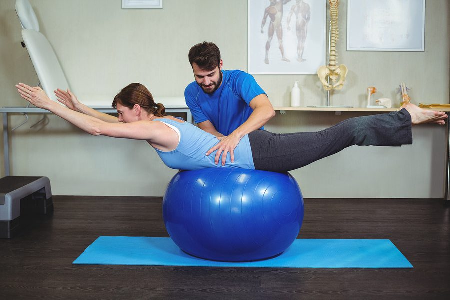
Core exercises will strengthen muscles that support the spine. Planking is an excellent exercise. Lay with your chest on the floor. Support your body weight on your toes and your elbows/forearms. Lift your body off the ground and maintain this position as long as possible.
5. Lose Weight.
Reduce extra body weight, which will remove stress from the spine and pressure from the vertebral discs.
Spinal Alignment
6. Spine Distraction TherapyThis therapy can be done at home to relieve stress on the vertebral discs. Lay with your upper body on a bed or soft surface. Let your legs hang off. The surface needs to be high enough that your knees don�t touch the ground. Allow gravity to pull your lower body down, which will expand the space between the vertebrae and allow the discs to return to a normal position.
Soldiers routinely carry between 50 and 100 pounds of gear i.e. body armor, weapons, flack vests, rucksacks, and ammo.
This weight is directly affecting the spine and increasing spine related problems for military men and women. Carrying gear weighing over 10% of a persons body weight is known to cause damage to the spine and vertebral disc.
For Answers to any questions you may have please call Dr. Jimenez at 915-850-0900
Carpal tunnel syndrome is a condition involving numerous symptoms affecting the hands and wrists. This painful condition is caused when the median nerve, found within the carpal tunnel of the wrist along with other tendons in the hand, becomes compressed.
The median nerve functions by controlling nerve sensations and impulses from the palm area of the fingers and the thumb as well as to the muscles in the hand, providing feeling and movement. When the tendons become irritated, causing inflammation and swelling, an impingement of the median nerve occurs, resulting in carpal tunnel syndrome. The pinched nerve may then develop a variety of symptoms most commonly associated to the condition.
Symptoms of Carpal Tunnel Syndrome
Trauma from an accident to the hand or wrist and/or overuse from repetitive activities, such as constant typing on a keyboard, are common reasons carpal tunnel syndrome develops. The added pressure begins to irritate and compress the median nerve, leading to the impairing symptoms of pain, among others.
The most common symptoms include pain, numbness, a tingling sensation, and weakness on the hands, wrists, and forearms. Pain and numbness in these areas is usually intense. The discomfort from these symptoms can affect an individual�s everyday lifestyles. Gripping objects may become difficult as carpal tunnel syndrome progresses over time due to loss of muscle strength. The tingling sensations radiating from the hand is often described as a feeling of �pins and needles� through the hands, wrists, and forearms.
IFM's Find A Practitioner tool is the largest referral network in Functional Medicine, created to help patients locate Functional Medicine practitioners anywhere in the world. IFM Certified Practitioners are listed first in the search results, given their extensive education in Functional Medicine








 Soldiers routinely carry between 50 and 100 pounds of gear i.e. body armor, weapons, flack vests, rucksacks, and ammo.�This weight is directly affecting their spine and increasing spine related problems for military men and women. Carrying gear weighing over 10% of a persons body weight is known to cause damage to the spine and vertebral discs. It is no surprise that the main complaint from soldiers returning home would be neck & back pain, followed by hip and knee problems. The VA has seen over 396,000 veterans already for these musculoskeletal problems, which is roughly 31% of the returning soldiers. It is assumed many more have not sought medical care or have gone to private medical center.
 absorbers between the spinal vertebrae. If these discs become worn and degenerated the spine can no longer function correctly. Compensation and adaptation sets into the spine and other structures such as the hips, knees, and muscles of the back.