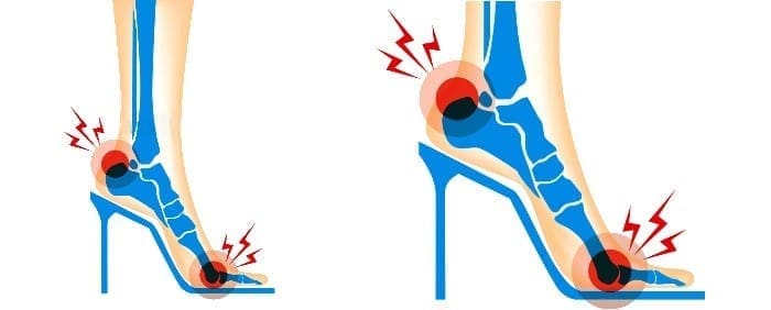
Unfortunately, there are no stretches for� Root canals or Kidney stones. But there are stretches for Piriformis syndrome.
These ailments are painful and no fun! Piriformis syndrome�is especially a pain, in the butt, no pun intended.
Seriously, people suffering from Piriformis syndrome have frequent and sometimes severe pain and numbness through the buttocks and down their legs. This occurs when the Piriformis muscle spasms. When this happens, it�can end up also aggravating the sciatic nerve, which compounds the pain with tingling and numbness.
The Piriformis�is a short, small�muscle deep inside our hips, and helps rotate our legs both outward and inward. Because of its proximity to the sciatic nerve, this little body part can cause big problems, and hinder our ability to run, or even walk, through our daily activities with ease.
Fortunately, there are a variety of exercises that help stretch and relax the Piriformis muscle, giving sufferers much-needed relief from the pain and numbness it causes. If you are dealing with Piriformis syndrome, try these stretches to get yourself back on your feet and moving, pain-free.
Stretches
The “Knee Up” Stretch
Lie on the floor, use a mat or thick rug for comfort, on your back. Stretch both legs out, with your arms to your sides.�Bend one of your legs at the knee and use your hand to pull it toward the opposite shoulder Hold for ten to thirty seconds.�Straighten out that leg, and then do the same motion with the opposite leg and shoulder.
The “Cross Arm” Stretch
Arrange yourself in a sitting position. The bottoms of your feet should be touching each other, with your arms crossed and your hands resting on the opposite leg. Push both knees down toward the floor until you feel the stretch inside your thighs. Hold the position for a count of 30, then relax for a few seconds and repeat up to five more times.
The “Standing” Stretch
This is a handy exercise you can do in the bathroom at work, in a hotel, or anywhere that you would rather not lie on the floor.
Begin in a standing position with both feet flat on the floor, with your feet a shoulder-length apart.��Don’t lock your knees, and keep your back straight.�Lift one knee up and grasp it with both hands. Make sure you keep the knee parallel to the corresponding hip. Use your hands to pull your knee toward the opposite shoulder until you feel a pull along the side of your buttocks. Hold up to one minute, or as long as you can balance. Repeat the action with the other leg. Try to do it three times for each leg.
The “Sit and Bend” Stretch
This is another convenient�exercise that doesn’t require getting on the floor. Choose a solid, straight-backed chair that doesn’t roll,�sit, and bring one leg across the other, resting your ankle on your other leg. Slowly lean forward until you feel a pull along the crossed leg. Hold for up to 30 seconds. Repeat with the other leg, stretching each side 3 times.
Remember that consistency is key. Perform these exercises at least once a day, every day, until your pain and numbness is gone.
These four simple exercises�help you make great strides in recovering from Piriformis syndrome. However, if you perform these for several days and still have pain, or experience pain while attempting the stretches, it is a good idea to make an appointment with a professional chiropractor. He or she will be able to evaluate your condition and offer a comprehensive treatment plan to treat the Piriformis so it doesn’t get worse, or cause additional issues with your sciatic nerve.
You try to stand up from a seated position and feel a stab of pain in your lower back. It may even shoot through your hip, buttock, or down the back of your thigh. The pain may even get worse then you walk uphill or sit for a long period of time. While these symptoms could mean a pinched nerve, lumbar disc herniation, hip bursitis, or degenerative hip disease, it could also be sacroiliac joint dysfunction.
What Is Sacroiliac Joint Dysfunction?
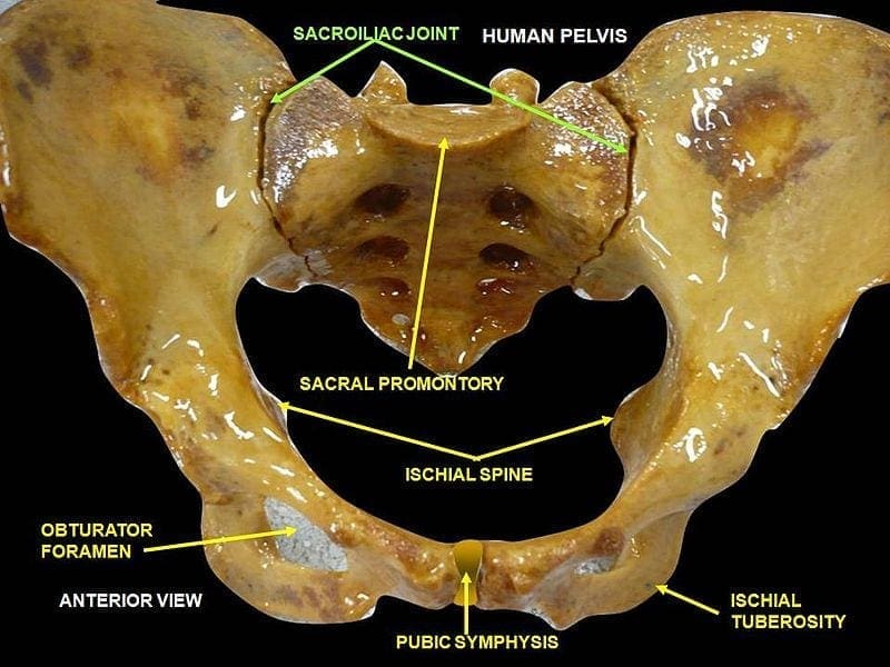
The sacroiliac (SI) joint is located in the pelvis. It is very strong as it is a weight bearing joint connecting the pelvis to the sacrum. It is surrounded by tough ligaments that reinforce it, providing added support.
There is an SI joint located on each side of the sacrum and they work together, moving as a single unit to act as a shock absorber for the spine and for transmitting force of the upper body. Just like any other joint in the body, the SI joint can be injured or diseased, causing it to become unstable and inflamed, causing pain and limited mobility.
What Causes SI Joint Inflammation?
While doctors have not established how the pain is generated, it is believed that it is due to a change in the normal motion of the joint. This could occur due to:
Hypermobility (Instability or Too Much Movement) � This can cause the pain to reside in the lower back. It can also be felt in the hip or both the hip and lower back and may even radiate into the groin.
Hypomobility (Fixation or Too Little Movement) � This can cause the pain to reside in the lower back or buttocks and may radiate down one leg, usually in the back of the thigh. It usually doesn�t reach the knee, but sometimes can even reach the ankle and foot. In this way, the condition mimics sciatica.
Sacroiliac joint dysfunction typically affects women who are young or middle aged. Older women and men are rarely affected although it does happen.
What Are Treatment Options For Sacroiliac Joint Pain?
When SI joint pain is initially diagnosed the treatment is usually fairly conservative. Medication, physical therapy, and injections are used by doctors for pain management.
NSAIDs and other similar medications decrease inflammation and reduce pain, while physical therapy can readjust the SI joint in cases where it is dislocated or immobilized. It also includes exercises that stabilize the joint for pain management over the long term.
Steroid injections directly into the sacroiliac joint can help with the reduction of inflammation and pain while making physical therapy more effective. When steroid injections are effective but the effects are temporary there is another non-surgical treatment that is sometimes used called RFA, or radiofrequency ablation.
In cases where the conservative methods do not achieve the desired results there are surgical options that provide pain reduction and stabilization on a more permanent scale. SI fusion involves fusing the joint, providing relief.
However, there is a treatment option that is non-invasive, doesn�t involve steroids or medications that could have harmful side effects � chiropractic.
Spinal manipulation � This is the traditional chiropractic adjustment that is also known as high-velocity, low-amplitude (HVLA) thrust.
Spinal mobilization � This is a less forceful, gentle chiropractic adjustment also known as low-velocity, low-amplitude thrust.
Chiropractic is proven to be an effective, non-invasive, gentle method for relieving the pain and inflammation of SI joint dysfunction. No medication, no surgery, just relief.
So if you�ve been suffering from sacroiliac joint dysfunction, give us a call! Our Doctor of Chiropractic is here to help!
Injury Medical Clinic: Sciatica Treatments & Recoveries
Arthritis pain is a complex phenomenon involving intricate neurophysiological processing at all levels of the pain pathway. The treatment options available to alleviate joint pain are fairly limited, and most arthritis patients report only modest pain relief with current treatments. A better understanding of the neural mechanisms responsible for musculoskeletal pain and identifying new targets will help develop future pharmacological therapies. This article reviews some of the latest research into factors that contribute to joint pain and covers areas such as cannabinoids, proteinase-activated receptors, sodium channels, cytokines, and transient receptor potential channels. The emerging hypothesis that osteoarthritis may have a neuropathic component is also discussed.
Introduction
The world health organization ranks musculoskeletal disorders as the most frequent cause of disability in the modern world, affecting one in three adults [1]. Even more alarming is that the prevalence of these diseases is rising while our knowledge of their underlying causes is fairly rudimentary.
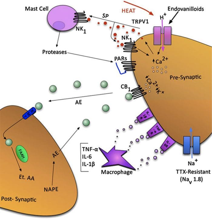
Fig. 1 A schematic illustrating some of the targets known to modulate joint pain. Neuromodulators can be released from nerve terminals as well as mast cells and macrophages to alter afferent mechanosensitivity. Endovanilloids, acid, and noxious heat can activate transient receptor potential vanilloid type 1 (TRPV1) ion channels leading to the release of algogenic substance P (SP), which subsequently binds to neurokinin-1 (NK1) receptors. Proteases can cleave and stimulate protease-activated receptors (PARs). Thus far, PAR2and PAR4have been shown to sensitize joint primary afferents. The endocannabinoid anandamide (AE) is produced on demand and synthesized from N-arachidonoyl phosphatidylethanolamine (NAPE) under the enzymatic action of phospholipases. A portion of AE then binds to cannabinoid-1 (CB1) receptors leading to neuronal desensitization. Unbound AE is rapidly taken up by an anandamide membrane transporter (AMT)before being broken down by a fatty acid amide hydrolase (FAAH)into ethanolamine (Et) and arachidonic acid (AA). The cytokines tumor necrosis factor-?(TNF-?), interleukin-6 (IL-6) and interleukin1-beta (IL-1?) Can bind to their respective receptors to enhance pain transmission. Finally, tetrodotoxin (TTX)-resistant sodium channels (Nav1.8) are involved in neuronal sensitization.
Patients yearn for their chronic pain to disappear; however, currently prescribed analgesics are largely ineffective and are accompanied by a wide range of unwanted side effects. As such, millions of people worldwide are suffering from the debilitating effects of joint pain, for which there is no satisfactory treatment [2].
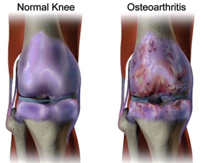
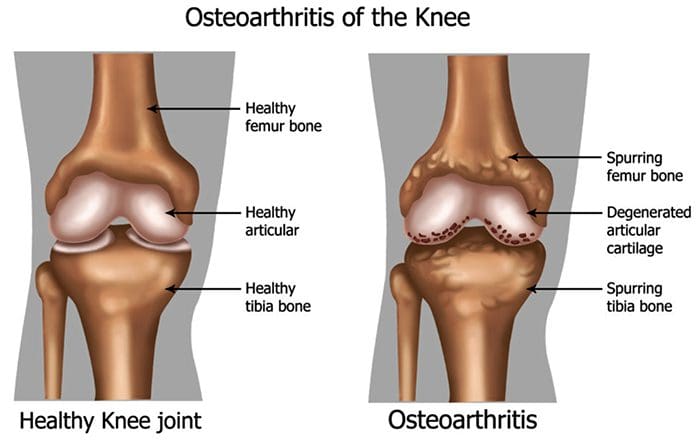
More than 100 different forms of arthritis have osteoarthritis (OA) being the most common. OA is a progressively degenerative joint disease that causes chronic pain and loss of function. Commonly, OA is the inability of the joint to repair damage effectively in response to excessive forces being placed on it. The biological and psychosocial factors that comprise chronic OA pain are not well understood, although ongoing research unravels the complex nature of disease symptoms [2]. Current therapeutics, such as non-steroidal anti-inflammatory drugs (NSAIDs), provide some symptomatic relief, reducing the pain for short periods of time, but do not alleviate pain across the patient’s lifespan. Furthermore, high-dose NSAIDs cannot be taken repeatedly over many years, as this can lead to renal toxicity and gastrointestinal bleeding.
Traditionally, arthritis research has focused largely on the articular cartilage as a primary target for the therapeutic development of novel OA drugs for disease modification. This chondrogenic focus has shed new light on the intricate biochemical and biomechanical factors that influence chondrocyte behavior in diseased joints. However, as the articular cartilage is aneural and avascular, this tissue is unlikely to be the source of OA pain. This fact, coupled with the findings that there is no correlation between the damage of articular cartilage and pain in OA patients [3,4] or preclinical models of OA [5], has caused a shift in focus to develop drugs for effective pain control. This article will review the latest findings in joint pain research and highlight some of the emerging targets that may be the future of arthritis pain management (summarized in Fig. 1)
Cytokines
The actions of various cytokines in joint neurophysiology studies have featured quite prominently recently. Interleukin-6 (IL-6), for example, is a cytokine that typically binds to the membrane-bound IL-6 receptor (IL-6R). IL-6 can also signal by binding with a soluble IL-6R (SIL-6R) to produce an IL-6/sIL-6R complex. This IL-6/sIL-6R complex subsequent lybinds to a transmembrane glycoprotein subunit 130(gp130), thereby allowing IL-6 to signal in cells that do not constitutively express membrane-bound IL-6R [25,26]. IL-6 and SIL-6R are key players in systemic inflammation and arthritis, as upregulation of both has been found in RA patients’ serum and synovial fluid [27,29]. Recently, Vazquez et al.observed that co-administration of IL-6/sIL-6R into rat knees caused inflammation-evoked pain, as revealed by an increase in the response of spinal dorsal horn neurons to mechanical stimulation of the knee and other parts of the hindlimb [30]. Spinal neuron hyperexcitability was also seen when IL-6/sIL-6R was applied locally to the spinal cord. Spinal application of soluble gp130 (which would mop up IL-6/sIL-6R complexes, thereby reducing trans-signaling) inhibited IL-6/sIL-6R-induced central sensitization. However, acute application of soluble gp130 alone did not reduce the neuronal responses to already established joint inflammation.
The transient receptor potential (TRP) channels are non-selective cation channels that act as integrators of various physiological and pathophysiological processes. In addition to thermosensation, chemosensation, and mechanosensation, TRP channels are involved in the modulation of pain and inflammation. For example, TRP vanilloid-1 (TRPV1) ion channels have been shown to contribute to joint inflammatory pain as thermal hyperalgesia was not evocable in TRPV1 mono arthritic mice [31]. Similarly, TRP ankyrin-1 (TRPA1)ion channels are involved in arthritic mechano hypersensitivity as blockade of the receptor with selective antagonists attenuated mechanical pain in the Freunds complete adjuvant model inflammation [32,33]. Further evidence thatTRPV1 may be involved in the neurotransmission of OA pain comes from studies in which neuronal TRPV1 expression is elevated in the sodium monoiodoacetate model of OA [34]. In addition, systemic administration of the TRPV1 antagonist A-889425 reduced the evoked and spontaneous activity of spinal-wide dynamic range and nociception-specific neurons in the monoiodoacetate model [35]. These data suggest that endovanilloids could be involved in central sensitization processes associated with OA pain.
There are currently known to be at least four polymorphisms in the gene that encodes TRPV1, leading to an alteration in the structure of the ion channel and impaired function. One particular polymorphism (rs8065080) alters the sensitivity of TRPV1 to capsaicin, and individuals carrying this polymorphism are less sensitive to thermal hyperalgesia [36]. A recent study examined whether OA patients with the rs8065080 polymorphism experienced altered pain perception based on this genetic anomaly. The research team found that patients with asymptomatic knee OA were more likely to carry the rs8065080 gene than patients with painful joints [37]. This observation indicates that OA patients with normal functioning; TRPV1 channels have an increased risk of joint pain and re-affirms the potential involvement of TRPV1 in OA pain perception.
Conclusion
While the hurdle of treating arthritis pain effectively remains, great leaps are being made in our understanding of the neurophysiological processes responsible for the generation of joint pain. New targets are being discovered continually, while the mechanisms behind known pathways are being further defined and refined. Targeting one specific receptor or ion channel is unlikely to be the solution to normalizing joint pain, but rather a polypharmacy approach is indicated in which various mediators are used in combination during specific phases of the disease. Unraveling the functional circuitry at each level of the pain pathway will also improve our knowledge of how joint pain is generated. For example, identifying the peripheral mediators of joint pain will allow us to control nociception within the joint and likely avoid the central side effects of systemically administered pharmacotherapeutics.
FACETOGENIC PAIN
FACET SYNDROME & FACETOGENIC PAIN
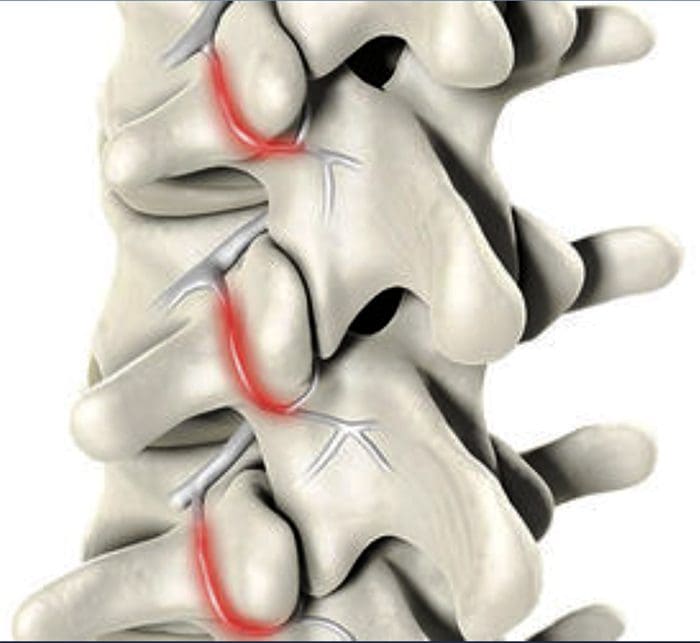
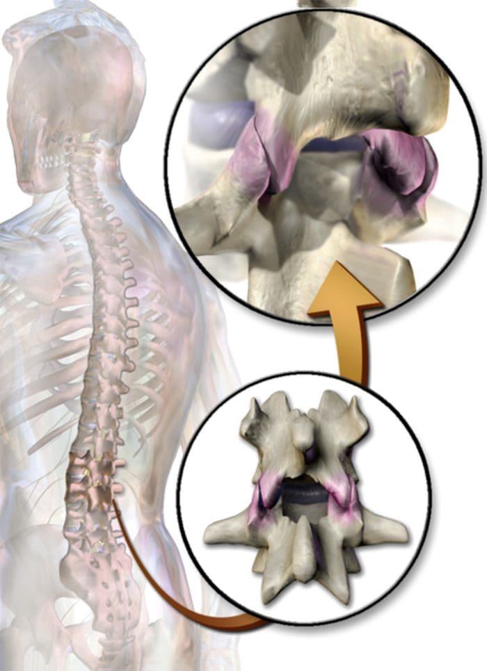
Facet syndrome is an articular disorder related to the lumbar facet joints and their innervations and produces both local and radiating facetogenic pain.
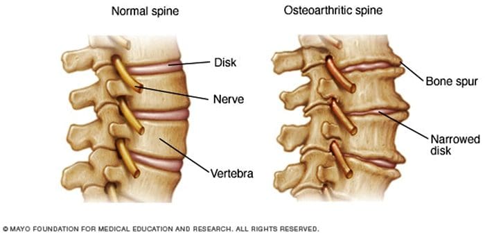
Excessive rotation, extension, or flexion of the spine (repeated overuse) can result in degenerative changes to the joint’s cartilage. In addition, itt may involve degenerative changes to other structures, including the intervertebral disc.
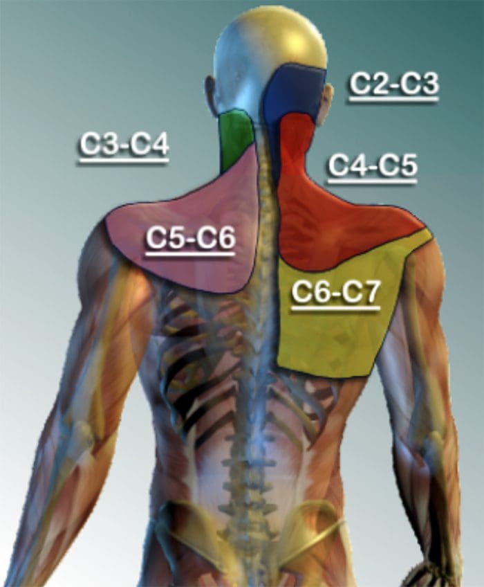
CERVICAL FACET SYNDROME & FACETOGENIC PAIN
Axial neck pain (rarely radiating past the shoulders), most common unilaterally.
Pain with and/or limitation of extension and rotation
Tenderness upon palpation
Radiating facetogenic pain locally or into the shoulders or upper back, and rarely radiate in the front or down an arm or into the fingers as a herniated disc might.
LUMBAR FACET SYNDROME & FACETOGENIC PAIN
Pain or tenderness in the lower back.
Local tenderness/stiffness alongside the spine in the lower back.
Pain, stiffness, or difficulty with certain movements (such as standing up straight or getting up from a chair.
Pain upon hyperextension
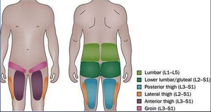
Referred pain from upper lumbar facet joints can extend into the flank, hip, and upper lateral thigh.
Referred pain from lower lumbar facet joints can penetrate deep into the thigh, laterally and/or posteriorly.
L4-L5 and L5-S1 facet joints can refer to pain extending into the distal lateral leg, and in rare instances, to the foot
EVIDENCE-BASED MEDICINE
Evidence-based Interventional Pain Medicine according to Clinical Diagnoses
12. Pain Originating from the Lumbar Facet Joints
Abstract
Although the existence of a facet syndrome had long been questioned, it is now generally accepted as a clinical entity. Depending on the diagnostic criteria, the zygapophysial joints account for between 5% and 15% of cases of chronic, axial low back pain. Most commonly, facetogenic pain results from repetitive stress and/or cumulative low-level trauma, leading to inflammation and stretching of the joint capsule. The most frequent complaint is axial low back pain with referred pain perceived in the flank, hip, and thigh. No physical examination findings are pathognomonic for diagnosis. The strongest indicator for lumbar facetogenic pain is pain reduction after anesthetic blocks of the rami mediales (medial branches) of the rami dorsales that innervate the facet joints. Because false-positive and, possibly, false-negative results may occur, results must be interpreted carefully. In patients with injection-confirmed zygapophysial joint pain, procedural interventions can be undertaken in the context of a multidisciplinary, multimodal treatment regimen that includes pharmacotherapy, physical therapy, and regular exercise, and, if indicated, psychotherapy. Currently, the gold standard for treating facetogenic pain is radiofrequency treatment (1 B+). The evidence supporting intra-articular corticosteroids is limited; hence, this should be reserved for those who do not respond to radiofrequency treatment (2 B1).
Facetogenic Pain emanating from the lumbar facet joints is a common cause of low back pain in the adult population. Goldthwaite was the first to describe the syndrome in 1911, and Ghormley is generally credited with coining the term �facet syndrome� in 1933. Facetogenic pain is defined as pain that arises from any structure that is part of the facet joints, including the fibrous capsule, synovial membrane, hyaline cartilage, and bone.35
More commonly, it is the result of repetitive stress and/or cumulative low-level trauma. This leads to inflammation, which can cause the facet joint to be filled with fluid and swell, resulting in stretching of the joint capsule and subsequent pain generation.27 Inflammatory changes around the facet joint can also irritate the spinal nerve via foraminal narrowing, resulting in sciatica. In addition, Igarashi et al.28 found that inflammatory cytokines released through the ventral joint capsule in patients with zygapophysial joint degeneration may be partially responsible for the neuropathic symptoms in individuals with spinal stenosis. Predisposing factors for zygapophysial joint pain include spondylolisthesis/lysis, degenerative disc disease, and advanced age.5
I.C ADDITIONAL TESTS
The prevalence rate of pathological changes in the facet joints on radiological examination depends on the mean age of the subjects, the radiological technique used, and the definition of abnormality. Degenerative facet joints can be best visualized via computed tomography (CT) examination.49
NEUROPATHIC PAIN
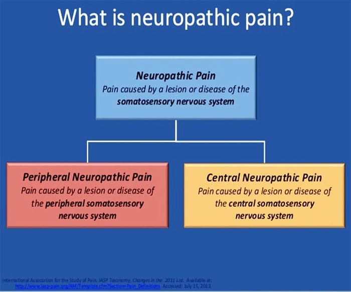
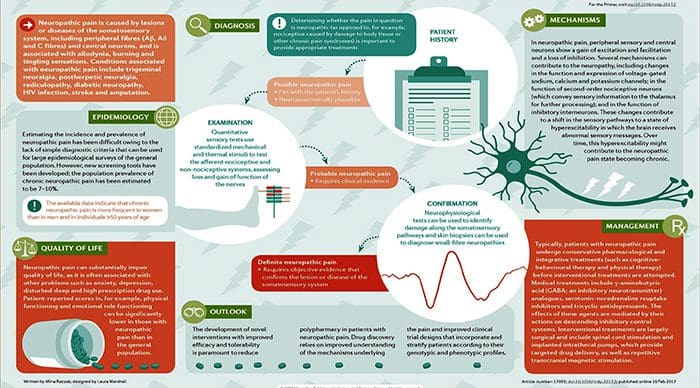
Pain initiated or caused by a primary lesion or dysfunction in the somatosensory nervous system.
Neuropathic pain is usually chronic, difficult to treat, and often resistant to standard analgesic management.
Abstract
Neuropathic pain is caused by a lesion or disease of the somatosensory system, including peripheral fibers (A?, A? and C fibers) and central neurons, and affects 7-10% of the general population. Multiple causes of neuropathic pain have been described. Its incidence is likely to increase due to the aging global population, increased diabetes mellitus, and improved survival from cancer after chemotherapy. Indeed, imbalances between excitatory and inhibitory somatosensory signaling, alterations in ion channels, and variability in how pain messages are modulated in the central nervous system all have been implicated in neuropathic pain. Furthermore, the burden of chronic neuropathic pain seems to be related to the complexity of neuropathic symptoms, poor outcomes, and difficult treatment decisions. Importantly, quality of life is impaired in patients with neuropathic pain due to increased drug prescriptions and visits to health care providers and the morbidity from the pain itself and the inciting disease. Despite challenges, progress in understanding the pathophysiology of neuropathic pain is spurring the development of new diagnostic procedures and personalized interventions, which emphasize the need for a multidisciplinary approach to the management of neuropathic pain.
PATHOGENESIS OF NEUROPATHIC PAIN
PERIPHERAL MECHANISMS
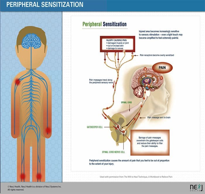
After a peripheral nerve lesion, neurons become more sensitive and develop abnormal excitability and elevated sensitivity to stimulation.
This is known as…Peripheral Sensitization!
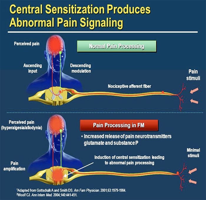
CENTRAL MECHANISMS
As a consequence of ongoing spontaneous activity in the periphery, neurons develop an increased background activity, enlarged receptive fields, and increased responses to afferent impulses, including normal tactile stimuli. This is known as…Central Sensitization!
Chronic neuropathic pain is more frequent in women (8% versus 5.7% in men) and in patients >50 years of age (8.9% versus 5.6% in those <49 years of age), and most commonly affects the lower back and lower limbs, neck and upper limbs24. Lumbar and cervical painful radiculopathies are probably the most frequent cause of chronic neuropathic pain. Consistent with these data, a survey of >12,000 patients with chronic pain with both nociceptive and neuropathic pain types, referred to pain specialists in Germany, revealed that 40% of all patients experienced at least some characteristics of neuropathic pain (such as burning sensations, numbness, and tingling); patients with chronic back pain and radiculopathy were particularly affected25.
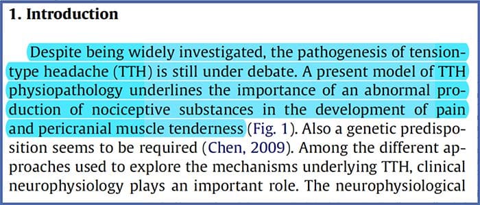
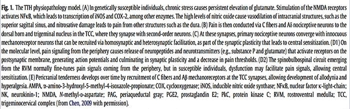
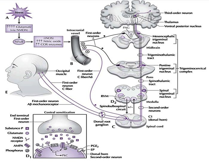
The contribution of clinical neurophysiology to the comprehension of the tension-type headache mechanisms.
Abstract
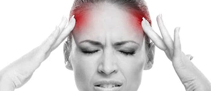
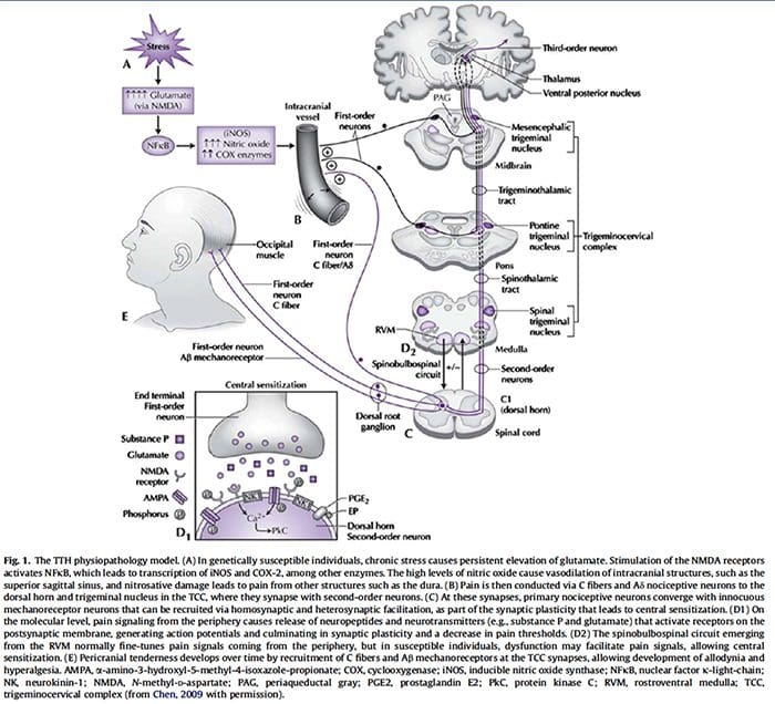
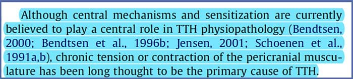
So far, clinical neurophysiological studies on tension-type headache (TTH) have been conducted with two main purposes: (1) to establish whether some neurophysiological parameters may act as markers of TTH, and (2) to investigate the physiopathology of TTH. Regarding the first point, the present results are disappointing since some abnormalities found in TTH patients may also be frequently observed in migraineurs. On the other hand, clinical neurophysiology has played an important role in the debate about the pathogenesis of TTH. Studies on the exteroceptive suppression of the temporalis muscle contraction have detected a dysfunction of the brainstem excitability and suprasegmental control. A similar conclusion has been reached using trigeminocervical reflexes, whose abnormalities in TTH have suggested a reduced inhibitory activity of brainstem interneurons, reflecting abnormal endogenous pain control mechanisms. Interestingly, the neural excitability abnormality in TTH seems to be a generalized phenomenon, not limited to the cranial districts. Defective DNIC-like mechanisms have indeed been evidenced also in somatic districts by nociceptive flexion reflex studies. Unfortunately, most neurophysiological studies on TTH are marred by serious methodological flaws, which should be avoided in future research to clarify the TTH mechanisms better.
References:
Neurophysiology of arthritis pain. McDougall JJ1 Linton P.
Pain originating from the lumbar facet joints. van Kleef M1,Vanelderen P,Cohen SP,Lataster A,Van Zundert J,Mekhail N.
Neuropathic painLuana Colloca,1Taylor Ludman,1Didier Bouhassira,2Ralf Baron,3Anthony H. Dickenson,4David Yarnitsky,5Roy Freeman,6Andrea Truini,7Nadine Attal, Nanna B. Finnerup,9Christopher Eccleston,10,11Eija Kalso,12David L. Bennett,13Robert H. Dworkin,14and Srinivasa N. Raja15
The contribution of clinical neurophysiology to the comprehension of the tension-type headache mechanisms. Rossi P1, Vollono C, Valeriani M, Sandrini G.
Heel Spur: Blanca, born and raised in El Paso, TX, has been suffering from heel spurs for about two years. As a registered nurse, her symptoms significantly affected her ability to work and her overall quality of life. Determined to improve her health, Blanca considered chiropractic care. Once she started treatment with Dr. Alex Jimenez, however, Blanca experienced tremendous relief from her heel spurs, almost instantly. Blanca highly recommends chiropractic care with Dr. Alex Jimenez as the non-surgical choice for treatment of heel spurs.
Chiropractic Heel Spur Treatment
A heel spur is a calcium residue resulting in a bony protrusion on the bottom of the heel bone. Although heel spurs are often painless, they can lead to heel pain. They are often associated with plantar fasciitis, a painful inflammation of the fibrous band of connective tissue (plantar fascia) that runs across the bottom of the foot and also connects the heel bone to the ball of the foot. Heel spurs are usually caused by strains on foot muscles and ligaments, stretching of the plantar fascia, and repeated tearing of the membrane which covers the heel bone. Heel spurs are particularly common among athletes.
We are blessed to present to you�El Paso�s Premier Wellness & Injury Care Clinic.
As El Paso�s Chiropractic Rehabilitation Clinic & Integrated Medicine Center,�we passionately are focused treating patients after frustrating injuries and chronic pain syndromes. We focus on improving your ability through flexibility, mobility and agility programs tailored for all age groups and disabilities.
If you have enjoyed this video and/or we have helped you in any way please feel free to subscribe and share us.
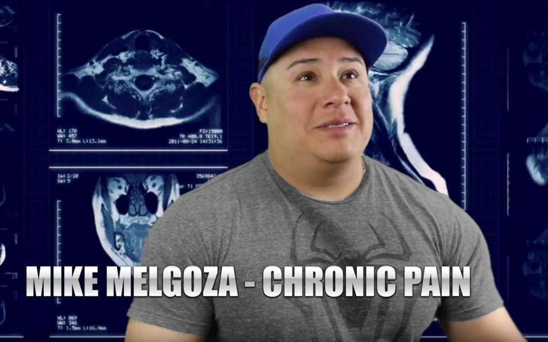
Doctor of Chiropractic Near Me: Mike Melgoza is an active individual who engages in a variety of strenuous physical activities on a regular basis, as a result, however, he began to experience chronic pain symptoms due to improper technique and repetitive movements. Although Mike Melgoza works out of town, he visits Dr. Alex Jimenez every time he begins to experience pain to receive chiropractic care. Mike Melgoza recommends Dr. Alex Jimenez as the non-surgical choice for chronic pain. Mike Melgoza trusts Dr. Jimenez to care for his health.
Doctor Of Chiropractic Near Me
Before you go to a chiropractor to deal with your chronic pain symptoms, it’s important to understand what exactly is causing your pain. Your physician will perform a physical exam as well as some tests to help them diagnose the source of the patient’s pain. As soon as you’re diagnosed with a pain illness, your chiropractor will create a treatment program. Your treatment plan may include spinal manipulation, manual therapies, and therapeutic exercises. Work with your chiropractor to develop a treatment plan. Once your pain is fully addressed, you should be able to slowly resume daily activities.
We are blessed to present to you�El Paso�s Premier Wellness & Injury Care Clinic.
As El Paso�s Chiropractic Rehabilitation Clinic & Integrated Medicine Center,�we passionately are focused treating patients after frustrating injuries and chronic pain syndromes. We focus on improving your ability through flexibility, mobility and agility programs tailored for all age groups and disabilities.
If you have enjoyed this video and/or we have helped you in any way please feel free to subscribe and share us.
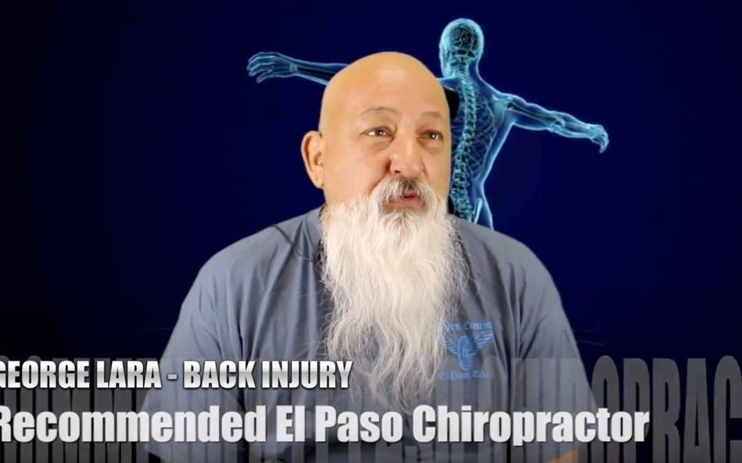
Chiropractor Near Me: Due to his previous back injury history, George Lara suffered a recent back injury which he was unfortunately unable to recover from through traditional treatment. That’s when the VA recommended him to seek chiropractic care with Dr. Alex Jimenez, ultimately changing his quality of life. George Lara had degenerative disc disease, or DDD, and sciatic nerve pain before receiving treatment with Dr. Alex Jimenez. George Lara describes how much his life has improved with chiropractic care and he’s grateful of the services and care he received. George Lara recommends Dr. Alex Jimenez as the non-surgical choice for back pain, among other spine health issues.
Chiropractor Near Me
Chiropractic care is a means to diagnose and treat health problems that affect the nerves, muscles, bones, and joints of the body. A healthcare provider who supplies chiropractic care is known as a chiropractor. Adjustment of the spine, known as manipulation, is the basis of care. Chiropractors also use other kinds of treatments. Your physician will ask about your goals for your health history and therapy. It’s important to inform your physician about any physical problems you may have which make it difficult for you to do particular things.
We are blessed to present to you�El Paso�s Premier Wellness & Injury Care Clinic.
As El Paso�s Chiropractic Rehabilitation Clinic & Integrated Medicine Center,�we passionately are focused treating patients after frustrating injuries and chronic pain syndromes. We focus on improving your ability through flexibility, mobility and agility programs tailored for all age groups and disabilities.
If you have enjoyed this video and/or we have helped you in any way please feel free to subscribe and share us.
Getting a good night�s sleep is absolutely integral to good spinal health. Sometimes, though that isn�t possible. According to the National Sleep Foundation, 92 percent of people believe that a�comfortable mattress is important for good, restful sleep. A bad mattress, or one that is old, or one that is simply wrong for your body can contribute to sleep deprivation, lower back pain, headaches, stiff neck, and anxiety and depression. With so much at stake, it�s easy to see just how important it is to select a good mattress.
Ask About How The Mattress Is Made
Learn about the construction� and what the different components mean for your comfort. Different mattresses have different coils and they are arranged differently. The padding can vary in thickness. The depth can range from 7 inches to 18 inches on the average. Understanding the various components can make it easier for you to find the one that is right for you.
Look For Comfort, As Well As, Support
A good mattress is comfortable and has good support. Support is good but if you don�t have comfort then it won�t be effective.
If it is too firm (too much support) it will cause pain on your body�s pressure points. You want your hips and shoulders to slightly sink into the mattress. However, if you prefer a mattress that is firmer to support your back, you can get one with padding on top.
Don�t Let Price Be The Determining Factor
You naturally want to get the most for your dollar, but remember that you get what you pay for. A cheap mattress can translate to a poor quality one.
Look for quality and value rather than price. If money is an issue, do some comparison shopping to find the mattress you want for the best price.
Sales are another way to save money on a purchase, but look out for advertising gimmicks. Know the meaning of the terms that are used and know what you are looking for before you go for that so-called great deal.
Educate Yourself On The Different Mattress Types
Do you want a memory foam or would latex work better for you? What exactly is an innerspring mattress? Are adjustable beds really all they are cracked up to be? Do some research and brush up on the different�types of mattresses�so that you can approach your shopping trip with confidence and as an educated consumer. It will definitely work in your favor.
In The End, It�s All About Personal Preference
There is no mattress that is a one size (or type) fits all. Different people will respond differently to mattresses. The best thing to do is try them out. Spend at least 20 minutes laying down before you make the decision to purchase or not.
Finally, if you find that your�quality of sleep�has recently gotten worse, that you are tossing and turning or wake up with pain in your back, neck, or head, it could be time to change your mattress � or pillow. If you can see your mattress sagging, that could be another indication that it is time to get a new one.
Mattresses are designed to withstand a certain degree of wear and tear, but they don�t last forever. The quality, the weight and other factors contribute to how quickly it wears out. So if you notice any of the warning signs it may be time to get a new mattress so that you can get back to peaceful, restful sleep.
Injury Medical Clinic: Back Pain Care & Treatments
IFM's Find A Practitioner tool is the largest referral network in Functional Medicine, created to help patients locate Functional Medicine practitioners anywhere in the world. IFM Certified Practitioners are listed first in the search results, given their extensive education in Functional Medicine





 Abstract