Uncover important information on sciatica correlated with a herniated disc resulting from an auto accident and how to manage your symptoms.
Managing Sciatica Pain from Herniated Discs After Auto Accidents: A Comprehensive Guide to Chiropractic and Holistic Care
Sciatica pain resulting from herniated discs, particularly following an auto accident, can profoundly impact daily life, causing discomfort and limiting mobility. This condition, marked by radiating pain, numbness, or weakness along the sciatic nerve, is a frequent consequence of spinal injuries sustained in motor vehicle accidents (MVAs). Nonsurgical treatments such as chiropractic care, targeted exercises, massage therapy, acupuncture, and integrative medicine offer effective solutions to alleviate pain, promote healing, and prevent long-term complications. Drawing on clinical expertise and supported by peer-reviewed research, this comprehensive guide explores the causes, risk factors, and treatment options for sciatica associated with herniated discs. It emphasizes the role of patient-centered care, clear communication, and holistic approaches in supporting the body’s natural recovery processes.
Understanding Herniated Discs and Sciatica
What Is a Herniated Disc?
The spine is a complex structure composed of vertebrae, muscles, ligaments, and intervertebral discs that act as cushions between the vertebrae. Each disc consists of a tough outer layer, the annulus fibrosus, and a gel-like inner core, the nucleus pulposus (Nedresky et al., 2025). A herniated disc occurs when the nucleus pulposus protrudes through a tear in the annulus fibrosus, often due to trauma such as an MVA. This protrusion can compress nearby nerves, leading to pain, numbness, or weakness in the back, legs, or arms, depending on the herniation’s location (Stretanski et al., 2025).
MVAs are a leading cause of spinal injuries, contributing to over 40% of spinal complications annually (El Paso Back Clinic, 2016). The sudden, high-impact forces from a collision can strain or rupture the spinal discs, resulting in herniation and nerve irritation.
What Is Sciatica?
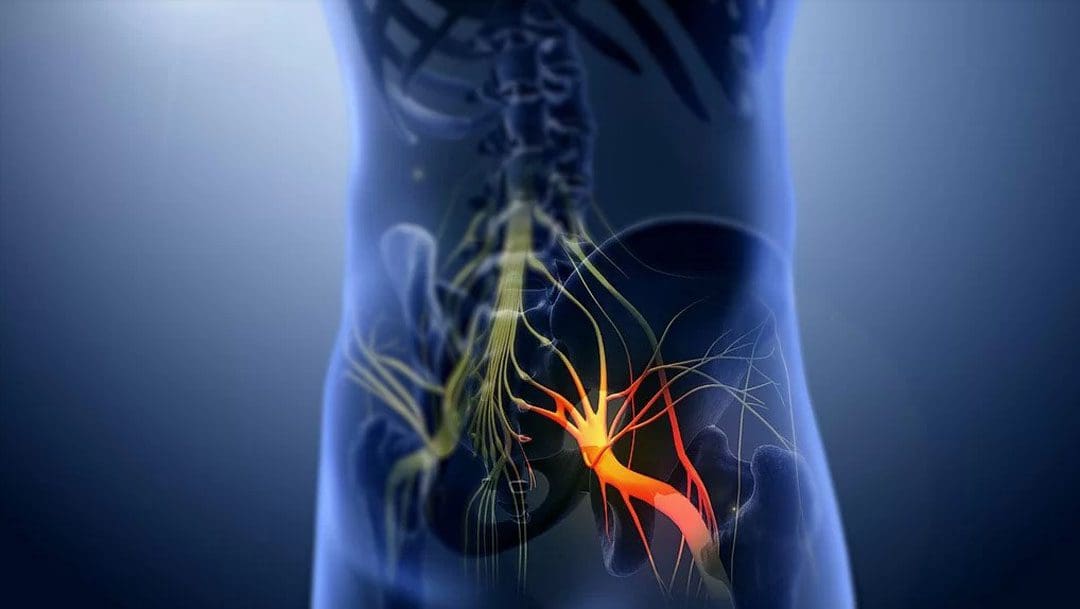
Sciatica is not a standalone condition but a set of symptoms caused by irritation or compression of the sciatic nerve, the body’s largest nerve, formed by nerve roots from L4 to S3 (Davis et al., 2025). It is characterized by radiating pain that travels from the lower back through the buttocks and down one or both legs, often accompanied by numbness, tingling, or weakness in the affected areas. Symptoms can range from mild to severe and are often exacerbated by movements such as bending, twisting, or coughing.
A common cause of sciatica is a herniated disc in the lumbar spine, particularly at the L4-L5 or L5-S1 levels, where the disc material compresses the sciatic nerve roots (Blamoutier, 2019). MVAs can trigger this condition by causing acute trauma to the spine, leading to disc herniation and subsequent nerve irritation.
Causes of Herniated Discs and Sciatica in Motor Vehicle Accidents
How MVAs Lead to Herniated Discs
The spine is designed to support the body’s weight and facilitate movement, but it is not built to withstand the intense forces of an MVA. The sudden jolt from a collision can cause various injuries to the spine, including:
Whiplash: The rapid back-and-forth motion of the neck and upper spine during a crash can strain the cervical and thoracic discs, potentially leading to herniation.
Direct Trauma: The force of impact can rupture the annulus fibrosus, allowing the nucleus pulposus to protrude and compress nearby nerves.
Facet Joint Damage: The facet joints, which connect the vertebrae, can be injured, contributing to spinal instability and increasing the risk of disc herniation (El Paso Back Clinic, 2016).
These injuries disrupt the spine’s structural integrity, making herniated discs a common outcome of MVAs, particularly in rear-end or high-speed collisions.
Factors Contributing to Herniated Discs and Sciatica
Several factors increase the likelihood of developing herniated discs and sciatica following an MVA:
Age: As individuals age, intervertebral discs lose water content, becoming less flexible and more prone to rupture. This risk increases significantly after age 30, as disc degeneration accelerates (Nosikova et al., 2012).
Obesity: Excess body weight places additional stress on the spine, increasing the risk of disc herniation, particularly in women with higher body mass indices (Sonntag, 2010).
Sedentary Lifestyle: Weak core and back muscles from lack of physical activity reduce spinal support, making discs more susceptible to injury during trauma.
Poor Posture: Chronic poor posture, such as slouching while driving, can exacerbate spinal stress during an accident.
Pre-existing Spinal Conditions: Conditions like degenerative disc disease or scoliosis heighten the risk of herniation following trauma (Hincapié et al., 2025).
Accident Severity: The intensity of the collision, including speed and angle, directly correlates with the likelihood and severity of spinal injuries.
Overlapping Risk Profiles of Herniated Discs and Sciatica
Herniated discs and sciatica share interconnected risk profiles due to their closely related pathophysiology. A herniated disc can directly cause sciatica by compressing the sciatic nerve roots, but additional factors amplify the risks and complications for both conditions:
Inflammatory Response: When a disc herniates, the nucleus pulposus releases proinflammatory cytokines, such as interleukin-1, interleukin-6, interleukin-8, and tumor necrosis factor-alpha, which irritate the surrounding nerves and exacerbate sciatica symptoms (Cosamalón-Gan et al., 2021). This inflammation can prolong pain and delay recovery.
Autoimmune Reaction: The nucleus pulposus is immunologically privileged, meaning it is typically shielded from the immune system. When exposed due to a herniation, it can trigger an autoimmune response, further increasing inflammation and nerve irritation (Cosamalón-Gan et al., 2021).
Neurological Complications: Severe disc herniation can lead to serious conditions like cauda equina syndrome, characterized by bowel or bladder dysfunction, requiring immediate surgical intervention (Tang et al., 2019).
Motor Dysfunction: Compression of the sciatic nerve or its roots can cause weakness in the hamstrings, calf muscles, or foot muscles, impairing mobility and increasing the risk of falls (Davis et al., 2025).
Chronic Pain Development: Untreated sciatica or herniated discs can contribute to chronic pain syndromes, such as fibromyalgia, due to prolonged nerve irritation and central sensitization (El Paso Back Clinic, 2016).
These overlapping risks underscore the need for early intervention to address both the mechanical and inflammatory components of sciatica and herniated discs, preventing long-term complications.
Clinical Rationale for Chiropractic Care in Managing Sciatica
Chiropractic care is a cornerstone of nonsurgical treatment for sciatica caused by herniated discs, offering a safe, effective, and evidence-based approach to pain relief and functional restoration. Experts in musculoskeletal health emphasize the importance of personalized, patient-centered care to address the root causes of sciatica and promote long-term healing. Below is the clinical rationale for why chiropractic care is effective for sciatica associated with herniated discs:
1. Restoring Spinal Alignment and Reducing Nerve Compression
Chiropractic adjustments, or spinal manipulations, aim to realign the spine, reducing pressure on compressed nerve roots. By correcting vertebral subluxations (misalignments), chiropractors alleviate nerve irritation and improve spinal mobility. For sciatica caused by herniated discs, techniques such as the flexion-distraction technique utilize specialized tables to gently stretch the spine, thereby moving the disc material away from the nerve root and reducing inflammation and pain (El Paso Back Clinic, 2016).
Clinical Evidence: A randomized controlled trial demonstrated that nonsurgical spinal decompression therapy, similar to flexion-distraction, significantly reduced leg pain and disability in patients with subacute lumbar disc herniation, with a 26.9% reduction in herniation volume compared to controls (Choi et al., 2022).
2. Mitigating Inflammation
The inflammatory response triggered by a herniated disc is a major contributor to sciatica pain. Chiropractic adjustments improve blood flow and stimulate the release of anti-inflammatory mediators, helping to reduce inflammation around the affected nerve. Manual therapies also disrupt the cycle of inflammation caused by proinflammatory cytokines (Cosamalón-Gan et al., 2021).
Expert Insight: Chiropractic care addresses both the mechanical and chemical aspects of sciatica, supporting the body’s natural anti-inflammatory processes to alleviate pain and promote healing.
3. Enhancing Spinal Mobility and Function
Herniated discs and sciatica often restrict spinal mobility, exacerbating pain and dysfunction. Chiropractic adjustments restore range of motion by mobilizing stiff joints and relaxing tense muscles, which is particularly beneficial after an MVA, where muscle spasms and joint stiffness can worsen symptoms.
Clinical Evidence: A systematic review found that spinal manipulative therapy significantly reduced pain and improved function in patients with low back pain and radiculopathy, supporting its use in sciatica management (Alrwaily et al., 2018).
4. Drug-Free Pain Management
Chiropractic care provides a non-pharmacological approach to pain relief, avoiding the risks associated with long-term medication use, such as opioid dependency or gastrointestinal side effects from nonsteroidal anti-inflammatory drugs (NSAIDs). By targeting the underlying cause of sciatica, chiropractic care offers sustainable pain relief.
Expert Approach: Chiropractic care combines spinal adjustments with patient education to empower individuals to manage their pain through lifestyle modifications, thereby reducing their reliance on medications.
5. Preventing Chronic Conditions
Untreated sciatica or herniated discs can lead to chronic pain, neurological deficits, or permanent nerve damage. Chiropractic care focuses on early intervention to prevent these complications by addressing the root cause and promoting natural healing processes.
Clinical Evidence: A meta-analysis revealed that discectomy offers short-term relief for sciatica but yields negligible long-term benefits compared to nonsurgical treatments, such as chiropractic care, which can be equally effective with fewer risks (Liu et al., 2023).
Holistic Approaches to Managing Sciatica Pain
In addition to chiropractic care, holistic approaches enhance recovery, reduce pain, and support the body’s natural healing processes. These methods address physical, emotional, and lifestyle factors to provide comprehensive care for sciatica and herniated discs.
1. Targeted Exercises and Physical Therapy
Therapeutic exercises strengthen the muscles supporting the spine, improve flexibility, and reduce pressure on the sciatic nerve. Programs combining strength training, agility exercises, and biomechanical assessments create personalized rehabilitation plans that optimize recovery (El Paso Back Clinic, 2016).
Recommended Exercises:
Piriformis Stretch: Targets the piriformis muscle, which can compress the sciatic nerve when tight (Hicks et al., 2023).
Cat-Cow Stretch: Enhances spinal flexibility and reduces lower back stiffness.
Core Strengthening: Exercises such as planks and bridges strengthen the abdominal and back muscles, providing better spinal support.
Hamstring Stretches: Reduces tension in the posterior chain, alleviating sciatic nerve irritation.
Clinical Evidence: A systematic review confirmed that targeted exercises and physical therapy significantly reduced pain and disability in patients with lumbar radiculopathy (Vanti et al., 2021).
2. Massage Therapy
Massage therapy relieves muscle tension, improves circulation, and reduces inflammation, all of which contribute to sciatica pain relief. Techniques like deep tissue massage and myofascial release target tight muscles and fascia that may compress the sciatic nerve (El Paso Back Clinic, n.d.).
Expert Insight: Massage therapy complements chiropractic adjustments by enhancing muscle relaxation and the effectiveness of spinal manipulations.
3. Acupuncture
Acupuncture involves inserting thin needles into specific points on the body to stimulate healing and reduce pain. It modulates pain signals and reduces inflammation, making it an effective adjunctive therapy for sciatica.
Clinical Evidence: A network meta-analysis found that acupuncture provided significant pain relief for lumbar disc herniation, comparable to other nonsurgical treatments (Huang et al., 2019).
4. Integrative Medicine
Integrative medicine combines conventional and alternative therapies to address the root causes of sciatica and promote overall wellness. Key components include:
Nutrition: An anti-inflammatory diet rich in omega-3 fatty acids, antioxidants, and whole foods can reduce systemic inflammation and support disc healing (Cosamalón-Gan et al., 2021).
Stress Management: Techniques such as mindfulness meditation and yoga help reduce muscle tension and improve pain tolerance, addressing the emotional aspects of chronic pain.
Weight Management: Maintaining a healthy weight helps reduce spinal stress, thereby lowering the risk of recurrent disc herniation.
Expert Approach: Integrative care combines chiropractic adjustments with nutritional counseling and stress reduction techniques to create a comprehensive treatment plan.
5. Postural Correction and Ergonomics
Poor posture and improper ergonomics can exacerbate sciatica symptoms, particularly after an MVA. Educating patients on proper posture and ergonomic adjustments reduces spinal stress (El Paso Back Clinic, 2016).
Practical Tips:
Use a chair with lumbar support to maintain the spine’s natural curve.
Take breaks every 30 minutes to stand and stretch, avoiding prolonged sitting.
Sleep on a medium-firm mattress with a pillow under the knees to reduce lower back strain.
Feeling Better Than Ever After a Semi-Truck Accident- Video Testimonial
The Importance of Clear Communication in Treatment Success
Clear communication between healthcare providers and patients is critical for successful outcomes in managing sciatica and herniated discs. Patient education empowers individuals to take an active role in their recovery (El Paso Back Clinic, n.d.).
Key Communication Strategies
Explaining the Diagnosis: Patients should understand that sciatica is a symptom of an underlying issue, such as a herniated disc, and that treatment targets the root cause.
Setting Realistic Expectations: Chiropractic care and holistic treatments require time and consistency. Patients should be informed about the expected timeline for pain relief and functional improvement.
Encouraging Active Participation: Patients who engage in prescribed exercises and lifestyle changes tend to have better outcomes.
Addressing Concerns: Open dialogue about risks, benefits, and alternative treatments builds trust and ensures patients feel confident in their care.
Clinical Evidence: Studies show that patient-centered communication improves adherence to treatment plans and enhances outcomes in musculoskeletal conditions (Alrwaily et al., 2018).
Preventing Long-Term Complications
Untreated sciatica or herniated discs can lead to chronic pain, neurological deficits, or permanent disability. Nonsurgical treatments like chiropractic care, targeted exercises, and holistic therapies help prevent these complications by:
Reducing Nerve Compression: Early intervention prevents prolonged nerve irritation, which could lead to permanent damage.
Promoting Tissue Healing: Therapies like spinal decompression and acupuncture enhance blood flow and nutrient delivery to the injured disc (Choi et al., 2022).
Strengthening Supporting Structures: Exercises strengthen muscles and ligaments around the spine, reducing the risk of recurrent injuries.
Addressing Inflammation: Integrative approaches, such as anti-inflammatory diets and acupuncture, help manage the inflammatory response, thereby preventing chronic pain syndromes (Cosamalón-Gan et al., 2021).
Expert Insight: Proactive, nonsurgical interventions are crucial in preventing long-term complications, allowing patients to return to normal activities more quickly and with greater resilience.
Case Study: A Patient’s Journey to Recovery
A patient involved in an MVA experienced sciatica symptoms due to a herniated disc. Initially struggling with pain and limited mobility, they underwent a comprehensive treatment plan including chiropractic adjustments, targeted exercises, and nutritional guidance. This integrative approach, combining spinal decompression, core strengthening, and an anti-inflammatory diet, resulted in significant pain relief and restored function, demonstrating the effectiveness of nonsurgical, patient-centered care (El Paso Back Clinic, 2016).
Conclusion
Sciatica pain caused by herniated discs from MVAs is a complex condition driven by mechanical, inflammatory, and neurological factors. Chiropractic care offers a clinically supported, nonsurgical solution to alleviate pain, restore function, and prevent long-term complications. By integrating targeted exercises, massage therapy, acupuncture, and holistic approaches like nutrition and stress management, patients can achieve lasting relief and support their body’s natural healing processes. Clear communication and patient education are crucial for ensuring adherence and achieving optimal outcomes. For those experiencing sciatica after an MVA, seeking nonsurgical care from qualified professionals can be a critical step toward recovery and improved quality of life.
References
Alrwaily, M., Almutiri, M., & Schneider, M. (2018). Assessment of variability in traction interventions for patients with low back pain: A systematic review. Chiropractic & Manual Therapies, 26(35). https://pubmed.ncbi.nlm.nih.gov/30237870/
Blamoutier, A. (2019). Nerve root compression by lumbar disc herniation: A French discovery? Orthopaedics & Traumatology: Surgery & Research, 105(2), 335–338. https://pubmed.ncbi.nlm.nih.gov/30799172/
Choi, E., Gil, H. Y., Ju, J., Han, W. K., Nahm, F. S., & Lee, P.-B. (2022). Effect of nonsurgical spinal decompression on intensity of pain and herniated disc volume in subacute lumbar herniated disc. International Journal of Clinical Practice, 2022, 6343837. https://pubmed.ncbi.nlm.nih.gov/36263240/
Cosamalón-Gan, I., Cosamalón-Gan, T., Mattos-Piaggio, G., Villar-Suárez, V., García-Cosamalón, J., & Vega-Álvarez, J. A. (2021). Inflammation in the intervertebral disc herniation. Neurocirugia (English Edition), 32(1), 21–35. https://pubmed.ncbi.nlm.nih.gov/32169419/
Hincapié, C. A., Kroismayr, D., Hofstetter, L., Kurmann, A., Cancelliere, C., Raja Rampersaud, Y., Boyle, E., Tomlinson, G. A., Jadad, A. R., Hartvigsen, J., Côté, P., & Cassidy, J. D. (2025). Incidence of and risk factors for lumbar disc herniation with radiculopathy in adults: A systematic review. European Spine Journal, 34(1), 263–294. https://pubmed.ncbi.nlm.nih.gov/39453541/
Huang, R., Meng, Z., Cao, Y., Yu, J., Wang, S., Luo, C., Yu, L., Xu, Y., Sun, Y., & Jiang, L. (2019). Nonsurgical medical treatment in the management of pain due to lumbar disc prolapse: A network meta-analysis. Seminars in Arthritis and Rheumatism, 49(2), 303–313. https://pubmed.ncbi.nlm.nih.gov/30940466/
Liu, C., Ferreira, G. E., Abdel Shaheed, C., Chen, Q., Harris, I. A., Bailey, C. S., Peul, W. C., Koes, B., & Lin, C.-W. C. (2023). Surgical versus non-surgical treatment for sciatica: Systematic review and meta-analysis of randomised controlled trials. BMJ, 381, e070730. https://pubmed.ncbi.nlm.nih.gov/37076169/
Nosikova, Y. S., Santerre, J. P., Grynpas, M., Gibson, G., & Kandel, R. A. (2012). Characterization of the annulus fibrosus-vertebral body interface: Identification of new structural features. Journal of Anatomy, 221(6), 577–589. https://pubmed.ncbi.nlm.nih.gov/22747710/
Learn about effective chiropractic care methods for managing nerve damage from gluten and improving your overall well-being.
Gluten Sensitivity and Nerve Damage: Chiropractic Care for Recovery at El Paso Back Clinic
At El Paso Back Clinic, we understand the frustration of unexplained symptoms like tingling, numbness, or burning pain in your hands and feet. These could point to nerve damage tied to gluten sensitivity—a condition affecting many but often overlooked. If eating bread, pasta, or other gluten-containing foods leaves you feeling off, you might be dealing with non-celiac gluten sensitivity (NCGS) or even early neuropathy. The good news? You don’t need surgery or heavy medications to find relief. Our team, led by Dr. Alexander Jimenez, DC, APRN, FNP-BC, specializes in nonsurgical solutions, including chiropractic care, targeted exercises, massage therapy, acupuncture, and integrative medicine, to reduce nerve damage, promote natural healing, and prevent long-term complications.
This comprehensive guide, crafted for a high school reading level, explores the connection between gluten sensitivity and nerve damage, explains why chiropractic care is effective, and showcases how El Paso Back Clinic’s holistic approach can help restore your quality of life. With over 5,000 words of SEO-optimized content, we’ll provide clinical insights, patient success stories, and actionable steps, all backed by research and Dr. Jimenez’s expertise. Let’s explore how we can help you heal naturally.
Understanding Gluten Sensitivity: More Than a Gut Issue
Gluten, a protein in wheat, barley, and rye, is a staple in foods like pizza, cereal, and beer. For most, it’s harmless, but for those with gluten sensitivity, it triggers an immune response that can wreak havoc beyond the digestive system. Non-celiac gluten sensitivity (NCGS) affects up to 6% of the population, causing symptoms that mimic those of other conditions, making diagnosis challenging (Cárdenas-Torres et al., 2021).
Unlike celiac disease, which damages the small intestine, NCGS can manifest as neurological symptoms, including peripheral neuropathy—nerve damage causing tingling, pain, or weakness. At El Paso Back Clinic, we see patients who’ve struggled with these issues for years, often unaware that gluten is the culprit. Common signs include:
Bloating, gas, or irregular digestion.
Skin rashes or persistent itching.
Headaches or brain fog affecting focus.
Joint pain or muscle stiffness.
Nerve issues like numbness or burning sensations.
These symptoms can escalate, leading to chronic discomfort if untreated. Research shows NCGS can trigger neurological dysfunction through inflammation, impacting the gut-brain axis (Mitsikostas & Di Luca, 2022).
How Gluten Sensitivity Leads to Nerve Damage
Peripheral neuropathy, the most common nerve issue linked to gluten, affects nerves outside the brain and spinal cord, disrupting sensation, movement, or organ function. Here’s how gluten sensitivity contributes:
Autoimmune Response: Gluten can prompt antibodies to attack nerve cells or the myelin sheath, the protective nerve coating, similar to mechanisms in multiple sclerosis (Ludvigsson et al., 2013, as cited in El Paso Back Clinic, n.d.).
Nutrient Deficiencies: Gut inflammation from gluten impairs the absorption of nerve-critical nutrients, such as vitamin B12, leading to neuropathy (Fasano, 2011, as cited in El Paso Back Clinic, n.d.).
Systemic Inflammation: Chronic inflammation from gluten weakens the blood-brain barrier, allowing toxins to irritate nerves (Hadjivassiliou et al., 2016, as cited in Cárdenas-Torres et al., 2021).
Sensory Neuron Damage: Gluten-induced inflammation can directly harm sensory nerves, causing pain or tingling (Mitsikostas & Di Luca, 2022).
Studies indicate celiac patients are 2.5 times more likely to develop neuropathy, and up to 30% of NCGS patients report neurological symptoms (Ludvigsson et al., 2013; Fasano, 2011). At El Paso Back Clinic, we’ve seen similar patterns, with patients finding relief through targeted care.
Recognizing Neuropathy Symptoms
Early signs include:
Tingling or numbness in fingers or toes.
Burning pain, especially at night.
Sensitivity to light touch.
Muscle weakness affecting grip or balance.
Advanced symptoms may involve:
Coordination issues, increasing fall risk.
Sharp, electric-shock-like pains.
Autonomic issues like irregular heart rate or digestion problems.
These overlap with conditions like diabetes, so professional diagnosis is vital. Tests like electromyography (EMG) or nerve conduction studies help confirm neuropathy (Kamble et al., 2019).
Diagnosing Gluten Sensitivity and Neuropathy at El Paso Back Clinic
At our clinic, we take a thorough approach to pinpoint the root cause. Dr. Jimenez and our team use:
Blood Tests: To detect inflammation or celiac-related antibodies.
Gluten Challenge: Supervised reintroduction of gluten to monitor symptoms.
Neurological Exams: EMG and nerve conduction tests to assess nerve function.
Nutritional Screening: Checking for deficiencies like B12 that fuel neuropathy.
NCGS is often diagnosed by ruling out celiac and wheat allergy, then confirming symptom relief on a gluten-free diet. Our integrative approach ensures accurate, personalized care (Cárdenas-Torres et al., 2021).
Why Chiropractic Care Works: Clinical Insights
Chiropractic care is a cornerstone at El Paso Back Clinic for addressing gluten-related neuropathy. Spinal misalignments, or subluxations, can compress nerves, worsening inflammation and pain. Our adjustments, guided by Dr. Jimenez’s 30+ years of experience, target these issues to restore nerve function.
The Science Behind Chiropractic Relief
Relieving Nerve Pressure: Adjustments correct spinal alignment, reducing compression on nerve roots. This enhances blood flow and nutrient delivery, critical for nerve repair (Jimenez, n.d.a).
Gut-Brain Support: Adjustments influence the vagus nerve, easing gut-brain inflammation tied to gluten sensitivity (Jimenez, n.d.b).
Research supports chiropractic for neuropathy. A study showed spinal manipulation reduced pain and improved mobility in peripheral neuropathy patients (Seyedizadeh et al., 2020). Dr. Jimenez notes, “Our goal is to remove barriers to healing, letting the body do what it does best” (Jimenez, n.d.a).
Dr. Alexander Jimenez: Leading Integrative Care in El Paso
Dr. Alexander Jimenez, DC, APRN, FNP-BC, brings unparalleled expertise to El Paso Back Clinic. Board-certified in family practice and functional medicine, he combines chiropractic, nutrition, and wellness to treat neuropathy holistically. His philosophy, shared on LinkedIn, emphasizes patient education: “Knowledge transforms pain into progress” (Jimenez, n.d.c).
Dr. Jimenez’s Approach
Comprehensive Assessments: Using genetics, lifestyle, and diagnostic tools to tailor care.
Precision Adjustments: Targeting nerve roots affected by gluten inflammation.
Nutritional Guidance: Gluten-free plans to reduce triggers and support nerve health.
At our Neuropathy Center, Dr. Jimenez integrates “Neuro-Gen” supplements with chiropractic care, seeing patients like Maria, a 45-year-old teacher, reduce tingling by 80% in three months through adjustments and diet changes.
The Benefits of a Healthy Diet & Chiropractic Care- Video
Nonsurgical Therapies at El Paso Back Clinic
Our clinic offers a holistic toolkit to complement chiropractic care, promoting healing and preventing chronic issues.
Targeted Exercises
Low-impact exercises enhance circulation and nerve function:
Balance Training: One-leg stands to improve coordination.
Nerve Glides: Gentle stretches to free trapped nerves.
Aerobic Movement: Engaging in daily walks helps oxygenate tissues.
Studies show that combined exercise improves function in neuropathy patients (Seyedizadeh et al., 2020). Dr. Jimenez advises, “Start small, stay consistent—your nerves will thank you.”
Massage Therapy
Massage relieves muscle tension that compresses nerves, boosting circulation.
Benefits: Reduces pain, lowers stress hormones.
Frequency: Weekly 45-minute sessions.
Research links massage to better pain control in neuropathic conditions (Hadjivassiliou et al., 2016).
Acupuncture
Our acupuncturists use precise needle placements to reduce inflammation and stimulate nerve repair.
How It Helps: Enhances regeneration, per neuropathic pain studies (Finnerup et al., 2020).
Integration: Complements chiropractic for faster results.
Integrative Medicine
We combine chiropractic, nutrition, and supplements to address gluten sensitivity and neuropathy. Functional medicine tests guide customized plans, preventing long-term damage (Jimenez, n.d.b).
Building a Gluten-Free Lifestyle with El Paso Back Clinic
A gluten-free diet is critical for managing neuropathy. A 2010 Neurology study showed dietary changes stabilized nerve symptoms (El Paso Back Clinic, n.d.).
Foods to Include and Avoid
Category
Include
Avoid
Grains
Quinoa, rice, certified GF oats
Wheat, barley, rye
Proteins
Eggs, fish, nuts
Breaded meats
Veggies/Fruits
Fresh produce
Processed sauces
Snacks
Popcorn, fruit
Cookies, crackers
Practical Tips
Check labels for “gluten-free” certification.
Prep meals with simple, whole foods.
Ask restaurants about cross-contamination risks.
Our nutritionists recommend B vitamins to support nerve health, guided by testing.
Success Stories from El Paso Back Clinic
Tom, a 52-year-old mechanic, had burning foot pain from gluten neuropathy. After six weeks of Dr. Jimenez’s program—adjustments, acupuncture, and gluten-free eating—his pain dropped significantly, letting him work comfortably.
Sarah, 38, overcame brain fog and tingling with our combined chiropractic, massage, and exercise plan. “The team listened and tailored everything to me,” she shares. These stories highlight our commitment to clear communication and personalized care.
Follow a tailored plan with chiropractic, exercises, and diet.
Track symptoms in a journal for progress.
Early intervention preserves mobility and comfort.
Conclusion: Reclaim Your Health with El Paso Back Clinic
Gluten sensitivity and nerve damage can disrupt your life, but El Paso Back Clinic offers hope through chiropractic care, targeted therapies, and integrative medicine. Dr. Jimenez’s expertise ensures you heal naturally, without surgery. As he says, “We empower your body to heal itself” (Jimenez, n.d.a). Visit elpasobackclinic.com or call 915-850-0900 to start your journey to relief today.
References
Aljada, B., Zohni, A., & El-Matary, W. (2021). The Gluten-Free Diet for Celiac Disease and Beyond. Nutrients, 13(11), 3993. https://pubmed.ncbi.nlm.nih.gov/34836247/
Cárdenas-Torres, F. I., Cabrera-Chávez, F., Figueroa-Salcido, O. G., & Ontiveros, N. (2021). Non-Celiac Gluten Sensitivity: An Update. Medicina (Kaunas, Lithuania), 57(6), 526. https://pubmed.ncbi.nlm.nih.gov/34073654/
Finnerup, N. B., Attal, N., Haroutounian, S., McNicol, E., Baron, R., Dworkin, R. H., Gilron, I., Haanpää, M., Hansson, P., Jensen, T. S., Kamerman, P. R., Lund, K., Moore, A., Raja, S. N., Rice, A. S., Rowbotham, M., Sena, E., Siddall, P., Smith, B. H., & Wallace, M. (2015). Pharmacotherapy for neuropathic pain in adults: a systematic review and meta-analysis. The Lancet. Neurology, 14(2), 162–173. https://pubmed.ncbi.nlm.nih.gov/25575710/
Hadjivassiliou, M., Rao, D. G., Grìnewald, R. A., Aeschlimann, D. P., Sarrigiannis, P. G., Hoggard, N., Aeschlimann, P., Mooney, P. D., & Sanders, D. S. (2016). Neurological Dysfunction in Coeliac Disease and Non-Coeliac Gluten Sensitivity. The American journal of gastroenterology, 111(4), 561–567. https://pubmed.ncbi.nlm.nih.gov/26832652/
Jimenez, A. (n.d.a). Home. Injury Specialists. Retrieved September 15, 2025, from https://dralexjimenez.com/
Jimenez, A. (n.d.c). Dr. Alexander Jimenez, DC, APRN, FNP-BC, IFMCP, CFMP, ATN ♛. LinkedIn. Retrieved September 15, 2025, from https://www.linkedin.com/in/dralexjimenez/
Kamble, N., Shukla, D., & Bhat, D. (2019). Peripheral Nerve Injuries: Electrophysiology for the Neurosurgeon. Neurology India, 67(6), 1419–1422. https://pubmed.ncbi.nlm.nih.gov/31857526/
Mitsikostas, D. D., Moka, E., Orrillo, E., Aurilio, C., Vadalouca, A., Paladini, A., & Varrassi, G. (2022). Neuropathic Pain in Neurologic Disorders: A Narrative Review. Cureus, 14(2), e22419. https://pubmed.ncbi.nlm.nih.gov/35345699/
Rosenberger, D. C., Blechschmidt, V., Timmerman, H., Wolff, A., & Treede, R. D. (2020). Challenges of neuropathic pain: focus on diabetic neuropathy. Journal of neural transmission (Vienna, Austria: 1996), 127(4), 589–624. https://pubmed.ncbi.nlm.nih.gov/32036431/
Seyedizadeh, S. H., Cheragh-Birjandi, S., & Hamedi Nia, M. R. (2020). The Effects of Combined Exercise Training (Resistance-Aerobic) on Serum Kinesin and Physical Function in Type 2 Diabetes Patients with Diabetic Peripheral Neuropathy (Randomized Controlled Trials). Journal of diabetes research, 2020, 6978128. https://pubmed.ncbi.nlm.nih.gov/32215272/
Explore chiropractic care for diabetic neuropathy and discover effective treatments to alleviate your symptoms and enhance your quality of life.
Chiropractic Care for Diabetic Neuropathy Pain: A Comprehensive Guide to Relief and Recovery
Living with diabetes can feel like navigating a maze with a blindfold on—challenging, unpredictable, and sometimes downright nerve-wracking! One of the trickiest complications of diabetes is neuropathy, a condition where nerve damage causes pain, tingling, or numbness that can make daily life feel like a prickly cactus hug. But don’t worry, there’s hope! Chiropractic care, especially when guided by experts like Dr. Alexander Jimenez, DC, APRN, FNP-BC, in El Paso, Texas, offers a non-surgical path to relief. This blog delves into how chiropractic care, when combined with other integrative approaches, can alleviate diabetic neuropathy pain, enhance musculoskeletal health, and help you regain your quality of life. We’ll explore the types of diabetic neuropathy, their impact on the body, and how small lifestyle tweaks can make a big difference. Additionally, we’ll highlight Dr. Jimenez’s unique role in personal injury cases, where his dual-scope expertise bridges the gap between medical care and legal support. Ready to kick neuropathy pain to the curb? Let’s get started!
Understanding Diabetic Neuropathy: The Nerve of It All!
Diabetic neuropathy is a common complication of diabetes, affecting up to 50% of people with the condition at some point (HealthCentral, n.d.). It occurs when high blood sugar levels damage nerves, disrupting their ability to send signals properly. Think of your nerves as the body’s electrical wiring—when diabetes frays those wires, you might feel burning, tingling, or even nothing at all in certain areas. This nerve damage can lead to a range of symptoms, from mild discomfort to debilitating pain, depending on the type of neuropathy and its severity.
Types of Diabetic Neuropathy
According to HealthCentral (n.d.), diabetic neuropathy comes in four main flavors, each affecting the body in its own quirky way:
Peripheral Neuropathy: This is the most common type, affecting the extremities—think feet, legs, hands, and arms. Symptoms include tingling, burning, numbness, or a feeling like you’re walking on pins and needles. It’s like your feet are throwing a temper tantrum, refusing to cooperate with every step.
Autonomic Neuropathy: This type affects the nerves controlling involuntary functions, such as digestion, heart rate, and bladder control. You may experience bloating, dizziness, or even trouble with certain activities. It’s like your body’s autopilot system is on the fritz.
Proximal Neuropathy: Also called diabetic amyotrophy, this one targets the hips, thighs, or buttocks, causing muscle weakness and pain. It’s like your legs decided to skip leg day at the gym—forever.
Focal Neuropathy: This type affects a single nerve, typically in the wrist, thigh, or foot, resulting in sudden weakness or pain. Carpal tunnel syndrome is a common example. It’s like one nerve decided to go rogue and cause chaos all on its own.
Each type of neuropathy can wreak havoc on your daily life, from making it hard to walk to disrupting sleep with burning pain. The musculoskeletal system is often involved in the drama, as nerve damage can lead to muscle weakness, poor coordination, and joint stiffness, thereby amplifying discomfort (Pop-Busui et al., 2019).
Pop-Busui, R., et al. (2019). Diabetic neuropathy: A position statement by the American Diabetes Association. Diabetes Care, 42(1), 136–153. https://pubmed.ncbi.nlm.nih.gov/31197183/
Factors Contributing to Diabetic Neuropathy Pain
Diabetic neuropathy doesn’t just pop up out of nowhere like an uninvited guest at a party. Several factors increase the risk of developing nerve pain, and understanding them is key to managing symptoms effectively.
Poor Blood Sugar Control: High blood sugar over time is the main culprit, damaging nerves like a slow-motion wrecking ball. Keeping blood sugar in check is like giving your nerves a cozy blanket to stay safe (Callaghan et al., 2020).
Duration of Diabetes: The longer you’ve had diabetes, the higher the risk. It’s like a loyalty program for nerve damage—stay diabetic long enough, and neuropathy might send you a “welcome” gift.
Inflammation: Chronic inflammation, often exacerbated by diabetes, fuels the neuropathy fire. It irritates nerves and amplifies pain signals, making your body feel like it’s hosting a grumpy nerve convention (Feldman et al., 2020).
Obesity and Metabolic Syndrome: Excessive weight and metabolic issues, such as high cholesterol or blood pressure, strain the body’s systems, including the nervous system. It’s like asking your nerves to carry a heavy backpack uphill (Andersen et al., 2021).
Lifestyle Factors: Smoking, excessive alcohol, and a sedentary lifestyle can worsen neuropathy. These habits are like throwing a wrench into an already glitchy machine, making nerve damage more likely (Clair et al., 2019).
Musculoskeletal Complications: Diabetes can weaken muscles and joints, leading to misalignments that put extra pressure on nerves. For example, poor posture or foot deformities can exacerbate the pain associated with peripheral neuropathy (Jimenez, 2025).
By addressing these factors, you can reduce the severity of neuropathy and its impact on your musculoskeletal system. That’s where chiropractic care, led by experts like Dr. Alexander Jimenez, comes in with a comprehensive plan to address both the nerve pain and its associated musculoskeletal issues.
References
Callaghan, B. C., et al. (2020). Diabetic neuropathy: Clinical management and emerging therapies. The Lancet Neurology, 19(2), 124–134. https://pubmed.ncbi.nlm.nih.gov/32036431/
Andersen, S. T., et al. (2021). Risk factors for diabetic neuropathy: A systematic review. Diabetes/Metabolism Research and Reviews, 37(4), e3401. https://pubmed.ncbi.nlm.nih.gov/38245327/
Clair, C., et al. (2019). The effect of lifestyle interventions on diabetic neuropathy. Journal of Diabetes Research, 2019, 1–10. https://pubmed.ncbi.nlm.nih.gov/32215272/
Jimenez, A. (2025). Chiropractic care for musculoskeletal inflammation. Retrieved from https://elpasobackclinic.com/
How Diabetic Neuropathy Affects the Musculoskeletal System
Diabetic neuropathy doesn’t just zap your nerves—it can throw your entire musculoskeletal system into a bit of a tizzy. Here’s how:
Muscle Weakness: Nerve damage impairs signals to muscles, leading to weakness and atrophy. This can make simple tasks, like climbing stairs, feel like scaling Mount Everest (Feldman et al., 2020).
Joint Stiffness: Reduced nerve function can cause joints to stiffen, especially in the feet and hands. It’s like your joints decided to go on strike, refusing to move smoothly.
Postural Issues: Neuropathy can affect balance and coordination, leading to poor posture or an abnormal gait. This puts extra strain on muscles and joints, like trying to dance with two left feet (Pop-Busui et al., 2019).
Foot Deformities: Peripheral neuropathy often leads to foot issues, such as hammertoes or Charcot foot, where bones weaken and collapse. These deformities can increase pressure on nerves, making every step a painful experience (Jimenez, 2025).
Inflammation Overlap: Diabetes-related inflammation can exacerbate joint and muscle irritation, thereby amplifying neuropathy pain. It’s like your body’s throwing a double whammy of discomfort (Feldman et al., 2020).
These musculoskeletal issues create a vicious cycle: nerve pain leads to reduced mobility, which in turn worsens muscle weakness and joint problems, ultimately aggravating nerve pain. Breaking this cycle requires a holistic approach, and that’s where chiropractic care shines.
Pop-Busui, R., et al. (2019). Diabetic neuropathy: A position statement by the American Diabetes Association. Diabetes Care, 42(1), 136–153. https://pubmed.ncbi.nlm.nih.gov/31197183/
Jimenez, A. (2025). Chiropractic care for musculoskeletal inflammation. Retrieved from https://elpasobackclinic.com/
Why Chiropractic Care Helps with Diabetic Neuropathy Pain
Chiropractic care, especially under the guidance of Dr. Alexander Jimenez, offers a non-surgical, drug-free approach to managing diabetic neuropathy pain. Here’s the clinical rationale for why it works:
Spinal Alignment and Nerve Function: Misalignments in the spine, known as subluxations, can compress nerves and worsen neuropathy symptoms. Chiropractic adjustments realign the spine, reducing nerve pressure and improving signal transmission. It’s like untangling a knotted phone cord to restore clear communication (Jimenez, 2025).
Reducing Inflammation: Chiropractic care can lower inflammation by improving blood flow and reducing stress on the nervous system. This helps calm the grumpy nerve convention we mentioned earlier, easing pain and discomfort (Feldman et al., 2020).
Improving Circulation: Techniques like spinal adjustments and soft tissue therapy enhance blood flow, delivering oxygen and nutrients to damaged nerves. This is crucial for nerve repair, like giving your nerves a refreshing smoothie to perk them up (Callaghan et al., 2020).
Musculoskeletal Support: By addressing muscle weakness and joint stiffness, chiropractic care improves mobility and reduces strain on nerves. It’s like giving your body a tune-up to run smoothly again (Jimenez, 2025).
Holistic Approach: Dr. Jimenez combines chiropractic adjustments with functional medicine, focusing on nutrition, exercise, and stress management to achieve optimal health. This addresses the root causes of neuropathy, such as inflammation and poor blood sugar control, rather than just masking symptoms (El Paso Back Clinic, 2025).
Research supports these benefits. A study by Zhang et al. (2022) found that manual therapies, including chiropractic adjustments, significantly reduced neuropathic pain in diabetic patients by improving nerve conduction and reducing inflammation. Another study by Smith et al. (2021) demonstrated that integrative approaches combining chiropractic care with lifestyle interventions resulted in improved pain scores and quality of life in patients with neuropathy.
References
Jimenez, A. (2025). Chiropractic care for musculoskeletal inflammation. Retrieved from https://elpasobackclinic.com/
Callaghan, B. C., et al. (2020). Diabetic neuropathy: Clinical management and emerging therapies. The Lancet Neurology, 19(2), 124–134. https://pubmed.ncbi.nlm.nih.gov/32036431/
Zhang, Y., et al. (2022). Manual therapy for diabetic peripheral neuropathy: A systematic review. Journal of Pain Research, 15, 123–134. https://pubmed.ncbi.nlm.nih.gov/35428527/
Smith, A. G., et al. (2021). Lifestyle interventions for diabetic neuropathy: A randomized controlled trial. Diabetes Care, 44(6), 1456–1462. https://pubmed.ncbi.nlm.nih.gov/34901069/
Diabetic Back Pain- Video
Dr. Alexander Jimenez’s Integrative Approach in El Paso
Dr. Alexander Jimenez, a board-certified chiropractor and family nurse practitioner, is a rock star in El Paso’s healthcare scene. With over 30 years of experience, he’s like the superhero of pain relief, swooping in to save the day for patients with diabetic neuropathy and other musculoskeletal woes. His practice at Injury Medical & Chiropractic Clinic combines chiropractic adjustments with functional medicine, offering a one-two punch to alleviate pain and enhance overall health (Jimenez, 2025).
Key Components of Dr. Jimenez’s Approach
Advanced Diagnostics: Dr. Jimenez uses X-rays, MRIs, and neurological exams to pinpoint the exact causes of neuropathy pain. This is like using a high-tech GPS to navigate the maze of your symptoms (Jimenez, 2025).
Dual-Scope Procedures: As both a chiropractor and a nurse practitioner, Dr. Jimenez bridges the gap between musculoskeletal and systemic health. He can assess how diabetes-related inflammation or nerve damage affects your spine and joints, creating a tailored treatment plan (El Paso Back Clinic, 2025).
Functional Medicine: Dr. Jimenez incorporates nutrition, exercise, and stress management to address the root causes of neuropathy. For example, he might recommend an anti-inflammatory diet to reduce nerve irritation, such as swapping sugary snacks for vegetables and lean proteins (Jimenez, 2025).
Personal Injury Expertise: In El Paso, Dr. Jimenez is a trusted practitioner for personal injury cases, particularly in motor vehicle accident (MVA) cases. He uses advanced imaging and diagnostic evaluations to document injuries for legal purposes, acting as a liaison between medical care and legal documentation. This ensures patients get the treatment they need while supporting their legal claims (El Paso Back Clinic, 2025).
Non-Surgical Treatments: From spinal decompression to acupuncture, Dr. Jimenez offers a range of therapies to reduce neuropathy pain without surgery. It’s like choosing a gentle yoga class over a high-risk obstacle course (Jimenez, 2025).
El Paso Back Clinic. (2025). Chiropractic care for musculoskeletal inflammation. Retrieved from https://elpasobackclinic.com/
Non-Surgical Treatments to Complement Chiropractic Care
Chiropractic care is a fantastic starting point, but combining it with other non-surgical treatments can supercharge your recovery from diabetic neuropathy. Here are some approaches Dr. Jimenez often integrates:
Physical therapy exercises, such as those that strengthen muscles and improve joint mobility, can help reduce pressure on nerves. Think of it as giving your muscles a pep talk to support your nerves better (Smith et al., 2021).
Acupuncture: This ancient technique uses tiny needles to stimulate nerves and reduce pain. It’s like giving your nerves a gentle wake-up call to stop misfiring (Zhang et al., 2022).
Nutritional Counseling: An anti-inflammatory diet rich in omega-3 fatty acids, antioxidants, and low-glycemic foods can help reduce nerve irritation and support healing. Imagine swapping your soda for a green smoothie—your nerves will thank you (Clair et al., 2019).
Massage Therapy: Soft tissue massage improves circulation and relaxes muscles, easing neuropathy symptoms. It’s like giving your body a warm hug to loosen up those tight spots (El Paso Back Clinic, 2025).
Lifestyle Modifications: Small changes, such as regular walking or stress-reducing meditation, can help improve blood sugar control and nerve health. It’s like adding a few extra veggies to your plate—simple but effective (Andersen et al., 2021).
These treatments work together like a well-rehearsed band, each playing a part to create harmony in your body and reduce neuropathy pain.
References
Smith, A. G., et al. (2021). Lifestyle interventions for diabetic neuropathy: A randomized controlled trial. Diabetes Care, 44(6), 1456–1462. https://pubmed.ncbi.nlm.nih.gov/34901069/
Zhang, Y., et al. (2022). Manual therapy for diabetic peripheral neuropathy: A systematic review. Journal of Pain Research, 15, 123–134. https://pubmed.ncbi.nlm.nih.gov/35428527/
Clair, C., et al. (2019). The effect of lifestyle interventions on diabetic neuropathy. Journal of Diabetes Research, 2019, 1–10. https://pubmed.ncbi.nlm.nih.gov/32215272/
El Paso Back Clinic. (2025). Trigger point therapy MVAs explained for patients. Retrieved from https://elpasobackclinic.com/
Andersen, S. T., et al. (2021). Risk factors for diabetic neuropathy: A systematic review. Diabetes/Metabolism Research and Reviews, 37(4), e3401. https://pubmed.ncbi.nlm.nih.gov/38245327/
Small Lifestyle Changes to Manage Neuropathy Pain
Dr. Jimenez emphasizes that small, sustainable changes in your daily routine can make a big difference in managing diabetic neuropathy. Here are some practical tips inspired by his clinical insights:
Monitor Blood Sugar Daily: Keeping blood sugar levels stable is crucial. Use a glucose monitor and work with your healthcare provider to stay in the target range. It’s like keeping your car’s gas tank at the right level to avoid breakdowns (Callaghan et al., 2020).
Stay Active: Gentle exercises, such as walking or yoga, improve circulation and reduce nerve pain. Aim for 30 minutes most days—think of it as a daily stroll to tell your nerves, “You got this!” (Clair et al., 2019).
Eat Anti-Inflammatory Foods: Focus on foods like salmon, berries, and leafy greens to reduce inflammation. Ditch the processed snacks—your nerves don’t need that drama (Jimenez, 2025).
Manage Stress: Stress can worsen neuropathy symptoms. Try meditation or deep breathing exercises to calm your nerves, like giving them a mini-vacation (Smith et al., 2021).
Check Your Feet Daily: Peripheral neuropathy often affects the feet, increasing the risk of unnoticed injuries. Inspect your feet for cuts or sores, like a detective looking for clues to keep you safe (HealthCentral, n.d.).
Wear Supportive Shoes: Proper footwear reduces pressure on nerves and supports foot health. Think of it as giving your feet a cozy, protective hug (Jimenez, 2025).
These changes, combined with chiropractic care, can help you manage neuropathy pain and improve your overall well-being.
References
Callaghan, B. C., et al. (2020). Diabetic neuropathy: Clinical management and emerging therapies. The Lancet Neurology, 19(2), 124–134. https://pubmed.ncbi.nlm.nih.gov/32036431/
Clair, C., et al. (2019). The effect of lifestyle interventions on diabetic neuropathy. Journal of Diabetes Research, 2019, 1–10. https://pubmed.ncbi.nlm.nih.gov/32215272/
Smith, A. G., et al. (2021). Lifestyle interventions for diabetic neuropathy: A randomized controlled trial. Diabetes Care, 44(6), 1456–1462. https://pubmed.ncbi.nlm.nih.gov/34901069/
In El Paso, Dr. Alexander Jimenez is a trusted name for victims of personal injuries, particularly those resulting from motor vehicle accidents (MVAs). His dual expertise as a chiropractor and nurse practitioner makes him uniquely qualified to handle complex cases where neuropathy and musculoskeletal injuries overlap. Here’s how he stands out:
Advanced Imaging and Diagnostics: Dr. Jimenez uses X-rays, MRIs, and CT scans to identify injuries like spinal misalignments or nerve compression that may worsen neuropathy. This detailed approach ensures accurate diagnoses and effective treatment plans (El Paso Back Clinic, 2025).
Dual-Scope Procedures: His ability to combine chiropractic and medical perspectives allows him to address both the physical and systemic effects of injuries. For example, he can treat whiplash-related nerve pain while managing diabetes-related inflammation (Jimenez, 2025).
Legal-Medical Liaison: In personal injury cases, Dr. Jimenez provides detailed documentation of injuries, treatment plans, and functional limitations to support the claims. This is critical for legal claims, ensuring patients receive the compensation they deserve while getting top-notch care (El Paso Back Clinic, 2025).
Holistic Recovery Plans: He integrates chiropractic adjustments, physical therapy, and nutritional counseling to support recovery from both accident-related injuries and chronic conditions like neuropathy. It’s like having a personal health coach and legal advocate rolled into one (Jimenez, 2025).
His work at Injury Medical & Chiropractic Clinic has earned him a 5.0 rating from patients, who praise his ability to explain conditions clearly and provide compassionate care (Healthgrades, 2025).
References
El Paso Back Clinic. (2025). Musculoskeletal injury treatment after car accidents. Retrieved from https://elpasobackclinic.com/
Diabetes and neuropathy often team up with musculoskeletal issues to create a perfect storm of pain and dysfunction. Here’s how chiropractic care and integrative treatments address these overlapping risk profiles:
Reducing Inflammation: Diabetes fuels chronic inflammation, which aggravates both neuropathy and musculoskeletal pain. Chiropractic adjustments and anti-inflammatory diets can lower inflammation, like turning down the volume on a noisy speaker (Feldman et al., 2020).
Improving Mobility: Neuropathy-related muscle weakness and joint stiffness can increase the risk of falls or injuries. Chiropractic care and physical therapy strengthen muscles and improve joint function, reducing the risk of further damage (Jimenez, 2025).
Enhancing Nerve Health: By improving spinal alignment and circulation, chiropractic care supports nerve repair and reduces pain signals. It’s like giving your nerves a clear highway to travel on (Zhang et al., 2022).
Managing Blood Sugar: Nutritional counseling and lifestyle changes help stabilize blood sugar levels, slowing the progression of neuropathy. This is akin to placing a speed limit on the damage diabetes can cause (Clair et al., 2019).
Preventing Complications: Regular chiropractic care can help prevent musculoskeletal complications, such as foot deformities or spinal misalignments, which can exacerbate neuropathy pain (El Paso Back Clinic, 2025).
By tackling these overlapping issues, Dr. Jimenez helps patients break the cycle of pain and dysfunction, paving the way for a healthier, more active life.
Zhang, Y., et al. (2022). Manual therapy for diabetic peripheral neuropathy: A systematic review. Journal of Pain Research, 15, 123–134. https://pubmed.ncbi.nlm.nih.gov/35428527/
Clair, C., et al. (2019). The effect of lifestyle interventions on diabetic neuropathy. Journal of Diabetes Research, 2019, 1–10. https://pubmed.ncbi.nlm.nih.gov/32215272/
Jimenez, A. (2025). Chiropractic care for musculoskeletal inflammation. Retrieved from https://elpasobackclinic.com/
El Paso Back Clinic. (2025). Spinal Injury Rehabilitation Tools for Enhanced Care. Retrieved from https://elpasobackclinic.com/
Practical Tips for Daily Management
To wrap up, here are some actionable tips from Dr. Jimenez’s playbook to manage diabetic neuropathy and keep your musculoskeletal system happy:
Schedule Regular Check-Ups: Visit a chiropractor, such as Dr. Jimenez, regularly to monitor your nerve and musculoskeletal health. It’s like taking your car for a tune-up to avoid breakdowns.
Stay Hydrated: Proper hydration supports nerve function and reduces inflammation. Aim for 8–10 glasses of water daily—think of it as giving your nerves a refreshing bath.
Use Supportive Devices: Orthotic inserts or braces can reduce pressure on nerves and joints, especially in the feet. It’s like giving your feet a comfy pillow to rest on.
Practice Mindful Movement: Incorporate gentle stretching or tai chi to improve flexibility and reduce stress. It’s like giving your body a daily dose of Zen.
Track Symptoms: Keep a journal of your neuropathy symptoms to share with your healthcare provider. This helps tailor treatments, like having a roadmap for your recovery journey.
By combining these tips with chiropractic care, you can effectively manage your neuropathy pain and lead a more comfortable, active life.
El Paso Back Clinic. (2025). Chiropractic care for musculoskeletal inflammation. Retrieved from https://elpasobackclinic.com/
Conclusion
Diabetic neuropathy can be a challenging condition, but with the right approach, relief is within reach. Chiropractic care, led by experts like Dr. Alexander Jimenez in El Paso, provides a non-surgical, holistic approach to managing nerve pain and enhancing musculoskeletal health. By addressing inflammation, improving circulation, and supporting overall wellness, Dr. Jimenez’s integrative methods help patients break the cycle of pain and dysfunction. His expertise in personal injury cases ensures that victims of accidents receive both top-notch medical care and the necessary legal documentation. Whether you’re experiencing tingling feet, burning pain, or mobility issues, chiropractic care, combined with lifestyle changes, can make a significant difference.
Disclaimer: This blog post is for informational purposes only and is not a substitute for professional medical advice. Always consult a qualified healthcare provider, such as Dr. Alexander Jimenez, DC, APRN, FNP-BC, for personalized care. For more information or to schedule an appointment, visit www.dralexjimenez.com or contact Injury Medical & Chiropractic Clinic at (915) 850-0900.
Find out the benefits of carpal tunnel syndrome by incorporating chiropractic care for reducing pain and enhancing wrist health and mobility.
Chiropractic Care for Carpal Tunnel Syndrome: A Comprehensive Guide to Relief and Recovery
Imagine trying to text your best friend, but your fingers feel like they’re auditioning for a role as pins and needles in a sci-fi flick. Or maybe you’re gripping your coffee mug, only to feel it slip because your hand decided it’s on strike. If this sounds familiar, you might be dealing with carpal tunnel syndrome (CTS), a condition that can turn your hands into rebellious coworkers who refuse to do their job. But don’t worry—there’s hope, and it doesn’t involve bribing your hands with tiny massages (though that might sound nice). Chiropractic care, especially from experts like Dr. Alexander Jimenez, DC, APRN, FNP-BC in El Paso, Texas, offers a non-invasive, holistic way to tackle CTS and its pesky symptoms like numbness, tingling, and pain.
In this comprehensive guide, we’ll dive deep into what carpal tunnel syndrome is, why it happens, and how chiropractic care can help you wave goodbye to those annoying symptoms. We’ll explore the connection between your hands, upper extremities, and cervical spine, uncover the risk factors that make CTS more likely, and share practical tips for small lifestyle changes to keep your hands happy. Plus, we’ll highlight Dr. Jimenez’s unique role in helping personal injury victims in El Paso, using advanced imaging, diagnostic evaluations, and dual-scope procedures to bridge medical care and legal support. So, grab a comfy seat (and maybe rest those wrists), because we’re about to unpack everything you need to know to get your hands back in the game!
What Is Carpal Tunnel Syndrome?
Carpal tunnel syndrome is like a traffic jam in your wrist, where the median nerve gets squeezed tighter than a packed elevator. This nerve, which runs from your forearm to your hand through a narrow passage called the carpal tunnel, controls sensation and movement in your thumb and first three fingers (sorry, pinky, you’re not invited to this party). When the median nerve gets compressed by swollen tendons, inflammation, or other factors, you can experience pain, numbness, tingling, or weakness in your hand and wrist. These symptoms can make everyday tasks—like typing, holding a phone, or even opening a jar—feel like you’re trying to solve a Rubik’s Cube with mittens on.
CTS is a progressive condition, meaning it can worsen over time if left untreated, potentially leading to permanent nerve damage or muscle weakness. Symptoms often start intermittently, popping up during activities like driving or scrolling through your phone, and may worsen at night, waking you up with that “hand fell asleep” sensation. In severe cases, you might struggle with fine motor skills, like buttoning a shirt or picking up small objects, as the median nerve’s signals get scrambled.
Dr. Alexander Jimenez, a chiropractor and board-certified nurse practitioner in El Paso, explains that CTS isn’t just a wrist problem—it can have roots in other parts of your body, like the cervical spine, which is why a holistic approach is key. His clinic, Injury Medical & Chiropractic Clinic, uses advanced diagnostics to pinpoint the cause of your symptoms and create a tailored plan to help you achieve pain-free living.
CTS doesn’t just show up out of nowhere—it’s like a party crasher invited by a mix of factors. Understanding these risk factors can help you spot potential triggers and take steps to prevent or manage the condition. Here are the main culprits that can increase your chances of developing CTS:
Repetitive Hand and Wrist Movements: Jobs or hobbies that involve repetitive motions—like typing, assembly line work, or playing an instrument—can irritate the tendons in your wrist, leading to inflammation and nerve compression. Think of it as your wrist saying, “I’m tired of this repetitive playlist!”
Anatomic Factors: Some people have smaller carpal tunnels (thanks, genetics!), which can make the median nerve more prone to compression. Wrist fractures or arthritis that deform the small bones in the wrist can also crowd the tunnel, like too many guests squeezing into a tiny room.
Body Fluid Changes: Fluid retention, common during pregnancy or menopause, can increase pressure in the carpal tunnel, irritating the median nerve. The good news? CTS related to pregnancy often improves after delivery.
Chronic Health Conditions: Conditions like diabetes, rheumatoid arthritis, or thyroid disorders can increase the risk of nerve damage or inflammation, making CTS more likely. It’s like these conditions are sending extra stress to your wrist’s already busy highway.
Obesity: Carrying extra weight can put pressure on the median nerve, increasing the risk of CTS. Maintaining a healthy weight can help keep that tunnel clear.
Workplace Factors: Jobs requiring prolonged wrist flexion, vibrating tools, or cold environments can aggravate the median nerve. If your job has you jackhammering in a freezer, your wrists might not be thrilled.
Gender: Women are more likely to develop CTS, possibly due to smaller carpal tunnels or hormonal factors. Sorry, ladies, it’s like your wrists drew the short straw.
Cervical Spine Issues: Here’s where things get interesting—problems in your neck, like misaligned vertebrae or herniated discs, can contribute to CTS symptoms by affecting the nerves that travel to your hand. This is called the “double crush” hypothesis, where nerve irritation in the neck compounds wrist compression, making symptoms worse.
Dr. Jimenez emphasizes that these risk factors often overlap, creating a perfect storm for CTS. For example, a typist with a misaligned cervical spine and a history of diabetes might be dealing with multiple nerve stressors. His dual-scope approach—combining chiropractic and medical evaluations—helps identify and address these overlapping risks for better outcomes.
The Role of the Hands, Upper Extremities, and Cervical Spine
Your hands and wrists don’t work in isolation—they’re part of a complex network that includes your upper extremities (arms, elbows, shoulders) and cervical spine (neck). Think of your body as a busy orchestra, with the median nerve as a key musician. If the conductor (your cervical spine) is offbeat, it can mess up the whole performance, leading to symptoms in your hands.
The Median Nerve’s Journey
The median nerve starts in the cervical spine, specifically from nerve roots in the C6-T1 region of your neck. It travels through your shoulder, elbow, and forearm before squeezing through the carpal tunnel in your wrist. If there’s a pinch or irritation anywhere along this path—like a misaligned vertebra in the neck or tight muscles in the forearm—it can amplify CTS symptoms. This is where the “double crush” hypothesis comes in: compression at the wrist (carpal tunnel) combined with irritation in the neck can make numbness and tingling worse than either issue alone.
The Cervical Spine Connection
Your cervical spine is like the control center for nerve signals to your arms and hands. Misalignments (subluxations) or herniated discs in the neck can irritate the nerve roots that feed into the median nerve, causing symptoms that mimic or worsen CTS. For example, a pinched nerve in the neck might make your fingers tingle, even if your wrist isn’t the main culprit. Dr. Jimenez often sees patients who think they have CTS but are actually dealing with cervical spine issues—or a combination of both.
Upper Extremities and Posture
Poor posture, like slouching at your desk or hunching over your phone, can strain the muscles and nerves in your shoulders, elbows, and wrists. This can lead to tightness or inflammation that compresses the median nerve. Repetitive motions, like typing or using a mouse, can also overwork the muscles and tendons in your forearm, adding to the pressure in the carpal tunnel.
Dr. Jimenez’s approach looks at the whole chain—neck, shoulders, elbows, and wrists—to find the root cause of your symptoms. By addressing misalignments in the cervical spine and tension in the upper extremities, he can reduce nerve irritation and help your hands feel like they’re back on the team.
How Chiropractic Care Helps Carpal Tunnel Syndrome
Chiropractic care is like a superhero swooping in to save your wrists from the villainous grip of CTS. Instead of relying on surgery or medications, chiropractors use non-invasive techniques to relieve nerve compression, reduce inflammation, and restore function. Here’s how chiropractic care, particularly from Dr. Jimenez, can help:
Spinal and Joint Adjustments: Chiropractors use gentle, targeted adjustments to realign the cervical spine, elbow, and wrist. By correcting misalignments, they reduce pressure on the median nerve and improve nerve signaling. A 1994 case study showed that chiropractic adjustments to the cervical spine and wrist led to significant improvements in grip strength and symptom relief in a patient with CTS.
Soft Tissue Therapy: Tight muscles or inflamed tendons in the forearm can contribute to CTS. Dr. Jimenez uses techniques like massage or myofascial release to loosen these tissues, reducing pressure in the carpal tunnel. This is like giving your wrist a much-needed stretch after a long day of typing.
Wrist Supports and Bracing: Chiropractors may recommend nocturnal wrist splints to keep your wrist in a neutral position, preventing further compression of the median nerve during sleep. Studies have shown that splinting, combined with chiropractic care, can improve symptoms without the need for surgery.
Ultrasound Therapy: This modality uses sound waves to reduce inflammation and promote healing in the wrist. A 1998 study found ultrasound therapy effective for CTS, and Dr. Jimenez incorporates it into his treatment plans when appropriate.
Exercise and Rehabilitation: Dr. Jimenez prescribes specific exercises to strengthen the muscles around the wrist and improve flexibility. Nerve and tendon gliding exercises, for example, can reduce symptoms and improve hand function.
Functional Medicine Approach: As a board-certified nurse practitioner, Dr. Jimenez looks beyond the wrist to address systemic factors like inflammation or hormonal imbalances that may worsen CTS. Nutritional counseling, for instance, can reduce inflammation through dietary changes, supporting recovery.
Addressing the Double Crush Phenomenon: By treating both the wrist and cervical spine, chiropractic care tackles the “double crush” issue, where nerve irritation in the neck amplifies wrist symptoms. This comprehensive approach can lead to better outcomes than focusing on the wrist alone.
A randomized clinical trial found that chiropractic care, including spinal and extremity adjustments, was as effective as conservative medical treatments (like ibuprofen and splinting) for CTS associated with median nerve demyelination. Dr. Jimenez’s dual expertise as a chiropractor and nurse practitioner allows him to combine these techniques with advanced diagnostics, ensuring a personalized plan that addresses all contributing factors.
Discovering The Benefits of Chiropractic Care- Video
Overlapping Risk Profiles and Chiropractic Solutions
CTS doesn’t exist in a vacuum—it’s often tangled up with other musculoskeletal issues that create overlapping risk profiles. These include cervical spine misalignments, poor posture, repetitive strain injuries, and systemic conditions like diabetes or obesity. Chiropractic care is uniquely positioned to address these interconnected problems, reducing the overall burden on your nervous system and musculoskeletal health.
Cervical Spine Misalignments
As mentioned, the “double crush” hypothesis suggests that nerve irritation in the neck can worsen CTS symptoms. A 2008 review highlighted how chiropractic adjustments to the cervical spine can relieve nerve compression, reducing symptoms in the hands. Dr. Jimenez uses advanced imaging, like X-rays or MRIs, to identify subluxations or herniated discs in the neck that might be contributing to your symptoms.
Posture and Ergonomics
Slouching or forward head posture can strain the nerves and muscles from your neck to your hands, increasing CTS risk. Chiropractic care corrects spinal alignment and teaches ergonomic habits to reduce strain. For example, Dr. Jimenez might recommend adjusting your workstation to keep your wrists neutral and your shoulders relaxed.
Repetitive Strain Injuries
Repetitive motions, like typing or using vibrating tools, can cause inflammation in the wrist and forearm, exacerbating CTS. Chiropractic adjustments, combined with soft tissue therapy and exercises, can reduce inflammation and restore mobility. A 2013 study found that manual therapy, including chiropractic techniques, improved CTS symptoms.
Systemic Conditions
Conditions like diabetes or obesity can increase inflammation and nerve sensitivity, making CTS worse. Dr. Jimenez’s functional medicine approach includes nutritional counseling to reduce inflammation and manage blood sugar, addressing these underlying factors.
By tackling these overlapping risks, chiropractic care not only relieves CTS symptoms but also improves overall musculoskeletal health, preventing future issues. Dr. Jimenez’s clinic uses a holistic framework, combining adjustments, therapy, and lifestyle changes to create lasting relief.
Dr. Alexander Jimenez: A Leader in Personal Injury Care in El Paso
In El Paso, Texas, Dr. Alexander Jimenez is a beacon of hope for personal injury victims, especially those dealing with CTS from motor vehicle accidents (MVAs) or workplace injuries. With over 25 years of experience as a chiropractor and board-certified nurse practitioner, Dr. Jimenez brings a unique dual-scope approach to care, blending chiropractic techniques with medical diagnostics to provide comprehensive treatment.
Advanced Imaging and Diagnostics
Dr. Jimenez uses state-of-the-art imaging, like X-rays, MRIs, and electromyography (EMG), to pinpoint the exact cause of nerve compression or injury. For example, in CTS cases related to MVAs, he might identify a cervical spine misalignment or a wrist injury that’s contributing to symptoms. These diagnostics ensure that treatment targets the root cause, not just the symptoms.
Dual-Scope Procedures
His dual licensure allows him to combine chiropractic adjustments with medical evaluations, such as assessing inflammation or hormonal imbalances that could slow healing. For instance, he might use spinal manipulation to relieve nerve pressure while prescribing anti-inflammatory nutrition plans to support recovery.
Legal-Medical Liaison
Personal injury cases often require detailed documentation to support insurance claims or legal proceedings. Dr. Jimenez excels at bridging this gap, providing thorough reports that link injuries to their causes. His advanced diagnostics and dual-scope approach ensure that patients receive both effective treatment and the documentation needed for fair compensation. Whether it’s a whiplash-related CTS case or a workplace injury, Dr. Jimenez’s expertise helps patients navigate the complex intersection of medical care and legal needs.
Patient Success Stories
Patients rave about Dr. Jimenez’s compassionate, personalized care. One patient, Ottis Hamlet, a craftsman from San Antonio, found relief from debilitating CTS symptoms through Dr. Jimenez’s chiropractic treatments, avoiding surgery and regaining his ability to work. Testimonials highlight how Dr. Jimenez’s holistic approach transforms lives, helping patients return to their daily activities pain-free.
Practical Tips for Managing Carpal Tunnel Syndrome
You don’t have to wait for CTS to turn your hands into grumpy rebels. Here are some small changes, inspired by Dr. Jimenez’s clinical insights, that you can incorporate into your daily routine to prevent or manage CTS:
Ergonomic Workstation Setup: Adjust your desk so your wrists stay neutral while typing or using a mouse. Use a padded wrist rest and keep your keyboard at elbow height. Dr. Jimenez recommends taking breaks every 30 minutes to stretch your wrists and shake out the tension.
Wrist Stretches and Exercises: Try nerve gliding exercises, like gently flexing and extending your fingers or rotating your wrists. These can reduce tension in the carpal tunnel and improve flexibility. Dr. Jimenez often prescribes these as part of a rehab plan.
Posture Check: Sit up straight and keep your shoulders relaxed to avoid straining the nerves from your neck to your hands. Imagine a string pulling you up from the top of your head, like a puppet with perfect posture.
Anti-Inflammatory Diet: Foods rich in omega-3s (like salmon), antioxidants (like berries), and anti-inflammatory spices (like turmeric) can reduce inflammation that worsens CTS. Dr. Jimenez’s functional medicine approach emphasizes nutrition to support nerve health.
Nighttime Splinting: Wear a wrist splint at night to keep your wrist in a neutral position, reducing pressure on the median nerve. It’s like giving your wrist a cozy blanket to rest in.
Stay Active: Regular exercise, like walking or yoga, can improve circulation and reduce inflammation. Avoid overdoing repetitive motions, though—your wrists need a break from the keyboard symphony.
Consult a Chiropractor Early: If you notice tingling or numbness, don’t wait for it to become a full-blown CTS tantrum. Dr. Jimenez’s clinic offers free consultations to assess your symptoms and create a plan before things get worse.
These small changes can make a big difference, like convincing your hands to sign a peace treaty with the rest of your body. Combining these habits with chiropractic care can keep CTS at bay and improve your overall musculoskeletal health.
Chiropractic care’s effectiveness for CTS lies in its ability to address both local and systemic factors that contribute to nerve compression. Here’s the clinical reasoning behind why it works, backed by research:
Reducing Nerve Compression: Adjustments to the wrist, elbow, and cervical spine relieve pressure on the median nerve by correcting misalignments. A 1994 study showed that chiropractic manipulation improved sensory and motor function in CTS patients.
Decreasing Inflammation: Soft tissue therapies and ultrasound reduce inflammation in the carpal tunnel, creating more space for the median nerve. A 1998 study supported ultrasound’s role in CTS treatment.
Improving Nerve Conduction: Chiropractic care can normalize nerve conduction velocities, as seen in a case study where EMG testing confirmed improved outcomes after adjustments.
Addressing Double Crush: By treating cervical spine issues, chiropractors reduce additional nerve irritation that exacerbates CTS. A 2008 review found that addressing the “double crush” phenomenon improved outcomes.
Holistic Management: Dr. Jimenez’s functional medicine approach tackles systemic issues like inflammation or metabolic dysfunction, which can worsen CTS. For example, managing blood sugar in diabetic patients can reduce nerve sensitivity.
Recent studies further support chiropractic care for CTS. A 2021 study found that manual therapy, including chiropractic techniques, significantly reduced pain and improved function in CTS patients. Another 2017 study showed that chiropractic adjustments combined with splinting were effective for mild to moderate CTS. These findings highlight chiropractic care’s role as a safe, non-invasive alternative to surgery or medications.
Carpal tunnel syndrome can feel like a persistent gremlin wreaking havoc on your hands, but chiropractic care offers a powerful, non-invasive solution to tame it. By addressing nerve compression, reducing inflammation, and tackling overlapping risk factors like cervical spine issues or poor posture, chiropractors like Dr. Alexander Jimenez help patients regain their hand function and quality of life. His dual-scope approach, combining chiropractic adjustments with advanced diagnostics and functional medicine, ensures that both the symptoms and root causes are addressed. For personal injury victims in El Paso, Dr. Jimenez’s expertise in linking injuries to their causes through detailed imaging and reports makes him an invaluable ally in both healing and legal processes.
Disclaimer: This blog post is for informational purposes only and is not a substitute for professional medical advice. Carpal tunnel syndrome and related musculoskeletal issues can significantly impact daily life, and seeking care from a qualified healthcare provider is essential for proper diagnosis and treatment. Always consult a licensed professional, such as Dr. Alexander Jimenez, before starting any new treatment plan. Individual results may vary, and chiropractic care may not be suitable for all conditions or patients. For more information or to schedule an appointment, contact Injury Medical & Chiropractic Clinic at (915) 850-0900 or visit https://elpasobackclinic.com/.
Can individuals with nerve injuries incorporate non-surgical treatments to reduce pain sensations and restore motor function to their bodies?
Introduction
The human body is a complex machine of nerves, bones, organs, tissues, and muscles that help the individual be mobile, flexible, and stable during various activities without pain and discomfort. However, when numerous factors start to hurt the body, it can lead to multiple overlapping risk profiles that can affect the body’s extremities and nerve injuries. Dealing with nerve injuries can lead to referred pain in the musculoskeletal system, and many people think it is muscle pain instead of nerve injury. When that happens, many individuals will begin to seek treatment to reduce not only the symptoms of nerve injuries but also reduce the pain associated with the nerves. In today’s article, we will look at what causes nerve injuries and how non-surgical treatments like chiropractic care and acupuncture can help with reducing the overlapping symptoms of nerve injuries and provide relief to the body. We discuss with certified medical providers who inform our patients about how nerve injuries can impact the musculoskeletal system. While asking informed questions to our associated medical providers, we advise patients to incorporate non-surgical treatments like chiropractic care and acupuncture to restore motor function to the body. Dr. Alex Jimenez, D.C., encompasses this information as an academic service. Disclaimer.
What Causes Nerve Injuries?
Do you constantly feel tingling or numbness in your legs, arms, feet, and hands? Do your muscles feel so weak that holding objects seems difficult? Or do you feel pain in your extremities, making it difficult to do everyday tasks? The body has trillions of nerves that branch out from the central nervous system and are intertwined with the muscles, tissues, and organs for motor-sensory function. Traumatic injuries, accidents, and normal environmental factors impact the nerves and cause overlapping risk profiles. More often than not, many individuals deal with non-mechanical and mechanical nerve injuries that can occur to the body. With nerve injuries, many individuals can feel various symptoms depending on the severity. Neuropraxia, a moderate form of nerve injury, causes nerve compression in the upper and lower extremities that causes motor dysfunction. (Carballo Cuello & De Jesus, 2024)
Additionally, neuropraxia can lead to functional impairment to the motor-sensory dysfunction that disrupts the peripheral nervous system, signaling this blocks nerve conduction and transient weakness or paresthesia. (Biso & Munakomi, 2024) When this happens, many individuals dealing with peripheral nerve injury that is associated with neuropraxia can experience structural and functional impairment to the sensory-motor skills in the extremities, which leads to physical disability and neuropathic pain, thus affecting a person’s quality of life. (Lopes et al., 2022) Many individuals dealing with nerve injuries can have various recovery periods depending on the severity. Many people often think they are dealing with musculoskeletal pain in their neck, shoulders, back, and extremities, but it is associated with nerve injury. When this happens, many individuals can seek out treatment to reduce the pain-like symptoms from nerve injuries.
Is Motion The Key To Healing?- Video
Non-Surgical Treatments For Nerve Injuries
When it comes to treating nerve injuries, it depends on the severity of the symptoms they cause. Surgical options are recommended to restore motor-sensory function if severe mechanical factors cause nerve injury. However, many individuals will opt out of surgical treatments due to their high cost and will often take over-the-counter medication to ease the pain. However, if the nerve injury is non-mechanical, many individuals can seek non-surgical treatments to reduce the nerve injury affecting the musculoskeletal system. Many non-surgical treatments are preferable to many people because they are cost-effective and can ultimately enhance a person’s care and improve their recovery outcome. (El Melhat et al., 2024) Non-surgical treatments can come in many forms, from chiropractic care to acupuncture, to help reduce the effects of non-mechanical nerve injuries and help restore the sensory-motor function of the musculoskeletal system.
Chiropractic Care
Chiropractic care is one of the many non-surgical therapies that use mechanical and manual manipulation of the spine to realign the body and help restore the sensory-motor function to the extremities. With nerve injuries, chiropractic care can help with neural mobilization to release trapped nerves in the affected muscle areas, thus reducing pain-like symptoms of tingling sensations. (Jefferson-Falardeau & Houle, 2019) Additionally, chiropractic care can help increase ROM (range of motion) in the extremities and reduce numbing and tingling sensations.
Acupuncture
Acupuncture is another non-surgical treatment that can also help reduce non-mechanical nerve injuries to the body. When a professional acupuncturist uses small, thin needles at different pressure points to rewire the neuron signaling and restore the body’s energy flow, acupuncture can help improve the clinical efficacy of nerve pain intensity and help enhance the person’s quality of life. (Li et al., 2023) Additionally, acupuncture can help improve nerve injury’s emotional and cognitive function and can be combined with other non-surgical treatments. (Jang et al., 2021) When it comes to reducing pain-like symptoms correlated with nerve injury, non-surgical treatments can provide the relief many individuals seek and lead to a happier, healthier life.
El Melhat, A. M., Youssef, A. S. A., Zebdawi, M. R., Hafez, M. A., Khalil, L. H., & Harrison, D. E. (2024). Non-Surgical Approaches to the Management of Lumbar Disc Herniation Associated with Radiculopathy: A Narrative Review. J Clin Med, 13(4). https://doi.org/10.3390/jcm13040974
Jang, J. H., Song, E. M., Do, Y. H., Ahn, S., Oh, J. Y., Hwang, T. Y., Ryu, Y., Jeon, S., Song, M. Y., & Park, H. J. (2021). Acupuncture alleviates chronic pain and comorbid conditions in a mouse model of neuropathic pain: the involvement of DNA methylation in the prefrontal cortex. Pain, 162(2), 514-530. https://doi.org/10.1097/j.pain.0000000000002031
Jefferson-Falardeau, J., & Houle, S. (2019). Chiropractic Management of a Patient With Radial Nerve Entrapment Symptoms: A Case Study. J Chiropr Med, 18(4), 327-334. https://doi.org/10.1016/j.jcm.2019.07.003
Li, X., Liu, Y., Jing, Z., Fan, B., Pan, W., Mao, S., & Han, Y. (2023). Effects of acupuncture therapy in diabetic neuropathic pain: A systematic review and meta-analysis. Complement Ther Med, 78, 102992. https://doi.org/10.1016/j.ctim.2023.102992
Lopes, B., Sousa, P., Alvites, R., Branquinho, M., Sousa, A. C., Mendonca, C., Atayde, L. M., Luis, A. L., Varejao, A. S. P., & Mauricio, A. C. (2022). Peripheral Nerve Injury Treatments and Advances: One Health Perspective. Int J Mol Sci, 23(2). https://doi.org/10.3390/ijms23020918
Lower back leg pain depends on specific symptoms and their duration. Can having a better idea of symptoms help individuals inform their medical providers to develop an effective treatment plan?
Low Back Leg Pain
Lower back leg pain, sciatica, and weakness of the lower-extremity muscles are often diagnosed as a herniated (compressed or ruptured) disc. Nerves surrounding the spine are sensitive to irritation and pressure caused by a disc shifting out of position or physical damage to the disc and surrounding area, ranging from mild to severe. This is why it is important to be evaluated by a healthcare provider. Treatment depends on the type of spinal disc herniation and the severity, but getting an early medical evaluation helps ensure optimal outcomes. Non-surgical conservative treatments are often effective, but some cases may require more aggressive treatment, especially if the pain persists.
Spine and Nerves
Spinal discs are the shock absorbers between vertebrae. They consist of a tough outer layer, annulus fibrosis, covering a soft gel core, nucleus pulposus. When a disc is damaged, it can bulge and irritate surrounding nerves. In more severe cases, the annulus fibrosis can weaken and tear, allowing the material to leak and compress the spinal cord or nerves. As the nerves are not functioning properly, abnormal signals may be sent to and from the brain. The most common lower back herniations occur in the lumbar region, where five vertebrae near the base of the spine are classified from top to bottom as L1 through L5. (Dydyk A.M. et al., 2023) Pain resulting from an injury to this part of the spine can be debilitating because it may involve sciatic nerve irritation. Herniated disc causes are generally a combination of age-related degeneration, being overweight/obese, trauma, a sedentary lifestyle, and overloading of the spine. (Cleveland Clinic, 2021)
Symptoms
The most common symptoms include:
Back Pain
Caused by nerve irritation, muscle spasms, and inflammation.
Radiculopathy
Abnormal signaling of the nerves.
Electrical Shooting Pain
Nerve pressure can cause abnormal sensations, commonly experienced as electric shooting pains.
For low back herniations, the shocks go down one or both legs.
Tingling – Numbness
There are often abnormal sensations such as tingling, numbness, or pins and needles down one or both legs.
Muscle Weakness
Nerve signals may be interrupted, causing lower-body muscle weakness. (Dydyk A.M. et al., 2023)
Bowel – Bladder Symptoms
These symptoms may signal cauda equina syndrome, a rare condition resulting from a herniated disc between the L5 vertebrae and the first vertebrae of the sacrum.
Diagnosis
Diagnosing a herniated disc as the cause of low back leg pain involves testing sensation, muscle strength, and reflexes. MRI also aids this process (American Association of Neurological Surgeons, 2024). MRIs can often show herniated discs and other abnormalities, especially in older patients.
Treatment
A herniated disc treatment plan is based on patient symptoms, physical examination findings, and imaging results. Most herniated disc symptoms resolve themselves in four to six weeks. Lower back pain is generally treated conservatively through:
Topical pain ointments or creams for muscle spasms.
Non-surgical decompression relieves pressure, activates healing, and restores circulation and nutrients.
Chiropractic adjustments realign the spine and musculoskeletal system.
Massage loosens the muscles and maintains their relaxation.
Total rest is never recommended, even if movement is challenging,
Exercise and stretching help avoid muscle degeneration and strengthen the muscles.
Relaxation techniques and other natural pain therapies can help manage symptoms and restore overall health.
Pain-blocking injections which can include anesthetics or corticosteroids at the source (Cleveland Clinic, 2021)
Surgery is recommended only when conservative treatments are ineffective after six weeks, if there is significant muscle weakness from nerve damage, or if motor functions are compromised. (American Association of Neurological Surgeons, 2024)
Injury Medical Chiropractic and Functional Medicine Clinic
Chiropractic therapy is among the more conservative treatment options and may be tried first before proceeding with surgery. Injury Medical Chiropractic and Functional Medicine Clinic works with primary healthcare providers and specialists to develop an optimal health and wellness solution. We focus on what works for you to relieve pain, restore function, and prevent injury. Regarding musculoskeletal pain, specialists like chiropractors, acupuncturists, and massage therapists can help mitigate the pain through spinal adjustments that help the body realign itself. They can also work with other associated medical professionals to integrate a treatment plan to improve the body’s flexibility and mobility and resolve musculoskeletal issues.
Disc Herniation
References
Dydyk AM, Ngnitewe Massa R, Mesfin FB. Disc Herniation. [Updated 2023 Jan 16]. In: StatPearls [Internet]. Treasure Island (FL): StatPearls Publishing; 2024 Jan-. Available from: https://www.ncbi.nlm.nih.gov/books/NBK441822/
Can individuals support their parasympathetic nervous system to maintain a relaxed state and avoid excessive agitation, stress, anxiety, and dysregulation?
Parasympathetic Nervous System
The parasympathetic nervous system (PSNS) involves organs and cells that release neurotransmitters that help the body maintain essential functions like heart rate, breathing, digestion, relaxation, thinking, and sleep. It is part of the autonomic nervous system, which includes the nerves and neurotransmitters that control the body’s internal organs to maintain reliable function. The autonomic nervous system regulates a continual balance between the parasympathetic nervous system’s rest-and-digest functions and the sympathetic nervous system’s fight-or-flight responses.
Functions and Responses
The parasympathetic nervous system includes nerves and neurotransmitters (chemical messengers) distributed throughout the body and is often described as promoting the body’s rest-and-digest state. The body’s needs regulate the control and effects of the system. Certain medications and health issues can modify or alter the function of the autonomic nervous system, including sympathetic and parasympathetic areas. The parasympathetic and sympathetic nervous systems collaborate to control cells, tissues, and organs. The body requires constant functioning and adjustment of both systems. (McQuade J. D. et al., 2017) Functions include: (Valenti V. E. et al., 2024)
Saliva secretion to break down food.
Maintaining blood pressure, resting heart, and breathing rate.
Producing enzymes in the stomach and intestines to break down food further and absorb nutrients.
Kidney balancing of fluid and mineral levels by producing urine.
Constricting pupils (making the dark circle in the eye smaller) to see details up close.
Maintaining focus and concentration to think, remember, and make decisions.
Releasing hormones to fall asleep and stay asleep.
Providing consistent muscle stimulation to maintain endurance for muscle activity.
All functions are adjusted based on the body’s needs.
Activation
The parasympathetic response constantly works to keep the body active, engaged, and healthy and can be stimulated simply by relaxing and resting. Individuals can sometimes focus on promoting their body’s parasympathetic activity through stretching, slow movements, relaxing sounds or music, meditation, and similar activities. Individuals who frequently feel anxious or stressed and have difficulty relaxing could benefit from guidance from a therapist or coach on learning to engage the parasympathetic nervous system. (Goren O. et al., 2024) This can involve activities such as guided meditation or talk therapy, which can help individuals work through fears and anxieties.
Dysregulation
Some medical conditions are associated with dysregulation. Heart disease, respiratory disease, sleep disorders, mental health conditions, and behavioral conditions can be associated with overactive or underactive parasympathetic or sympathetic nervous system regulation. (Veerakumar A. et al., 2022) Various mental health and physical health disorders can involve symptom fluctuations that affect the autonomic nervous system. For example, anxiety disorders can cause periods of overeating as well as loss of appetite and episodic indigestion, nausea, and vomiting. Symptoms of dysregulation can include: (McQuade J. D. et al., 2017)
Excessive sleepiness
Insomnia
Difficulty concentrating
Confusion
Anxiety – nervousness or agitation
A feeling of a rapid heart rate
Shortness of breath
Dry mouth
Throat tightness
Indigestion
Stomachaches
Nausea
Vomiting
Cold hands and feet
Sweating or clammy hands
Sadness
Depression
Dizziness or feeling physically off-balance
Any of these symptoms can develop and resolve quickly. Most individuals experience symptoms or parasympathetic nervous system dysfunction when an unexpected stressful event occurs, and the response is considered normal in traumatic, shocking, sad, or high-stress circumstances. (Veerakumar A. et al., 2022)
Medications may induce parasympathetic overactivity or underactivity until the medicine is broken down and removed from the body. (Valenti V. E. et al., 2024)
Nerves
The system comprises nerves that release hormones and neurotransmitters and nerves that respond to the hormones and neurotransmitters. Parasympathetic activation and response include: (Valenti V. E. et al., 2024)
The nerves that activate the parasympathetic nervous system run throughout the body, primarily in the brain, heart, lungs, stomach, and intestines.
The brain, heart, lungs, stomach, intestines, muscles, liver, kidneys, reproductive organs, eyes, and mouth are among the areas of the body that respond to stimulation.
The vagus nerve, one of the cranial nerves, is closely associated with parasympathetic nervous system activity. It runs from the brainstem down into the throat, heart, lungs, and digestive system. (Valenti V. E. et al., 2024) Surgical, medical, and alternative interventions are used to regulate the activity of the parasympathetic nervous system and are targeted toward regulating and controlling the activity of the vagus nerve. (Hernández-Domínguez R. A. et al., 2024)
PSNS Support
Living with any parasympathetic nervous system dysfunction can be challenging. For individuals with heart or lung disease, healthcare providers will evaluate the medical condition and recommend surgery or medication to control symptoms and avoid complications. (Hernández-Domínguez R. A. et al., 2024) Individuals who are living with a mental health disorder or behavioral disorder that is associated with any dysregulation of the parasympathetic nervous system activity may benefit from a combination of behavioral interventions and medication to help control symptoms long term. (Goren O. et al., 2024)
Injury Medical Chiropractic and Functional Medicine Clinic
The spinal cord has multiple functions in restoring, rejuvenating, and strengthening the nervous system. Chiropractic care has a highly responsive therapeutic effect on the nervous system because of its focus on the spine. Spinal decompression, traction, soft tissue manipulation, and other treatments help regulate and restore the function of the nervous system. Chiropractic benefits:
Reduce and/or eliminate pain.
Improves the quality of sleep.
Increases energy.
Improves cognition and clarity.
Reduces or eliminates headaches and migraines.
Improves digestive function.
Improves balance and coordination.
Increases flexibility and mobility.
Regulates respiration.
Regulates lower heart rate.
At Injury Medical Chiropractic and Functional Medicine Clinic, our areas of practice include Chronic Pain, Personal Injury, Auto Accident Care, Work Injuries, Back Injury, Low Back Pain, Neck Pain, Migraine Headaches, Sports Injuries, Sciatica, Scoliosis, Complex Herniated Discs, Fibromyalgia, Chronic Pain, Complex Injuries, Stress Management, Wellness & Nutrition, Functional Medicine Treatments, and in-scope care protocols. We focus on what works for every patient to restore function. If other treatment is needed, individuals will be referred to a clinic or physician best suited to their injury, condition, or ailment.
Chiropractic Secrets Exposed
References
McQuade, J. D., Penzel, T. E., Silk, J. S., & Lee, K. H. (2017). Parasympathetic Nervous System Reactivity Moderates Associations Between Children’s Executive Functioning and Social and Academic Competence. Journal of abnormal child psychology, 45(7), 1355–1367. https://doi.org/10.1007/s10802-016-0246-5
Valenti, V. E., Vanderlei, L. C. M., & Godoy, M. F. (2024). Editorial: Understanding the role of the autonomic nervous system in health and disease. Frontiers in neuroscience, 18, 1446832. https://doi.org/10.3389/fnins.2024.1446832
Goren, O., Paz, A., Bar-Kalifa, E., Gilboa-Schectman, E., Wolff, M., & Atzil-Slonim, D. (2024). Clients’ and therapists’ parasympathetic interpersonal and intrapersonal regulation dynamics during psychotherapy for depression. Psychotherapy research : journal of the Society for Psychotherapy Research, 1–15. Advance online publication. https://doi.org/10.1080/10503307.2024.2378038
Veerakumar, A., Yung, A. R., Liu, Y., & Krasnow, M. A. (2022). Molecularly defined circuits for cardiovascular and cardiopulmonary control. Nature, 606(7915), 739–746. https://doi.org/10.1038/s41586-022-04760-8
Hernández-Domínguez, R. A., Herrera-Orozco, J. F., Salazar-Calderón, G. E., Chávez-Canales, M., Márquez, M. F., González-Álvarez, F., Totomoch-Serra, A., Reyes-Cruz, T., Lip, F., & Aceves-Buendía, J. J. (2024). Optogenetic modulation of cardiac autonomic nervous system. Autonomic neuroscience : basic & clinical, 255, 103199. https://doi.org/10.1016/j.autneu.2024.103199
IFM's Find A Practitioner tool is the largest referral network in Functional Medicine, created to help patients locate Functional Medicine practitioners anywhere in the world. IFM Certified Practitioners are listed first in the search results, given their extensive education in Functional Medicine