For individuals dealing with chronic pain, can undergoing a nerve block procedure help alleviate and manage symptoms?
Nerve Blocks
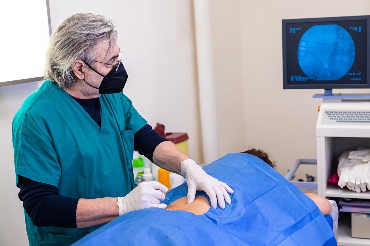
A nerve block is a procedure done to interrupt/block pain signals due to nerve dysfunction or injury. They can be used for diagnostic or treatment purposes, and their effects can be short or long-term, depending on the type being used.
A temporary nerve block may involve the application or injection that stops pain signals from transmitting for a short time.
For example, in pregnancy, an epidural injection can be used during labor and delivery.
Permanent nerve blocks involve cutting/severing or removing certain parts of a nerve to stop pain signals.
These are used in cases with severe injuries or other chronic pain conditions that have not improved with other treatment approaches.
Treatment Usage
When healthcare providers diagnose a chronic pain condition caused by nerve injury or dysfunction, they may use a nerve block to locate the area generating pain signals. They may perform electromyography and/or a nerve conduction velocity/NCV test to pinpoint the cause of chronic nerve pain. Nerve blocks can also treat chronic neuropathic pain, such as pain caused by nerve damage or compression. Nerve blocks are regularly used to treat back and neck pain caused by herniated discs or spinal stenosis. (Johns Hopkins Medicine. 2024)
Types
Three types include:
Local
Neurolytic
Surgical
All three can be used for conditions that cause chronic pain. However, neurolytic and surgical blocks are permanent and are only used for severe pain that has worsened with other treatments unable to provide relief.
Temporary Blocks
A local block is done by injecting or applying local anesthetics, like lidocaine, to a certain area.
An epidural is a local nerve block that injects steroids or analgesics into an area around the spinal cord.
These are common during pregnancy, labor, and delivery.
Epidurals can also be used to treat chronic neck or back pain due to a compressed spinal nerve.
Local blocks are usually temporary, but in a treatment plan, they can be repeated over time to manage chronic pain from conditions like arthritis, sciatica, and migraines. (NYU Langone Health. 2023)
Permanent Blocks
A neurolytic block uses alcohol, phenol, or thermal agents to treat chronic nerve pain. (National Institute of Neurological Disorders and Stroke. 2023) These procedures damage certain areas of the nerve pathway on purpose so that pain signals cannot be transmitted. A neurolytic block is mainly used for severe chronic pain cases, like pain from cancer or complex regional pain syndrome/CRPS. They are sometimes used to treat ongoing pain from chronic pancreatitis and pain in the chest wall after surgery. (Johns Hopkins Medicine. 2024) (Alberto M. Cappellari et al., 2018)
The neurosurgeon performs a surgical nerve block that involves surgically removing or damaging specific areas of the nerve. (National Institute of Neurological Disorders and Stroke. 2023) A surgical nerve block is only used for severe pain cases, such as cancer pain or trigeminal neuralgia.
Although neurolytic and surgical nerve blocks are permanent procedures, pain symptoms, and sensations can come back if the nerves are able to regrow and repair themselves. (Eun Ji Choi et al., 2016) However, symptoms and sensations may not return months or years after the procedure.
These procedures can have the potential risk of permanent nerve damage. (Anthem BlueCross. 2023) Nerves are sensitive and regenerate slowly, so a tiny error can cause side effects. (D O’Flaherty et al., 2018) Common side effects include:
Muscle paralysis
Weakness
Frequent numbness
In rare cases, the block could irritate the nerve and cause added pain.
Skilled and licensed health practitioners like surgeons, pain management physicians, anesthesiologists, and dentists are trained to perform these procedures carefully.
There is always a risk of nerve damage or injury, but the majority of nerve blocks safely and successfully decrease and help manage chronic pain. (Anthem BlueCross. 2023)
What to Expect
Individuals may feel numbness or soreness and/or notice redness or irritation near or around the area that is temporary.
There can also be swelling, which compresses the nerve and requires time to improve. (Stanford Medicine. 2024)
Individuals may be asked to rest for a certain amount of time after the procedure.
Depending on the type of procedure, individuals may have to spend a few days in a hospital.
Some pain may still be present, but that does not mean the procedure did not work.
Individuals should consult with a healthcare provider about the risks and benefits to ensure it is the right treatment.
NYU Langone Health. (2023). Nerve block for migraine (Education and Research, Issue. https://nyulangone.org/conditions/migraine/treatments/nerve-block-for-migraine
National Institute of Neurological Disorders and Stroke. (2023). Pain. Retrieved from https://www.ninds.nih.gov/health-information/disorders/pain#3084_9
Cappellari, A. M., Tiberio, F., Alicandro, G., Spagnoli, D., & Grimoldi, N. (2018). Intercostal Neurolysis for The Treatment of Postsurgical Thoracic Pain: a Case Series. Muscle & nerve, 58(5), 671–675. https://doi.org/10.1002/mus.26298
Choi, E. J., Choi, Y. M., Jang, E. J., Kim, J. Y., Kim, T. K., & Kim, K. H. (2016). Neural Ablation and Regeneration in Pain Practice. The Korean journal of pain, 29(1), 3–11. https://doi.org/10.3344/kjp.2016.29.1.3
Hospital for Special Surgery. (2023). Regional anesthesia. https://www.hss.edu/condition-list_regional-anesthesia.asp
Anthem BlueCross. (2023). Peripheral nerve blocks for treatment of neuropathic pain. (Medical Policy, Issue. https://www.anthem.com/dam/medpolicies/abc/active/policies/mp_pw_c181196.html
O’Flaherty, D., McCartney, C. J. L., & Ng, S. C. (2018). Nerve injury after peripheral nerve blockade-current understanding and guidelines. BJA education, 18(12), 384–390. https://doi.org/10.1016/j.bjae.2018.09.004
Stanford Medicine. (2024). Common patient questions about nerve blocks. (For Patients, Issue. https://med.stanford.edu/ra-apm/for-patients/nerve-block-questions.html
Individuals experiencing pain symptoms like shooting, stabbing, or electrical sensations to the latissimus dorsi of the upper back could be caused by a nerve injury to the thoracodorsal nerve. Can knowing the anatomy and symptoms help healthcare providers develop an effective treatment plan?
Thoracodorsal Nerve
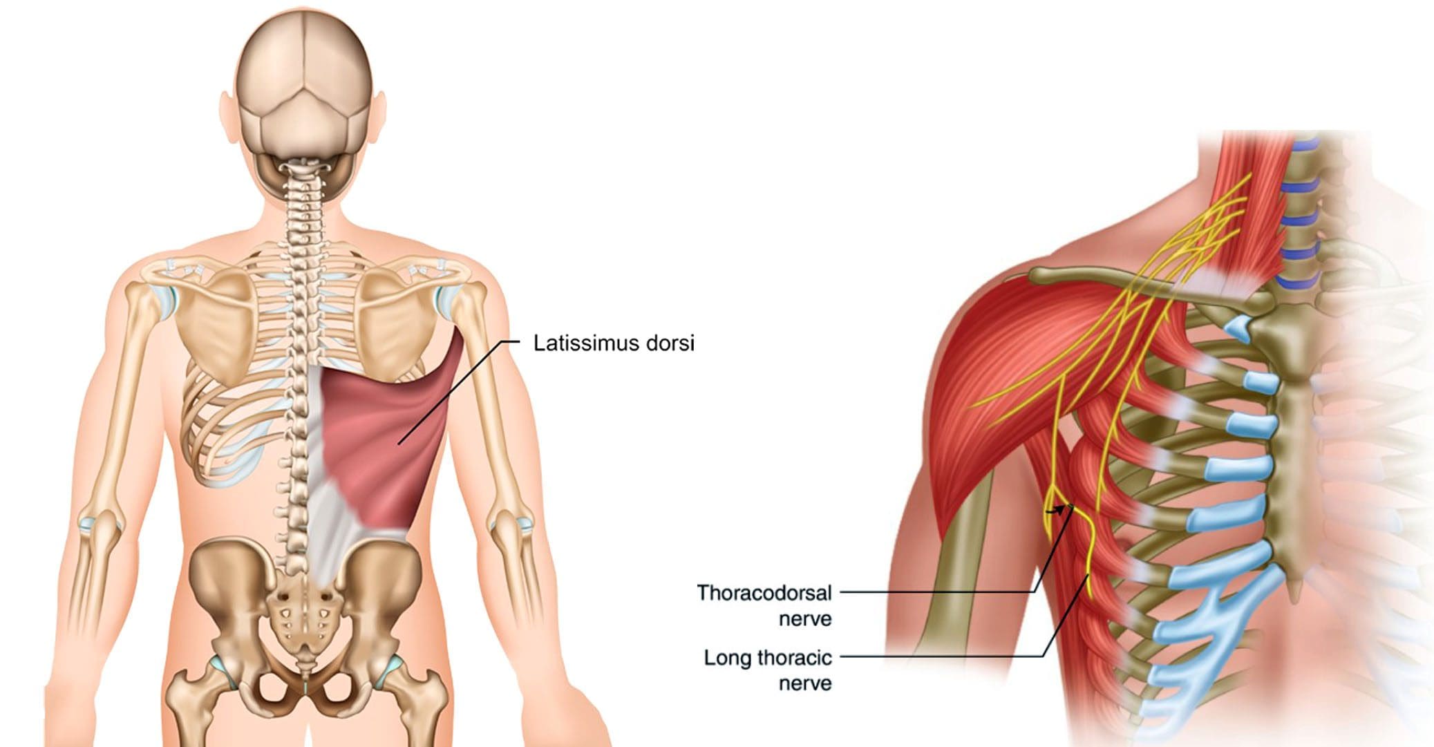
Also known as the middle subscapular nerve or the long subscapular nerve, it branches out from a part of the brachial plexus and provides motor innervation/function to the latissimus dorsi muscle.
Anatomy
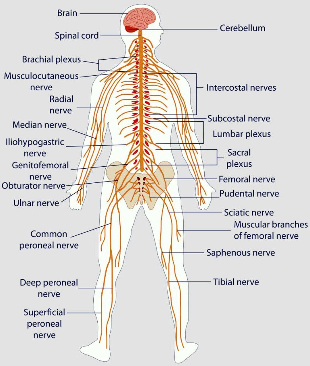
The brachial plexus is a network of nerves that stem from the spinal cord in the neck. The nerves supply most of the sensation and movement of the arms and hands, with one on each side. Its five roots come from the spaces between the fifth through eighth cervical vertebrae and the first thoracic vertebra. From there, they form a larger structure, then divide, re-combine, and divide again to form smaller nerves and nerve structures as they travel down the armpit. Through the neck and chest, the nerves eventually join and form three cords that include:
Lateral cord
Medial cord
Posterior cord
The posterior cord produces major and minor branches that include:
Axillary nerve
Radial nerve
The minor branches include:
Superior subscapular nerve
Inferior subscapular nerve
Thoracodorsal nerve
Structure and Position
The thoracodorsal nerve branches off the posterior cord in the armpit and travels down, following the subscapular artery, to the latissimus dorsi muscle.
It connects to the upper arm, stretches across the back of the armpit, forming the axillary arch, and then expands into a large triangle that wraps around the ribs and the back.
The thoracodorsal nerve lies deep in the latissimus dorsi, and the lower edge typically reaches close to the waist.
Variations
There is a standard location and course of the thoracodorsal nerve, but individual nerves are not the same in everyone.
The nerve typically branches off the posterior cord of the brachial plexus from three different points.
The lats can have a rare anatomical variation known as a Langer’s arch, which is an extra part that connects to muscles or connective tissue of the upper arm beneath the common connecting point.
In individuals with this abnormality, the thoracodorsal nerve supplies function/innervation) to the arch. (Ahmed M. Al Maksoud et al., 2015)
Function
The latissimus dorsi muscle cannot function without the thoracodorsal nerve. The muscle and nerve help:
Stabilize the back.
Pull the body weight up when climbing, swimming, or doing pull-ups.
Assist with breathing by expanding the rib cage during inhalation and contracting when exhaling. (Encyclopaedia Britannica. 2023)
Rotate the arm inward.
Pull the arm toward the center of the body.
Extend the shoulders by working with the teres major, teres minor, and posterior deltoid muscles.
Bring down the shoulder girdle by arching the spine.
Pain that can be shooting, stabbing, or electrical sensations.
Numbness, tingling.
Weakness and loss of function in the associated muscles and body parts, including wrist and finger drop.
Because of the nerve’s path through the armpit, doctors have to be cautious of the anatomical variants so they don’t inadvertently damage a nerve during breast cancer procedures, including axillary dissection.
The procedure is performed to examine or remove lymph nodes and is used in staging breast cancer and in treatment.
According to a study, 11% of individuals with axillary lymph node dissection suffered damage to the nerve. (Roser Belmonte et al., 2015)
Breast Reconstruction
In breast reconstruction surgery, the lats can be used as a flap over the implant.
Depending on the circumstances, the thoracodorsal nerve can be left intact or severed.
There is some evidence that leaving the nerve intact can cause the muscle to contract and dislocate the implant.
An intact thoracodorsal nerve may also cause atrophy of the muscle, which can lead to shoulder and arm weakness.
Graft Uses
A portion of the thoracodorsal nerve is commonly used in nerve graft reconstruction to restore function after injury, which includes the following:
Musculocutaneous nerve
Accessory nerve
Axillary nerve
The nerve can also be used to restore nerve function to the triceps muscle in the arm.
Rehabilitation
If the thoracodorsal nerve is injured or damaged, treatments can include:
Braces or splints.
Physical therapy to improve range of motion, flexibility, and muscle strength.
If there is compression, surgery may be required to alleviate the pressure.
Exploring Integrative Medicine
References
Chu B, Bordoni B. Anatomy, Thorax, Thoracodorsal Nerves. [Updated 2023 Jul 24]. In: StatPearls [Internet]. Treasure Island (FL): StatPearls Publishing; 2023 Jan-. Available from: https://www.ncbi.nlm.nih.gov/books/NBK539761/
Al Maksoud, A. M., Barsoum, A. K., & Moneer, M. M. (2015). Langer’s arch: a rare anomaly affects axillary lymphadenectomy. Journal of surgical case reports, 2015(12), rjv159. https://doi.org/10.1093/jscr/rjv159
Britannica, The Editors of Encyclopaedia. “latissimus dorsi“. Encyclopedia Britannica, 30 Nov. 2023, https://www.britannica.com/science/latissimus-dorsi. Accessed 2 January 2024.
Belmonte, R., Monleon, S., Bofill, N., Alvarado, M. L., Espadaler, J., & Royo, I. (2015). Long thoracic nerve injury in breast cancer patients treated with axillary lymph node dissection. Supportive care in cancer : official journal of the Multinational Association of Supportive Care in Cancer, 23(1), 169–175. https://doi.org/10.1007/s00520-014-2338-5
Kwon, S. T., Chang, H., & Oh, M. (2011). Anatomic basis of interfascicular nerve splitting of innervated partial latissimus dorsi muscle flap. Journal of plastic, reconstructive & aesthetic surgery : JPRAS, 64(5), e109–e114. https://doi.org/10.1016/j.bjps.2010.12.008
Facet hypertrophy is an incurable, chronic disease that affects the facet joints in the spine. Can recognizing symptoms, help in diagnosis, and treatment?
Facet Hypertrophy
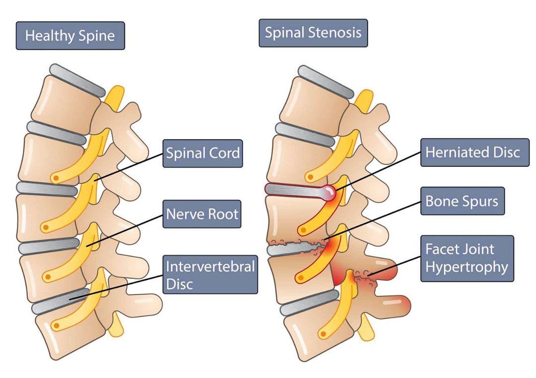
Facet hypertrophy causes the facet joints in the spine to enlarge. They are found where the vertebrae come into contact on the back of the vertebrae that form the backbone. These joints stabilize the spine when twisting and bending. Hypertrophy results when damage wears down the cartilage that cushions the bones that meet in the joint. This can include:
Aging
Wear and tear
Arthritis
Other joint diseases can damage facet joints.
Swelling, new bone growth, and bone spurs can occur as the joint tries to repair the damaged cartilage. The swelling and new bone growth can narrow the spinal canal and compress surrounding nerves, causing pain and other sensation symptoms. This ailment does not have a cure and worsens over time. The objective of treatment is to manage the pain symptoms and slow down the disease’s progress.
Types
Facet hypertrophy can be described as unilateral or bilateral.
Symptoms can have a wide range of intensity, from a dull ache to chronic, disabling pain. The location of symptoms depends on the affected joint and the nerves involved, Pain manifests when the enlarged joints and new bone growth compresses the nearby nerves. The result leads to nerve damage and the following symptoms: (Weill Cornell Medicine Brain & Spine Center. 2023) (Cedars Sinai. 2022)
Stiffness, especially when standing up or getting out of a chair.
Inability to stand straight when walking.
Inability to look up to the left or right without turning the whole body.
Reduced range of motion and mobility.
Numbness or a tingling sensation of pins and needles.
Radiating pain from the affected joint into the buttocks, hips, and upper thigh when the affected joint/s are in the lower back.
Radiating pain from the affected joint into the shoulder, neck, and back of the head when the affected joint/s are in the upper back.
Headaches when the affected joint is in the neck.
Causes
A common cause is the age-related degeneration of the joints, called spondylosis. Research indicates that more than 80% of individuals who are 40 or older have radiologic evidence of spondylosis, even though they may not have symptoms. (The University of Toledo Medical Center. N.D.) The following conditions can also increase the risk of facet hypertrophy (Weill Cornell Medicine Brain & Spine Center. 2023)
Unhealthy posture
Being overweight or obese
Sedentary lifestyle
Injury or trauma to the spine
Inflammatory conditions like rheumatoid arthritis or ankylosing spondylitis
Osteoarthritis
Genetic predisposition to the condition
Diagnosis
Diagnosis can be challenging when neck or back pain is the main complaint, as symptoms can mimic conditions such as sciatica from a herniated disc or hip arthritis. (Weill Cornell Medicine Brain & Spine Center. 2023)
A healthcare provider will conduct a complete physical examination, medical history, and discussion of symptoms.
CT scans with or without myelogram – the use of contrast dye in the space around the spinal cord.
MRI
X-rays with or without a myelogram
A diagnosis is confirmed by injecting a diagnostic block that involves administering an anesthetic injection, sometimes with an anti-inflammatory like cortisone, into the joint or nerves near the affected joint. Two injections are given at different times to confirm the effect. (Romain Perolat et al., 2018)
If immediate relief improves after each injection, the facet joint is confirmed as the source of the pain symptoms.
If the block does not decrease the pain, the facet joint is probably not the source of the pain symptoms. (Brigham and Women’s Hospital. 2023)
Treatment
There is no cure for facet hypertrophy.
The goal of treatment is to make the pain more manageable.
Conservative treatment is usually successful in making a difference.
Nonsteroidal anti-inflammatory – aspirin, ibuprofen, and naproxen.
Muscle relaxers – cyclobenzaprine or metaxalone.
Steroid injection into the facet joints.
Injection of platelet-rich plasma/PRP into the joints.
Medial Branch or Facet Block
A medial branch block injects local anesthetic near the medial nerves that connect to an inflamed joint.
Medial nerves are small nerves outside the joint space near the nerve that transmit signals and other impulses to the brain.
A facet block injects the medication outside the joint space near the nerve that supplies the joint called the medial branch.
Neurolysis
Neurolysis, also known as rhizotomy or neurotomy, is a procedure that destroys affected nerve fibers to relieve pain, reduce disability, and reduce the need for analgesics. This treatment can relieve pain for six to 12 months until the nerve regenerates, where further treatments may be necessary. (Matthew Smuck et al., 2012) Neurolysis can be performed using one of the following techniques (Romain Perolat et al., 2018)
Radiofrequency ablation RFA – the application of heat through radiofrequency.
Cryoneurolysis – the application of cold temperatures to the targeted nerve.
Chemical neurolysis – applying chemical agents, like a combination of phenol and alcohol.
Severing the nerves with surgical instrumentation.
Surgery
When one or more facet joints are severely damaged, they can become nonfunctional and painful. Surgery may be recommended when symptoms are not relieved by other therapies. (Ali Fahir Ozer, et al., 2015)
Prognosis
Facet hypertrophy is a chronic condition that progresses with age and does not affect life expectancy. (Weill Cornell Medicine Brain & Spine Center. 2023) The disorder is incurable, but symptoms can be managed with conservative therapies
A healthcare provider can help develop a treatment plan based on the extent and location of the joint affected.
Early diagnosis and treatment can help individuals achieve the best results.
Maintaining an active lifestyle and healthy weight can help prevent further joint stress. Individuals may be recommended to do regular stretching and strengthening exercises to lower inflammation, reduce stress, and improve overall health.
Facet Syndrome Treatment
References
Perolat, R., Kastler, A., Nicot, B., Pellat, J. M., Tahon, F., Attye, A., Heck, O., Boubagra, K., Grand, S., & Krainik, A. (2018). Facet joint syndrome: from diagnosis to interventional management. Insights into imaging, 9(5), 773–789. https://doi.org/10.1007/s13244-018-0638-x
Smuck, M., Crisostomo, R. A., Trivedi, K., & Agrawal, D. (2012). Success of initial and repeated medial branch neurotomy for zygapophysial joint pain: a systematic review. PM & R : the journal of injury, function, and rehabilitation, 4(9), 686–692. https://doi.org/10.1016/j.pmrj.2012.06.007
Ozer, A. F., Suzer, T., Sasani, M., Oktenoglu, T., Cezayirli, P., Marandi, H. J., & Erbulut, D. U. (2015). Simple facet joint repair with dynamic pedicular system: Technical note and case series. Journal of craniovertebral junction & spine, 6(2), 65–68. https://doi.org/10.4103/0974-8237.156049
Individuals’ feet will heat up when walking or running; however, burning feet could be a symptom of medical conditions like athlete’s foot or a nerve injury or damage. Can awareness of these symptoms help identify solutions to relieve and heal the underlying condition?
Burning Feet
Walkers and runners often experience heat in their feet. This is natural from the increased circulation, heart rate, warm or hot sidewalks, and pavement. But the feet could experience an abnormal hot or burning sensation. Usually, the overheating is caused by socks and shoes and fatigue after a long workout. The first self-care steps include trying new or specialized footwear and workout adjustments. If burning feet persist or there are signs of infection, tingling, numbness, or pain, individuals should see their healthcare provider. (Mayo Clinic. 2018)
Footwear
The shoes and how they are worn may be the cause.
First, look at the material of the shoes. They could be shoes and/or insoles that don’t circulate air. They can get hot and sweaty without proper air circulation around the feet.
When choosing running shoes, consider a mesh material that allows airflow to keep the feet cool.
Consider getting fitted for shoes that are the right size, as the feet swell when running or walking.
If the shoes are too small, air can’t circulate, creating more friction between the foot and the shoe.
Shoes that are too large can also contribute to friction as the feet move around too much.
Insoles could also contribute.
Some insoles can make the feet hot, even if the shoes are breathable.
Swap the insoles from another pair of shoes to see if they are contributing, and if so, look into new insoles.
Tips to help prevent hot feet:
Topical Ointments
Use an anti-blister/chafing topical cream to lubricate and protect the feet.
This will reduce friction and prevent blisters.
Lace Properly
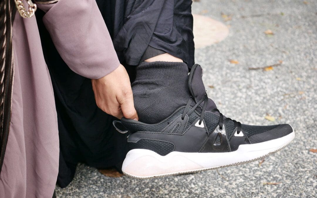
Individuals may be lacing the shoes too tight, constricting circulation, or irritating the nerves at the top of the foot.
Individuals should be able to slide one finger under the knot.
Remember that the feet will swell as walking or running commences
Individuals may need to loosen their laces after warming up.
Individuals are recommended to learn lacing techniques that will ensure they are not too tight over the sensitive areas.
Cushioning
Fatigue from long workouts or long days standing/moving can result in burning feet.
Individuals may need added cushioning in the shoes.
Look for work and athletic shoes that have added cushioning.
Shoe Allergies
Individuals may have an allergic reaction or a sensitivity to the fabric, adhesives, dyes, or other chemicals. (Cleveland Clinic. 2023) The chemicals used in production vary for leather compared to fabric and are different by brand and manufacturer.
A shoe material allergy may also result in burning, itching, and swelling.
It’s recommended to note whether symptoms only happen when wearing a specific pair of shoes.
Recommendations are to try different kinds and brands of shoes.
Socks
The sock fabric could be contributing to hot or burning feet. Steps to take can include:
Avoid cotton
Cotton is a natural fiber but is not recommended for walking and running as it holds sweat that can keep the feet wet.
It is recommended to use socks made of Cool-Max and other artificial fibers that wick sweat away and cool them down.
Wool
Wool socks can also cause itching and burning sensations.
Consider athletic socks made from itch-free wool.
Mindfulness
Individuals could be sensitive to other fabrics or dyes in socks.
Take note of which socks cause hot or burning feet symptoms.
Individuals could also be sensitive to laundry products and are recommended to try a different brand or type.
Medical Conditions
In addition to shoes and socks, medical conditions could cause and contribute to symptoms.
Athlete’s Foot
Athlete’s foot is a fungal infection.
Individuals may feel a burning sensation in the affected area.
Typically, it is itchy, red, scaling, or cracking.
Rotate shoes.
The fungus grows in damp places, therefore, it is recommended to rotate shoes to allow them to dry out between workouts.
Wash and dry the feet after walking or running.
Try home and over-the-counter solutions, powders, and remedies to treat athlete’s foot.
Peripheral Neuropathy
Individuals frequently experiencing burning feet apart from when they have been exercising could be due to nerve damage known as peripheral neuropathy. (National Institute of Neurological Disorders and Stroke. 2023) Peripheral neuropathy symptoms include pins and needles, numbness, tickling, tingling, and/or burning sensations.
Examination
Diabetes is one of the most common causes of peripheral neuropathy.
Diabetes can come on at any age.
Individuals need to learn how to protect their feet, as exercise is recommended for diabetes.
Other conditions that can produce peripheral neuropathy include:
Vitamin B-12 deficiency
Alcohol abuse
Circulatory disorders
AIDS
Heavy metal poisoning
Massage and Movement
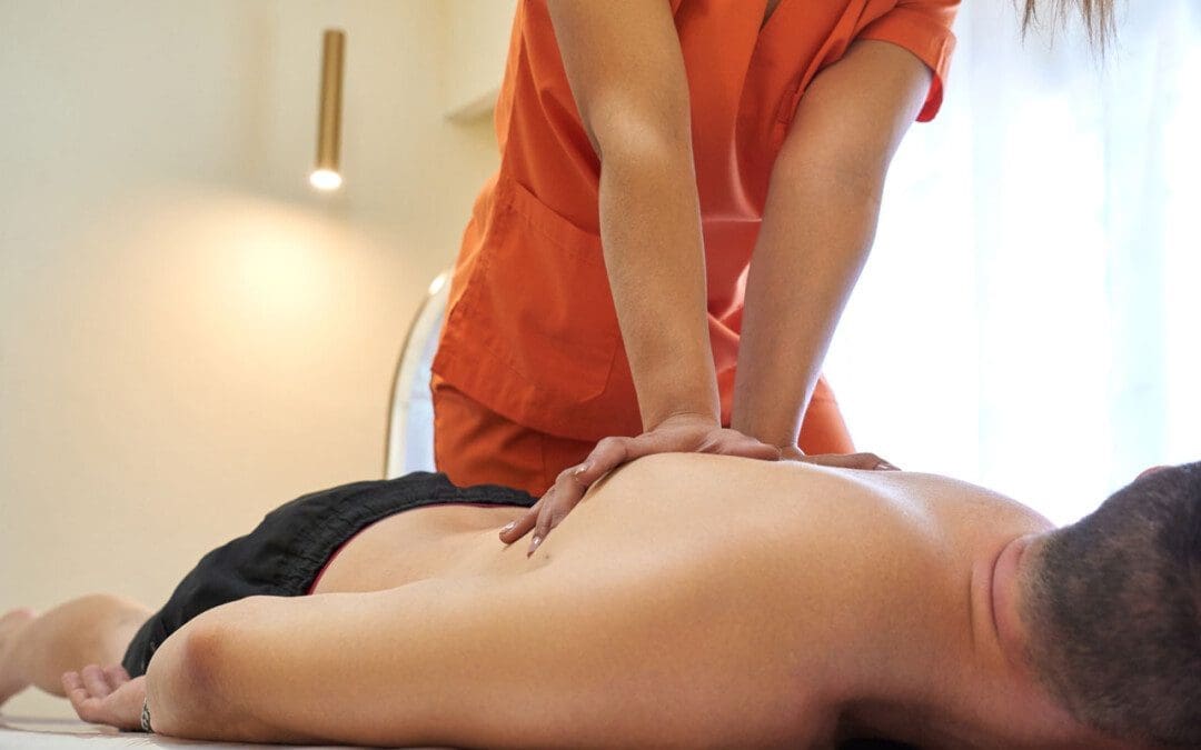
Massaging the feet also increases circulation.
Exercise such as walking is recommended for peripheral neuropathy as it improves circulation to the feet.
Individuals feeling tingling or pins and needles sensations that overtake the arms or legs could be experiencing paresthesia, which occurs when a nerve has been compressed or damaged. Can knowing the symptoms and causes help in diagnosis and treatment?
Paresthesia Body Sensations
The numbness or tingling feeling when an arm, leg, or foot has fallen asleep is not so much about blood circulation but nerve function.
Paresthesia is an abnormal sensation felt in the body due to the compression or irritation of nerves.
It can be a mechanical cause like a compressed/pinched nerve.
Or it may be due to a medical condition, injury, or illness.
The symptoms typically last for 30 minutes or less.
Shaking the affected limb often relieves the sensations.
Paresthesia usually affects only one arm or leg at a time.
However, both arms and legs can be affected, depending on the cause.
Consult a healthcare provider if the symptoms last for more than 30 minutes. Treatment may be required if paresthesia body sensations are brought on by a serious underlying cause.
Causes
Sitting with incorrect and unhealthy postures can compress a nerve and generate symptoms. However, some causes are more concerning and can include:
If the symptoms don’t go away after 30 minutes or keep returning for unknown reasons, call a healthcare provider to find out what is causing the abnormal sensations. A worsening case should be monitored by a healthcare provider.
Peripheral neuropathy caused by diabetes usually begins with a feeling of paresthesia in the foot/feet and can worsen and lead to other complications.
A healthcare provider will work with the individual to understand the symptoms and perform the appropriate diagnostic tests to determine the cause. A healthcare provider will choose the tests based on a physical examination. Common diagnostic procedures include: (Merck Manual Professional Version. 2022)
Magnetic resonance imaging – MRI of the spine, brain, or extremities.
X-ray to rule out bone abnormalities, like a fracture.
Blood tests.
Electromyography – EMG studies.
Nerve conduction velocity – NCV test.
If paresthesia is accompanied by back or neck pain, a healthcare provider may suspect a compressed/pinched spinal nerve.
If the individual has a history of diabetes that is poorly controlled, they may suspect peripheral neuropathy.
Treatment
Treatment for paresthesia depends on the diagnosis. A healthcare provider can help determine the best course of action for the specific condition.
Nervous System
If symptoms are triggered by a central nervous condition like MS, individuals will work closely with their healthcare provider to get the appropriate treatment.
If paresthesia is caused by compression of a spinal nerve, like sciatica, individuals may be referred to a chiropractor and physical therapy team to release the nerve and pressure. (Julie M. Fritz, et al., 2021)
A physical therapist may prescribe spinal exercises to relieve compression of the nerve and restore normal sensations and motion.
Strengthening exercises to restore flexibility and mobility may be prescribed if weakness presents along with paresthesia body sensations.
Herniated Disc
If a herniated disc is causing the abnormal sensations, and there has been no improvement with conservative measures, a healthcare provider may suggest surgery to relieve pressure on the nerve/s. (American Association of Neurological Surgeons. 2023)
In surgical procedures like a laminectomy or discectomy, the objective is to restore nerve function.
Post-surgery, individuals may be recommended to a physical therapist to help regain mobility.
National Institute of Neurological Disorders and Stroke. (2023) Paresthesia.
American Association of Neurological Surgeons. (2023) Herniated disc.
National Institute of Diabetes and Digestive and Kidney Diseases. (2018) Peripheral neuropathy.
Merck Manual Professional Version. (2022) Numbness.
Razazian, N., Yavari, Z., Farnia, V., Azizi, A., Kordavani, L., Bahmani, D. S., Holsboer-Trachsler, E., & Brand, S. (2016). Exercising Impacts on Fatigue, Depression, and Paresthesia in Female Patients with Multiple Sclerosis. Medicine and science in sports and exercise, 48(5), 796–803. https://doi.org/10.1249/MSS.0000000000000834
Fritz, J. M., Lane, E., McFadden, M., Brennan, G., Magel, J. S., Thackeray, A., Minick, K., Meier, W., & Greene, T. (2021). Physical Therapy Referral From Primary Care for Acute Back Pain With Sciatica : A Randomized Controlled Trial. Annals of internal medicine, 174(1), 8–17. https://doi.org/10.7326/M20-4187
Individuals diagnosed with peripheral neuropathy, or with small fiber neuropathy, can understanding symptoms and causes help with potential treatments?
Small Fiber Neuropathy
Small fiber neuropathy is a specific classification of neuropathy, as there are different types, which are nerve injury, damage, disease, and/or dysfunction. Symptoms can result in pain, loss of sensation, and digestive and urinary symptoms. Most cases of neuropathy like peripheral neuropathy involve small and large fibers. Common causes include long-term diabetes, nutritional deficiencies, alcohol consumption, and chemotherapy.
Small fiber neuropathy is diagnosed after diagnostic testing showing it is clear that the small nerve fibers are involved.
The small nerve fibers detect sensation, temperature, and pain and help regulate involuntary functions.
Isolated small-fiber neuropathy is rare, but research is ongoing on the type of nerve damage and potential treatments. (Stephen A. Johnson, et al., 2021)
Small fiber neuropathy is not specifically dangerous but is a sign/symptom of an underlying cause/condition that is damaging the body’s nerves.
Pain – symptoms can range from mild or moderate discomfort to severe distress and can happen at any time.
Loss of sensation.
Because the small nerve fibers help with digestion, blood pressure, and bladder control – symptoms of autonomic dysfunction can vary and can include:
Constipation, diarrhea, incontinence, urinary retention – the inability to completely drain the bladder.
If there is progressing nerve damage, the intensity of the pain can decrease, but the loss of normal sensation and autonomic symptoms can worsen. (Josef Finsterer, Fulvio A. Scorza. 2022)
Hypersensitivity to touch and pain sensations can cause pain without a trigger.
The loss of sensation can make individuals unable to accurately detect sensations of touch, temperature, and pain in affected areas, which can lead to various types of injuries.
Although more research is needed, certain disorders that were not considered neuropathies may have small fiber neuropathy components involved.
A study suggested that neurogenic rosacea, a skin condition, could have some elements of small fiber neuropathy. (Min Li, et al., 2023)
These small nerve fibers are distributed throughout the body including the tops of the fingers and toes, trunk, and internal organs.
These fibers are usually located in the superficial areas of the body, such as close to the skin’s surface. (Mohammad A. Khoshnoodi, et al., 2016)
The small nerve fibers that get damaged are involved in transmitting pain and temperature sensations.
Most nerves have a special type of insulation called myelin that protects them and increases the speed of nerve impulses.
Small nerve fibers may have a thin sheath, making them more susceptible to injury and damage at earlier stages of conditions and diseases. (Heidrun H. Krämer, et al., 2023)
Individuals At Risk
Most types of peripheral neuropathy cause damage to the small and large peripheral nerve fibers. Because of this, most neuropathies are a mix of small-fiber and large-fiber neuropathy. Common risk factors for mixed fiber neuropathy include: (Stephen A. Johnson, et al., 2021)
Diabetes
Nutritional deficiencies
Overconsumption of alcohol
Autoimmune disorders
Medication toxicity
Isolated small-fiber neuropathy is rare, but there are conditions that are known to contribute to the cause and include: (Stephen A. Johnson, et al., 2021)
Sjogren Syndrome
This autoimmune disorder causes dry eyes and mouth, dental problems, and joint pain.
It can also cause nerve damage throughout the body.
Fabry Disease
This condition causes a buildup of certain fats/lipids in the body that can lead to neurological effects.
Amyloidosis
This is a rare disorder that causes a buildup of proteins in the body.
The proteins can damage tissues like the heart or nerves.
Lewy Body Disease
This is a neurological disorder that causes dementia and impaired movement and can lead to nerve damage.
Lupus
This is an autoimmune disease that affects joints, skin, and sometimes nerve tissue.
Viral Infection
These infections typically cause a cold or gastrointestinal/GI upsetness.
Less often they can cause other effects like small fiber neuropathy.
These conditions have been seen to cause isolated small-fiber neuropathy or begin as small-fiber neuropathy before progressing to the large nerve fibers. They can also begin as a mixed neuropathy, with small and large fibers.
Progression
Often the damage progresses at a relatively moderate rate, leading to added symptoms within months or years. The fiber nerves that are affected by the underlying condition usually progressively deteriorate, regardless of where they are located. (Mohammad A. Khoshnoodi, et al., 2016) Medications can help alleviate damage to the peripheral nerves. For individuals that are diagnosed in the early stage, it is possible to stop the progression, and potentially prevent involvement of the large fibers.
Treatments
Treatment toward preventing the progression requires controlling the underlying medical condition with treatment options depending on the cause. Treatments that can help prevent the progression include:
Blood sugar control for individuals with diabetes.
Immune suppression for control of autoimmune diseases.
Plasmapheresis – blood is taken and the plasma is treated and returned or exchanged for the treatment of autoimmune diseases.
Symptom Treatment
Individuals can get treatment for the symptoms that will not reverse or cure the condition but can help with temporary relief. Symptomatic treatment can include: (Josef Finsterer, Fulvio A. Scorza. 2022)
Pain management can include medications and/or topical analgesics.
Physical therapy – stretching, massage, decompression, and adjustments to keep the body relaxed and flexible.
Rehabilitation to help improve coordination, which can be impaired by loss of sensation.
Medications to relieve GI symptoms.
Wearing specialized clothes such as neuropathy socks to help with foot pain symptoms.
Treatment and medical management of neuropathies usually involve a neurologist. A neurologist may prescribe medication to help alleviate pain symptoms and provide medical interventions like immunotherapy if there is concern that an autoimmune process could be the cause. Additionally, treatment could include the care of a physical medicine and rehabilitation physician or a physical therapy team to provide stretches and exercises to help strengthen the body and maintain mobility and flexibility.
Peripheral Neuropathy Myths & Facts
References
Johnson, S. A., Shouman, K., Shelly, S., Sandroni, P., Berini, S. E., Dyck, P. J. B., Hoffman, E. M., Mandrekar, J., Niu, Z., Lamb, C. J., Low, P. A., Singer, W., Mauermann, M. L., Mills, J., Dubey, D., Staff, N. P., & Klein, C. J. (2021). Small Fiber Neuropathy Incidence, Prevalence, Longitudinal Impairments, and Disability. Neurology, 97(22), e2236–e2247. https://doi.org/10.1212/WNL.0000000000012894
Finsterer, J., & Scorza, F. A. (2022). Small fiber neuropathy. Acta neurologica Scandinavica, 145(5), 493–503. https://doi.org/10.1111/ane.13591
Krämer, H. H., Bücker, P., Jeibmann, A., Richter, H., Rosenbohm, A., Jeske, J., Baka, P., Geber, C., Wassenberg, M., Fangerau, T., Karst, U., Schänzer, A., & van Thriel, C. (2023). Gadolinium contrast agents: dermal deposits and potential effects on epidermal small nerve fibers. Journal of neurology, 270(8), 3981–3991. https://doi.org/10.1007/s00415-023-11740-z
Li, M., Tao, M., Zhang, Y., Pan, R., Gu, D., & Xu, Y. (2023). Neurogenic rosacea could be a small fiber neuropathy. Frontiers in pain research (Lausanne, Switzerland), 4, 1122134. https://doi.org/10.3389/fpain.2023.1122134
Khoshnoodi, M. A., Truelove, S., Burakgazi, A., Hoke, A., Mammen, A. L., & Polydefkis, M. (2016). Longitudinal Assessment of Small Fiber Neuropathy: Evidence of a Non-Length-Dependent Distal Axonopathy. JAMA neurology, 73(6), 684–690. https://doi.org/10.1001/jamaneurol.2016.0057
Hamstring muscle injuries are common, especially in athletes and individuals with physically demanding jobs. Is there a better chance of full recovery with surgical repair and post-op rehabilitation?
Hamstring Muscle Tear
Most often, hamstring muscle injuries are partial tears of the muscle. These types of injuries are muscle strains that occur when the muscle fibers are stretched beyond their normal limits. Complete tears of the hamstring muscle are unusual, but they do occur in both athletes and non-athletes. Determining the optimal treatment plan depends on:
The severity of the tendon tear
The expectations of the injured individual.
Incomplete tears are when the hamstring muscle is stretched too far, but not completely detached.
Complete tears usually occur at the top of the muscle where the tendon tears away from the pelvis.
A complete tear usually occurs when there is a sudden flexion of the hip and extension of the knee joint – when the muscle contracts in this position, it gets stretched beyond its limits.
Basic hamstring strains can be treated with simple steps – rest, ice, anti-inflammatory medications, and conservative therapies.
Symptoms
Symptoms of a hamstring muscle strain can include pain, bruising, swelling, and movement difficulty. (American Academy of Orthopaedic Surgeons. 2021) Individuals who sustain this injury typically experience sudden sharp pain. Signs of a tear can include:
Sharp pain where the buttock and thigh meet.
Difficulty walking.
Sitting can be difficult as the edge of a chair can place pressure directly on the injury.
Spasms and cramping sensations in the back of the thigh.
Weakness in the leg, specifically when bending the knee or lifting the leg behind the body.
Numbness or burning sensations as a result of sciatic nerve irritation.
Swelling and bruising in the back of the thigh – over time it can travel down to the back of the knee and calf and possibly into the foot.
With a complete hamstring tear, there is usually significant swelling and bruising that develops in the back of the thigh.
Diagnosis
The symptoms can be difficult to spot in the early stages which is why X-rays of the hip or thigh are usually obtained.
In some situations, a fragment of bone can get pulled off the pelvis along with the hamstring muscle attachment. MRI testing can be performed to evaluate the attachment and can define critical features of a complete hamstring muscle tear, including: (American Academy of Orthopaedic Surgeons. 2021)
The number of tendons involved.
Complete versus incomplete tearing.
The amount of retraction – the amount the tendons have pulled back.
This will guide the development of treatment.
Treatment
The treatment of a complete tear will depend on different factors. The other variable is the patient and their expectations.
Treatment is more aggressive in younger individuals like high-level athletes.
Treatment is less aggressive in middle-aged individuals.
Often a single tendon tear can be treated non-surgically.
When one tendon is involved, it is typically not pulled very far from its normal attachment and will develop scar tissue in a positive position.
Conversely, when three tendons have been torn, they usually pull more than a few centimeters away from the bone. These cases have better results with surgical repair. (UW Health. 2017)
Surgeons will use patient characteristics – high-level athletes or less physically active individuals – to guide treatment recommendations.
Rehabilitation
Rehabilitation following surgery can take 3-6 months or longer.
The first six weeks limit weight-bearing with the use of crutches.
Patients may be recommended to wear a brace to reduce tension on the repaired hamstring tendons.
Strengthening does not begin until three months post-op, and even light activities are usually delayed. (UW Health. 2017)
Because this injury can have a long recovery time, some individuals may choose nonsurgical treatment.
Sometimes these individuals experience symptoms of discomfort from sitting and may exhibit long-term weakness of the hamstring muscle.
Full recovery from a complete hamstring muscle injury takes time. Studies have shown high-level athletes are able to resume competitive sports after the repair and rehabilitation of an acute hamstring muscle injury. (Samuel K. Chu, Monica E. Rho. 2016)
Delaying surgical treatment may not always lead to optimal results.
When the tendon is torn away from its normal attachment, it begins to scar around the surrounding soft tissues.
When there is a delay of more than a few weeks following the initial injury, regaining the full length of the tendon and muscle can be challenging.
This could delay the rehabilitation process and may limit the potential for full recovery. (Ho Yoon Kwak, et al., 2011)
With severe injuries, there is a better chance of full recovery with surgical repair but could involve a long recovery and commitment to a post-op rehabilitation plan.
Chu, S. K., & Rho, M. E. (2016). Hamstring Injuries in the Athlete: Diagnosis, Treatment, and Return to Play. Current sports medicine reports, 15(3), 184–190. https://doi.org/10.1249/JSR.0000000000000264
Kwak, H. Y., Bae, S. W., Choi, Y. S., & Jang, M. S. (2011). Early surgical repair of acute complete rupture of the proximal hamstring tendons. Clinics in orthopedic surgery, 3(3), 249–253. https://doi.org/10.4055/cios.2011.3.3.249
IFM's Find A Practitioner tool is the largest referral network in Functional Medicine, created to help patients locate Functional Medicine practitioners anywhere in the world. IFM Certified Practitioners are listed first in the search results, given their extensive education in Functional Medicine