The sciatic nerve is formed through a combination of motor and sensory fibers based on the spinal nerves of the lower back L4 to S3, known as the lumbosacral plexus. It is the largest and longest nerve in the human body and about as wide as an adult thumb. It begins at the base of the spine, runs along the back of each leg, and ends at the foot supplying the areas with fresh blood and nutrients. There are sciatic nerve branches that consist of primary branches and smaller branches.
Sciatic Nerve Branches
The nerve splits into two main branches near the back of the knee called the popliteal fossa.
This fossa is located slightly above the joint behind the knee.
The popliteal fossa is a diamond-shaped space that acts as the conduit for the blood vessels and nerves.
Primary branches
From the popliteal fossa:
The tibial nerve continues down the back of the calf to the heel and bottom of the foot.
The common peroneal nerve, aka common fibular nerve, travels sideways along the outer part of the knee to the outer border of the lower leg and foot.
Both nerves convert into small sensory nerves in the calf that supply the outer side of each foot.
The sciatic nerve breaks off into smaller branches, known as collaterals, that include:
These are muscle branches that supply the muscles in the thigh, including the hamstring group and the adductor magnus muscles along the inner thigh.
Other small branches supply the leg and foot muscles.
Articular branches supply the back of the hip joint, the back and side of the knee joint.
The sciatic nerve does not supply structures in the buttocks; however, pain commonly radiates/spreads into this area when the nerve is impaired, impinged, and inflamed.
Blood Supply
The delivery of nutrients to the sciatic nerve is done through blood vessels that also contribute to the nerve’s function. Any interruption of blood flow to the sciatic nerve can cause pain and dysfunction. The sciatic nerve and the sciatic nerve branches receive their blood supply from two sources that include:
The extrinsic system is made up of nearby arteries and veins.
The intrinsic system includes arteries and veins that run along the nerve and are embedded deep in a sheath known as the epineurium of connective tissue that envelops the nerve.
The intrinsic blood supply can be affected by conditions like diabetes, which can contribute to symptoms associated with diabetic neuropathy.
Both systems connect at various junction points.
Nerve Function
The combination of sensory and motor fibers that make up the sciatic nerve provides the essential functions in the lower limbs allowing the body to:
Stand
Walk
Run
Climb
Lift
A healthy sciatic nerve is well protected around the low back and buttock muscles where it starts, and it cannot be palpated or felt by touching or pressing on the area. When the nerve gets inflamed, injured, or pinched, the leg can feel stiff and inflexible when trying to move and can lead to pain, weakness, and tingling in the lower back, buttock, leg/s, and feet.
Anatomical Variations of the Nerve
Individuals can have variations in the anatomical structure of the sciatic nerve. These variations are considered normal, but they can increase the risk of developing sciatica brought on by impingement, entrapment, or irritation of the nerve root/s. Variations in sciatic nerve branches include:
The nerve divides above the piriformis muscle; one portion passes through the piriformis, with the other portion exiting the pelvis below the muscle. This is the most common variation.
The nerve divides above the piriformis muscle; one portion passes through the piriformis, with the other portion exiting the pelvis above the muscle.
The nerve divides above the piriformis, with one portion traveling in front while the other travels behind it.
Undivided sciatic nerve exits through the piriformis muscle.
Undivided sciatic nerve exits from behind the top part of the piriformis.
Around 10% of individuals have a nerve that divides above the popliteal fossa and does not merge but courses down in two separate branches.
The sciatic nerve and the sciatic nerve branches are significant components of the body. It supplies motor functions to move the legs and feet and provides sensory functions along the nerve path. Keeping the sciatic nerve healthy is key in helping to prevent back and spinal issues. Chiropractic can help realign the sciatic nerve and educate on maintaining the nerve’s health.
Body Composition
Fitness Motivation
New workout routine
Individuals that don’t feel like returning to previous workout routines are recommended to try out other fitness options. If the gym isn’t cutting it or there is burnout with the current routine, switch things up. This can include:
Virtual group classes.
1-on-1 personal training.
Outdoor activities.
All are valid options to explore if in a rut with the current routine.
The important thing is to find what works for you.
Allow the body to rest
Individuals may want to push it to the limit to get back into shape, but rest days are essential for healthy muscle development and improved performance.
Noticing the body is more sore and exhausted after a workout is an indication that the body needs rest. This also includes:
Maintaining proper hydration.
Stretching out the muscles regularly.
Taking days off from exercising are necessary to:
Prevent muscle fatigue.
Reduce the risk of injury.
Allow for adequate muscle recovery.
Long term commitment is key
It can be discouraging to commit to a workout schedule only to notice minor changes to strength and fitness.
However, small improvements do accumulate over time.
Small increases over time can have a huge impact on overall strength and fitness.
Keep the bigger picture in mind to remain positive.
References
Davis D, Vasudevan A. Sciatica. [Updated 2019 Feb 28]. In: StatPearls [Internet]. Treasure Island (FL): StatPearls Publishing; 2019 Jan-. Available from: https://www.ncbi.nlm.nih.gov/books/NBK507908/
Barral J, Croibier A. Manual Therapy for the Peripheral Nerves. Elsevier Health Sciences; 2007.
Ryan MM, Jones HR Jr. Mononeuropathies. In: Neuromuscular Disorders of Infancy, Childhood, and Adolescence. Elsevier; 2015:243-273. doi:10.1016/b978-0-12-417044-5.00014-7
Walking for a healthy back. This simple form of exercise can:
Trim the waistline.
Elevate mood.
Reduce the risk of chronic disease.
Improve back health.
Chiropractors recommend walking because of the ease of the workout and the health benefits it provides. It is a simple, low-impact exercise that can significantly improve the body’s overall health in a short amount of time. It improves back health by:
Strengthening the muscles that support the spine.
Improves posture.
Facilitates strong circulation.
Improves bone strength.
Walking For A Healthy Back
Strengthens Muscles
Walking engages all of the muscles which keep the body upright, including the core, leg, and back muscles. Muscle strength increases, providing optimal support of the spine.
Optimize Bone Health
Bone is living tissue like the muscles, and exercise stimulates bone the same way as muscle, gradually increasing strength.
Studies have found that walking improves bone density and reduces bone loss.
Poor posture is one of the most common reasons why individuals have back pain.
Poor posture affects mobility and places a significant amount of strain on the back.
Walking a few times each week engages and strengthens the back muscles keeping the body straight.
Reduces Weight
Many individuals have lower back pain that is caused by excess weight.
The added weight causes the front of the body to shift forward, placing additional strain on the lower back.
Walking reduces the load on the lower back.
Improves Flexibility and Range of Motion
Combined with stretching, walking improves flexibility and range of motion, making it easier to perform everyday activities reducing the risk of back injuries.
Improves Circulation to the Spinal structures
Walking improves blood circulation, delivers nutrients to the soft tissues, and removes toxins.
Added Benefits include:
Stress relief.
Better sleep.
Improved skin tone.
Lower risk of diabetes.
Reduced risk of depression.
Improved cardiovascular health that lowers the risk of:
High blood pressure.
Heart disease.
Stroke.
Before Exercising
Before beginning an exercise program, it’s essential to consult a doctor or chiropractor for individuals that have not exercised for a while or are dealing with underlying condition/s. They will educate and recommend how much exercise is appropriate given their current fitness level and overall health. To maximize the benefits of walking and prevent injuries:
Use High-Quality Tennis or Walking Shoes
Walking is much more enjoyable and safer when the body is comfortable.
Improving comfort levels is by using a pair of high-quality walking shoes or trainers.
They will provide proper support, cushioning, and adequate traction.
Maintain Proper Posture
Stay aware of body position when walking. A few key points to keep in mind:
Place the heel down first.
Then roll through each part of the foot, ending on the point of the toes.
Keep the shoulders back and head up.
Lift from the hips to reduce the impact on the lower joints.
Keep a slight bend in the arms and smoothly swing them back and forth.
Turn Walking Into a Healthy Habit
In the beginning, aim for at least 5 to 7 walks each week that last 25 minutes.
Speed does not matter as the objective is to get out and walk.
Once walking starts turning into a healthy habit with improvements in health, then start walking faster and longer.
Interval Walking
Interval walking involves short periods of high-intensity walking followed by alonger period of slow walking.
This increases cardiovascular fitness and muscle strength.
Begin with a 1-minute interval of fast walking.
This is followed by 2 minutes of slower walking.
Take On Easy Obstacles
Makes the walks more challenging by walking up or down a hill.
They will help strengthen the arms, shoulders, and upper back.
Body Composition
Building Lean Body Mass
Lean Body Mass is the body’s total weight minus the fat. This includes all the weight of the muscles, organs, and total body water. The best way to develop muscle and Lean Body Mass is to adopt a resistance training program. As stronger muscles are developed, the size and amount of the muscle cells increases. The muscles then require more intracellular water, which allows them to function at optimal levels. As the muscles grow and take in more water, Lean Body Mass increases.
References
Morris, J N, and A E Hardman. “Walking to health.” Sports medicine (Auckland, N.Z.) vol. 23,5 (1997): 306-32. doi:10.2165/00007256-199723050-00004
Nauman, Javaid et al. “Walking in the Fast Lane: High-Intensity Walking for Improved Fitness and Health Outcomes.” Mayo Clinic proceedings vol. 94,12 (2019): 2378-2380. doi:10.1016/j.mayocp.2019.10.020
Vanti, Carla et al. “The effectiveness of walking versus exercise on pain and function in chronic low back pain: a systematic review and meta-analysis of randomized trials.” Disability and rehabilitation vol. 41,6 (2019): 622-632. doi:10.1080/09638288.2017.1410730
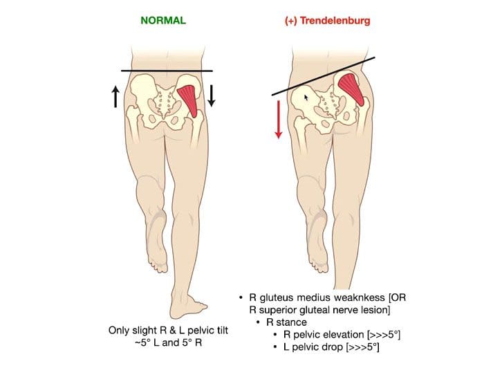
A Trendelenburg gait is an abnormal walking gait resulting from a defective or weakened hip abductor.The gluteal musculature is the primary musculature that includes the gluteus medius and gluteus minimus muscles. Weakness in these muscles causes sagging/dropping of the pelvis on the opposite side while walking. There will be a noticeable side-to-side motion if the glutes are too weak to support the body’s weight when walking. It can look as though the individual is limping or missing a step. Individuals can minimize the effects with foot orthotics, core strengthening, chiropractic, and physical therapy.
Trendelenburg Gait Causes
This gait often results from straining the hip abductor muscles during physical activity. Exercises specifically for the glutes done improperly are a common cause. When improper exercise form is the cause, the abnormal gait usually goes away as muscle inflammation fades. The gait can also present after total hip replacement surgery, as the procedure requires incisions in the gluteus medius muscle. This can weaken the muscle causing an abnormal gait. Weakness in these muscles can also be caused by:
Nerve damage or dysfunction in the nerves that run through the gluteal minimus and medius muscles.
Osteoarthritis is a type of arthritis that occurs when joint cartilage starts to wear down.
Muscular dystrophy is a condition that causes the muscles and bones to become weak over time.
Poliomyelitisis a condition associated with polio that weakens the muscles.
Cleidocranial dysostosis is a condition present from birth that can cause your bones to develop improperly.
Symptoms
The walking gait is made up of two phases:
Swing – When one leg moves forward.
Stance – The other leg stays still and maintains balance.
The main symptom of Trendelenburg gait can be seen when one leg swings forward and the hip drops down and move outward. This is because the hip abductor of the other leg is too weak to support the weight. Individuals may lean back or to the side slightly when walking to maintain balance, or they may lift the foot higher off the ground with each step to avoid losing balance or tripping as the pelvis shifts unevenly.
Diagnosis
Abnormal hip movement during a swing of one or both legs can give a doctor enough evidence to diagnose a Trendelenburg gait. A doctor will observe the individual’s walk in front and behind to get a detailed view. A doctor will also use the Trendelenburg test to diagnose the condition. The doctor will instruct the individual to lift one leg for 30 seconds. If the individual cannot keep the hips parallel with the ground while lifting, it could indicate Trendelenburg gait. X-rays of the hip will be used to identify any causes of weakness in the gluteus minimus or medius.
Treatment Options
Treatment options will depend on the severity and cause of the gait.
Medication
If the gait is causing pain, over-the-counter nonsteroidal anti-inflammatory NSAIDs, like ibuprofen or acetaminophen, will help ease symptoms.
In severe cases, a doctor may prescribe cortisone injections to help reduce pain.
Foot Orthotics
A doctor could also recommend using a foot orthotic in one or both shoes to compensate the hip abductor muscle weakness.
Chiropractic, Physical Therapy, and Exercise
Chiropractic and physical therapy can help adjust, realign, and strengthen the muscles to regain control of the Trendelenburg gait. The chiropractor or physical therapist will move the legs in various directions to help the joints become more accustomed to moving in certain directions and increase muscle strength and resistance. Exercises that can strengthen the hip abductor muscles include:
Lie on the side and extend the leg straight out.
Lie on the floor and move one leg up, over the other, and back in the opposite direction.
Step sideways and onto an elevated surface, then back down again.
Talk with a doctor or chiropractor before beginning any new exercise routine so they can recommend specific exercises and educate on proper form.
Complications
If left untreated, moderate-to-severe cases of Trendelenburg gait can become debilitating, leading to severe complications. These include:
Pinched nerves.
Sciatica.
Pain, stiffness, or grinding in the hips.
Loss of range of motion in the hips and gait.
Losing the ability to walk, which could require the use of a walker or wheelchair.
Trendelenburg gait is treatable with special shoes, orthotics, and exercises designed to strengthen the hip abductor muscles. Chiropractic and physical therapy can help limit the condition’s impact on the body’s health, the ability to walk, and reduce the risk of complications.
Body Composition
Heart-Healthy Foods
Citrus
The bright and tangy fruits are packed with vitamins and unique plant compounds known as polyphenols that can help lower blood pressure naturally.
However, it’s important to note that grapefruit and grapefruit juice could interact with certain prescription medications.
Beans and Lentils
Foods high in magnesium, potassium, and fiber can help maintain healthy blood pressure.
This is where beans and legumes come in, as they are high in fiber, potassium, and magnesium.
Individuals that swapped beans and lentils noticed a lower blood pressure, whether or not they had been diagnosed with hypertension.
Pumpkin Seeds
These seeds are packed with potassium, magnesium, and arginine.
Arginine is an amino acid used to make nitric oxide, which helps the blood vessels relax and dilate, allowing lower blood pressure.
A study found that postmenopausal women who took 3 grams of pumpkin seed oil daily for six weeks saw a significant decrease in their systolic blood pressure.
Garlic
Garlic contains nitric oxide, which has been shown to relax blood vessels.
Kyolic garlic, in particular, has been shown to help with arterial stiffness and can improve cholesterol levels.
References
Feyh, Andrew et al. “Role of Dietary Components in Modulating Hypertension.” Journal of Clinical & experimental cardiology vol. 7,4 (2016): 433. doi:10.4172/2155-9880.1000433
Gandbhir, Viraj N., et al. “Trendelenburg Gait.” StatPearls, StatPearls Publishing, 19 August 2021.
Giangarra CE, et al. (2018). Clinical orthopedic rehabilitation: A team approach.sciencedirect.com/science/book/9780323393706
Gilliss AC, et al. (2010). Use of osteopathic manipulative treatment to manage compensated Trendelenburg gait caused by sacroiliac somatic dysfunction.
jaoa.org/article.aspx?articleid=2093879
Maricelli JW, et al. (2016). Trendelenburg-like gait, instability and altered step patterns in a mouse model for limb-girdle muscular dystrophy 2i. DOI:
10.1371/journal.pone.0161984
Mayo Clinic Staff. (2017). Osteoarthritis.mayoclinic.org/diseases-conditions/osteoarthritis/home/ovc-20198248
Michalopolous N, et al. (2016). A personalized monitoring and recommendation framework for kinetic dysfunctions: The Trendelenburg gait. DOI: 10.1145/3003733.3003786
Reaching, twisting, walking, and driving are everyday activities that require upper and lower back strength. An aching back can easily affect daily activities, generate frustration, anger, and affect all-around health. The more back muscle strength an individual has, the more they can accomplish far more without injury. Immense power is not required to protect the body from a back injury. All that is needed is regular, consistent physical activity and exercise. A balance of body strength is vital for preventing injury. However, overdoing one fitness exercise or physical activity can imbalance musculature, leading to injury. Because the back/spine is the central part of the body, complete and proper care is necessary for optimal health and wellness. For individuals experiencing sore, aching, and tired muscles, here are some exercises that will help in the process.
Alternating Arm and Leg Extensions
Alternating extensions help build strength and coordination in the core areas. The back muscles increase their efficiency by creating muscle memory that supports the work shared by all the torso muscles. Upper and lower back muscles must work together to maintain a healthy balance and not overwork each other, causing strain and fatigue.
Start by placing hands and knees on the floor with the head directly between shoulders and facing toward the floor.
Feet are directly in line behind the buttocks and resting on the floor.
Hips and shoulders rest above the knees and hands.
Raise the right hand straight ahead with the arm at full length.
At the time same time, raise the left leg straight behind the body.
Try to keep the arm and leg as straight as possible.
Hold for 10 seconds.
Switch sides.
Repeat three to eight times, depending on strength level.
If it is difficult, a modified option is to raise the arm and leg separately.
Plank Hold
These can help build back muscles and strengthen the arms, legs, and the front torso area. Plank holds are a recommended starting point. Plank holds can be done on the elbows, palms of the hands, or closed fist hands. The key is to keep the shoulders, hips, and ankles straight like a wood plank parallel to the floor.
Place hands and feet directly on the floor like doing a push–up.
Toes should be on the floor.
Keep the abdominals tight and buttocks lifted to prevent straining the lower back.
Face straight down.
Hold for a count of 10.
Repeat three times.
For those with an aching back, keeping the hips level with the shoulders could be challenging at the beginning.
With practice, it will become easier; then, the individual is recommended to increase the length of time until 30 seconds is achieved.
Then increase the challenge to try more than three repetitions.
A modification for beginners is to start with the body resting on the floor, stomach down.
Then raise the body into the start position from the floor.
Hip Raises
Hip raises help to strengthen the lower back muscles to unite and support the lower half of the body. Training the body to work cooperatively is critical for reducing the aching and pain from muscle imbalance.
Rest the body flat on the floor, facing upward.
Place the hands flat at the body’s sides.
Knees should be about shoulder-width apart.
Keep the feet flat on the floor
Pull the feet toward the buttocks.
Look straight up.
Raise the hips as high as possible while pressing down with the hands.
Physical activity that keeps blood moving throughout the body. Examples include yoga, gardening, and dancing.
While the back is healing, go at a gentle even pace for any activity. Jerking and quickly stopping can be hard on joints and discs. When injured, the other muscles try to compensate to avoid causing a flare-up that could worsen the injury and/or create a new injury.
Aching Back Muscles
Strength-building exercises are great for preventing injury and avoiding re-injury. However, avoid overreaching or overstretching with any of the activities. Continuous aching or painful back muscles could indicate something else is occurring that could be:
The beginning of an arthritic condition causing inflammation.
Back muscle tear/s.
Pregnancy.
Body Composition
Sarcopenia – Loss of Skeletal Muscle Mass and Strenght Causes
Decreased Physical Activity
Physical inactivity is one of the primary contributors to sarcopenia.
Sedentariness can exacerbate the effects of sarcopenia.
Regular resistance exercise can help maintain muscle mass and build muscular strength.
Decrease in motor neurons
Aging is accompanied by a loss of motor neurons caused by cell death.
This can lead to a decrease in muscle fibers and size.
This decrease leads to:
Impaired performance
Reduced functional capacity
Decreased ability to perform everyday tasks.
Increase in Pro-inflammatory Cytokines
Poor diet and exercise also promote the storage of visceral fat.
This type of fat tissue produces pro-inflammatory cytokines.
This can accelerate muscle breakdown.
Obesity and muscle weakness are associated with high levels of pro-inflammatory cytokines.
References
Alfuth, M, and D Cornely. “Chronischer lumbaler Rückenschmerz : Vergleich zwischen Mobilisationstraining und Training der rumpfstabilisierenden Muskulatur” [Chronic low back pain : Comparison of mobilization and core stability exercises]. Der Orthopade vol. 45,7 (2016): 579-90. doi:10.1007/s00132-016-3233-1
Kim, Beomryong, and Jongeun Yim. “Core Stability and Hip Exercises Improve Physical Function and Activity in Patients with Non-Specific Low Back Pain: A Randomized Controlled Trial.” The Tohoku journal of experimental medicine vol. 251,3 (2020): 193-206. doi:10.1620/tjem.251.193
Smith, Benjamin E et al. “An update of stabilization exercises for low back pain: a systematic review with meta-analysis.” BMC musculoskeletal disorders vol. 15 416. 9 Dec. 2014, doi:10.1186/1471-2474-15-416
Suh, Jee Hyun et al. “The effect of lumbar stabilization and walking exercises on chronic low back pain: A randomized controlled trial.” Medicine vol. 98,26 (2019): e16173. doi:10.1097/MD.0000000000016173
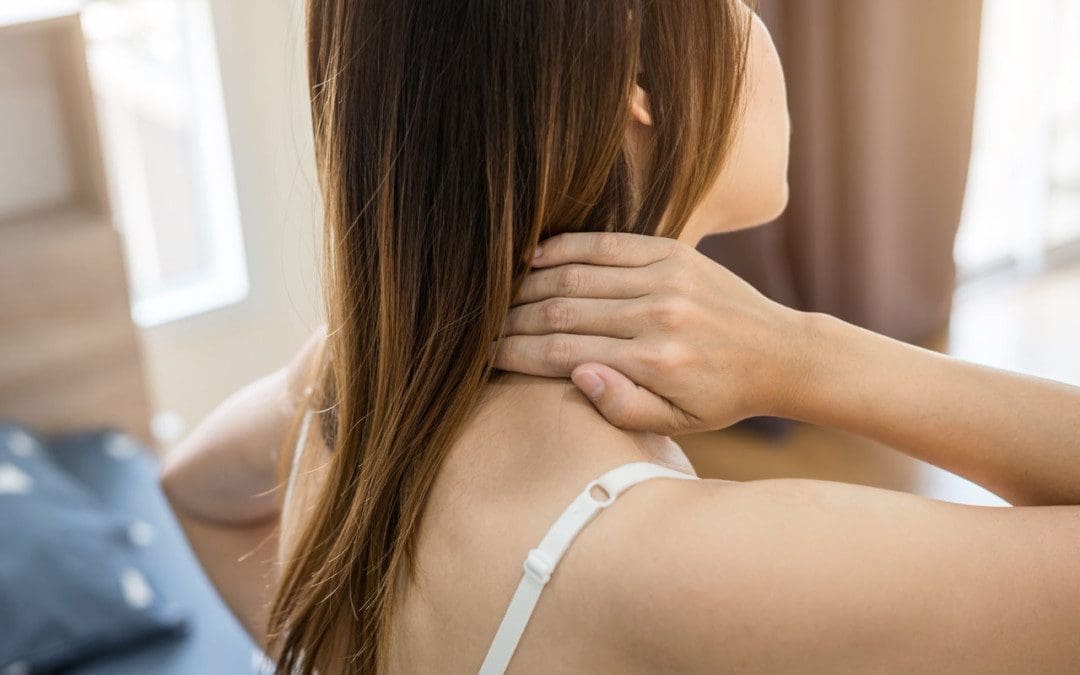
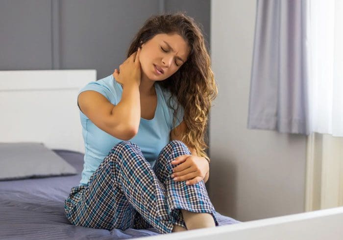
Waking up with neck soreness, stiffness, achiness, and pain can take a toll throughout the day. Individuals, that experience this regularly wonder what happened while laying down in bed? Individuals can wake up with one or a combination of these symptoms after sleeping. A few ways to prevent neck pain after sleeping and self-care to relieve any symptoms.
What Is Happening?
The spine keeps the body upright and moving and regularly resists gravity and other forces acting upon it. The neck, aka the cervical spine, is a little more delicate. The neck has the important job of holding up the head. The human head weighs around 10 to 12 lbs, and that’s using proper posture. According to a study, the head’s weight can increase up to 60 lbs. with a 60-degree tilt. This can happen from looking down at a phone for too long. All that weight makes the muscles that support the head and neck work overtime contributing to fatigued muscles.
Then when sleeping, cervical spinal misalignment starts to set in, producing torticollis. Torticollis, aka wry neck, is a condition where the neck gets twisted or tilted at an awkward angle. Babies can be born with it, known as congenital torticollis, and individuals can develop it from various sources. It can be temporary, chronic, and it can be caused by acute trauma. Torticollis is not considered a condition like ankylosing spondylitis but more like a symptom with overlapping sources.
The neck’s ligaments can become irritated and inflamed.
Neck muscle spasms can cause soreness and inflammation.
Either of these can be caused by sleeping in an awkward position or by using the wrong pillow.
Waking With Neck Pain
When waking up with neck pain, it could be that the pillow no longer provides sufficient support, the pillow is too thick, placing the neck in an awkward position, the individual’s sleeping position strains the muscles and ligaments, or a combination. It is usually a pillow that is too soft with no support that causes neck pain. Maintaining spinal alignment when sleeping is just as crucial as during the day, as it helps to prevent overly taxing the muscles and ligaments.
How to control posture when sleeping?
The pillow could be the answer. A firm pillow will keep the spine in a straight line from the atlas, which is the first cervical vertebra/C1, down to the coccyx or the tailbone. The way an individual sleeps also affects how they wake up. The most recommended sleeping position for individuals with morning neck pain is on the back. Back sleeping might not work for everyone as it can aggravate conditions like sleep apnea. If that is the case, sleeping on the side is the next recommended position. It is recommended to avoid sleeping on the stomach. The head could slip down the pillow edge causing the head to be in a tilted position. This can place added pressure on the nerves that start in the neck, leading to further neck pain or radiculopathy pain that spreads out to the arms or legs.
What To Do?
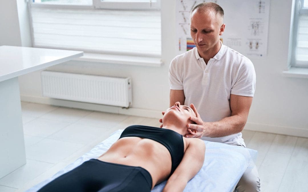
If neck pain presents after waking, get some ice or a cold pack on it. Try 20 minutes on, 20 minutes off. This will reduce inflammation. Also, over-the-counter nonsteroidal anti-inflammatory medications like ibuprofen can help. If neck pain continues, switch from ice to heat also 20 minutes on, 20 off. If the pain is caused by spasming muscle/s, heat can relax the area and increase blood circulation. A gentle massage on and around the area can help spread the circulation and ease the spasm.
Stretching the neck
Stretching the neck will keep the muscles loose and reduce the risk of ligament, muscle and tendon strains, and torticollis.
Body composition change and losing fat mass are also related to sleep. Losing fat requires the body to be in a caloric deficit. This means having the body use more energy than the body takes in. This is accomplished by restricting calories through diet or increasing calories used through physical activity/exercise. However, most individuals utilize a combination. This can be referred to as calories in/calories out. Losing sleep can sabotage fat loss goals by stealing both the calories in and calories out.
References
Hansraj, Kenneth K. “Assessment of stresses in the cervical spine caused by posture and position of the head.” Surgical technology international vol. 25 (2014): 277-9.
Preventing Neck Pain from Sleeping: National Sleep Foundation. (n.d.) “How to Prevent Neck Pain While Sleeping.” sleep.org/articles/prevent-neck-pain-while-sleeping/
Neck crepitus is a grinding sound that comes from moving or rotating the neck. Usually, it is not something to worry about, as the body is a sound system that generates various noises. For example, when hungry, the stomach rumbles. After digestion, the body releases the gasses through a burp. The bones can also generate neck cracking or popping sounds with regular movements. This unusual sensation is known as crepitus.
Crepitus
Crepitus or crepitation is a scientific term that describes joint movements sounds. Sounds can include:
Popping
Cracking
Snapping
Grinding
However, crepitus can happen in any moveable joints in the body. An example could be a neck cracking or popping sound when looking over the shoulder.
Why the Neck So Susceptible
The cervical spine consists of seven segments, and each segment has multiple joints that interact with the segments above and below it. The cervical spine is a flexible system that protects the neurologic structures while maintaining head and neck stability. This flexibility and the multiple joints at each level can wear down, leading to arthritis and neck crepitus.
Other Symptoms
Neck crepitus can present without other symptoms. But it can also be associated with other severe symptoms that include:
Neck pain
Instability
Weakness
Numbness
Diminished manual dexterity
Difficulty walking
Risk Increases With Age
Neck crepitus can present at any age; however, the risk increases as the body ages. Some individuals may have neck crepitus symptoms more often. For example, the neck cracking or popping sounds could present just a few times a month. However, other individuals could have cracking, popping sounds daily or even throughout the day. Neck crepitus can increase or decrease in frequency. Symptoms could present for several days before the sensations stop entirely.
Possible Causes
Neck crepitus can have various causes, and multiple factors can also overlap to generate these sensations.
Articular Pressure Changes
Natural lubricating lining and fluid are found within the body’s joints. Small gas bubbles can form within the synovial joints, including the facet joints. When the bubbles collapse, they are released, creating cracking noises in the joints. The sounds can happen with regular everyday movements. This also occurs when a chiropractor or physical therapist performs spinal manipulations.
Tendon or Ligament Movement
Tendons are the tissue that connects the muscles to the bones, and Ligaments connect the bones. A tendon in motion can also make noises when sliding around a bone or over another tendon or ligament. The cracking can be caused by tight tissues and muscles from aging or muscles that have become weak/deconditioned.
Bones Grinding
Osteoarthritis, known as spondylosis in the spine, can cause the facet joints that connect the vertebrae to degenerate. The protective cartilage wears down, and the vertebral bones start to rub against each other. This can produce a grinding noise. However, the grinding can result from disc degeneration, which reduces the cushioning between the vertebrae.
When to Consult A Physician
If neck crepitus presents without other symptoms, it’s usually not serious. When neck crepitus presents with other symptoms, it is recommended to contact a doctor. These symptoms include:
If pain spreads out and runs down the arm or there is difficulty completing fine motor tasks like writing your name or getting dressed, consult a doctor. These symptoms can be caused by spinal cord or nerve root compression. Sometimes, neck crepitus can show up after a different health issue. For example, if an individual notices neck sounds weeks after cervical spine surgery, the spine surgeon can determine if the two are connected. A recent fall or car accident could also cause symptoms to present. If the crepitus presents almost every time with joint movement, there could be compromised joint function.
Treatment and Prevention
There are various treatment options for neck crepitus. It is recommended to start with conservative treatment like physical therapy and chiropractic pain management. Imaging scans are necessary to see if there are signs of compression on the spinal cord or nerves. Treatment objectives are to remove the pressure from the neural structures and restore the spine’s stability. Cervical traction is another form of treatment. Consult a physician, spine specialist, or chiropractor to properly diagnose the issues, figure out what is going on, and develop a personalized treatment plan if necessary.
Body Composition
Sugar Replacements
Sugar substitutes can help with weight control and diabetes by allowing individuals to eat sweets without raising blood sugar levels. Sugar replacements are additives that add sweetness to food without the calories of sugar. Some sugar substitutes are synthetically made, while others are natural. Sugar replacements include:
Sucralose
This artificial sweetener comes from sucrose and contains no calories. It is highly sweeter than sugar and can be found in grocery stores.
Fructose
This sweetener comes in crystalline form or high-fructose corn syrup, which is often used for baking. Fructose is sweeter than sugar and has been linked to early diabetes.
Stevia
This sweetener is extracted from the stevia rebaudiana plant species. It is calorie-free and can help manage and improve cholesterol levels.
Aspartame
Only a tiny amount is necessary, as this artificial sweetener is 200 times sweeter than sugar. It contains four calories per gram.
Aspartame has been associated with cancer, dementia, and depression. But research has not found a direct correlation, and currently, recommended amounts are safe to consume.
References
Mohamad, I et al. “Swollen neck and crepitus after bouts of cough.” Malaysian family physician: the official journal of the Academy of Family Physicians of Malaysia vol. 8,3 49-50. 31 Dec. 2013
Nguyen, Andrew B et al. “Crepitus: an uncommon complication of a common procedure.” The Annals of thoracic surgery vol. 91,4 (2011): e63. doi:10.1016/j.athoracsur.2011.01.031
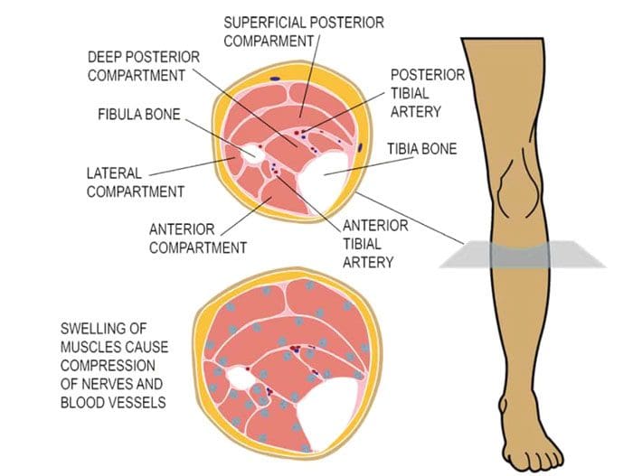
Compartment syndrome is a condition that causes pressure within a group of muscles to build up to dangerous levels. This pressure build-up begins to decrease blood flow, not allowing proper circulation, nutrients, and oxygen from getting to the nerves and muscle cells. The syndrome can be acute or chronic, and surgery can be required. Acute compartment syndrome is considered a medical emergency, usually caused by a severe injury and requires immediate treatment; otherwise, it can lead to permanent muscle damage. Chronic compartment syndrome or exertional compartment syndrome is usually not a medical emergency and is often caused by physical exertion.
The fascia does not stretch or expand because its job is to keep the tissues in place. If compartmental pressure builds up, swelling and bleeding may occur. When the tissues don’t have enough blood to provide the proper amount of oxygen and nutrients, the tissues begin to die, leading to permanent damage. Because the fascia does not stretch if there is swelling or bleeding within a compartment, this increases pressure on the:
Capillaries
Nerves
Muscles in that compartment.
Blood circulation does not reach the compartment to supply oxygen and nutrients.
Nerve and muscle cells get damaged.
Compartment syndrome most often takes place in the lower leg’s anterior/front calf compartment.
However, it can also develop in other areas like the:
Legs
Arms
Hands
Feet
Buttocks
Acute
The typical symptom is pain, specifically when the muscle in the compartment is stretched.
The pain is more intense than the injury itself.
Flexing, contracting, or stretching the muscles increases the pain.
Tingling or burning sensations may present.
Muscle tightness or fullness sensation like bloating.
Numbness or paralysis are late symptoms that usually indicate severe to permanent tissue injury.
The acute syndrome develops after a severe injury, like an automobile accident or from a broken bone. Injuries and conditions that can cause acute compartment syndrome include:
Fractures
Muscle contusion/bruise that goes beyond just a bump. Two examples include a motorcycle falling on the rider’s leg or a football player getting hit in the leg intensely.
Constricting bandages – Casts and bandages that are too tight can cause the blockage of blood. If symptoms develop, remove or loosen any constricting bandages. If it is from a cast, contact the doctor immediately.
Anabolic steroids – Taking steroids is a possible factor in compartment syndrome.
Blood circulation restoration after a blockage.
When sleeping, a blood vessel can get blocked. Lying for a long time in a position that causes a limb to go to sleep, then shifting, moving, or getting up can contribute to the condition. This type of development can happen in individuals with neurological damage or who do not realize what is occurring. This can happen after intense intoxication with alcohol and/or drugs.
Surgical repair of a damaged blood vessel that was blocked can result in compartment swelling.
Permanent disability and tissue death can result unless the pressure is relieved.
Chronic Physical Exertion
The pain and swelling from the chronic condition are caused by vigorous physical activity/exercise. It most often occurs in the leg. Individuals that participate in activities with repetitive motions have an increased risk. Physical activities/sports include:
Running
Biking
Swimming
This is usually not dangerous and is often relieved by discontinuing the specific exercise/s or physical activity for a while. Symptoms include:
Pain during exercise.
Cramping during exercise.
Numbness
Moving the foot is difficult.
Muscle bulge can be seen.
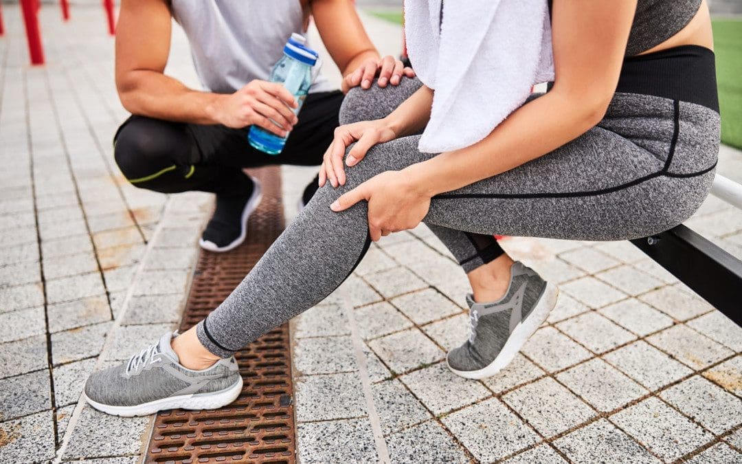
Chiropractic Treatment
Leg pain should not be ignored for long as the problems could escalate into severe/dangerous territory. Chiropractic treatment is highly effective in the detection and treatment of leg pain. Chiropractors are experts in the neuromusculoskeletal system. Their expertise in promoting physical function applies to the whole body’s systems, including the:
Muscles
Bones
Ligaments
Nerves
Tendons
They are trained to diagnose and treat developing and chronic musculoskeletal problems and know when to seek specialized medical care when necessary.
Body Composition
Can’t Individuals Just Exercise More and Eat Whatever They Want?
No individuals cannot just exercise/move more and eat whatever they want if they are serious about losing excess weight. A healthy diet and exercise are essential parts of the formula for effective weight loss. One study shows that being aware of diet in quality and quantity overtakes just exercising when achieving and maintaining healthy body composition changes as a vital part of maintaining a healthy lifestyle. Evaluating the effects of diet, exercise, or a combination of both revealed that long-term success was most significant in the mix of diet and exercise. Individuals can exercise vigorously, but losing weight can be very difficult if they have unhealthy eating habits or cannot stick to a healthy diet. The individual can develop other health problems from an unhealthy diet.
References
Braver, Richard T. “Chronic Exertional Compartment Syndrome.” Clinics in podiatric medicine and surgery vol. 33,2 (2016): 219-33. doi:10.1016/j.cpm.2015.12.002
Joubert, Sonia V, and Manuel A Duarte. “Chronic Exertional Compartment Syndrome in a Healthy Young Man.” Journal of chiropractic medicine vol. 15,2 (2016): 139-44. doi:10.1016/j.jcm.2016.04.007
Schmidt, Andrew H. “Acute compartment syndrome.” Injury vol. 48 Suppl 1 (2017): S22-S25. doi:10.1016/j.injury.2017.04.024
Vajapey, Sravya, and Timothy L Miller. “Evaluation, diagnosis, and treatment of chronic exertional compartment syndrome: a review of current literature.” The Physician and sportsmedicine vol. 45,4 (2017): 391-398. doi:10.1080/00913847.2017.1384289
IFM's Find A Practitioner tool is the largest referral network in Functional Medicine, created to help patients locate Functional Medicine practitioners anywhere in the world. IFM Certified Practitioners are listed first in the search results, given their extensive education in Functional Medicine