For individuals going through post surgery, injury rehabilitation, illness and/or chronic condition management, can physical therapy isometric exercises help?
Isometric Exercise
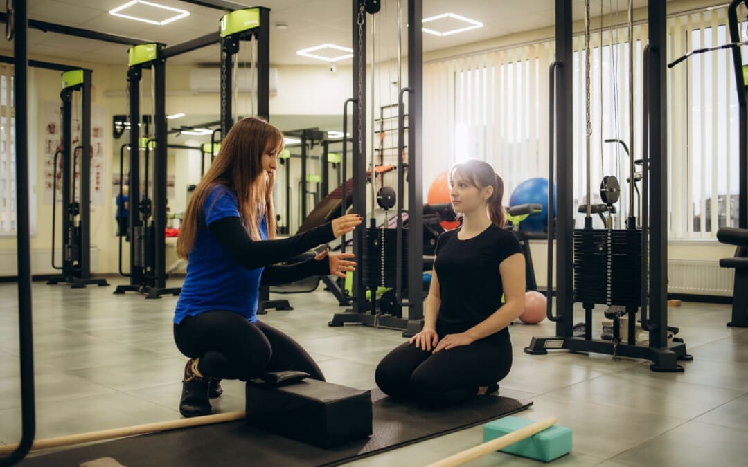
Isometric exercises are used in physical therapy to help build muscle endurance, improve range of motion, relieve pain, and reduce blood pressure more effectively than other types of exercise. Because they don’t involve joint movement, they are a solid starting point for rehabilitation and are suitable for individuals with a limited range of motion. They can be performed by pushing against an immovable object, like a wall, or by having a therapist provide resistance. Examples of isometric exercises include:
A physical therapist/PT may have a patient perform isometric exercises after injury or illness. During an isometric contraction, the muscle does not change in length, and there is no motion around the joint surrounding the muscle/s. (Rhyu H. S. et al., 2015)
When To Use
Isometric muscular contractions can be used at any time during physical rehabilitation and strengthening or a home exercise program and are regularly used with the following (Rhyu H. S. et al., 2015)
Post-surgery
When muscles cannot contract forcefully enough to move the joint it surrounds.
To help increase neuromuscular input to a specific muscle/s.
When injury or condition frailty makes other forms of exercise dangerous and not beneficial.
A healthcare provider or physical therapist should be consulted first if isometrics are used in a rehabilitation program.
Benefits
The benefits of using isometric exercise after injury or surgery may include the following:
No special equipment is necessary to perform isometric exercises.
The ability to safely contract a muscle while protecting a surgical incision or scar tissue.
The muscles can be strengthened in a specific range of motion around a joint. (NikolaidouO. et al., 2017)
A physical therapist can help determine whether isometric exercise benefits the specific condition.
Effectiveness
Isometric exercise is very effective after injury or surgery. However, when a muscle is contracted isometrically, it gains strength in a very small area and with a short range of motion. For example, an isometric shoulder external rotation performed with the arm at the side will only strengthen the rotator cuff muscles in the specific position that the arm is in. (NikolaidouO. et al., 2017).
Strength gains are specific to the joint’s position during the exercise.
Individuals who want to strengthen their gluteal muscles in their hip using isometrics would have to contract their glute muscles in one specific position for several reps.
Once several reps of the exercise in one position have been performed, the individual moves their hip joint into a new position and repeats the gluteal contractions in the new position.
This makes the exercise time-consuming, but it is perfect for injury rehabilitation, preventing and avoiding worsening or further injuries.
How to Perform
To perform isometric exercises, all that is needed is something stable to push against. (Rhyu H. S. et al., 2015) For example, to strengthen the shoulder muscles:
Stand next to a wall and try to lift an arm out to the side.
Allow the hand to press against the wall so no motion occurs at the shoulder joint.
Once pressed against the wall, hold the contraction for 5 to 6 seconds and slowly release it.
Perform 6 to 10 repetitions of the exercise.
This could be one set of completed isometric exercises for the shoulder muscles.
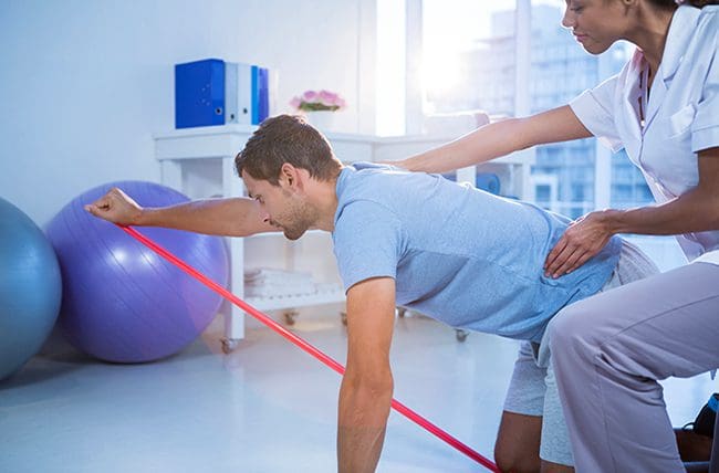
Elastic resistance bands or tubing can also be used to perform isometric exercises. Hold the tubing in a specific position and then move the body away from the anchor point instead of moving the joint. The muscles will contract against the increased resistance of the elastic tubing, and no motion will occur at the joint. A physical therapist can show and train on how to perform isometric exercises with the bands.
Neuromuscular Stimulation
Isometric exercise can strengthen muscles and help improve the neuromuscular recruitment of the muscles being trained. This enhances muscle contraction and expedites gains in muscle recruitment while protecting the joint. Isometric exercise can also be used during physical therapy using neuromuscular electrical stimulation (NMES). (Fouré A. et al., 2014) For example, a PT may use NMES to improve muscular function for individuals who have difficulty contracting their quadriceps after knee surgery and may be instructed to perform isometric quad-setting exercises during the session.
Injury Medical Chiropractic and Functional Medicine Clinic
A physical therapist can use isometric exercises to help individuals injured or have had surgery and are experiencing difficulty with normal functional mobility by improving their strength during recovery. The exercises can safely enhance the function and stability of the muscles and return individuals to the previous level of activity and function. Injury Medical Chiropractic and Functional Medicine Clinic works with primary healthcare providers and specialists to develop an optimal health and wellness solution. We focus on what works for you to relieve pain, restore function, and prevent injury. Regarding musculoskeletal pain, specialists like chiropractors, acupuncturists, and massage therapists can help mitigate the pain through spinal adjustments that help the body realign itself. They can also work with other medical professionals to integrate a treatment plan to resolve musculoskeletal issues.
Personal Injury Rehabilitation
References
Rhyu, H. S., Park, H. K., Park, J. S., & Park, H. S. (2015). The effects of isometric exercise types on pain and muscle activity in patients with low back pain. Journal of Exercise Rehabilitation, 11(4), 211–214. https://doi.org/10.12965/jer.150224
Nikolaidou, O., Migkou, S., & Karampalis, C. (2017). Rehabilitation after Rotator Cuff Repair. The Open Orthopaedics Journal, 11, 154–162. https://doi.org/10.2174/1874325001711010154
Fouré, A., Nosaka, K., Wegrzyk, J., Duhamel, G., Le Troter, A., Boudinet, H., Mattei, J. P., Vilmen, C., Jubeau, M., Bendahan, D., & Gondin, J. (2014). Time course of central and peripheral alterations after isometric neuromuscular electrical stimulation-induced muscle damage. PloS one, 9(9), e107298. https://doi.org/10.1371/journal.pone.0107298
“Various problems with the sacrum make up or contribute to a significant portion of lower back problems. Can understanding the anatomy and function help prevent and treat back injuries?”
The Sacrum
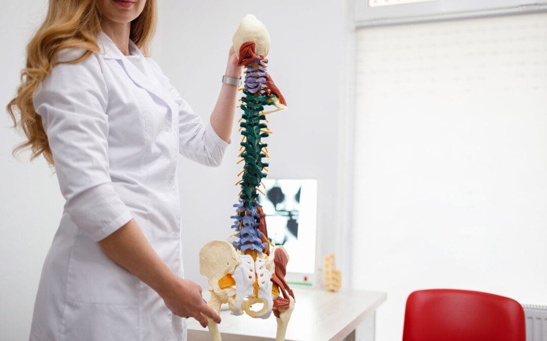
The sacrum is a bone shaped like an upside-down triangle located at the base of the spine that helps support the upper body when sitting or standing and provides pelvic girdle flexibility during childbirth. It comprises five vertebrae that fuse during adulthood and connect to the pelvis. This bone takes and endures all of the body’s pressure and stress from everyday activities and movements.
Formation
Humans are born with four to six sacral vertebrae. However, fusion does not take place in all sacral vertebrae simultaneously:
Fusion starts with the S1 and S2.
As the individual gets older, the overall shape of the sacrum begins to solidify, and the vertebrae fuse into a single structure.
The process usually starts in the mid-teens and finishes in the early to mid-twenties.
It is believed to start earlier in females than males.
The sacrum in a female is wider and shorter and has a more curved top or the pelvic inlet.
The male sacrum is longer, narrower, and flatter.
Structure
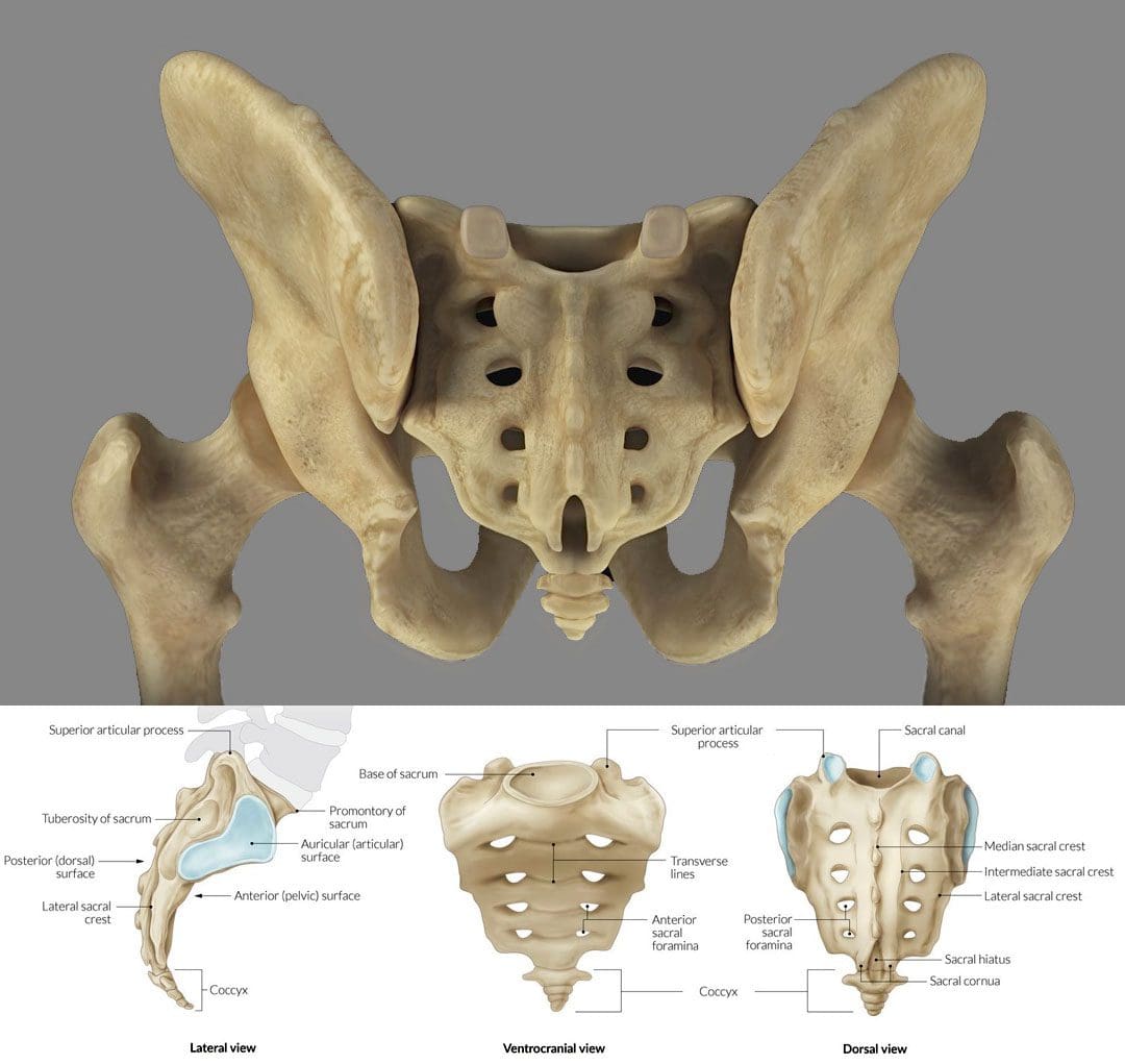
The sacrum is an irregular bone that makes up the back/posterior third of the pelvic girdle. There is a ridge across the front/anterior portion of the S1 vertebra known as the sacral promontory. Small holes/foramen on both sides of the sacrum are left over after the vertebrae fuse together. Depending on the number of vertebrae, there can be three to five foramen on each side, though there are usually four. (E. Nastoulis, et al., 2019)
Each anterior foramen is typically wider than the posterior or dorsal/backside foramen.
Each sacral foramina/plural of foramen provides a channel for the sacral nerves and blood vessels.
Small ridges develop between each of the fused vertebrae, known as transverse ridges or lines.
The top of the sacrum is called the base and is connected to the largest and lowest of the lumbar vertebrae – L5.
The bottom is connected to the tailbone/coccyx, known as the apex.
The sacral canal is hollow, runs from the base to the apex, and serves as a channel at the end of the spinal cord.
The sides of the sacrum connect to the right and left hip/iliac bones. The attachment point is the auricular surface.
Right behind the auricular surface is the sacral tuberosity, which serves as an attachment area for the ligaments that hold the pelvic girdle together.
Location
The sacrum is at the level of the lower back, just above the intergluteal cleft or where the buttocks split. The cleft starts at around the level of the tailbone or coccyx. The sacrum is curved forward and ends at the coccyx, with the curvature being more pronounced in females than males. It connects to the L5 lumbar vertebra by way of the lumbosacral joint. The disc between these two vertebrae is a common source of low back pain.
On either side of the lumbosacral joint are wing-like structures known as the sacral ala, which connect to the iliac bones and form the top of the sacroiliac joint.
These wings provide stability and strength for walking and standing.
Anatomical Variations
The most common anatomical variation applies to the number of vertebrae. The most common is five, but anomalies have been documented, including individuals with four or six sacral vertebrae. (E. Nastoulis, et al., 2019)
Other variations involve the sacrum’s surface and curvature, where the curvature differs widely between individuals.
In some cases, the first and second vertebrae do not fuse and remain separately articulated.
Failure of the canal to completely close during formation is a condition known as spina bifida.
Function
Studies on the sacrum are ongoing, but some proven functions include:
It serves as an anchor point for the spinal column to attach to the pelvis.
It provides stability for the body’s core.
It acts as a platform for the spinal column to rest on when sitting.
It facilitates childbirth, providing pelvic girdle flexibility.
It supports upper body weight when sitting or standing.
It provides extra stability for walking, balance, and mobility.
Conditions
The sacrum can be a main source or focal point for lower back pain. It is estimated that 28% of men and 31.6% of women aged 18 years or older have experienced low back pain in the past three months. (Centers for Disease Control and Prevention. 2020) Conditions that can cause sacrum pain symptoms include.
Sacroiliitis
This is a common condition of sacroiliac/SI joint inflammation.
A doctor only makes the diagnosis when all other possible causes of pain have been ruled out, known as a diagnosis of exclusion.
About half of all chordomas form in the sacrum, but the tumors can also develop elsewhere in the vertebral column or at the base of the skull. (National Library of Medicine. 2015)
Spina Bifida
Individuals can be born with conditions that affect the sacrum.
Spina bifida is a congenital condition that can arise from the malformation of the sacral canal.
Unlocking the Secrets of Inflammation
References
Gruss, L. T., & Schmitt, D. (2015). The evolution of the human pelvis: changing adaptations to bipedalism, obstetrics and thermoregulation. Philosophical transactions of the Royal Society of London. Series B, Biological sciences, 370(1663), 20140063. https://doi.org/10.1098/rstb.2014.0063
Nastoulis, E., Karakasi, M. V., Pavlidis, P., Thomaidis, V., & Fiska, A. (2019). Anatomy and clinical significance of sacral variations: a systematic review. Folia morphologica, 78(4), 651–667. https://doi.org/10.5603/FM.a2019.0040
Barros, G., McGrath, L., & Gelfenbeyn, M. (2019). Sacroiliac Joint Dysfunction in Patients With Low Back Pain. Federal practitioner : for the health care professionals of the VA, DoD, and PHS, 36(8), 370–375.
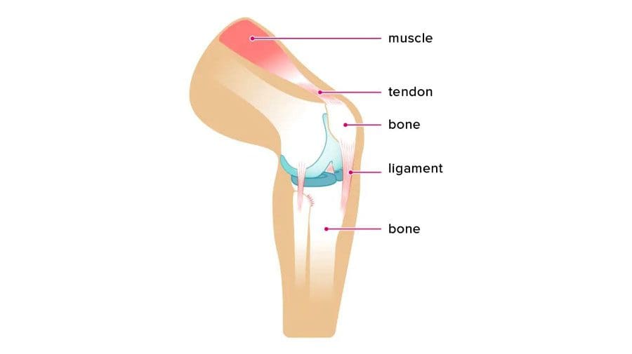
Tendons and Ligaments: A tendon is a fibrous flexible, strong tissue similar to a rope that attaches the muscles to the bones. Tendons allow for the movement of the body’s limbs and help prevent muscle injury by absorbing muscles’ impact when running, jumping, or performing other actions. Ligaments are bands of solid elastic tissue that connect bone to bone, hold structures together and keep them stable, support the joints and limit their movement.
Tendons and Ligaments
Tendons are strong and non-flexible.
Ligaments are flexible and elastic.
Both comprise collagen and living cells, essential in joints and bones and integral to locomotion.
Tendons allow body movement by transmitting force from muscle to bone, allowing the body to stand, walk, and jump.
Ligaments work by allowing for the full range of motion.
Ligaments are around the knees, ankles, elbows, shoulders, and other joints.
Connective Tissue
The collagen connective tissue that makes up tendons and ligaments is the same; their patterns are different.
Tendon fibers are laid out in a parallel pattern.
Tendon connective tissue needs to have more elasticity to help move the muscles.
Ligament fibers are laid out in a crisscross pattern.
Ligament connective tissue stabilizes and strengthens the bones’ joint structure.
Tendon Injury
A tendon that gets overstretched or torn is known as a strain. Common areas affected by strains are the:
Leg
Foot
Back
Strains often result from repetitive work movements, intense physical activity, and sports. Individuals who overuse their bodies without proper rest and muscle repair recovery have an increased risk of injury. Symptoms include:
Inflammation
Swelling
Pain
Cramping
Weakness
Ligament Injury
A ligament that gets overstretched or torn results in a sprain. Sprains can happen suddenly from a fall, awkward movement, or trauma. Sprains commonly occur in the:
Ankle
Knee
Wrist
Examples include:
Misstep causing the ankle to twist in an awkward position, snapping a ligament and causing unstableness or wobbliness.
There could be a popping sensation or the feeling of a tear when the injury occurs.
Wrist sprains often happen when reaching out and extending the hands to break a fall, and the wrist hyperextending back.
The hyperextension overstretches the ligament.
Symptoms of a sprained ligament include:
Inflammation
Swelling
Bruising
Pain
The joint may feel loose or weak and unable to take on weight.
The intensity of symptoms varies depending on whether the ligament is overextended or torn. Sprains are classified by grade:
Grade 1 – a mild sprain with slight stretching of the ligament.
Grade 2 – a moderate ligament tear, but not a complete tear.
Grade 3 – a complete ligament tear, making the joint unstable.
Chiropractic Care
Tendons and ligaments do not receive full blood circulation like other soft tissues. Depending on the severity of the injury, and the slower transfer of oxygen and nutrients, ligament and tendon injuries can take six to twelve weeks to heal, and repeatedly stressing the injured area from overuse can extend recovery. Chiropractic adjustments, and massage therapy, combined with corrective exercises and stretches, will reduce inflammation, decrease pain, improve the range of motion, increase nerve and muscle function, and strengthen the muscles. Chiropractic treatment involves:
Childress, Marc A, and Anthony Beutler. “Management of chronic tendon injuries.” American family physician vol. 87,7 (2013): 486-90.
Fenwick, Steven A et al. “The vasculature and its role in the damaged and healing tendon.” Arthritis research vol. 4,4 (2002): 252-60. doi:10.1186/ar416
Leong, Natalie L et al. “Tendon and Ligament Healing and Current Approaches to Tendon and Ligament Regeneration.” Journal of orthopedic research: official publication of the Orthopaedic Research Society vol. 38,1 (2020): 7-12. doi:10.1002/jor.24475
Scalcione, Luke R et al. “The athlete’s hand: ligament and tendon injury.” Seminars in musculoskeletal radiology vol. 16,4 (2012): 338-49. doi:10.1055/s-0032-1327007
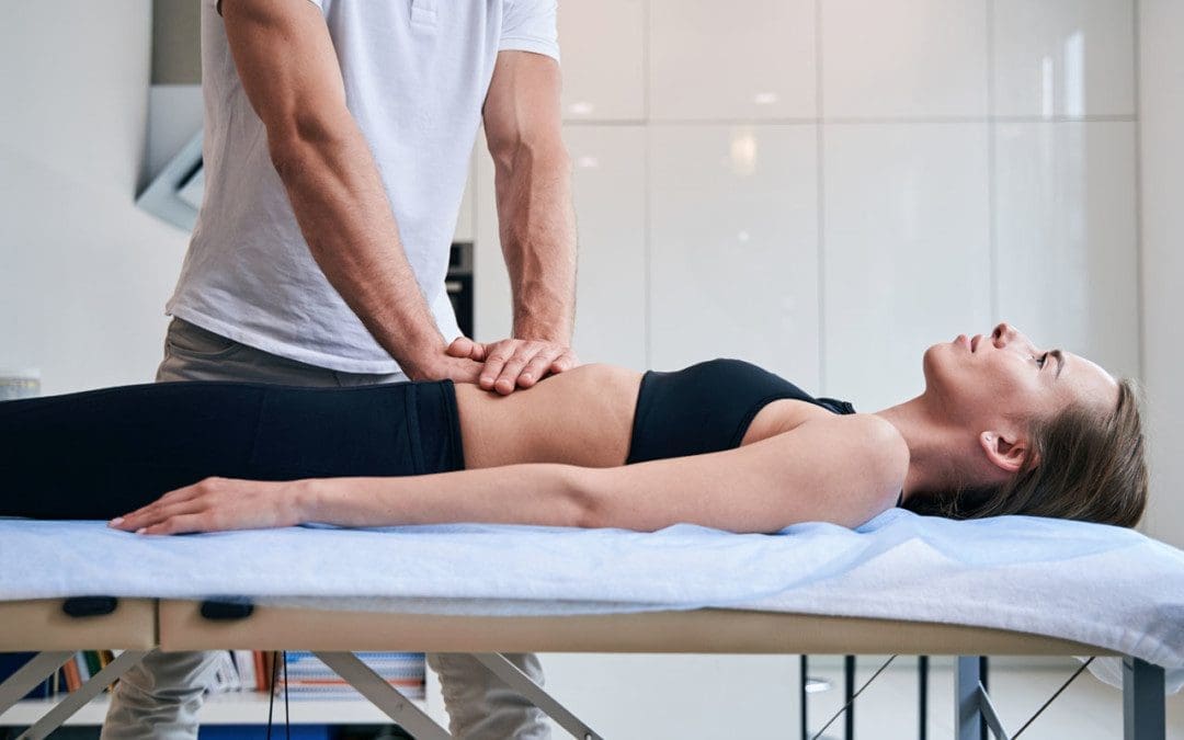
Orthopedic massage is part of injury rehabilitation that focuses on the muscles and soft tissues surrounding the joints and ligaments. Pain could be caused post-surgery, an acute injury, or overuse/repetitive motion injury from work or sports. The objective is to:
Decrease pain
Release tension
Restore balance
Increase mobility and flexibility
Preparing the body to return to everyday routines and activities.
Whatever the cause of the muscle damage or injury, an orthopedic massage will lengthen and soften the muscles and ligaments, allowing for a better range of motion of the affected joints.
Orthopedic Massage
All massage techniques can improve joint movement and function. Orthopedic massage is specifically designed to return the joints to their normal position function and alleviate pain with movement.
Swedish massage focuses on overall relaxation.
Deep tissue massage reduces deep muscle pain and strain.
Orthopedic massage therapists have an extensive understanding of anatomy, soft tissues, and misalignment of the musculoskeletal system that can cause pain and injury. It is similar to sports massage targeting damaged areas for recovery and rehabilitation from conditions and injuries. Sports massage helps the individual strengthen and retrain the damaged areas back to optimal performance and prevent injury. Orthopedic massage utilizes:
Massage benefits many symptoms and conditions. It has been shown to help with:
Sprains
Pulled muscles
Torn ligaments
Carpal-tunnel syndrome
Frozen shoulder
Tennis elbow
Tendinitis
Sciatica
Bulging discs
Post-surgery
Techniques
A therapist will look at the range of motion, flexibility, and rotation of the tissues. This will help determine what muscle groups and tendons are involved and which techniques to use. Massage therapists use an assortment of approaches to loosen muscles and tendons. These include:
Active Engagement
This is used to reach deep, hard-to-reach muscles by applying pressure and massaging lengthwise in a perpendicular motion.
It is beneficial for whiplash and/or back pain.
Positional Release
This is a gentle treatment for inflamed muscles and tissues highly sensitive to other techniques.
Soft tissues are manipulated into comfortable positions and held in place for a specific time.
This lengthens and softens tissues to bring pain relief.
Nerve Mobilization
Also known as neural mobilization, this method addresses strained nerves and pain sources.
Muscle Energy Release
The therapist provides resistance while the individual voluntarily contracts muscles.
Effective with low back pain.
Trigger Point Therapy
Pressure intervals are held on trigger areas to release lactic acid and promote circulation.
Myofascial Release
Gentle pressure is applied to stretch fascia tissues.
Body Composition
Brittle Bones
The reason bones become weaker is that bone tissue is living tissue that constantly forms new bone material and absorbs the old bone material. As the body ages, the rate at which bone is reabsorbed becomes faster than newly formed bone material. One reason for rapid bone loss is lack of exercise and physical activity. The Mayo Clinic has stated that individuals that spend a great deal of time sitting, whether at home or work, have an increased risk of osteoporosis than more active individuals. Sitting too much with little to no activity can lead to weakened bones. Just like the muscles, bones get stronger when they are in use. Walking, running, jumping, and getting the body moving along with using some resistance, can increase the strength and durability of the bones.
References
Kim, Seung-Kook et al. “Clinical outcomes and cost-effectiveness of massage chair therapy versus basic physiotherapy in lower back pain patients: A randomized controlled trial.” Medicine vol. 99,12 (2020): e19514. doi:10.1097/MD.0000000000019514
Klein, Ifat et al. “Lymphatic treatments after orthopedic surgery or injury: A systematic review.” Journal of bodywork and movement therapies vol. 24,4 (2020): 109-117. doi:10.1016/j.jbmt.2020.06.034
Loew, Laurianne M et al. “Deep, transverse friction massage for treating lateral elbow or lateral knee tendinitis.” The Cochrane database of systematic reviews vol. 2014,11 CD003528. 8 Nov. 2014, doi:10.1002/14651858.CD003528.pub2
Majewski-Schrage, Tricia, and Kelli Snyder. “The Effectiveness of Manual Lymphatic Drainage in Patients With Orthopedic Injuries.” Journal of sport rehabilitation vol. 25,1 (2016): 91-7. doi:10.1123/jsr.2014-0222
Moving every part of the body freely, without pain or stiffness, is necessary for a high quality of life. As the body ages, it begins to lose its natural flexibility. One of the most common problems with mobility and flexibility is tight and misaligned backs, shoulders, necks, and legs that can cause pain when moving. This means having a limited range of motion that can cause negative body compensation patterns that can lead to further dysfunction and injury. Maintaining healthy mobility requires a conscious effort to keep every joint, muscle, ligament, and tendon in shape. Chiropractic treatment can restore range of motion and strengthen the body.
Restore Range of Motion
Range of motion or R.O.M. is the measurement of movement around a joint or body part expressed in degrees. It is tied with the flexibility around a joint and plays a role in moving well without pain or discomfort. After an injury, trauma, or medical problem, the range of motion can be limited. Individuals with back, neck, shoulder, and leg pain feel stiff, tight, and sore in these areas and cannot move freely. Range of motion is vital for physical activity, athletic activity, and preventing injuries. When an individual pushes the body too hard and tries to move in an uncomfortable way, they can cause a tear or sprain, leading to added inflammation, stiffness, and further limited mobility.
Factors That Contribute To A Lack Of Flexibility
Age
Body age impacts flexibility. As the body gets older, it becomes stiff and can begin to present with pain, which restricts movements.
Limited Physical Activity or Exercise
Being sedentary with minimal physical activity contributes to a lack of flexibility, muscle loss, disrupted circulation, and weight gain.
Work
An individual’s profession can affect the body’s flexibility. A job that has little to no movement regularly, like being seated for most of the time, will contribute to reduced flexibility.
Obesity
Carrying additional body weight can significantly limit movement and decrease flexibility.
Flexibility Improvement
Staying Active
Regular physical activity/exercise will help maintain body health and flexibility. Activities can include:
Sports
Walking
Jogging
Weight lifting
Swimming
Yoga
Regular Stretching
Regular stretching will keep the muscles loose and the joints flexible. Incorporate stretching into a daily routine throughout the day and a wind-down stretch before going to bed.
Maintaining Proper Hydration
When the body is dehydrated, it causes the muscles to stiffen and tighten up, decreasing elasticity. Staying hydrated will help maintain flexibility by re-lubricating the muscles, ligaments, and tendons.
Healthy Diet
Losing excess weight and maintaining a healthy weight range through proper nutrition will reduce inflammation, improve mobility and flexibility.
Chiropractic Restoration
When normal movement is not possible, discomfort and pain will worsen as the muscles become tighter, causing the tendons and ligaments to shorten and stick together, placing added stress on the areas, leading to pain and inflammation. The body was made to be in motion, and when it does not move and stretch out, it stiffens up. Trying to use the muscles even when they are stiff and strained can make the condition worse, limiting the range of motion further causing the slightest movements to cause discomfort and pain. A chiropractor can provide adjustments, soft and deep-tissue massage to the tight areas to loosen the muscles, improve circulation, flexibility, mobility, and restore range of motion.
Body Composition
Myth Eating at Night Causes Fat Gain
The myth is eating right before sleeping causes the body to turn whatever was eaten straight into fat. However, the fact is that it is not about when an individual eats but rather the calorie intake and exercise level. According to the C.D.C., it’s the calories that are burned over a 24-hour period that determine fat gain/loss, and not when those calories are taken in. Far from being a fat gain guarantee, healthy nighttime meals were shown to:
They had no effect on weight gain among overweight and obese individuals that participated in a high-intensity cardiovascular exercise program during the day.
What can make the myth true is when eating and drinking foods/drinks with a high caloric content: This includes:
An extra 500-1000 calories after 8 pm is easy to add if not careful. Remember, it’s about the calories themselves, not the time.
References
Marcano-Fernández, Francesc et al. “Physical outcome measures: The role of strength and range of motion in orthopedic research.” Injury vol. 51 Suppl 2 (2020): S106-S110. doi:10.1016/j.injury.2019.11.017
Mortazavi, Fatemeh, and Ali Nadian-Ghomsheh. “Stability of Kinect for a range of motion analysis in static stretching exercises.” PloS one vol. 13,7 e0200992. 24 Jul. 2018, doi:10.1371/journal.pone.0200992
O’Sullivan, Kieran et al. “The effect of warm-up, static stretching and dynamic stretching on hamstring flexibility in previously injured subjects.” B.M.C. musculoskeletal disorders vol. 10 37. 16 Apr. 2009, doi:10.1186/1471-2474-10-37
Simão, Roberto et al. “The influence of strength, flexibility, and simultaneous training on flexibility and strength gains.” Journal of strength and conditioning research vol. 25,5 (2011): 1333-8. doi:10.1519/JSC.0b013e3181da85bf
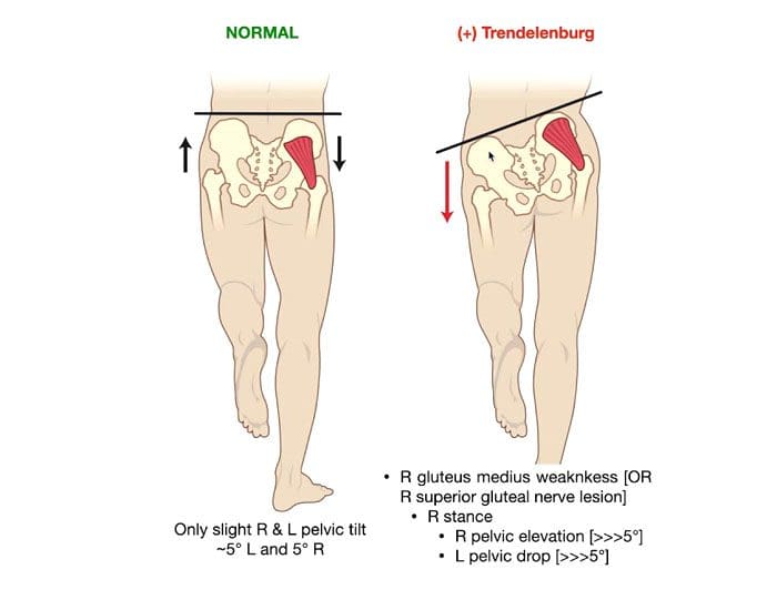
A Trendelenburg gait is an abnormal walking gait resulting from a defective or weakened hip abductor.The gluteal musculature is the primary musculature that includes the gluteus medius and gluteus minimus muscles. Weakness in these muscles causes sagging/dropping of the pelvis on the opposite side while walking. There will be a noticeable side-to-side motion if the glutes are too weak to support the body’s weight when walking. It can look as though the individual is limping or missing a step. Individuals can minimize the effects with foot orthotics, core strengthening, chiropractic, and physical therapy.
Trendelenburg Gait Causes
This gait often results from straining the hip abductor muscles during physical activity. Exercises specifically for the glutes done improperly are a common cause. When improper exercise form is the cause, the abnormal gait usually goes away as muscle inflammation fades. The gait can also present after total hip replacement surgery, as the procedure requires incisions in the gluteus medius muscle. This can weaken the muscle causing an abnormal gait. Weakness in these muscles can also be caused by:
Nerve damage or dysfunction in the nerves that run through the gluteal minimus and medius muscles.
Osteoarthritis is a type of arthritis that occurs when joint cartilage starts to wear down.
Muscular dystrophy is a condition that causes the muscles and bones to become weak over time.
Poliomyelitisis a condition associated with polio that weakens the muscles.
Cleidocranial dysostosis is a condition present from birth that can cause your bones to develop improperly.
Symptoms
The walking gait is made up of two phases:
Swing – When one leg moves forward.
Stance – The other leg stays still and maintains balance.
The main symptom of Trendelenburg gait can be seen when one leg swings forward and the hip drops down and move outward. This is because the hip abductor of the other leg is too weak to support the weight. Individuals may lean back or to the side slightly when walking to maintain balance, or they may lift the foot higher off the ground with each step to avoid losing balance or tripping as the pelvis shifts unevenly.
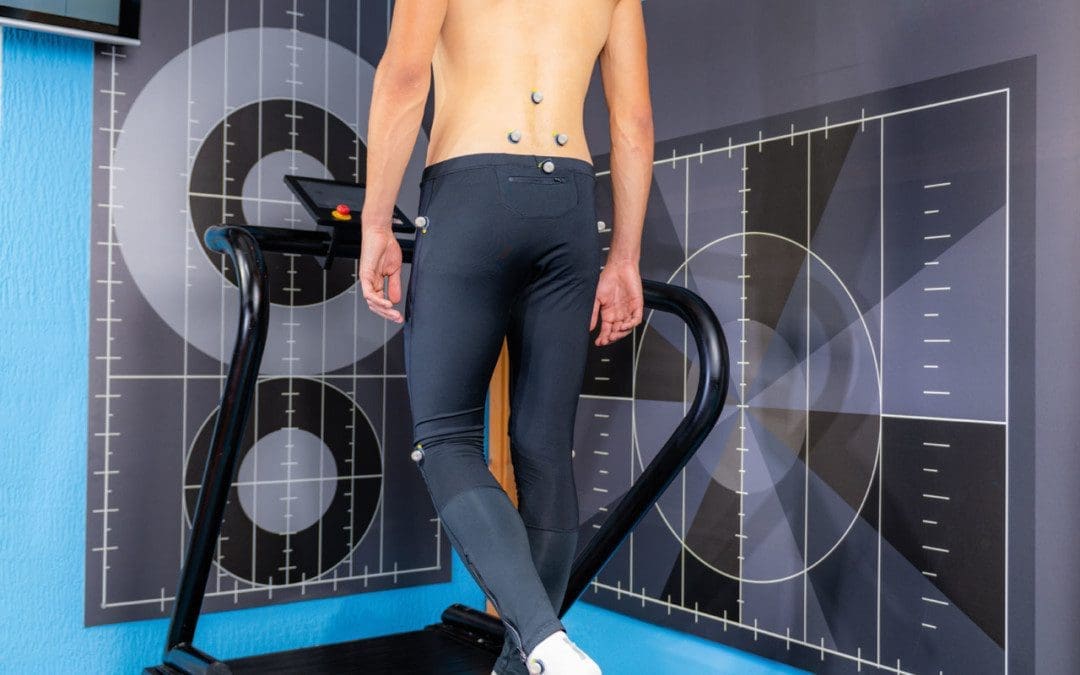
Diagnosis
Abnormal hip movement during a swing of one or both legs can give a doctor enough evidence to diagnose a Trendelenburg gait. A doctor will observe the individual’s walk in front and behind to get a detailed view. A doctor will also use the Trendelenburg test to diagnose the condition. The doctor will instruct the individual to lift one leg for 30 seconds. If the individual cannot keep the hips parallel with the ground while lifting, it could indicate Trendelenburg gait. X-rays of the hip will be used to identify any causes of weakness in the gluteus minimus or medius.
Treatment Options
Treatment options will depend on the severity and cause of the gait.
Medication
If the gait is causing pain, over-the-counter nonsteroidal anti-inflammatory NSAIDs, like ibuprofen or acetaminophen, will help ease symptoms.
In severe cases, a doctor may prescribe cortisone injections to help reduce pain.
Foot Orthotics
A doctor could also recommend using a foot orthotic in one or both shoes to compensate the hip abductor muscle weakness.
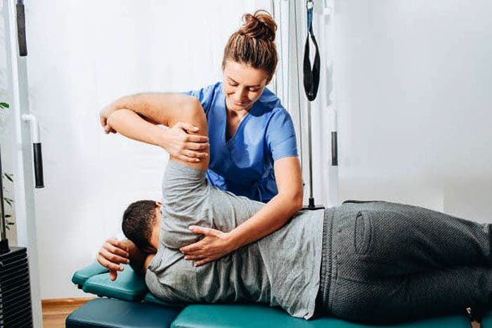
Chiropractic, Physical Therapy, and Exercise
Chiropractic and physical therapy can help adjust, realign, and strengthen the muscles to regain control of the Trendelenburg gait. The chiropractor or physical therapist will move the legs in various directions to help the joints become more accustomed to moving in certain directions and increase muscle strength and resistance. Exercises that can strengthen the hip abductor muscles include:
Lie on the side and extend the leg straight out.
Lie on the floor and move one leg up, over the other, and back in the opposite direction.
Step sideways and onto an elevated surface, then back down again.
Talk with a doctor or chiropractor before beginning any new exercise routine so they can recommend specific exercises and educate on proper form.
Complications
If left untreated, moderate-to-severe cases of Trendelenburg gait can become debilitating, leading to severe complications. These include:
Pinched nerves.
Sciatica.
Pain, stiffness, or grinding in the hips.
Loss of range of motion in the hips and gait.
Losing the ability to walk, which could require the use of a walker or wheelchair.
Trendelenburg gait is treatable with special shoes, orthotics, and exercises designed to strengthen the hip abductor muscles. Chiropractic and physical therapy can help limit the condition’s impact on the body’s health, the ability to walk, and reduce the risk of complications.
Body Composition
Heart-Healthy Foods
Citrus
The bright and tangy fruits are packed with vitamins and unique plant compounds known as polyphenols that can help lower blood pressure naturally.
However, it’s important to note that grapefruit and grapefruit juice could interact with certain prescription medications.
Beans and Lentils
Foods high in magnesium, potassium, and fiber can help maintain healthy blood pressure.
This is where beans and legumes come in, as they are high in fiber, potassium, and magnesium.
Individuals that swapped beans and lentils noticed a lower blood pressure, whether or not they had been diagnosed with hypertension.
Pumpkin Seeds
These seeds are packed with potassium, magnesium, and arginine.
Arginine is an amino acid used to make nitric oxide, which helps the blood vessels relax and dilate, allowing lower blood pressure.
A study found that postmenopausal women who took 3 grams of pumpkin seed oil daily for six weeks saw a significant decrease in their systolic blood pressure.
Garlic
Garlic contains nitric oxide, which has been shown to relax blood vessels.
Kyolic garlic, in particular, has been shown to help with arterial stiffness and can improve cholesterol levels.
References
Feyh, Andrew et al. “Role of Dietary Components in Modulating Hypertension.” Journal of Clinical & experimental cardiology vol. 7,4 (2016): 433. doi:10.4172/2155-9880.1000433
Gandbhir, Viraj N., et al. “Trendelenburg Gait.” StatPearls, StatPearls Publishing, 19 August 2021.
Giangarra CE, et al. (2018). Clinical orthopedic rehabilitation: A team approach.sciencedirect.com/science/book/9780323393706
Gilliss AC, et al. (2010). Use of osteopathic manipulative treatment to manage compensated Trendelenburg gait caused by sacroiliac somatic dysfunction.
jaoa.org/article.aspx?articleid=2093879
Maricelli JW, et al. (2016). Trendelenburg-like gait, instability and altered step patterns in a mouse model for limb-girdle muscular dystrophy 2i. DOI:
10.1371/journal.pone.0161984
Mayo Clinic Staff. (2017). Osteoarthritis.mayoclinic.org/diseases-conditions/osteoarthritis/home/ovc-20198248
Michalopolous N, et al. (2016). A personalized monitoring and recommendation framework for kinetic dysfunctions: The Trendelenburg gait. DOI: 10.1145/3003733.3003786
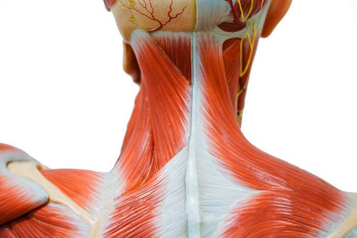
Muscle tension in the neck is a common musculoskeletal disorder. The neck is made up of flexible muscles that support the weight of the head. The muscles can experience injury and irritation from overuse and poor posture habits. Worn joints or compressed nerves can cause neck pain, but muscle spasms or soft tissue injuries commonly cause neck tension. Neck tension can present suddenly or progress slowly. Sleeping in an awkward position or straining the neck while engaged/involved in some activity can cause muscles to tense up. Chronic neck tension that comes and goes over the course of weeks or months could have a cause that goes unnoticed, like teeth grinding or being in a hunched position for extended periods.
Symptoms of neck tension
Symptoms can come on suddenly or progressively. These include:
Stiffness
Tightness
Spasms
Turning the head is difficult
Discomfort and/or pain worsens with certain positions
Causes
Because the neck can move in many directions, there are various causes of tension in the neck. These include:
Repetitive motion or overuse injuries
Individuals whose work requires repetitive movements like scanning objects, looking up and behind constantly can strain the muscles.
Improper posture
An adult’s head weighs 10 to 11 pounds. If the weight is not properly distributed and supported with a healthy posture, the neck muscles have to work harder, causing strain.
Computer workstation habits
Individuals that sit at a desk or workstation for most of the day or night can develop hunching habits that they may overlook. This can definitely cause neck muscles to strain.
Phone habits
Constantly looking down at the phone is a common cause of tension in the neck and text neck.
Grinding teeth
When individuals grind or clench their teeth, pressure is placed on the muscles in the neck and jaw. This pressure strains the muscles, causing pain. There are exercises to promote more relaxed jaw muscles.
Physical activities and sports
Working out in a way that engages the neck muscles or whipping the head around during a game or some physical activity can cause minor neck injury and strain.
Sleep position habits
When sleeping, the head and neck should be aligned with the rest of the body. Using large pillows that elevate the neck too much can cause tension to build up while sleeping.
Heavy purses, backpacks, shoulder bags
Lifting and carrying any heavy object can throw the body out of alignment. This can cause strain on one side of the neck, building tension.
These are mild to moderate headaches that typically affect the forehead. However, these types of headaches can cause neck tension and tenderness.
Prevention
Making simple adjustments can help relieve, manage, and prevent tension in the neck and shoulders. These include:
Ergonomics
Consider a standing desk. Adjust the workstation so that proper posture along with comfort is maintained. Try different adjustments like the height of the chair, desk, and computer.
Be aware of body posture.
Stay aware of the body’s posture when sitting and standing. Keep the ears, shoulders, and hips in a straight line. Consider phone posture reminders and devicesto check in with how you’re holding yourself throughout the day.
Take breaks throughout the day.
Take breaks that will move the body and stretch the neck and upper body. This benefits the muscles, eyes, and mental health.
Sleep position
Improve sleeping positions with a smaller, flatter, firmer pillow.
Reduce weight from the shoulders
Utilize a rolling bag instead of carrying heavy bags and backpacks, and only carry what is necessary.
Movement
Try to get 30 minutes of moderate exercise/physical activity a day to keep the body in healthy condition.
Meditation and stretching
Practicing yoga or meditation along with stretching out helps reduce psychological and physical stress. Yoga can count as daily exercise.
Doctor or Dentist
If chronic neck tension is presenting, see a doctor or chiropractor. Consult a dentist about teeth grinding or temporomandibular joint TMJ disorder treatments.
Neck stretches
To relieve tension in the neck, try some neck stretches.
Gently pull your head to the right, so the ear almost touches the shoulder.
Hold for 30 seconds
Repeat on the opposite side.
Body Composition
The Immune System
The Immune System is essential in maintaining health. Its objective is to:
Neutralize pathogenic microorganisms like bacteria that enter the body and threaten homeostasis.
Eliminate harmful substances from the environment.
Fight against cells that cause illnesses like cancer.
Innate and adaptive immune processes.
The innate system includes exterior defenses, like the skin, proteins, and white blood cells.
Any organisms that escape the first line of defense have to then face the adaptive system. This is made up of T and B cells.
The adaptive immune system is constantly adapting and evolving to identify changes in pathogens change over time.
These systems work together to provide resistance and the elimination of long-term survival of infectious agents in the body.
References
Chaplin, David D. “Overview of the immune response.” The Journal of allergy and clinical immunology vol. 125,2 Suppl 2 (2010): S3-23. doi:10.1016/j.jaci.2009.12.980
Hawk, Cheryl et al. “Best Practices for Chiropractic Management of Patients with Chronic Musculoskeletal Pain: A Clinical Practice Guideline.” Journal of alternative and complementary medicine (New York, N.Y.) vol. 26,10 (2020): 884-901. doi:10.1089/acm.2020.0181
Hughes, Stephen Fôn et al. “The role of phagocytic leukocytes following flexible ureterorenoscopy, for the treatment of kidney stones: an observational, clinical pilots-study.” European journal of medical research vol. 25,1 68. 11 Dec. 2020, doi:10.1186/s40001-020-00466-7
IFM's Find A Practitioner tool is the largest referral network in Functional Medicine, created to help patients locate Functional Medicine practitioners anywhere in the world. IFM Certified Practitioners are listed first in the search results, given their extensive education in Functional Medicine