For individuals experiencing sacroiliac joint/SIJ dysfunction and pain, could applying kinesiology tape help bring relief and manage symptoms?
Kinesiology Tape For Sacroiliac Joint Pain
A lower back ailment that is common during pregnancy. The pain is usually on one or both sides of the back, just above the buttocks, that comes and goes and can limit the ability to bend, sit, and perform various physical activities. (Moayad Al-Subahi et al., 2017) The therapeutic tape provides support while allowing for movement and may help treat and manage sacroiliac joint/SIJ pain by:
Decreasing muscle spasms.
Facilitating muscular function.
Increasing blood circulation to and around the pain site.
Decreasing muscle trigger points.
Mechanism
Some studies have found that taping the SI joint has benefits that include:
One theory is it helps lift and hold the overlying tissues off of the SI joint, which helps decrease the pressure around it.
Another theory is that lifting the tissues helps create a pressure differential under the tape, like non-surgical decompression, allowing increased circulation to the tissues surrounding the sacroiliac joint.
This floods the area with blood and nutrients, creating an optimal healing environment.
Application
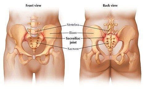
A sacroiliac joint on the right and left sides connects the pelvis to the sacrum or the lowest part of the spine. To apply the kinesiology tape correctly, locate the lowest part of the back within the pelvic area. (Francisco Selva et al., 2019) Ask a friend or family member for help if you can’t reach the area.
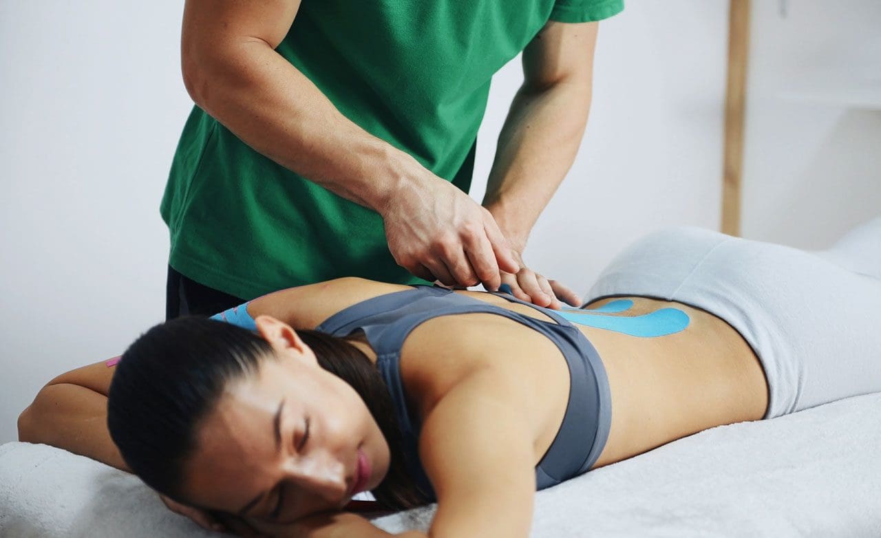
Taping steps:
Cut three strips of tape, each 4 to 6 inches long.
Sit in a chair and bend the body slightly forward.
If someone is helping, you can stand and slightly bend forward.
Remove the lift-off strip in the middle and stretch the tape to expose several inches, leaving the ends covered.
Apply the exposed tape at an angle over the SI joint, like making the first line of an X, just above the buttocks, with full stretch on the tape.
Peel the lift-off strips from the ends and adhere them with no stretching.
Repeat the application steps with a second strip, adhering at a 45-degree angle to the first strip, making the X over the sacroiliac joint.
Repeat this with the final strip horizontally across the X made from the first two pieces.
There should be a tape pattern of star shape over the sacroiliac joint.
Kinesiology tape can stay over the sacroiliac joint for three to five days.
Watch for signs of irritation around the tape.
Remove the tape if the skin becomes irritated, and consult your primary healthcare provider, physical therapist, or chiropractor for other treatment options.
Some individuals with specific conditions should avoid using the tape and get confirmation that it’s safe.
Individuals with severe sacroiliac pain where self-management is not working should see a healthcare provider, physical therapist, and or chiropractor for an evaluation and to learn therapeutic exercises and treatments to help manage the condition.
Sciatica During Pregnancy
References
Al-Subahi, M., Alayat, M., Alshehri, M. A., Helal, O., Alhasan, H., Alalawi, A., Takrouni, A., & Alfaqeh, A. (2017). The effectiveness of physiotherapy interventions for sacroiliac joint dysfunction: a systematic review. Journal of physical therapy science, 29(9), 1689–1694. https://doi.org/10.1589/jpts.29.1689
Do-Yun Shin and Ju-Young Heo. (2017). The Effects of Kinesiotaping Applied onto Erector Spinae and Sacroiliac Joint on Lumbar Flexibility. The Journal of Korean Physical Therapy, 307-315. https://doi.org/https://doi.org/10.18857/jkpt.2017.29.6.307
Selva, F., Pardo, A., Aguado, X., Montava, I., Gil-Santos, L., & Barrios, C. (2019). A study of reproducibility of kinesiology tape applications: review, reliability and validity. BMC musculoskeletal disorders, 20(1), 153. https://doi.org/10.1186/s12891-019-2533-0
For individuals dealing with knee pain symptoms from injury and/or arthritis, can incorporating an acupuncture and/or electroacupuncture treatment plan help in pain relief and management?
Acupuncture For Knee Pain
Acupuncture involves inserting very thin needles into the skin at specific acupoints on the body. It is based on the premise that the needles restore the flow of the body’s energy to activate and promote healing, relieve pain, and help the body relax.
Acupuncture can help address various health conditions, including knee pain caused by arthritis or injury.
Depending on the type and severity of pain, treatments can help reduce the pain for days or weeks.
Acupuncture is often used as a complementary therapy – treatment in addition to other treatment or therapy strategies like massage and chiropractic.
Acupuncture Benefits
Knee pain caused by osteoarthritis or injury can reduce flexibility, mobility, and quality of life. Acupuncture can help provide relief.
When the acupuncture needles are placed on the body, a signal is sent along the spinal cord to the brain, which triggers a release of endorphins/pain hormones. Medical researchers believe this helps reduce pain. (Qian-Qian Li et al., 2013) Acupuncture also helps decrease the production of cortisol, a hormone that helps control inflammation. (Qian-Qian Li et al., 2013) With reduced pain sensations and less inflammation after acupuncture treatments, knee function and mobility can be improved.
Various factors play a role in the pain relief experienced from acupuncture. Some evidence suggests that an individual’s expectations may impact the results of acupuncture treatment. (Stephanie L. Prady et al., 2015)
Researchers are currently assessing whether the expectation that acupuncture is beneficial contributes to a better outcome after treatment. (Zuoqin Yang et al., 2021)
In 2019, acupuncture was recommended in treating knee osteoarthritis in the American College of Rheumatology/Arthritis Foundation guidelines for hand, hip, and knee osteoarthritis pain management. (Sharon L. Kolasinski et al., 2020)
Research
Different clinical studies support acupuncture’s ability to help in knee pain relief and management.
One study found that acupuncture helps manage various conditions that cause chronic pain. (Andrew J. Vickers et al., 2012)
A scientific review analyzed previous studies on pain management interventions after knee surgery and found supporting evidence that the treatments delayed and reduced the use of medications for pain relief post-surgery. (Dario Tedesco et al., 2017)
Osteoarthritis
A systematic review analyzed randomized control studies to determine whether or not acupuncture reduced pain and improved joint function in individuals with chronic osteoarthritis knee pain. (Xianfeng Lin et al., 2016)
Individuals received six to twenty-three weekly acupuncture sessions for three to 36 weeks.
The analysis determined that acupuncture can improve short and long-term physical function and mobility and provide up to 13 weeks of pain relief in individuals with chronic knee pain caused by osteoarthritis.
Rheumatoid Arthritis
Rheumatoid arthritis is a chronic disease that affects joints, including the knee joint, causing pain and stiffness.
Acupuncture is beneficial in treating rheumatoid arthritis/RA.
A review found that acupuncture alone and in combination with other treatment modalities benefits individuals with RA. (Pei-Chi, Chou Heng-Yi Chu 2018)
Acupuncture is believed to have anti-inflammatory and antioxidant effects to help regulate immune system function.
Chronic Knee Pain
Various conditions and injuries can cause chronic knee pain, making mobility difficult.
Individuals with joint pain often turn to complementary therapies for pain relief management, with acupuncture being one of the popular modalities. (Michael Frass et al., 2012)
Working with a licensed, professional acupuncture practitioner can reduce the risk of unwanted side effects and complications.
Types
Other acupuncture options that may be offered include:
Electroacupuncture
A modified form of acupuncture where a mild electrical current passes through the needles, providing additional stimulation to the acupoints.
In one research study, individuals with knee osteoarthritis reported significant improvements in their pain, stiffness, and physical function after electroacupuncture treatment. (Ziyong Ju et al., 2015)
Auricular
Auricular or ear acupuncture works on acupoints in the ear corresponding to the body’s different parts.
A research review analyzed several studies on auricular acupuncture for pain relief and found that it can provide relief within 48 hours of pain onset. (M. Murakami et al., 2017)
Battlefield Acupuncture
The military and veteran healthcare facilities use a unique form of auricular acupuncture for pain management.
Studies show that it is effective at providing immediate pain relief, but more research is necessary to determine long-term pain relief effectiveness. (Anna Denee Montgomery, Ronovan Ottenbacher 2020)
Before trying acupuncture, consult a healthcare professional for guidance, as it may be integrated with other therapies and lifestyle adjustments.
Overcoming an ACL Injury
References
Li, Q. Q., Shi, G. X., Xu, Q., Wang, J., Liu, C. Z., & Wang, L. P. (2013). Acupuncture effect and central autonomic regulation. Evidence-based complementary and alternative medicine : eCAM, 2013, 267959. https://doi.org/10.1155/2013/267959
Prady, S. L., Burch, J., Vanderbloemen, L., Crouch, S., & MacPherson, H. (2015). Measuring expectations of benefit from treatment in acupuncture trials: a systematic review. Complementary therapies in medicine, 23(2), 185–199. https://doi.org/10.1016/j.ctim.2015.01.007
Yang, Z., Li, Y., Zou, Z., Zhao, Y., Zhang, W., Jiang, H., Hou, Y., Li, Y., & Zheng, Q. (2021). Does patient’s expectation benefit acupuncture treatment?: A protocol for systematic review and meta-analysis. Medicine, 100(1), e24178. https://doi.org/10.1097/MD.0000000000024178
Kolasinski, S. L., Neogi, T., Hochberg, M. C., Oatis, C., Guyatt, G., Block, J., Callahan, L., Copenhaver, C., Dodge, C., Felson, D., Gellar, K., Harvey, W. F., Hawker, G., Herzig, E., Kwoh, C. K., Nelson, A. E., Samuels, J., Scanzello, C., White, D., Wise, B., … Reston, J. (2020). 2019 American College of Rheumatology/Arthritis Foundation Guideline for the Management of Osteoarthritis of the Hand, Hip, and Knee. Arthritis care & research, 72(2), 149–162. https://doi.org/10.1002/acr.24131
Vickers, A. J., Cronin, A. M., Maschino, A. C., Lewith, G., MacPherson, H., Foster, N. E., Sherman, K. J., Witt, C. M., Linde, K., & Acupuncture Trialists’ Collaboration (2012). Acupuncture for chronic pain: individual patient data meta-analysis. Archives of internal medicine, 172(19), 1444–1453. https://doi.org/10.1001/archinternmed.2012.3654
Tedesco, D., Gori, D., Desai, K. R., Asch, S., Carroll, I. R., Curtin, C., McDonald, K. M., Fantini, M. P., & Hernandez-Boussard, T. (2017). Drug-Free Interventions to Reduce Pain or Opioid Consumption After Total Knee Arthroplasty: A Systematic Review and Meta-analysis. JAMA surgery, 152(10), e172872. https://doi.org/10.1001/jamasurg.2017.2872
Lin, X., Huang, K., Zhu, G., Huang, Z., Qin, A., & Fan, S. (2016). The Effects of Acupuncture on Chronic Knee Pain Due to Osteoarthritis: A Meta-Analysis. The Journal of bone and joint surgery. American volume, 98(18), 1578–1585. https://doi.org/10.2106/JBJS.15.00620
Chou, P. C., & Chu, H. Y. (2018). Clinical Efficacy of Acupuncture on Rheumatoid Arthritis and Associated Mechanisms: A Systemic Review. Evidence-based complementary and alternative medicine : eCAM, 2018, 8596918. https://doi.org/10.1155/2018/8596918
Frass, M., Strassl, R. P., Friehs, H., Müllner, M., Kundi, M., & Kaye, A. D. (2012). Use and acceptance of complementary and alternative medicine among the general population and medical personnel: a systematic review. Ochsner journal, 12(1), 45–56.
Hinman, R. S., McCrory, P., Pirotta, M., Relf, I., Forbes, A., Crossley, K. M., Williamson, E., Kyriakides, M., Novy, K., Metcalf, B. R., Harris, A., Reddy, P., Conaghan, P. G., & Bennell, K. L. (2014). Acupuncture for chronic knee pain: a randomized clinical trial. JAMA, 312(13), 1313–1322. https://doi.org/10.1001/jama.2014.12660
National Center for Complementary and Integrative Health. (2022). Acupuncture in depth. National Center for Complementary and Integrative Health. https://www.nccih.nih.gov/health/acupuncture-what-you-need-to-know
Harvard Medical School. (2023). Acupuncture: what is it? Harvard Health Publishing Harvard Medical School Blog. https://www.health.harvard.edu/a_to_z/acupuncture-a-to-z#:~:text=The%20most%20common%20side%20effects,injury%20to%20an%20internal%20organ.
Ju, Z., Guo, X., Jiang, X., Wang, X., Liu, S., He, J., Cui, H., & Wang, K. (2015). Electroacupuncture with different current intensities to treat knee osteoarthritis: a single-blinded controlled study. International journal of clinical and experimental medicine, 8(10), 18981–18989.
Murakami, M., Fox, L., & Dijkers, M. P. (2017). Ear Acupuncture for Immediate Pain Relief-A Systematic Review and Meta-Analysis of Randomized Controlled Trials. Pain medicine (Malden, Mass.), 18(3), 551–564. https://doi.org/10.1093/pm/pnw215
Montgomery, A. D., & Ottenbacher, R. (2020). Battlefield Acupuncture for Chronic Pain Management in Patients on Long-Term Opioid Therapy. Medical acupuncture, 32(1), 38–44. https://doi.org/10.1089/acu.2019.1382
For individuals experiencing tightness in the lower back and hamstrings, can utilizing the sit and reach test help determine an individual’s risk for future pain and injury?
Sit and Reach Test
Lower back and hamstring tightness and pain symptoms are usually brought on by muscle stiffness. The sit-and-reach test is one of the most common ways to measure lower back and hamstring flexibility. Exercise physiologists, physical therapists, chiropractors, and fitness trainers use the sit-and-reach test to measure lower back and hamstring flexibility to assess baseline flexibility. The test has been around since 1952 (Katharine F. Wells & Evelyn K. Dillon 2013) and has an extensive database of results across all age groups and genders.
Individuals can use the sit-and-reach test to compare flexibility to the average result for individuals of the same gender and age.
For healthcare providers, the test may be repeated after several weeks to determine flexibility progress.
Measurement
The test can be a valuable measurement of functional flexibility to sit with the legs straight in front and reach the toes. Jobs, sports, and everyday tasks regularly require bending over, reaching, and lifting objects. These are real-life examples of how having a healthy back and hamstring flexibility is vital in preventing pain symptoms and injuries. New flexibility assessments are currently being developed, and many trainers and therapists use their own versions with patients and clients. But even with more advanced specialized flexibility tests, the sit and reach test can be a functional testing tool for tracking general flexibility changes over time. (Daniel Mayorga-Vega et al., 2014)
Performing The Test
A special sit-and-reach testing box is used; however, individuals can make their own testing box by finding a heavy-duty box around 30cm or 11.811 inches tall. Set a measurement ruler/stick on top of the box so that 26 cm or 10.2362 inches of the ruler extends over the front edge toward the individual being tested. The 26cm mark should be at the edge of the box.
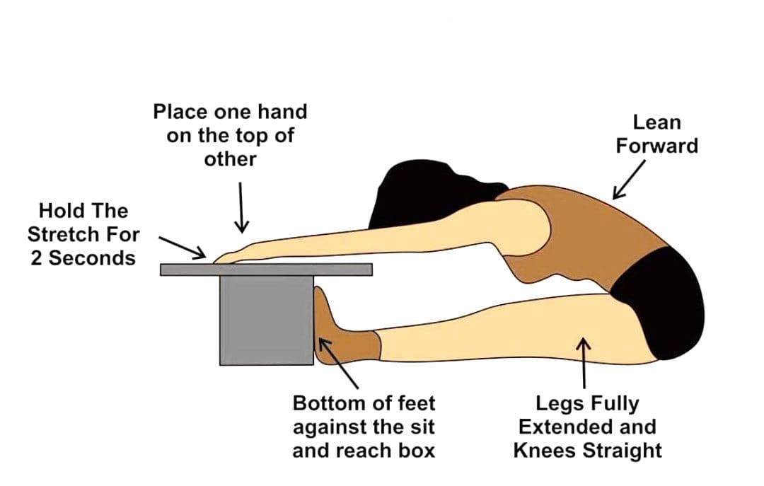
Get into position – Remove shoes and sit on the floor with legs stretched out in front with the knees straight and feet flat against the front end of the test box.
Start the movement – In a slow, steady motion, lean forward, keeping the knees straight, and slide the hands up the ruler as far as possible.
Stretch and repeat – Extend as far as possible, record the results, rest, and repeat three times.
Calculate the results – Average the results.
Results
Results compare flexibility over time to norms, or averages, for gender and age. Adequate flexibility is reaching the toes – the 26-cm mark on the ruler while keeping the legs straight.
Adult Women
37cm or 14.5669 inches or above: Excellent
33 to 36cm or 12.9921 inches: Above average
29 to 32cm or 11.4173 inches: Average
23 to 28cm or 9.05512 inches: Below average
Below 23cm or 8.66142 inches: Poor
Adult Men
34cm or 13.3858 inches or above: Excellent
28 to 33cm or 11.0236 inches: Above average
23 to 27cm or 9.05512 inches: Average
16 to 22cm or 6.29921 inches: Below average
Below 16cm or 5.90551 inches: Poor
Alternatives
Individuals can test their own hamstring and lower back flexibility with some easy at-home tests. Use these methods while working on flexibility, and keep a record to see improvements. (Brittany L. Hansberger et al., 2019) One alternative is the V-sit reach test.
To perform this, make a line on the floor with tape, then place a measuring tape perpendicular to the tape, making a cross.
Sit with the feet in a V shape, touching the tape, feet about a foot apart, with the measuring tape between the legs; the 0 end starts where the legs part.
Overlap hands with arms outstretched in front.
Repeat three times, leaning forward and reaching with hands out.
Then, repeat and take note of how far the hands could reach.
Individuals will need someone to measure the distance between their fingertips and the floor.
Warm up with a few practice stretches of standing and bending toward the floor.
Then, measure how far from the floor the fingertips are.
The ability to touch the floor is a good sign.
Improving Flexibility
Individuals with less than adequate flexibility are recommended to work on stretching the major muscle groups in both the upper and lower body on a regular basis to improve and maintain body flexibility.
Individuals can incorporate dynamic stretching, which consists of active movements utilizing a full range of motion as part of warming up for workouts, sports, or other activities.
Static stretching is recommended when cooling down after the muscles are warmed up and joints are lubricated.
The American College of Sports Medicine recommends 2 to 3 sessions a week of flexibility training and learning to stretch daily.
Stretches should be held for 15 to 30 seconds, then released and repeated 2 to 4 times. (Phil Page 2012)
This will take time and dedication, but with the help of trained specialists, regaining flexibility and full range of motion can be accomplished.
Benefits of Stretching
References
Katharine F. Wells & Evelyn K. Dillon (1952) The Sit and Reach—A Test of Back and Leg Flexibility, Research Quarterly. American Association for Health, Physical Education and Recreation, 23:1, 115-118, DOI: 10.1080/10671188.1952.10761965
Mayorga-Vega, D., Merino-Marban, R., & Viciana, J. (2014). Criterion-Related Validity of Sit-and-Reach Tests for Estimating Hamstring and Lumbar Extensibility: a Meta-Analysis. Journal of sports science & medicine, 13(1), 1–14.
Hansberger, B. L., Loutsch, R., Hancock, C., Bonser, R., Zeigel, A., & Baker, R. T. (2019). EVALUATING THE RELATIONSHIP BETWEEN CLINICAL ASSESSMENTS OF APPARENT HAMSTRING TIGHTNESS: A CORRELATIONAL ANALYSIS. International journal of sports physical therapy, 14(2), 253–263.
Page P. (2012). Current concepts in muscle stretching for exercise and rehabilitation. International journal of sports physical therapy, 7(1), 109–119.
Individuals in post-surgery recovery or dealing with illness or an injury can experience weakened muscles and endurance that can cause temporary loss of sleeping mobility and not being able to move around normally because of weakness, decreased range of motion, or pain. Can they benefit from physical therapy to help get back to normal functional mobility?
Sleeping Mobility
For individuals who are hospitalized or homebound from injury, illness, or surgical recovery, a physical therapist will assess various areas of functional mobility. These include transfers – from sitting to standing positions, walking, and sleeping mobility. Sleeping mobility is the ability to perform specific motions while in bed. A therapist can assess sleeping or bed mobility and recommend strategies and exercises to improve movements. (O’Sullivan, S. B., Schmitz, T. J. 2016) A therapist may have the individual use specific devices, like an over-the-bed trapeze or a sliding board, to help move around.
All of these movements require strength in different muscle groups. By checking out individual motions in sleeping mobility, a therapist can work out specific muscle groups that may be weak and require targeted exercises and stretches to restore mobility to normal. (O’Sullivan, S. B., Schmitz, T. J. 2016) Individuals visiting a therapist in an outpatient clinic or rehabilitation area may have the individual work on sleeping mobility on a treatment table. The same motions on the treatment table can be done in the bed.
Importance
The body is meant to move.
For individuals who cannot move comfortably on their bed, the body may suffer disuse atrophy or the wasting away of muscular strength, which can lead to increased difficulties. Not being able to move can also lead to pressure ulcers, especially for individuals who are severely deconditioned and/or remain in one position for a long period. Skin health may start to break down, leading to painful wounds that require specialized care. Being able to move around in bed can help prevent pressure ulcers. (Surajit Bhattacharya, R. K. Mishra. 2015)
Improvement
A physical therapist can prescribe specific exercises to strengthen muscle groups and improve sleeping mobility. The muscles include:
Shoulder and rotator cuff muscles.
Triceps and biceps in the arms.
Gluteus muscles of the hips.
Hamstrings
Quadriceps
Calf muscles
The shoulders, arms, hips, and legs work together when moving the body around the bed.
Various Exercises
To improve bed movement, physical therapy exercises can include:
Physical therapists are trained to assess these motions and functions and prescribe treatments to improve body movement. (O’Sullivan, S. B., Schmitz, T. J. 2016) Maintaining appropriate physical fitness can help the body stay active and mobile. Performing mobility exercises prescribed by a physical therapist can keep the right muscle groups working properly, and working with a physical therapist can ensure the exercises are correct for the condition and are performed properly.
Bhattacharya, S., & Mishra, R. K. (2015). Pressure ulcers: Current understanding and newer modalities of treatment. Indian journal of plastic surgery : official publication of the Association of Plastic Surgeons of India, 48(1), 4–16. https://doi.org/10.4103/0970-0358.155260
For individuals managing osteoarthritis, could massage therapy provide added treatment benefits?
Osteoarthritis Massage Therapy
Osteoarthritis happens when the cartilage between the joints wears away, causing stiffness and pain. Massage therapy is a treatment used to relieve various types of pain symptoms.
There are many types of massage therapy, that healthcare providers utilize to manipulate the muscles and other soft tissues to relieve symptoms, relax muscles, increase circulation, reduce inflammation, release trigger points, and restore mobility, flexibility, and function. (Ergonomic Trends. 2023)
Professional therapists can help relieve osteoarthritis joint pain by relaxing the surrounding muscles and other soft tissues to release stiffness. (Adam Perlman, et al., 2019)
Massage Objectives and Types
Massage therapists use their hands and fingers, forearms, elbows, and/or instruments to manipulate the body’s soft tissues. Soft tissues support and surround body structures and include muscle, fat, tendons, and ligaments.
The goal of osteoarthritis massage therapy is to relax muscles and soft tissues, increase blood and oxygen circulation, warm the affected area/s, relieve pain, and restore mobility and function.
Depending on the location of the muscles being massaged, individuals may be seated or lie down on a specialized table.
The amount of pressure and direction of movement depend on the body area.
Therapeutic oils and/or massage creams may be used to increase the therapy.
Types include:
Swedish
The therapist uses long strokes, kneading, and friction on the muscles.
Joints are moved to increase flexibility.
Deep Tissue
The therapist uses deep finger or instrument pressure, focusing on muscles that are tight or knotted.
Trigger Point
Trigger points represent a source of radiating pain symptoms.
The therapist focuses pressure on these myofascial tissue points using various strokes to release them.
Shiatsu
The therapist applies rhythmic pressure with their thumbs, fingers, and palms to redirect and increase energy or chi/qi.
A massage session lasts around 30–60 minutes depending on the severity of the condition and the number of sessions the patient has undergone. Chronic pain patients usually go through a series of specialized sessions that focus on specific areas and gradually build.
Risk Factors
Certain precautions must be taken before getting osteoarthritis massage therapy. Although there are a few serious risks, certain individuals are not suitable candidates and should not receive massage therapy. The conditions include: (Medical Massage Therapy Resource & Reference. 2023)
Damaged nerves.
Damaged blood vessels.
Infection and inflammation in the area to be massaged.
Open wounds.
Fever.
Taking a blood thinner.
Deep vein thrombosis – blood clots.
Bleeding disorders.
Osteoporosis – weak and brittle bones.
Recent fractures – broken bones.
Tumors.
Cancer.
Individuals who have recently undergone surgery.
Individuals with a skin condition that is contagious, like warts or herpes, or noncontagious, like psoriasis, could be aggravated by touch or pressure.
Individuals who have cancer, fragile skin, heart problems, or dermatomyositis are recommended to discuss osteoarthritis massage therapy with their healthcare provider.
Research on the effects of massage therapy on various health conditions is ongoing. Massage therapy promotes relaxation while reducing stress, which can help with chronic joint issues like osteoarthritis.
Perlman, A., Fogerite, S. G., Glass, O., Bechard, E., Ali, A., Njike, V. Y., Pieper, C., Dmitrieva, N. O., Luciano, A., Rosenberger, L., Keever, T., Milak, C., Finkelstein, E. A., Mahon, G., Campanile, G., Cotter, A., & Katz, D. L. (2019). Efficacy and Safety of Massage for Osteoarthritis of the Knee: a Randomized Clinical Trial. Journal of general internal medicine, 34(3), 379–386. https://doi.org/10.1007/s11606-018-4763-5
Individuals with plantar fasciitis may experience consistent flare-ups. Can knowing the causes help to find pain relief?
Plantar Fasciitis Flare-Up
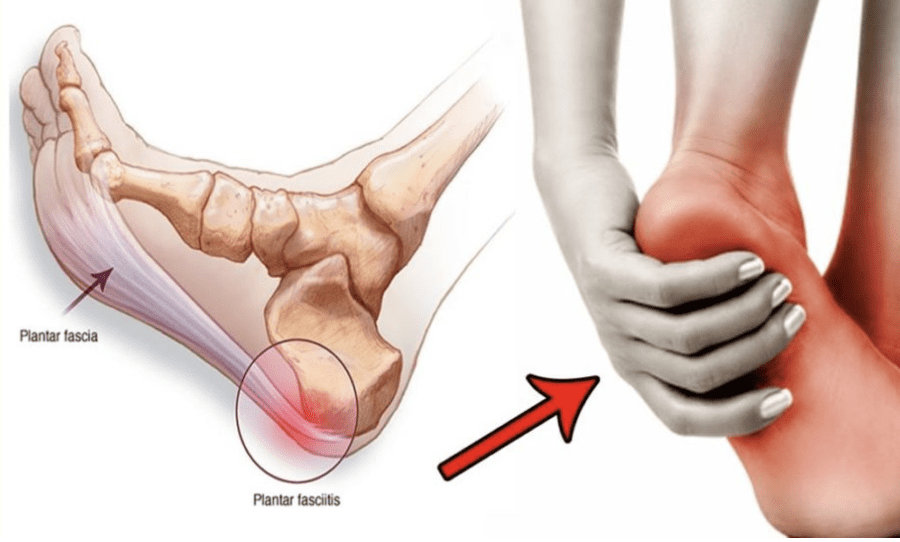
Plantar fasciitis is a common cause of heel and foot pain. The plantar fascia is a band of tissue that runs along the bottom of the foot and becomes inflamed. Certain factors can cause plantar fasciitis flare-ups, including:
Increased levels of physical activity.
Not stretching regularly.
Wearing shoes without proper support.
Weight gain.
Causes
A plantar fasciitis flare-up is often triggered by physical activity. (MedlinePlus. U.S. National Library of Medicine. 2022) It can also be brought on by underlying conditions, like increased body weight, arthritis, or the shape of the foot. (Johns Hopkins Medicine. 2023) Despite the root cause, there are activities and experiences that can contribute to and/or worsen the condition.
New Exercise Routine
Being highly physically active can exacerbate plantar fasciitis symptoms.
High heels, boots, or shoes that raise the heel above the toes.
Worn-out shoes like exercise workout shoes.
Not Stretching Properly or At All
Tight calves can increase pressure on the plantar fascia.
Stretching the calves, Achilles tendon/heel, and the bottom of the feet is highly recommended to help treat and prevent the condition. (Johns Hopkins Medicine. 2023)
Not stretching thoroughly or skipping stretches can worsen symptoms.
Individuals with plantar fasciitis are recommended to stretch before and after physical activities, exercise, before going to bed, and after waking up.
Working Through the Pain
Individuals may try to continue physical activities during a flare-up.
This is not recommended as doing so can cause more pain and worsen the condition.
When pain presents, it’s recommended to:
Stop all activities that strain the feet
Stay off the feet for at least a week.
Tearing the Plantar Fascia
The plantar fascia rarely tear completely from repeated stress known as a plantar fascia rupture.
Pascoe, S. C., & Mazzola, T. J. (2016). Acute Medial Plantar Fascia Tear. The Journal of orthopaedic and sports physical therapy, 46(6), 495. https://doi.org/10.2519/jospt.2016.0409
Can healthcare professionals help individuals with spinal pain by incorporating non-surgical spinal decompression to restore mobility?
Introduction
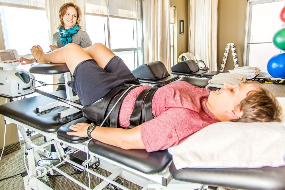
Many individuals don’t realize that putting unwanted pressure on their spines can lead to chronic pain within their spinal discs that is affecting their spinal mobility. This usually happens with demanding jobs requiring individuals to carry heavy objects, step wrong, or be physically inactive, which causes the surrounding back muscles to be overstretched and leads to referred pain that affects the upper and lower body portions. This can cause individuals to go to their primary doctors to get treated for back pain. This leads to them missing out on their busy work schedules and paying a high price to get treated. Back pain correlating with spinal issues can be a huge problem and make them feel miserable. Fortunately, numerous clinical options are cost-effective and personalized to many individuals dealing with spinal pain that is causing them to find the relief they deserve. Today’s article focuses on why spinal pain affects many people and how spinal decompression can help reduce spinal pain and restore spinal mobility. Coincidentally, we communicate with certified medical providers who incorporate our patients’ information to provide various treatment plans to reduce spinal pain affecting their backs. We also inform them that there are non-surgical options to reduce the pain-like symptoms associated with spinal issues in the body. We encourage our patients to ask amazing educational questions to our associated medical providers about their symptoms correlating with body pain in a safe and positive environment. Dr. Alex Jimenez, D.C., incorporates this information as an academic service. Disclaimer
Why Spinal Pain Is Affecting Many People?
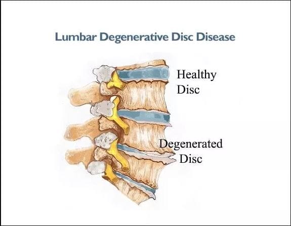
Have you often experienced pain from your back muscles that seem to ache after bending down constantly to pick up objects? Do you or your loved ones feel muscle stiffness in the back and experience numbness in your upper or lower body portions? Or are you experiencing temporary relief after stretching your back muscles, only for the pain to return? Many individuals with back pain never realize that their pain is within their spinal column. Since the spine is an S-curve shape with three different regions in the body, the spinal discs within each spinal segment can become compressed and become misaligned over time. This causes degenerative changes within the spine and can cause the three different spinal regions to develop pain-like issues in the body. When several environmental factors start to be the causes of degeneration of the spinal discs, it can affect the spinal structure. It can become a strong influence affecting their function, predisposing the disc to injuries. (Choi, 2009) At the same time, this can cause a significant impact when getting treated due to its high cost and can start normal age-related changes that cause pathophysiological issues to the vertebral body. (Gallucci et al., 2005)
When many individuals are dealing with spinal pain associated with herniated discs, it can not only cause discomfort but also mimic other musculoskeletal disorders that can cause radiating pain to different locations in the body. (Deyo et al., 1990) This, in turn, causes individuals to suffer constantly and research various treatments to reduce the pain they are experiencing. When spinal pain affects most individuals, many will seek cost-effective therapies to ease the pain they are experiencing and to be mindful of the daily habits they adopt over time and correct them.
Spinal Decompression In-Depth- Video
Do you often feel constant muscle aches and pains in your body that are your general areas of complaint? Do you feel your muscles pull uncomfortably after lifting or carrying a heavy object? Or do you feel constant stress in your neck, shoulders, or back? When many individuals are dealing with general pain, they often assume that it is just back pain when it could be a spinal issue that can be the root cause of the pain they are experiencing. When this happens, many individuals opt for non-surgical treatments due to its cost-effectiveness and how it can be personalized depending on the severity of the pain. One of the non-surgical treatments is spinal decompression/traction therapy. The video above gives an in-depth look at how spinal decompression can help reduce spinal pain associated with low back pain. Spinal pain can increase with age and be provoked by extreme lumbar extension, so incorporating spinal decompression can help reduce pain in the upper and lower extremities. (Katz et al., 2022)
How Spinal Decompression Can Reduce Spinal Pain
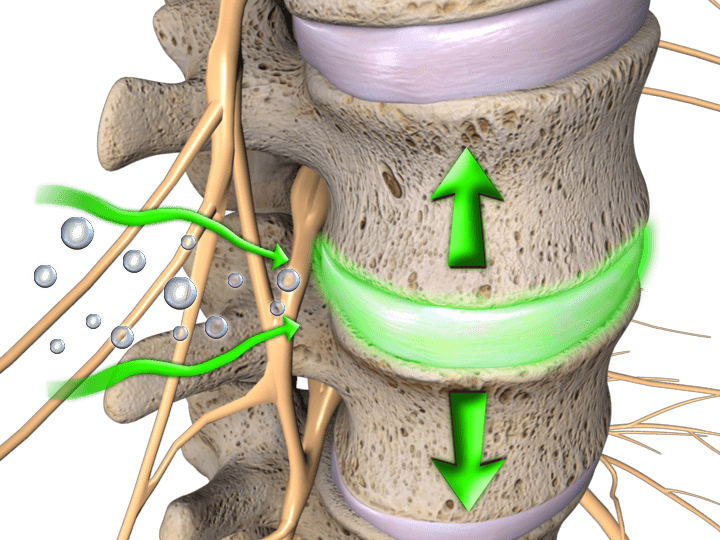
When individuals develop spinal issues, spinal decompression can help restore the spine to its original position and help the body naturally heal itself. When something is out of place within the spine, it is important to naturally restore it to its proper place to allow the affected muscles to heal. (Cyriax, 1950) Spinal decompression uses gentle traction to pull the spinal joints to let the spinal disc back in its original position and help increase fluid intake back in the spine. When people start incorporating spinal decompression into their health and wellness routine, they can reduce their spinal pain after a few consecutive treatments.
Spinal Decompression Restoring Spinal Mobility
Spinal decompression can also be incorporated with other non-surgical treatments to restore spinal mobility. When pain specialists utilize spinal decompression within their practices, they can help treat various musculoskeletal conditions, including spinal disorders, to allow the individual to regain spinal mobility. (Pettman, 2007) At the same time, pain specialists can use mechanical and manual manipulation to reduce the pain the individual feels. When spinal decompression starts to use gentle traction on the spine, it can help minimize radical pain correlated with nerve entrapment, create negative pressure within the spinal sections, and relieve musculoskeletal disorders causing pain. (Daniel, 2007) When people start thinking more about their health and wellness to reduce their pain, spinal decompression can be the answer through a personalized plan and can help many individuals find the relief they deserve.
Daniel, D. M. (2007). Non-surgical spinal decompression therapy: does the scientific literature support efficacy claims made in the advertising media? Chiropr Osteopat, 15, 7. https://doi.org/10.1186/1746-1340-15-7
Gallucci, M., Puglielli, E., Splendiani, A., Pistoia, F., & Spacca, G. (2005). Degenerative disorders of the spine. Eur Radiol, 15(3), 591-598. https://doi.org/10.1007/s00330-004-2618-4
Katz, J. N., Zimmerman, Z. E., Mass, H., & Makhni, M. C. (2022). Diagnosis and Management of Lumbar Spinal Stenosis: A Review. JAMA, 327(17), 1688-1699. https://doi.org/10.1001/jama.2022.5921
IFM's Find A Practitioner tool is the largest referral network in Functional Medicine, created to help patients locate Functional Medicine practitioners anywhere in the world. IFM Certified Practitioners are listed first in the search results, given their extensive education in Functional Medicine


 Taping steps: