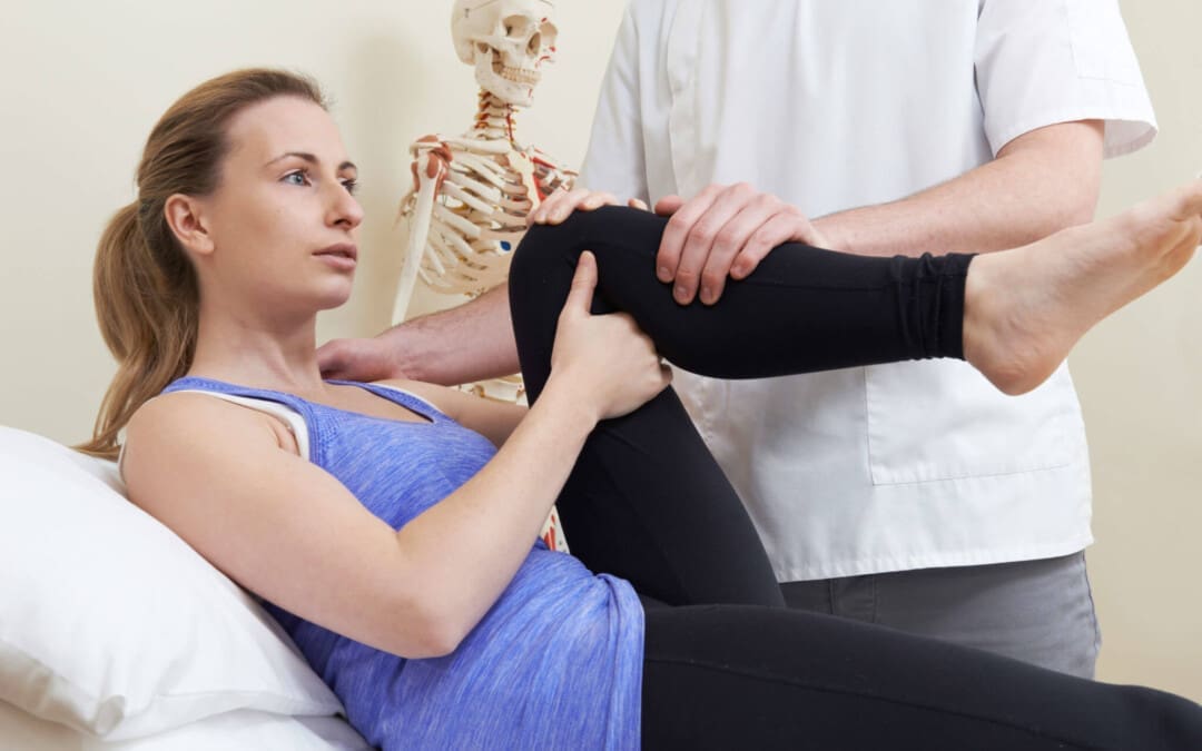
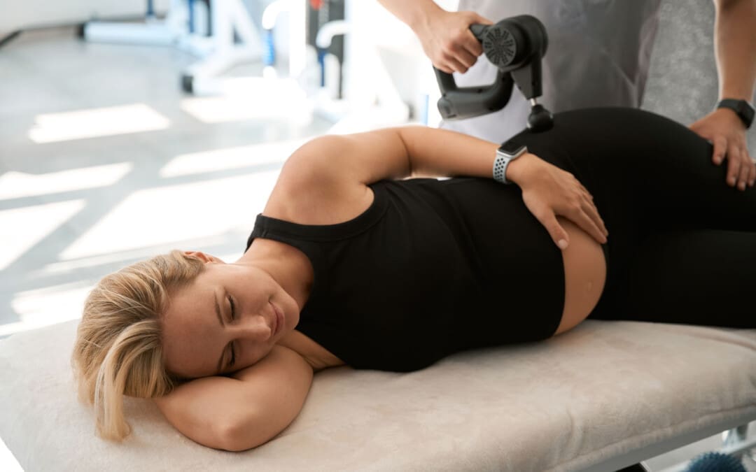
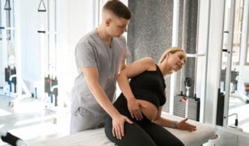
Stress on the lower back during pregnancy often leads to back (upper, middle, lower), sciatica, and leg pain. Can you use a massage gun while pregnant?
Pregnancy Massage Gun Use
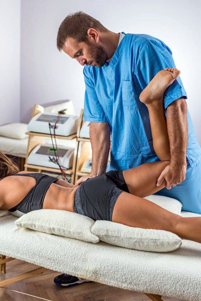
Pregnant women often experience back, hip, and leg pain along with symptoms such as swelling (edema), muscle spasms, cramps, and fatigue, which interfere with daily functions and sleep. Pregnancy massage gun use is a safe and helpful form of stress and tension relief, so long as healthcare providers and safety precautions are followed and sensitive areas are avoided. Massage guns use percussion and vibration to create a form of massage that can relieve tension, stiffness, and muscle soreness, break up tissue adhesions, and reduce inflammation. Moreover, pregnancy massage can provide relaxation and pain relief for muscles and joints that are adapting to a growing, changing body. Traditional massage therapy and massage gun therapy have been shown to help reduce muscle soreness and pain. (Imtiyaz S., Veqar Z., & Shareef M. Y. 2014) The benefits of pregnancy massage gun use include:
However, it is essential to discuss massage gun use with a healthcare professional and follow safety precautions when using massage guns during pregnancy.
Benefits
While there is no specific research on the benefits of massage guns for pregnant individuals, studies have demonstrated the general benefits of massage during pregnancy, including deep tissue massage. Therapeutic massage effectively reduces pregnancy discomforts and pain and is a safe and affordable method of pain relief. (El-Hosary EA, Abbas Soliman HF, El-Homosy SM. 2016) Researchers believe this relief helps improve the health of mother and baby in ways that include. (El-Hosary EA, Abbas Soliman HF, El-Homosy SM. 2016) (Mueller S. M., & Grunwald M. 2021)
Improves cardiovascular health
Overall circulation improves
Increases serotonin and dopamine levels
Decreases chronic back pain
Relieves muscle aches and joint pains
Improves sleep patterns
Increases energy
Reduces anxiety
Increases immune response
Enhances sense of well-being and mood
Reduces risk of preterm delivery
Massage Gun Safety
Pregnancy massage gun use is generally safe for those who do not have preexisting conditions. (Mueller S. M., & Grunwald M. 2021) However, there are no studies on the safety of massage guns or other massage tools for pregnant individuals. It is recommended to discuss the use of massage guns with a doctor before use.
Individuals with high-risk factors, such as bleeding, pre-term contractions, and preeclampsia, should avoid using massage guns unless a doctor clears them to do so.
Preeclampsia
This condition causes a rise in blood pressure that can be serious.
It typically develops around 20 weeks of pregnancy or later.
Deep Vein Thrombosis – DVT
Avoid using a massage gun if there is a history of deep vein thrombosis.
Blood volume increases during pregnancy, and leg circulation can be poor.
Certain hormones that prevent hemorrhage during delivery can cause blood to clot more easily.
Using a massage gun on areas with potential blood clots may release the clot, leading to a life-threatening embolism. (Sutham K. et al., 2020)
Compression socks are a better alternative for relieving leg pain and swelling, but follow the healthcare provider’s recommendations.
Placenta Previa, Accrete, or Abruption
These conditions involve the placenta, which can lead to bleeding.
Gestational Diabetes
High blood sugar levels do not necessarily mean individuals cannot use a massage gun during pregnancy.
However, speak to a doctor and monitor blood sugar before use.
How to Use
Although there are no specific expert directions for the use of massage guns during pregnancy, there are guidelines to consider while using the device. This includes:
Never place the massage gun directly over bones, nerves, or joints.
Avoid using the device around injured, swollen, or painful areas.
Avoid using a massage gun directly on the abdomen.
Start by trying a light 10- to 15-second pass over sore or tight areas.
Perform three to five sweeps over the location, then move on to another.
Be careful not to keep the gun on a single area too long, as you could overwork the muscle, leading to bruising and irritation.
The recommended time for leaving the massager in one area is two minutes.
Stop using the massage gun if there is unusual pain, sensations, or discomfort.
Injury Medical Chiropractic and Functional Medicine Clinic
Massage during pregnancy can help relieve symptoms such as anxiety, stress, pain, discomfort, tightness, and poor circulation. Regular massage can help improve sleep and can even benefit the baby. However, it is recommended to consult with a doctor about using a massage gun before trying it out. Injury Medical Chiropractic and Functional Medicine Clinic can help individuals recover and regain the benefits of quality rest through healthy sleep practices and lifestyle accommodations. We build optimal health and wellness solutions with primary healthcare providers and specialists. We focus on what works for you to relieve pain, restore function, prevent injury, and help mitigate issues through adjustments that help the body realign itself. They can also work with other medical professionals to integrate a treatment plan to resolve musculoskeletal problems.
Pregnancy and Sciatica: How Chiropractic Helped
References
Imtiyaz, S., Veqar, Z., & Shareef, M. Y. (2014). To Compare the Effect of Vibration Therapy and Massage in Prevention of Delayed Onset Muscle Soreness (DOMS). Journal of clinical and diagnostic research: JCDR, 8(1), 133–136. doi.org/10.7860/JCDR/2014/7294.3971
El-Hosary EA, Abbas Soliman HF, El-Homosy SM. (2016). Effect of Therapeutic Massage on Relieving Pregnancy Discomforts. IOSR Journal of Nursing and Health Science., 5(4), 57-64. doi.org/10.9790/1959-0504025764
Mueller, S. M., & Grunwald, M. (2021). Effects, Side Effects and Contraindications of Relaxation Massage during Pregnancy: A Systematic Review of Randomized Controlled Trials. Journal of Clinical Medicine, 10(16), 3485. doi.org/10.3390/jcm10163485
Sutham, K., Na-Nan, S., Paiboonsithiwong, S., Chaksuwat, P., & Tongsong, T. (2020). Leg massage during pregnancy with unrecognized deep vein thrombosis could be life-threatening: a case report. BMC pregnancy and childbirth, 20(1), 237. doi.org/10.1186/s12884-020-02924-w
Can performing the hip hinge exercise movement help individuals with lower back pain?
Hip Hinge Exercise
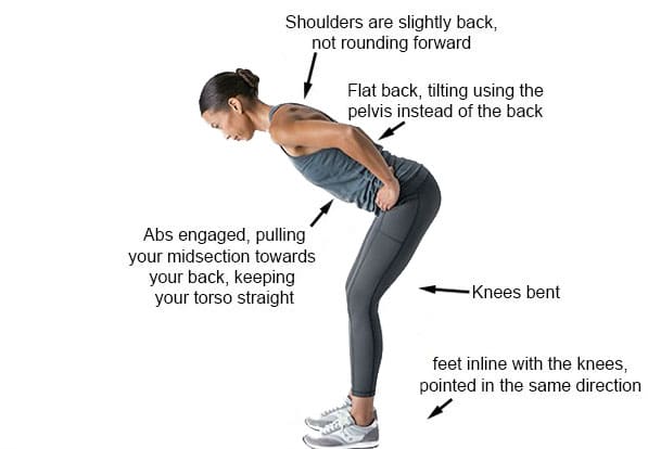
A hip hinge is a controlled movement that involves bending forward from the hips while keeping the spine neutral. The thoracic, lumbar, and pelvis stay neutral while bending forward. The movement comes from the hips, preventing the thoracic and lumbar spine from flexing or rounding. It is a fundamental movement that helps prevent back injuries and strengthens the glutes. It’s used in everyday activities, like picking up objects and sitting down.
The hip hinge exercise targets the posterior chain or back muscles, including the lower back, the glutes, and the hamstrings. It also strengthens the core or abdominal muscles to assist in the movement. When the body hinges at the hips, the bend occurs at the hips, and the spine stays neutral. When the lower back hinges or bends, this causes pain and reduces the range of motion.
Performing the Movement
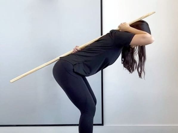
A wooden dowel, broom handle, or PVC pipe can be used as a guide to help achieve the proper positioning and learn the correct form. Place the dowel or pipe vertically on your back, anchoring it to the head, shoulder blades, and tailbone.
Grasp one end with your right hand in the natural curve of your neck and the other with your left hand in the small of your back. Ensure the dowel touches the back of your head, upper back, and the area where the lower back meets the sacrum. To perform the hip hinge:
Stand with your feet shoulder-width apart
Shift your weight to your heels and
Push your hips back while hinging your torso forward
Keep your chest open and back flat
Slightly bend your knees
Visualize sticking the butt out
The dowel should not lose contact with the three points as you hinge. If it does, the movement is incorrect.
Lower your torso until it’s midway between vertical and parallel to the floor.
Pause when your torso is about 45 degrees
Keep a slight bend in your knees during the downward and upward phases.
Reverse the movement by contracting your glutes and pushing your hips forward and upward to return to the starting position.
Repeat
Benefits
The hip hinge is a fundamental movement pattern that helps the body perform essential tasks such as bending over and picking things up without worry of pain or injury. It’s also required in strength training exercises like the deadlift, kettlebell swing, power clean, and more. The exercise can help strengthen the core, reduce back pain, improve balance, and improve flexion, extension, and trunk rotation. (Michaud F. et al., 2021) Stronger core muscles can increase fitness and athletic performance. (Clark D. R. et al., 2018)
Variations
It is a challenging movement that requires plenty of practice. Individuals who can’t perform it correctly after a few tries may need to modify the movement.
Wall Variation
Using a wall as a guide is an easy way to make the movement easier.
To do this, stand with your back to a wall, about three inches away.
Start hinging at the hips by sticking your butt out touching the wall.
Keep a neutral spine and a flat back.
Once you can do this several times, try stepping out another inch or two and perform the same modified motion. Stick with this until you are away from the wall and can do a full hinge without the wall.
With A Kettlebell
Once you master the basic hinge, you can elevate it using a kettlebell to make this move more difficult.
Start with the kettlebell swing exercise and progress to more challenging moves with the kettlebell.
Common Mistakes
Be aware of common mistakes to keep the move effective and reduce the risk of injury.
Treating the Move Like a Squat
The hip hinge is not the same as a squat.
This is a common misconception. When squatting, the knee joint determines the movement pattern.
But when hip hinging, the movement starts at the hips.
Not Engaging the Core Muscles
This exercise requires core engagement throughout the entire movement.
If these muscles relax, there is an increased risk of dipping the hips during the hinge, which can cause the lower back to dip and cause pain.
Using the Lower Back
Bending or hinging with the lower back rather than letting the hips generate the movement.
Using the wall as a guide can help reduce and eliminate excessive bending at the waist.
Lost Dowel Contact
If the dowel loses contact with one or more set-up positions on the back, the hinge is not being executed correctly.
If your head loses contact with the dowel, the neck is flexing too far forward.
If you lose contact with the sacrum or lower back area, the spine is flexing too much.
If you lose contact with the mid-back, the knees are bending rather than the hips.
Safety
Stop and check your form if you feel back pain during any part of the movement. The movement may need to be modified further or decrease how far the hinge is at the hips. If the pain continues, discontinue the exercise and talk with a doctor or a physical therapist before reattempting the exercise. The dowel is a great tool to help maintain a neutral spine. If you cannot perform the hip hinge while keeping the dowel in contact with the body, you might benefit from working with a personal trainer or physical therapist who can walk you through the steps with the correct form.
Injury Medical Chiropractic and Functional Medicine Clinic
Chiropractic care aims to help individuals improve movement with less pain due to condition, after injury, or surgery. A chiropractic physical therapy team can assess your condition and develop a customized treatment plan to expedite pain relief and improve mobility. Injury Medical Chiropractic and Functional Medicine Clinic works with primary healthcare providers and specialists to build optimal health and wellness solutions. We focus on what works for you to relieve pain, restore function, prevent injury, and help mitigate issues through adjustments that help the body realign itself. They can also work with other medical professionals to integrate a treatment plan to resolve musculoskeletal problems.
Chiropractic: The Secret to Unlocking Mobility
References
Michaud, F., Pérez Soto, M., Lugrís, U., & Cuadrado, J. (2021). Lower Back Injury Prevention and Sensitization of Hip Hinge with Neutral Spine Using Wearable Sensors during Lifting Exercises. Sensors (Basel, Switzerland), 21(16), 5487. doi.org/10.3390/s21165487
Clark, D. R., Lambert, M. I., & Hunter, A. M. (2018). Contemporary perspectives of core stability training for dynamic athletic performance: a survey of athletes, coaches, sports science and sports medicine practitioners. Sports medicine – open, 4(1), 32. doi.org/10.1186/s40798-018-0150-3
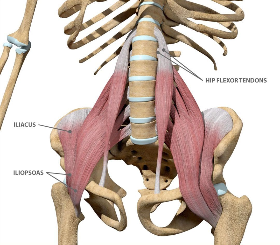
The iliacus muscle is a triangle-shaped muscle in the pelvic bone that flexes and rotates the thigh bone. It works with the other muscles in the hip and thigh to help bend, run, walk, sit, and maintain correct posture. Injuries and common medical conditions can affect its function, causing pain and stiffness. Can physical therapy help?
The Iliacus Muscle
The iliacus is one of the body’s most important hip flexor muscles. The iliacus and surrounding muscles work together to produce the stability and range of motion required for bending, dancing, sitting, and walking.
Anatomy
The iliacus muscle is part of a complex muscle system in the hip and pelvis. Two iliacus muscles on each side of the pelvic bone enable the thigh to flex and rotate. They are innervated by the femoral nerve, which provides movement and sensation to the lower limbs. (Bordoni B. and Varacallo M. 2023) The iliacus muscle sits on the wing-shaped ilium and fits into the curved surface of the ilium, called the iliac fossa. The top of the muscle is attached to the upper wings of the ilium or iliac crest. It extends past the hip joint, which connects to the upper thigh bone/femur at the lesser trochanter protrusion. The iliacus is part of a major trio of muscles called the iliopsoas, including the major psoas and minor psoas muscles. These muscles are also attached to the upper femur but extend upward, connecting to the lumbar/lower spine at several attachment points. The iliopsoas also interact with the quadratus lumborum muscle, the deepest muscle of the lower back that starts at the iliac crest and attaches to the lumbar spine at several points. The quadratus lumborum enables flexion and elevation of the spine, while the iliopsoas enable the flexion and rotation of the hip and thigh.
Functions
The iliacus muscle has many functions that include: (Physiopedia, 2024)
Flexing and rotating the femur.
Helps maintain proper body posture while standing and sitting.
Produces hip movement that enables walking, running, and climbing stairs.
Provides hip flexion – bringing the knee to the chest.
Enables the forward tilt of the pelvis and side-bending.
Conditions
Several conditions can affect the iliacus muscle, specifically from under and/or overuse injuries. These conditions, collectively known as Iliopsoas syndrome, are typically the result of overuse/repetitive strain or injuries. These include:
Iliopsoas tendinopathy – which affects tendons.
Iliopsoas bursitis – which affects cushioning sacs known as bursae.
Iliopsoas syndrome can affect anyone but is common in:
Individuals and athletes who repeatedly use movements that flex the hips.
Track-and-field athletes
Gymnasts
Dancers
Iliopsoas Bursitis
This is the inflammation of the cushioning sac or bursa under the iliacus muscle, which helps the muscle slide over the pelvic bone. Symptoms can range from mild discomfort to pain that radiates through parts of the leg and hips. Runners, skiers, and swimmers are commonly affected, and individuals who regularly have tight hips and individuals with different forms of arthritis can also be affected. Early treatment can prevent the symptoms from worsening. Mild cases can be treated with self-care and stretching to help relieve tightness, rest, ice application, and over-the-counter nonsteroidal anti-inflammatory drugs. In severe cases, treatment options that may be recommended include: (Physiopedia, 2024)
Physical therapy
Assistant walking devices to relieve pressure – for example, a cane.
Corticosteroid steroid injections
Prescription anti-inflammatory medications
Iliopsoas Tendinopathy
Another condition affecting the iliacus muscles is iliopsoas tendinopathy, sometimes called snapping hip syndrome, because individuals can hear an audible snapping sound (Davenport KL. 2019). The condition is often experienced by dancers who repeatedly flex and hyperextend their hips and can result in hip and groin pain that gets worse with kicking or rotation. Treatment of iliopsoas tendinopathy can include:
Retraining muscle imbalances with strengthening and stretching exercises.
If these fail to provide relief, corticosteroid injections may be used. A saline hydro dissection can relieve stress around the tendon by injecting fluids that cushion and release trapped tissues.
Tendon release surgery may be recommended when all other options have failed. The surgical release involves severing the tendon to reduce pain and improve the range of motion.
Rehabilitation
Core muscle strengthening is essential to the rehabilitation of iliacus muscle injuries. The iliopsoas is an integral component of the core group and can benefit from stretching and strengthening exercises (Yogateket, 2019)
Lunge stretches
Straight leg raises
Knee-to-chest stretches
Standing hip flexion with resistance bands
Certain yoga poses can also help and include variations on the bridge pose that encourage hip flexion. (Yoga International, 2024)
Injury Medical Chiropractic and Functional Medicine Clinic
Iliopsoas pain is often felt at the front of the hips, thigh, mid-back, and lower back. Chiropractic care can help with iliacus muscle injuries through:
Evaluation
A chiropractor can evaluate the condition and determine if the iliacus muscle is causing pain.
Treatment plan
A chiropractor can create a personalized treatment plan that may include exercise instructions, manipulation, and other therapies.
Rehabilitation
A chiropractor can create a rehabilitation program to expedite healing.
Injury Medical Chiropractic and Functional Medicine Clinic works with primary healthcare providers and specialists to develop an optimal health and wellness solution. We focus on what works for you to relieve pain, restore function, and prevent injury. Regarding musculoskeletal pain, specialists like chiropractors, acupuncturists, and massage therapists can help mitigate the pain through spinal adjustments that help the body realign itself. They can also work with other medical professionals to integrate a treatment plan to resolve musculoskeletal issues.
Hip Labral Tear and Chiropractic Care
References
Bordoni, B., & Varacallo, M. (2024). Anatomy, Bony Pelvis, and Lower Limb, Iliopsoas Muscle. In StatPearls. www.ncbi.nlm.nih.gov/pubmed/30285403
Individuals with hip bursitis often experience discomfort during physical activity, walking, and pain when lying on the affected side. What treatment options are available to control and manage the condition?
Hip Bursitis
Hip bursitis, also known as trochanteric bursitis, is a common condition that causes pain and discomfort in the hip and upper thigh along the outside of the hip joint. It occurs when one of the hip’s bursae, or fluid-filled sacs cushion joints, becomes inflamed. Treatment for hip bursitis is to control the inflammation caused by this condition.
Causes
Hip bursitis can be caused by injury or overuse of the hip, such as repetitive activities, twisting, or rapid joint movement. It can also be caused by a direct blow or fall to the side of the hip.
Symptoms
Pain from hip bursitis can be sharp at first and may feel dull and achy later.
It may be worse when standing up after sitting, moving, or using the hip.
Individuals may also notice pain when lying on the affected side or sitting for a long time.
Rest
This means a period of not participating in physical, exercise, and sports activities that aggravate symptoms. Any activity that causes hip pain should be avoided as this only contributes to inflammation of the bursa. (American Academy of Orthopaedic Surgeons, 2022) Modifying how particular activities are performed can help alleviate pressure on the inflamed bursa. Working with a physical therapist can also be recommended. They are experts in movement and alignment, and if certain muscles are overused compared to others, this can lead to unhealthy movement patterns, causing bursa irritation.
Anti-Inflammatory Medications
Nonsteroidal anti-inflammatory drugs, such as Motrin, Aleve, Naprosyn, etc., will help control inflammation (American Academy of Orthopaedic Surgeons, 2022). Anti-inflammatory medications can be extremely effective but should be taken cautiously. The instructions on the label need to be followed unless directed otherwise by a healthcare provider. Be aware of side effects and inform the healthcare provider if side effects present.
Cold Therapy
Applying ice to the hip area often helps alleviate the symptoms (National Library of Medicine, 2022). Ice can control inflammation by decreasing blood circulation to the area, especially after physical activity and exercise.
Aspiration
A needle is placed into the bursa to drain the fluid for those with a significant amount of fluid collected within the bursa. (National Library of Medicine, 2022) This is rarely needed in cases of hip bursitis, but when it is done, it can be combined with a cortisone injection.
Cortisone Injections
A cortisone injection may also be given into the bursa to alleviate pain. (American Academy of Orthopaedic Surgeons, 2022) The cortisone injection is helpful because it can be a diagnostic and therapeutic tool. In cases where hip bursitis may be one of several diagnoses being considered, cortisone can be given to see if it helps alleviate symptoms. Cortisone is a powerful anti-inflammatory medication that can be administered directly to the problem area. These injections are well-tolerated, but there can be possible side effects. Once the initial symptoms are under control, physical therapy strengthening and stretching exercises may be recommended.
Stretching
Most find relief by stretching the muscles and tendons over the outside of the hip, specifically the iliotibial band. The goal is for a better-conditioned muscle and tendon to glide more easily and not cause inflammation. Proper stretching techniques and posture are important in re-injury prevention.
Physical Therapy
Working with a physical therapist is an effective adjunct treatment for bursitis (American Academy of Orthopaedic Surgeons, 2022). Physical therapists correct muscle imbalances through stretching and exercise and improve alignment to prevent bursa irritation from reoccurring.
Surgery
Most patients get better with conservative treatment within about six weeks. Surgical treatment for hip bursitis is rarely needed (UCSF Health, 2024). Those who do not rest from their activities until the inflammation subsides often have a return of bursitis symptoms, and those who return too aggressively to activities and do not gradually build up also find that their symptoms return. In cases where surgery is needed, the healthcare provider may recommend an arthroscopic bursectomy. (American Academy of Orthopaedic Surgeons, 2022) The surgery is an outpatient minimally invasive procedure in which the bursa is removed through a small incision. After a short healing period, the individual can return to normal activity. Crutches may be used for a few days. Common complications are anesthetic-related complications and infection.
Injury Medical Chiropractic and Functional Medicine Clinic
As with any treatment program, always talk with your healthcare provider before initiating specific treatments. Fortunately, treatment of hip bursitis is generally accomplished with conservative therapies. Efforts to limit pressure directly on the bursa, alleviate inflammation, and restore normal movement to the hip joint will typically resolve symptoms. At Injury Medical Chiropractic and Functional Medicine Clinic, we focus on what works for you to relieve pain and restore function. Regarding musculoskeletal pain, specialists like chiropractors, acupuncturists, and massage therapists can help mitigate the pain through spinal adjustments that help the body realign itself. They can also work with other associated medical professionals to integrate into a treatment plan to improve the body’s flexibility and mobility, resolve musculoskeletal issues, and prevent future pain symptoms from reoccurring.
National Library of Medicine. (2022). Bursitis: Learn More – How can bursitis be treated? InformedHealth.org [Internet]. Cologne, Germany: Institute for Quality and Efficiency in Health Care (IQWiG). www.ncbi.nlm.nih.gov/books/NBK525763/
Can understanding the causes and symptoms of potential hip tendonitis help healthcare providers diagnose and treat the condition for individuals experiencing pain in the front of the hip with restricted hip flexibility that worsens during movement?
Hip Tendonitis
Hip tendonitis is inflammation of the iliopsoas tendon. It is most commonly caused by overuse of the hip flexors without adequate rest for recovery. The condition can occur when the hip muscles overpower the tendons attached to the hip bone, causing inflammation and irritation. This can lead to pain, tenderness, and mild swelling near the hip joint. Hip tendonitis can be diagnosed with a physical examination, and treatment can include:
Rest
Ice
NSAIDs
Stretching
Physical therapy
Chronic cases may require a cortisone injection into the iliopsoas tendon to decrease inflammation.
Surgical release of the iliopsoas tendon may be recommended to decrease tightness and pain.
There is a high prognosis for a full recovery.
Tendonitis
Inflammation in a muscle’s tendon leads to pain and tenderness that worsens the more the muscle is used. An overuse injury means the tendon becomes repeatedly stressed through repetitive muscle contractions, causing muscle and tendon fibers to micro-tear. If not enough rest is allowed for the micro-tears to heal, a chronic cycle of pain and inflammation develops within the affected tendon. Other tendons that are prone to developing the condition include:
The tendon of the wrist extensors/tennis elbow.
The tendon of the wrist flexors/golfer’s elbow.
The Achilles’ tendon/Achilles tendonitis.
The patellar tendon/jumper’s knee.
The tendons of the thumb/De Quervain’s tenosynovitis.
Bursitis
Bursae are small fluid-filled sacs that help cushion and decrease friction around joints.
Because the iliopsoas tendon overlays bursae, inflammation of the tendon can also cause bursitis or inflammation of the bursae surrounding the tendon.
Tendonitis and bursitis can and often occur together due to overlapping symptoms.
Causes
The iliopsoas originates in the pelvis and vertebrae of the lower spine and attaches to the top of the femur or thigh bone. It allows the hip joint movement that brings the leg closer to the front of the body, like lifting the leg to step up or jump. It also helps keep the torso stable when standing with one or both feet on the ground and rising from a lying position. Hip tendonitis most often results from physical activities that require repeated leg lifting when stepping, running, kicking, or jumping. This can include:
Running
Dancing
Gymnastics
Martial arts
Cycling
Playing soccer
Iliopsoas tendonitis can also occur after hip arthroscopy, a minimally invasive surgical procedure to repair structures inside the hip joint because of altered joint movement and muscle activation patterns after surgery. (Adib F. et al., 2018)
Symptoms
The primary symptoms of hip tendonitis include a soreness or deep ache in the front of the hip that worsens after physical activity and limits the range of motion because of the pain. Other symptoms include:
Tenderness to touch in the front of the hip.
The pain can feel like a dull ache.
Stiffness may also be present.
Hip flexor tightness.
Altered posture, with the pelvis rotated forward and an exaggerated curve in the lower back.
Lower back pain.
Discomfort after prolonged sitting.
Altered walking pattern characterized by shortened steps.
Diagnosis
Hip tendonitis is diagnosed through a physical examination and medical history reviews of individual symptoms.
Individuals may also have an X-ray of their hip performed to examine the joint alignment and determine if a fracture or arthritis is present.
Treatment
Initial treatment involves rest from physical activities, applying ice, and gentle stretching.
Nonsteroidal anti-inflammatory drugs/NSAIDs can ease pain and swelling, decrease inflammation, and reduce muscle spasms.
If chronic pain persists, individuals may receive a cortisone injection into their iliopsoas tendon. (Zhu Z. et al., 2020)
A personalized physical therapy program focusing on hip flexor stretching and strengthening, as well as strengthening the glutes and core, will help expedite an optimal recovery.
Surgery
For cases that do not improve after three months of treatment, surgery to lengthen the iliopsoas tendon, a procedure known as a tenotomy, may be performed. It involves making a small cut into a portion of the tendon, allowing the tendon to increase in length while decreasing tension as it heals back together. A tenotomy temporarily reduces the strength of the iliopsoas; however, this weakness usually resolves within three to six months after surgery. (Anderson C. N. 2016)
Chiropractic Care
Chiropractic care can be an effective treatment because it can help restore proper alignment and motion in the hip, reduce inflammation, and improve muscle and joint function. Treatments may include:
Spinal adjustments to realign the spine and other joints, reducing pressure on nerves and inflammation.
Non-surgical decompression
Manual therapy – massage, trigger point therapy, or spinal manipulation.
Acupuncture
Graston technique
Rehabilitative exercises like stretching, strengthening, and range of motion exercises.
Tendonitis generally has an excellent prognosis for full recovery as long as thorough rest from activities is taken to allow the inflamed tendon to heal. The postsurgical prognosis is positive for chronic and severe cases of iliopsoas tendonitis that require surgery.
Injury Medical Chiropractic and Functional Medicine Clinic works with primary healthcare providers and specialists to develop a customized treatment program through an integrated approach to treating injuries and chronic pain syndromes, improving flexibility, mobility, and agility to relieve pain and help individuals return to normal activities. If other treatments are needed, Dr. Jimenez has teamed up with top surgeons, clinical specialists, medical researchers, and rehabilitation providers to provide the most effective treatments.
Inflammation and Integrative Medicine
References
Adib, F., Johnson, A. J., Hennrikus, W. L., Nasreddine, A., Kocher, M., & Yen, Y. M. (2018). Iliopsoas tendonitis after hip arthroscopy: prevalence, risk factors and treatment algorithm. Journal of hip preservation surgery, 5(4), 362–369. doi.org/10.1093/jhps/hny049
Zhu, Z., Zhang, J., Sheng, J., Zhang, C., & Xie, Z. (2020). Low Back Pain Caused by Iliopsoas Tendinopathy Treated with Ultrasound-Guided Local Injection of Anesthetic and Steroid: A Retrospective Study. Journal of pain research, 13, 3023–3029. doi.org/10.2147/JPR.S281880
Anderson C. N. (2016). Iliopsoas: Pathology, Diagnosis, and Treatment. Clinics in sports medicine, 35(3), 419–433. doi.org/10.1016/j.csm.2016.02.009
Can physical therapies help treat a high steppage gait from injury or medical conditions and restore normal gait patterns for individuals who have or are developing one?
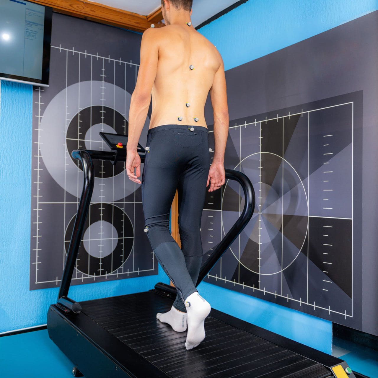
Walking or gait anthropometric analysis on a treadmill
Neuropathic Gait
Neuropathic gait, aka equine or high steppage gait, is a type of walking abnormality that causes individuals to raise their hips to lift their legs higher than normal. It occurs when individuals have a foot drop or ankle equinus due to loss of dorsiflexion. As a result, the foot hangs with the toes pointing down, causing the toes to drag while walking. The foot may appear floppy when it drops. Foot drop is caused by weakness or paralysis of the anterior tibialis muscle in front of the shin bone. The anterior tibialis muscle contracts to help flex the foot and ankle while walking, ensuring the foot clears the floor and doesn’t drag. Individuals with anterior tibialis weakness or paralysis may have a neuropathic gait and excessively bend the hip and knee while stepping forward, lifting their leg high off the floor to clear the foot to avoid tripping. A physical therapy team can help with a high steppage gait pattern after illness or injury.
Causes
Conditions that can cause anterior tibialis weakness or paralysis and a high steppage gait pattern include:
Sciatica
Pain caused by compression or irritation of the sciatic nerve starts in the lower back and travels down the back of the leg. (McCabe, F. J., McCabe, J. P. 2016)
Peroneal Nerve Injury
Damage to the peroneal nerve branches from the sciatic nerve that help move the lower leg and foot. (Johns Hopkins Medicine. 2024)
Multiple Sclerosis
An autoimmune disease that damages nerve cells in the brain and spinal cord. (Taylor, P. N. et al., 2016)
Balance exercises will help improve overall proprioception, or the sense of the body’s position and movement.
Neuromuscular electrical stimulation, or NMES, can help improve the function of the muscle. (Hollis, S., McClure, P. 2017)
The electrical stimulation artificially contracts the muscle to restore proper function.
For anterior tibialis weakness caused by sciatica, back decompression exercises may be prescribed to relieve pressure off the sciatic nerve.
The exercises release the nerve to restore normal signal transmission up and down the nerve in the lower back.
Neuromuscular electrical stimulation may also be used to help improve muscle function.
Assistive Walking Devices
A therapist may suggest using an assistive device to help the patient walk properly. This could include a wheeled walker or a quad cane. A temporary solution to anterior tibialis weakness is to elevate the foot while walking with an elastic band. Tie a band around the leg below the knee and secure it around the ball of the foot. When swinging the leg forward, the band pulls the foot up. Using it as a temporary solution may help maintain safe mobility. Sometimes, paralysis of the anterior tibialis muscle can become permanent. In this case, individuals may benefit from a special brace called an ankle-foot orthosis. The brace helps to lift the foot and toes off the ground.
For individuals concerned about losing their balance and falling, there are ways to improve walking patterns to stay safe. A healthcare provider may recommend physical therapy to correct gait, strengthen the anterior tibialis muscle, improve balance, and educate on injury prevention. Individuals should discuss symptoms and conditions with a primary physician, healthcare provider, or specialist to guide them in the right direction and determine the best treatment.
Injury Medical Chiropractic and Functional Medicine Clinic uses an integrated approach personalized to the individual that focuses on what works for them and treats injuries and chronic pain syndromes through personalized care plans that improve ability through flexibility, mobility, and agility programs to relieve pain. If other treatment is needed, Dr. Jimenez has teamed up with top surgeons, clinical specialists, medical researchers, and rehabilitation providers to provide the most effective treatments.
Control Foot Motion and Posture
References
McCabe, F. J., & McCabe, J. P. (2016). An Unusual Presentation of Right-Sided Sciatica with Foot Drop. Case reports in orthopedics, 2016, 9024368. doi.org/10.1155/2016/9024368
Kaykisiz, E. K., & Unluer, E. E. (2017). An Unexpected Reason for Isolated Foot Drop: Acute Stroke. Pakistan journal of medical sciences, 33(5), 1288–1290. doi.org/10.12669/pjms.335.13593
Taylor, P. N., Wilkinson Hart, I. A., Khan, M. S., & Slade-Sharman, D. E. (2016). Correction of Footdrop Due to Multiple Sclerosis Using the STIMuSTEP Implanted Dropped Foot Stimulator. International journal of MS care, 18(5), 239–247. doi.org/10.7224/1537-2073.2015-038
Hollis, S., & McClure, P. (2017). Intramuscular Electrical Stimulation for Muscle Activation of the Tibialis Anterior After Surgical Repair: A Case Report. The Journal of orthopaedic and sports physical therapy, 47(12), 965–969. doi.org/10.2519/jospt.2017.7368
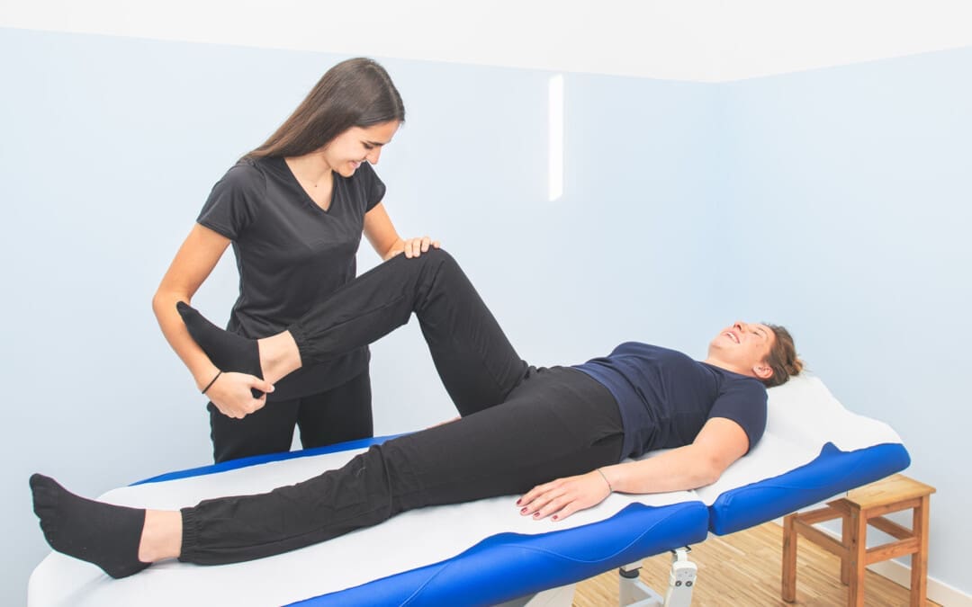
Can athletic individuals incorporate MET (muscle energy techniques) therapy to reduce the pain-like effects of adductor strain?
Introduction
The body’s lower extremities have an important role as they provide stability and mobility to the individual. Many athletes utilize their lower extremities by adding much power to exert the energy to win matches or competitions. The various muscles, soft tissues, ligaments, and joints help support the body’s skeletal structure and can succumb to injuries from repetitive motions or environmental factors. One of the muscles that can be affected by constant repetitive motions and environmental factors is the adductor muscles, which can cause many athletes to be in continuous pain and affect their performance during competitions. Luckily, there is a technique that many treatments offer to reduce muscle strain in the adductors and provide relief to the lower extremities. Today’s article looks at how adductor strain can affect many individuals, how MET therapy can help with an adductor strain, and its positive effect on athletic individuals. We discuss with certified medical providers who consolidate our patients’ information to assess the pain-like effects of an adductor strain in the lower extremities. We also inform and guide patients on how MET therapy can help stretch and strengthen tight adductor muscles to reduce strain and provide relief. We also encourage our patients to ask their associated medical providers many intricate and important questions about incorporating MET and other non-surgical therapies into their personalized treatment plan for a healthier lifestyle. Dr. Jimenez, D.C., includes this information as an academic service. Disclaimer.
How Does Adductor Strain Affect Individuals?
Do you feel tightness along your thighs and legs after a long day at work? Do you experience instability when walking from one location to another? Or do you feel pain when stretching your thighs that causes temporary relief? Many individuals experiencing pain in their lower extremities will often think it is hip pain, but their adductor muscles are in pain. The adductor muscles consist of three muscles that provide torque to the lower extremities by allowing them to move inward when a person is walking and help keep the trunk muscles steady. So, when many athletes begin to make constant repetitive motions while performing, it can cause issues for the adductors. As a common injury to many athletes, adductor strain can put exaggerated stress on the actual tendon, leading to biomechanical abnormalities affecting the musculoskeletal system. (Kiel & Kaiser, 2024a) Also, when athletes start to use constant repetitive motions during an increased volume or intensity of the training workload, it can cause stress factors in the lower extremities. (Kiel & Kaiser, 2024b) This, in turn, can have many individuals feel like they are experiencing hip and groin pain when it is, in fact, stress fractures in the adductor muscles causing myofascial pain.
So, for athletic individuals dealing with adductor strain, primary doctors need to differentiate between adductor strain and regular muscle strain in the lower extremities, as the pain symptoms sometimes have overlapping risk profiles with acute onset pain symptoms associated with distinct injury mechanisms. (McHugh et al., 2023) This is because when athletes overuse their adductor muscles, it causes pain, as many injuries within the adductors are associated with the hips and groin region. (Koscso et al., 2022) However, there are ways for athletes to find the relief they seek to reduce adductor strain and return to their routine.
Movement Medicine- Video
How MET Therapy Helps With Adductor Strain
For athletes and individuals engaged in physical activity, MET therapy can be a valuable part of the recovery process for adductor strain. MET (muscle energy technique) therapy, a form of osteopathic manipulative medicine, is used by pain specialists such as chiropractors, massage therapists, and sports physicians to alleviate pain symptoms in the musculoskeletal system. By using gentle, controlled muscle contractions, these specialists can improve musculoskeletal function by mobilizing joints, stretching tight muscles and fascia, and improving circulation and lymphatic flow. (Waxenbaum et al., 2024) Many pain specialists, including chiropractors and massage therapists, incorporate MET therapy into their practices due to its effectiveness in addressing muscular imbalances and alignment issues that contribute to pain and limited mobility in the lower extremities.
The Positive Effect Of MET Therapy
One of the positive effects of MET therapy for adductor strain is that when athletes and individuals start to utilize it as part of their recovery, their pain is reduced, and muscle mobility is increased since there are changes in the viscoelastic properties in the soft tissue. (Thomas et al., 2019) For the adductor muscles, MET therapy helps with:
Increasing muscle length & flexibility
Reduce muscle tension
Improving blood flow and promoting healing
Enhance joint function
MET therapy, when incorporated for pain relief for adductor strain, can put many individuals at ease as it actively focuses on muscle relaxation, lengthening, and strengthening the affected muscles. MET therapy can be combined with other therapies in a person’s personalized treatment plan to enhance mobility, be mindful of what is causing pain and discomfort to their bodies, and live a healthier lifestyle.
Koscso, J. M., McElheny, K., Carr, J. B., 2nd, & Hippensteel, K. J. (2022). Lower Extremity Muscle Injuries in the Overhead Athlete. Curr Rev Musculoskelet Med, 15(6), 500-512. doi.org/10.1007/s12178-022-09786-z
McHugh, M. P., Nicholas, S. J., & Tyler, T. F. (2023). Adductor Strains in Athletes. Int J Sports Phys Ther, 18(2), 288-292. doi.org/10.26603/001c.72626
Thomas, E., Cavallaro, A. R., Mani, D., Bianco, A., & Palma, A. (2019). The efficacy of muscle energy techniques in symptomatic and asymptomatic subjects: a systematic review. Chiropr Man Therap, 27, 35. doi.org/10.1186/s12998-019-0258-7
IFM's Find A Practitioner tool is the largest referral network in Functional Medicine, created to help patients locate Functional Medicine practitioners anywhere in the world. IFM Certified Practitioners are listed first in the search results, given their extensive education in Functional Medicine


 Causes