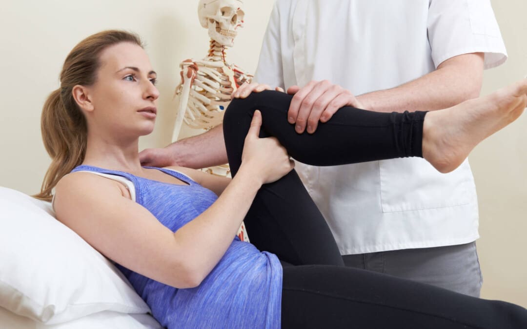
Can understanding the causes and symptoms of potential hip tendonitis help healthcare providers diagnose and treat the condition for individuals experiencing pain in the front of the hip with restricted hip flexibility that worsens during movement?
Hip Tendonitis
Hip tendonitis is inflammation of the iliopsoas tendon. It is most commonly caused by overuse of the hip flexors without adequate rest for recovery. The condition can occur when the hip muscles overpower the tendons attached to the hip bone, causing inflammation and irritation. This can lead to pain, tenderness, and mild swelling near the hip joint. Hip tendonitis can be diagnosed with a physical examination, and treatment can include:
Rest
Ice
NSAIDs
Stretching
Physical therapy
Chronic cases may require a cortisone injection into the iliopsoas tendon to decrease inflammation.
Surgical release of the iliopsoas tendon may be recommended to decrease tightness and pain.
There is a high prognosis for a full recovery.
Tendonitis
Inflammation in a muscle’s tendon leads to pain and tenderness that worsens the more the muscle is used. An overuse injury means the tendon becomes repeatedly stressed through repetitive muscle contractions, causing muscle and tendon fibers to micro-tear. If not enough rest is allowed for the micro-tears to heal, a chronic cycle of pain and inflammation develops within the affected tendon. Other tendons that are prone to developing the condition include:
The tendon of the wrist extensors/tennis elbow.
The tendon of the wrist flexors/golfer’s elbow.
The Achilles’ tendon/Achilles tendonitis.
The patellar tendon/jumper’s knee.
The tendons of the thumb/De Quervain’s tenosynovitis.
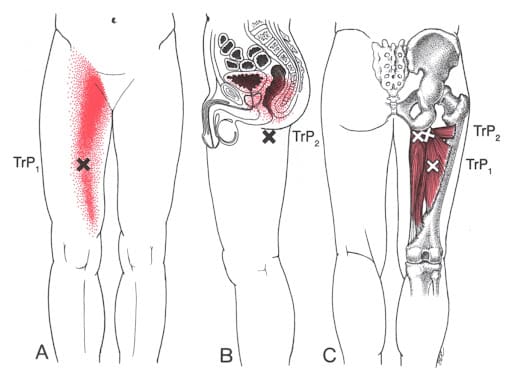
Bursitis
Bursae are small fluid-filled sacs that help cushion and decrease friction around joints.
Because the iliopsoas tendon overlays bursae, inflammation of the tendon can also cause bursitis or inflammation of the bursae surrounding the tendon.
Tendonitis and bursitis can and often occur together due to overlapping symptoms.
Causes
The iliopsoas originates in the pelvis and vertebrae of the lower spine and attaches to the top of the femur or thigh bone. It allows the hip joint movement that brings the leg closer to the front of the body, like lifting the leg to step up or jump. It also helps keep the torso stable when standing with one or both feet on the ground and rising from a lying position. Hip tendonitis most often results from physical activities that require repeated leg lifting when stepping, running, kicking, or jumping. This can include:
Running
Dancing
Gymnastics
Martial arts
Cycling
Playing soccer
Iliopsoas tendonitis can also occur after hip arthroscopy, a minimally invasive surgical procedure to repair structures inside the hip joint because of altered joint movement and muscle activation patterns after surgery. (Adib F. et al., 2018)
Symptoms
The primary symptoms of hip tendonitis include a soreness or deep ache in the front of the hip that worsens after physical activity and limits the range of motion because of the pain. Other symptoms include:
Tenderness to touch in the front of the hip.
The pain can feel like a dull ache.
Stiffness may also be present.
Hip flexor tightness.
Altered posture, with the pelvis rotated forward and an exaggerated curve in the lower back.
Lower back pain.
Discomfort after prolonged sitting.
Altered walking pattern characterized by shortened steps.
Diagnosis
Hip tendonitis is diagnosed through a physical examination and medical history reviews of individual symptoms.
Individuals may also have an X-ray of their hip performed to examine the joint alignment and determine if a fracture or arthritis is present.
Treatment
Initial treatment involves rest from physical activities, applying ice, and gentle stretching.
Nonsteroidal anti-inflammatory drugs/NSAIDs can ease pain and swelling, decrease inflammation, and reduce muscle spasms.
If chronic pain persists, individuals may receive a cortisone injection into their iliopsoas tendon. (Zhu Z. et al., 2020)
A personalized physical therapy program focusing on hip flexor stretching and strengthening, as well as strengthening the glutes and core, will help expedite an optimal recovery.
Surgery
For cases that do not improve after three months of treatment, surgery to lengthen the iliopsoas tendon, a procedure known as a tenotomy, may be performed. It involves making a small cut into a portion of the tendon, allowing the tendon to increase in length while decreasing tension as it heals back together. A tenotomy temporarily reduces the strength of the iliopsoas; however, this weakness usually resolves within three to six months after surgery. (Anderson C. N. 2016)
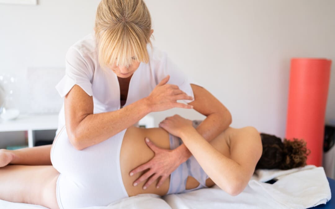
Chiropractic Care
Chiropractic care can be an effective treatment because it can help restore proper alignment and motion in the hip, reduce inflammation, and improve muscle and joint function. Treatments may include:
Spinal adjustments to realign the spine and other joints, reducing pressure on nerves and inflammation.
Non-surgical decompression
Manual therapy – massage, trigger point therapy, or spinal manipulation.
Acupuncture
Graston technique
Rehabilitative exercises like stretching, strengthening, and range of motion exercises.
Tendonitis generally has an excellent prognosis for full recovery as long as thorough rest from activities is taken to allow the inflamed tendon to heal. The postsurgical prognosis is positive for chronic and severe cases of iliopsoas tendonitis that require surgery.
Injury Medical Chiropractic and Functional Medicine Clinic works with primary healthcare providers and specialists to develop a customized treatment program through an integrated approach to treating injuries and chronic pain syndromes, improving flexibility, mobility, and agility to relieve pain and help individuals return to normal activities. If other treatments are needed, Dr. Jimenez has teamed up with top surgeons, clinical specialists, medical researchers, and rehabilitation providers to provide the most effective treatments.
Inflammation and Integrative Medicine
References
Adib, F., Johnson, A. J., Hennrikus, W. L., Nasreddine, A., Kocher, M., & Yen, Y. M. (2018). Iliopsoas tendonitis after hip arthroscopy: prevalence, risk factors and treatment algorithm. Journal of hip preservation surgery, 5(4), 362–369. https://doi.org/10.1093/jhps/hny049
Zhu, Z., Zhang, J., Sheng, J., Zhang, C., & Xie, Z. (2020). Low Back Pain Caused by Iliopsoas Tendinopathy Treated with Ultrasound-Guided Local Injection of Anesthetic and Steroid: A Retrospective Study. Journal of pain research, 13, 3023–3029. https://doi.org/10.2147/JPR.S281880
Anderson C. N. (2016). Iliopsoas: Pathology, Diagnosis, and Treatment. Clinics in sports medicine, 35(3), 419–433. https://doi.org/10.1016/j.csm.2016.02.009
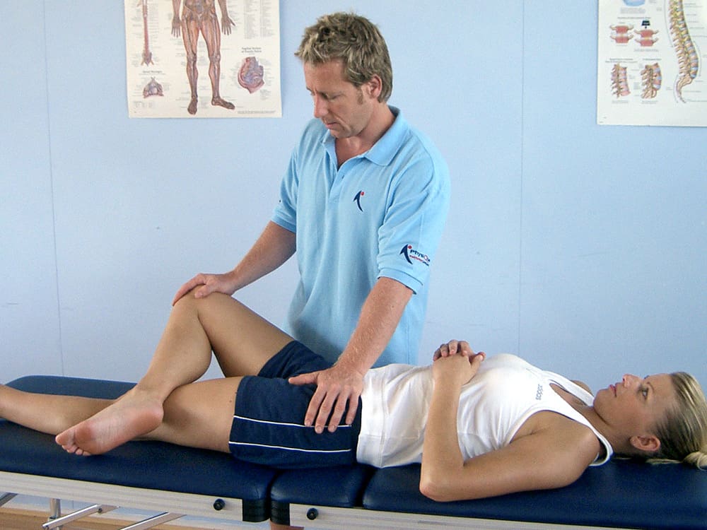
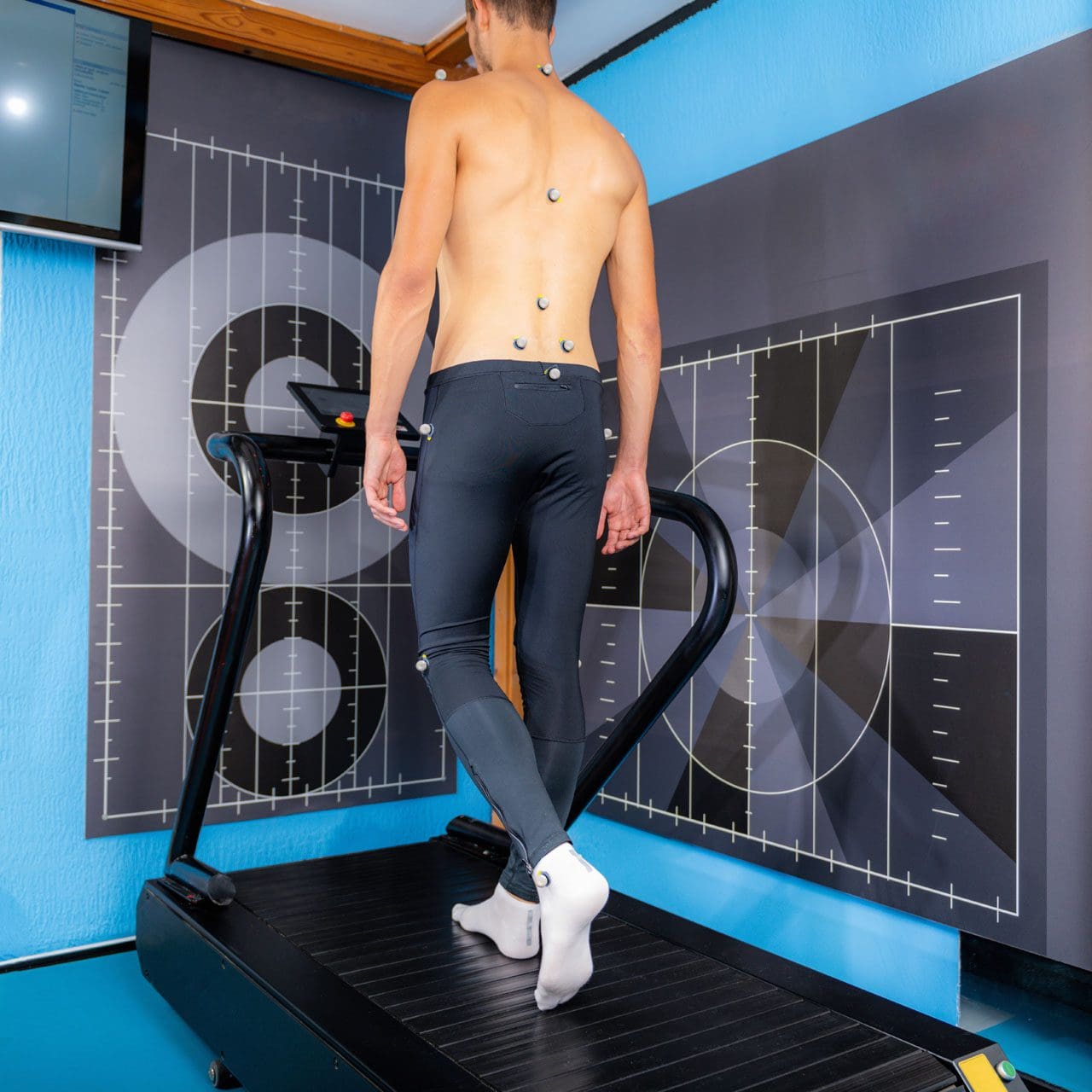
Can physical therapies help treat a high steppage gait from injury or medical conditions and restore normal gait patterns for individuals who have or are developing one?
Walking or gait anthropometric analysis on a treadmill
Neuropathic Gait
Neuropathic gait, aka equine or high steppage gait, is a type of walking abnormality that causes individuals to raise their hips to lift their legs higher than normal. It occurs when individuals have a foot drop or ankle equinus due to loss of dorsiflexion. As a result, the foot hangs with the toes pointing down, causing the toes to drag while walking. The foot may appear floppy when it drops. Foot drop is caused by weakness or paralysis of the anterior tibialis muscle in front of the shin bone. The anterior tibialis muscle contracts to help flex the foot and ankle while walking, ensuring the foot clears the floor and doesn’t drag. Individuals with anterior tibialis weakness or paralysis may have a neuropathic gait and excessively bend the hip and knee while stepping forward, lifting their leg high off the floor to clear the foot to avoid tripping. A physical therapy team can help with a high steppage gait pattern after illness or injury.
Causes
Conditions that can cause anterior tibialis weakness or paralysis and a high steppage gait pattern include:
Sciatica
Pain caused by compression or irritation of the sciatic nerve starts in the lower back and travels down the back of the leg. (McCabe, F. J., McCabe, J. P. 2016)
Peroneal Nerve Injury
Damage to the peroneal nerve branches from the sciatic nerve that help move the lower leg and foot. (Johns Hopkins Medicine. 2024)
Multiple Sclerosis
An autoimmune disease that damages nerve cells in the brain and spinal cord. (Taylor, P. N. et al., 2016)
Balance exercises will help improve overall proprioception, or the sense of the body’s position and movement.
Neuromuscular electrical stimulation, or NMES, can help improve the function of the muscle. (Hollis, S., McClure, P. 2017)
The electrical stimulation artificially contracts the muscle to restore proper function.
For anterior tibialis weakness caused by sciatica, back decompression exercises may be prescribed to relieve pressure off the sciatic nerve.
The exercises release the nerve to restore normal signal transmission up and down the nerve in the lower back.
Neuromuscular electrical stimulation may also be used to help improve muscle function.
Assistive Walking Devices
A therapist may suggest using an assistive device to help the patient walk properly. This could include a wheeled walker or a quad cane. A temporary solution to anterior tibialis weakness is to elevate the foot while walking with an elastic band. Tie a band around the leg below the knee and secure it around the ball of the foot. When swinging the leg forward, the band pulls the foot up. Using it as a temporary solution may help maintain safe mobility. Sometimes, paralysis of the anterior tibialis muscle can become permanent. In this case, individuals may benefit from a special brace called an ankle-foot orthosis. The brace helps to lift the foot and toes off the ground.
For individuals concerned about losing their balance and falling, there are ways to improve walking patterns to stay safe. A healthcare provider may recommend physical therapy to correct gait, strengthen the anterior tibialis muscle, improve balance, and educate on injury prevention. Individuals should discuss symptoms and conditions with a primary physician, healthcare provider, or specialist to guide them in the right direction and determine the best treatment.
Injury Medical Chiropractic and Functional Medicine Clinic uses an integrated approach personalized to the individual that focuses on what works for them and treats injuries and chronic pain syndromes through personalized care plans that improve ability through flexibility, mobility, and agility programs to relieve pain. If other treatment is needed, Dr. Jimenez has teamed up with top surgeons, clinical specialists, medical researchers, and rehabilitation providers to provide the most effective treatments.
Control Foot Motion and Posture
References
McCabe, F. J., & McCabe, J. P. (2016). An Unusual Presentation of Right-Sided Sciatica with Foot Drop. Case reports in orthopedics, 2016, 9024368. https://doi.org/10.1155/2016/9024368
Kaykisiz, E. K., & Unluer, E. E. (2017). An Unexpected Reason for Isolated Foot Drop: Acute Stroke. Pakistan journal of medical sciences, 33(5), 1288–1290. https://doi.org/10.12669/pjms.335.13593
Taylor, P. N., Wilkinson Hart, I. A., Khan, M. S., & Slade-Sharman, D. E. (2016). Correction of Footdrop Due to Multiple Sclerosis Using the STIMuSTEP Implanted Dropped Foot Stimulator. International journal of MS care, 18(5), 239–247. https://doi.org/10.7224/1537-2073.2015-038
Hollis, S., & McClure, P. (2017). Intramuscular Electrical Stimulation for Muscle Activation of the Tibialis Anterior After Surgical Repair: A Case Report. The Journal of orthopaedic and sports physical therapy, 47(12), 965–969. https://doi.org/10.2519/jospt.2017.7368
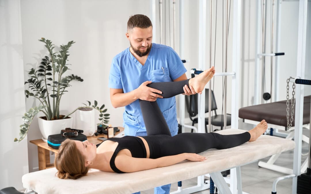
Can athletic individuals incorporate MET (muscle energy techniques) therapy to reduce the pain-like effects of adductor strain?
Introduction
The body’s lower extremities have an important role as they provide stability and mobility to the individual. Many athletes utilize their lower extremities by adding much power to exert the energy to win matches or competitions. The various muscles, soft tissues, ligaments, and joints help support the body’s skeletal structure and can succumb to injuries from repetitive motions or environmental factors. One of the muscles that can be affected by constant repetitive motions and environmental factors is the adductor muscles, which can cause many athletes to be in continuous pain and affect their performance during competitions. Luckily, there is a technique that many treatments offer to reduce muscle strain in the adductors and provide relief to the lower extremities. Today’s article looks at how adductor strain can affect many individuals, how MET therapy can help with an adductor strain, and its positive effect on athletic individuals. We discuss with certified medical providers who consolidate our patients’ information to assess the pain-like effects of an adductor strain in the lower extremities. We also inform and guide patients on how MET therapy can help stretch and strengthen tight adductor muscles to reduce strain and provide relief. We also encourage our patients to ask their associated medical providers many intricate and important questions about incorporating MET and other non-surgical therapies into their personalized treatment plan for a healthier lifestyle. Dr. Jimenez, D.C., includes this information as an academic service. Disclaimer.
How Does Adductor Strain Affect Individuals?
Do you feel tightness along your thighs and legs after a long day at work? Do you experience instability when walking from one location to another? Or do you feel pain when stretching your thighs that causes temporary relief? Many individuals experiencing pain in their lower extremities will often think it is hip pain, but their adductor muscles are in pain. The adductor muscles consist of three muscles that provide torque to the lower extremities by allowing them to move inward when a person is walking and help keep the trunk muscles steady. So, when many athletes begin to make constant repetitive motions while performing, it can cause issues for the adductors. As a common injury to many athletes, adductor strain can put exaggerated stress on the actual tendon, leading to biomechanical abnormalities affecting the musculoskeletal system. (Kiel & Kaiser, 2024a) Also, when athletes start to use constant repetitive motions during an increased volume or intensity of the training workload, it can cause stress factors in the lower extremities. (Kiel & Kaiser, 2024b) This, in turn, can have many individuals feel like they are experiencing hip and groin pain when it is, in fact, stress fractures in the adductor muscles causing myofascial pain.
So, for athletic individuals dealing with adductor strain, primary doctors need to differentiate between adductor strain and regular muscle strain in the lower extremities, as the pain symptoms sometimes have overlapping risk profiles with acute onset pain symptoms associated with distinct injury mechanisms. (McHugh et al., 2023) This is because when athletes overuse their adductor muscles, it causes pain, as many injuries within the adductors are associated with the hips and groin region. (Koscso et al., 2022) However, there are ways for athletes to find the relief they seek to reduce adductor strain and return to their routine.
Movement Medicine- Video
How MET Therapy Helps With Adductor Strain
For athletes and individuals engaged in physical activity, MET therapy can be a valuable part of the recovery process for adductor strain. MET (muscle energy technique) therapy, a form of osteopathic manipulative medicine, is used by pain specialists such as chiropractors, massage therapists, and sports physicians to alleviate pain symptoms in the musculoskeletal system. By using gentle, controlled muscle contractions, these specialists can improve musculoskeletal function by mobilizing joints, stretching tight muscles and fascia, and improving circulation and lymphatic flow. (Waxenbaum et al., 2024) Many pain specialists, including chiropractors and massage therapists, incorporate MET therapy into their practices due to its effectiveness in addressing muscular imbalances and alignment issues that contribute to pain and limited mobility in the lower extremities.
The Positive Effect Of MET Therapy
One of the positive effects of MET therapy for adductor strain is that when athletes and individuals start to utilize it as part of their recovery, their pain is reduced, and muscle mobility is increased since there are changes in the viscoelastic properties in the soft tissue. (Thomas et al., 2019) For the adductor muscles, MET therapy helps with:
Increasing muscle length & flexibility
Reduce muscle tension
Improving blood flow and promoting healing
Enhance joint function
MET therapy, when incorporated for pain relief for adductor strain, can put many individuals at ease as it actively focuses on muscle relaxation, lengthening, and strengthening the affected muscles. MET therapy can be combined with other therapies in a person’s personalized treatment plan to enhance mobility, be mindful of what is causing pain and discomfort to their bodies, and live a healthier lifestyle.
Koscso, J. M., McElheny, K., Carr, J. B., 2nd, & Hippensteel, K. J. (2022). Lower Extremity Muscle Injuries in the Overhead Athlete. Curr Rev Musculoskelet Med, 15(6), 500-512. https://doi.org/10.1007/s12178-022-09786-z
McHugh, M. P., Nicholas, S. J., & Tyler, T. F. (2023). Adductor Strains in Athletes. Int J Sports Phys Ther, 18(2), 288-292. https://doi.org/10.26603/001c.72626
Thomas, E., Cavallaro, A. R., Mani, D., Bianco, A., & Palma, A. (2019). The efficacy of muscle energy techniques in symptomatic and asymptomatic subjects: a systematic review. Chiropr Man Therap, 27, 35. https://doi.org/10.1186/s12998-019-0258-7
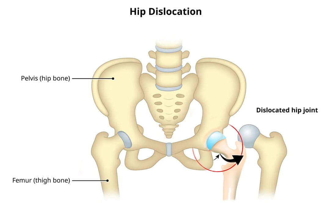
Can knowing treatment options for a dislocated hip help individuals expedite rehabilitation and recovery?
Dislocated Hip
A dislocated hip is an uncommon injury but can happen due to trauma or following hip replacement surgery. It usually occurs after severe trauma, including motor vehicle collisions, falls, and sometimes sports injuries. (Caylyne Arnold et al., 2017) A dislocated hip can also occur after hip replacement surgery. Other injuries like ligament tears, cartilage damage, and bone fractures can occur alongside the dislocation. Most hip dislocations are treated with a joint reduction procedure that resets the ball into the socket. It is usually done with sedation or general anesthesia. Rehabilitation takes time and could be a few months before full recovery. Physical therapy can help restore motion and strength in the hip.
What Is It?
If the hip is only partially dislocated, it’s called a hip subluxation. When this happens, the hip joint head only partially emerges from the socket. A dislocated hip is when the head or ball of the joint shifts or pops out of the socket. Because an artificial hip differs from a normal hip joint, the risk of dislocation increases after joint replacement. A study found that around 2% of individuals who undergo total hip replacement will experience hip dislocation within a year, with the cumulative risk increasing by approximately 1% over five years. (Jens Dargel et al., 2014) However, new technological prosthetics and surgical techniques are making this less common.
Hip Anatomy
The hip ball-and-socket joint is called the femoroacetabular joint.
The socket is called the acetabulum.
The ball is called the femoral head.
The bony anatomy and strong ligaments, muscles, and tendons help to create a stable joint. Significant force must be applied to the joint for a hip dislocation to occur. Some individuals report feeling a snapping sensation of the hip. This usually is not a hip dislocation but indicates a different disorder known as snapping hip syndrome. (Paul Walker et al., 2021)
Posterior Hip Dislocation
Around 90% of hip dislocations are posterior.
In this type, the ball is pushed backward from the socket.
A hip dislocation increases the risk of developing joint arthritis following the injury and can raise the risk of needing a hip replacement later in life. (Hsuan-Hsiao Ma et al., 2020)
Developmental Dislocation of the Hip
Some children are born with developmental dislocation of the hip or DDH.
Children with DDH have hip joints that did not form correctly during development.
This causes a loose fit in the socket.
In some cases, the hip joint is completely dislocated.
Joint reduction is the most common way to treat a dislocated hip. The procedure repositions the ball back into the socket and is usually done with sedation or under general anesthesia. Repositioning a hip requires significant force. A hip dislocation is considered an emergency, and reduction should be performed immediately after the dislocation to prevent permanent complications and invasive treatment. (Caylyne Arnold et al., 2017)
Once the ball is back in the socket, the healthcare provider will look for bone, cartilage, and ligament injuries.
Depending on what the healthcare provider finds, further treatment may be necessary.
Fractured or broken bones may need to be repaired to keep the ball within the socket.
Damaged cartilage may have to be removed.
Surgery
Surgery could be necessary to return the joint to its normal position. Hip arthroscopy can minimize the invasiveness of certain procedures. A surgeon inserts a microscopic camera into the hip joint to help the surgeon repair the injury using instruments inserted through other small incisions.
Hip replacement surgery replaces the ball and socket, a common and successful orthopedic surgical procedure. This surgery may be performed for various reasons, including trauma or arthritis, as it is common to develop early arthritis of the hip after this type of trauma. This is why many who have a dislocation ultimately need hip replacement surgery. As a major surgical procedure, it is not without risks. Possible complications include:
Infection
Aseptic loosening (the loosening of the joint without infection)
Hip dislocation
Recovery
Recovering from a hip dislocation is a long process. Individuals will need to walk with crutches or other devices early in recovery. Physical therapy will improve the range of motion and strengthen the muscles around the hip. Recovery time will depend on whether other injuries, such as fractures or tears, are present. If the hip joint was reduced and there were no other injuries, it may take six to ten weeks to recover to the point where weight can be placed on the leg. It could be between two and three months for a full recovery. Keeping weight off the leg is important until the surgeon or physical therapist gives the all-clear. Injury Medical Chiropractic and Functional Medicine Clinic will work with an individual’s primary healthcare provider and other surgeons or specialists to develop an optimal personalized treatment plan.
Chiropractic Solutions for Osteoarthritis
References
Arnold, C., Fayos, Z., Bruner, D., Arnold, D., Gupta, N., & Nusbaum, J. (2017). Managing dislocations of the hip, knee, and ankle in the emergency department [digest]. Emergency medicine practice, 19(12 Suppl Points & Pearls), 1–2.
Dargel, J., Oppermann, J., Brüggemann, G. P., & Eysel, P. (2014). Dislocation following total hip replacement. Deutsches Arzteblatt international, 111(51-52), 884–890. https://doi.org/10.3238/arztebl.2014.0884
Walker, P., Ellis, E., Scofield, J., Kongchum, T., Sherman, W. F., & Kaye, A. D. (2021). Snapping Hip Syndrome: A Comprehensive Update. Orthopedic reviews, 13(2), 25088. https://doi.org/10.52965/001c.25088
Cornwall, R., & Radomisli, T. E. (2000). Nerve injury in traumatic dislocation of the hip. Clinical orthopaedics and related research, (377), 84–91. https://doi.org/10.1097/00003086-200008000-00012
American Academy of Orthopaedic Surgeons. (2021). Hip dislocation. https://orthoinfo.aaos.org/en/diseases–conditions/hip-dislocation
Kellam, P., & Ostrum, R. F. (2016). Systematic Review and Meta-Analysis of Avascular Necrosis and Posttraumatic Arthritis After Traumatic Hip Dislocation. Journal of orthopaedic trauma, 30(1), 10–16. https://doi.org/10.1097/BOT.0000000000000419
Ma, H. H., Huang, C. C., Pai, F. Y., Chang, M. C., Chen, W. M., & Huang, T. F. (2020). Long-term results in the patients with traumatic hip fracture-dislocation: Important prognostic factors. Journal of the Chinese Medical Association : JCMA, 83(7), 686–689. https://doi.org/10.1097/JCMA.0000000000000366
American Academy of Orthopaedic Surgeons. (2022). Developmental dislocation (dysplasia) of the hip (DDH). https://orthoinfo.aaos.org/en/diseases–conditions/developmental-dislocation-dysplasia-of-the-hip-ddh/
Can physical therapy treatment protocols aimed at improving range of motion and flexibility around the hip and relieving inflammation around the sciatic nerve help individuals experiencing deep buttock pain or piriformis syndrome?
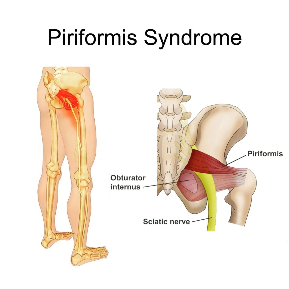
Deep Buttock Pain
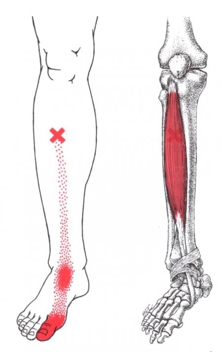
Piriformis syndrome, a.k .a. deep buttock pain, is described as sciatic nerve irritation from the piriformis muscle.
The piriformis is a small muscle behind the hip joint in the buttocks.
It is about one centimeter in diameter and functions in the hip joint’s external rotation or turning outward.
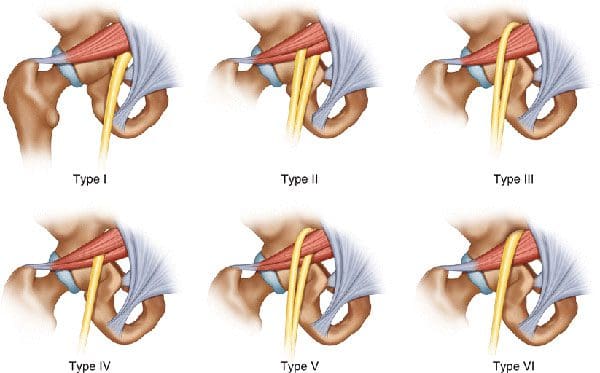
The piriformis muscle and tendon are close to the sciatic nerve, which supplies the lower extremities with motor and sensory functions.
Depending on an individual’s anatomic variation of the muscle and tendon:
The two cross over, under, or through each other behind the hip joint in the deep buttock.
This relationship is thought to irritate the nerve, leading to sciatica symptoms.
Piriformis Syndrome
When diagnosed with piriformis syndrome, it is thought that the muscle and tendon bind to and/or spasm around the nerve, causing irritation and pain symptoms.
The theory supported is that when the piriformis muscle and its tendon tighten, the sciatic nerve becomes compressed or pinched. This decreases blood circulation and irritates the nerve from the pressure. (Shane P. Cass 2015)
Tenderness with pressure on the piriformis muscle.
Discomfort in the back of the thigh.
Deep buttock pain behind the hip.
Electric sensations, shocks, and pains travel down the back of the lower extremity.
Numbness in the lower extremity.
Some individuals develop symptoms abruptly, while others go through a gradual increase.
Diagnosis
Doctors will order X-rays, MRIs, and nerve conduction studies, which is normal.
Because piriformis syndrome can be challenging to diagnose, some individuals with minor hip pain may receive a piriformis syndrome diagnosis even if they don’t have the condition. (Shane P. Cass 2015)
It is sometimes referred to as deep buttock pain. Other causes of this type of pain include back and spinal problems like:
Herniated discs
Spinal stenosis
Radiculopathy – sciatica
Hip bursitis
A piriformis syndrome diagnosis is usually given when these other causes are eliminated.
When the diagnosis is uncertain, an injection is administered in the area of the piriformis muscle. (Danilo Jankovic et al., 2013)
Different medications can be used, but the injection itself is used to help determine the specific location of the discomfort.
When an injection is given into the piriformis muscle or tendon, it is often administered by ultrasound guidance to ensure the needle delivers the medication to the correct location. (Elizabeth A. Bardowski, J. W. Thomas Byrd 2019)
Avoiding activities that cause symptoms for at least a few weeks.
Physical Therapy
Emphasize stretching and strengthening the hip rotator muscles.
Non-Surgical Decompression
Gently pulls the spine to release any compression, allowing optimal rehydration and circulation and taking the pressure off the sciatic nerve.
Therapeutic Massage Techniques
To relax and release muscle tension and increase circulation.
Acupuncture
To help relax the piriformis muscle, sciatic nerve, and surrounding area.
Relieve pain.
Chiropractic Adjustments
Realignment rebalances the spine and musculoskeletal system to alleviate pain.
Anti-Inflammatory Medication
To decrease inflammation around the tendon.
Cortisone Injections
Injections are used to decrease inflammation and swelling.
Botulinum Toxin Injection
Injections of botulinum toxin paralyze the muscle to relieve pain.
Surgery
Surgery can be performed in rare cases to loosen the piriformis tendon, known as a piriformis release. (Shane P. Cass 2015)
Surgery is a last resort when conservative treatments have been tried for at least 6 months with little to no relief.
Recovery can take several months.
Sciatica Causes and Treatment
References
Cass S. P. (2015). Piriformis syndrome: a cause of nondiscogenic sciatica. Current sports medicine reports, 14(1), 41–44. https://doi.org/10.1249/JSR.0000000000000110
Jankovic, D., Peng, P., & van Zundert, A. (2013). Brief review: piriformis syndrome: etiology, diagnosis, and management. Canadian journal of anaesthesia = Journal canadien d’anesthesie, 60(10), 1003–1012. https://doi.org/10.1007/s12630-013-0009-5
Bardowski, E. A., & Byrd, J. W. T. (2019). Piriformis Injection: An Ultrasound-Guided Technique. Arthroscopy techniques, 8(12), e1457–e1461. https://doi.org/10.1016/j.eats.2019.07.033
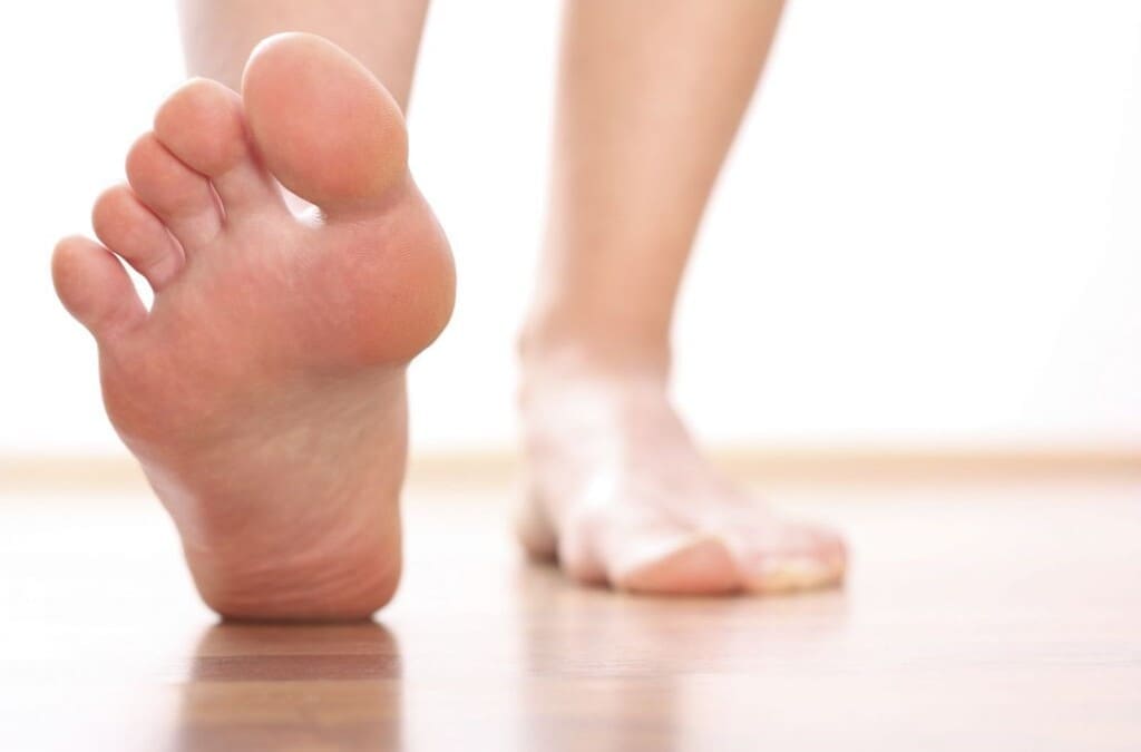
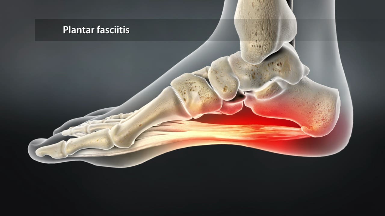
Can plantar fasciitis patients incorporate non-surgical treatments to reduce hip pain and restore mobility?
Introduction
Everyone is on their feet constantly as it helps people stay mobile and allows them to go from one location to another. Many people are constantly on their feet from childhood to adulthood. This is because the feet are part of the lower musculoskeletal extremities that stabilize the hips and allow sensory-motor function to the legs, thighs, and calves. The feet also have various muscles, tendons, and ligaments surrounding the skeletal structure to prevent pain and discomfort. However, when repetitive motions or injuries start to affect the feet, it can lead to plantar fasciitis and, over time, cause overlapping risk profiles that lead to hip pain. When people are experiencing these pain-like conditions, it can significantly affect their daily activities and overall quality of life. When this happens, many people seek various treatments to reduce the pain-like symptoms caused by plantar fasciitis and restore hip mobility. Today’s article looks at how plantar fasciitis correlates with hip pain, the connection between the feet and the hips, and how there are non-surgical solutions to reduce plantar fasciitis. We talk with certified medical providers who consolidate our patients’ information to assess how to mitigate plantar fasciitis and restore hip mobility. We also inform and guide patients on how numerous non-surgical treatments can help strengthen weak muscles associated with plantar fasciitis and help with restoring stabilization from hip pain. We encourage our patients to ask their associated medical providers intricate and important questions about incorporating small changes to reduce the pain-like effects caused by plantar fasciitis. Dr. Jimenez, D.C., includes this information as an academic service. Disclaimer.
How Plantar Fasciitis Correlates With Hip Pain
Do you experience pain in your heels constantly after a long walk? Do you feel stiffness in your hips when stretching? Or do you feel your shoes are causing tension and pain in your feet and calves? Often, many of these pain-like scenarios are due to people dealing with plantar fasciitis, characterized by heel pain due to inflammation or degenerative irritation of the plantar fascia, a band of thick tissues is running across the bottom of the foot and connecting to the heel bone to the toes in the lower extremities. This band of tissues plays an essential role in the body, providing normal biomechanics to the foot while supporting the arch and helping with shock absorption. (Buchanan et al., 2024) Plantar fasciitis can affect the stability of the lower extremities since the pain affects the feet and causes hip pain.
So, how would plantar fasciitis correlate with hip pain? With plantar fasciitis, many people are experiencing pain in their feet. It can lead to abnormal foot posture, lower extremity muscle weakness, and muscle stress that can reduce the stability of the legs and hip muscles. (Lee et al., 2022) With hip pain, many people can experience a gait dysfunction that causes muscle weakness in the lower extremities and causes the accessory muscles to perform the primary muscles’ jobs. To that point, this forces people to scrap the ground when walking. (Ahuja et al., 2020) This is because normal conditions like natural aging, muscle overuse, or trauma can cause pain-like symptoms to the hips, including discomfort on the thighs, groin, and buttock region, joint stiffness, and reduced range of motion. Hip pain can cause overlapping risk profiles that may include repetitive strain on the feet, thus leading to symptoms of sharp to dull aches on the heel.
The Connection Between The Feet and The Hips
It is important to understand that foot problems like plantar fasciitis can affect the hips and vice versa, as both body regions have a beautiful relationship within the musculoskeletal system. Plantar fasciitis on their feet can alter their gait function, potentially leading to hip pain over time. This is due to many environmental factors that can affect the hips and feet over time, leading to plantar fasciitis correlating with hip pain. From excessive weight-bearing activities to microtrauma in the hips or the plantar fascia, many people will often seek treatment to reduce the effects of plantar fasciitis correlated with hip pain by addressing how their range of motion is affecting the plantarflexion and their load on the force-absorbing plantar surface structures could be good starting points in the prevention and treatment of plantar fasciitis correlated with hip pain. (Hamstra-Wright et al., 2021)
What Is Plantar Fasciitis?-Video
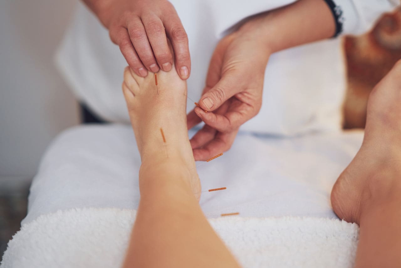
Non-Surgical Solutions To Reduce Plantar Fasciitis
When it comes to reducing plantar fasciitis in the body, many individuals will seek non-surgical treatments that can alleviate the pain from plantar fascia. Non-surgical treatments are cost-effective and can reduce the pain from plantar fasciitis and its associated symptoms, like hip pain. Some of the benefits of non-surgical treatments are promising, as they have a low risk of complications, good accessibility, and even a high capacity to relieve the mechanical load on the plantar fascia when doing regular activities. (Schuitema et al., 2020) Some of the non-surgical treatments that many people can incorporate include:
Stretching exercises
Orthotic devices
Chiropractic care
Massage therapy
Acupuncture/electroacupuncture
Spinal decompression
These non-surgical treatments not only help reduce plantar fasciitis but also help alleviate hip pain. For example, spinal decompression can help restore hip mobility by stretching the lumbar spine and relieving the lower extremities from numbness while strengthening tight muscles. (Takagi et al., 2023). Electroacupuncture can stimulate the body’s acupoints to release endorphins from the lower extremities to reduce inflammation of the plantar fascia. (Wang et al., 2019) When people begin to make small changes in their routine, like wearing proper footwear and not carrying or lifting heavy weighted objects, it can go a long way to prevent plantar fasciitis and hip pain from reoccurring can go a long way. Having a personalized treatment plan can ensure many individuals seeking non-surgical treatments have a better outcome on their health and mobility while preventing long-term complications.
References
Ahuja, V., Thapa, D., Patial, S., Chander, A., & Ahuja, A. (2020). Chronic hip pain in adults: Current knowledge and future prospective. J Anaesthesiol Clin Pharmacol, 36(4), 450-457. https://doi.org/10.4103/joacp.JOACP_170_19
Hamstra-Wright, K. L., Huxel Bliven, K. C., Bay, R. C., & Aydemir, B. (2021). Risk Factors for Plantar Fasciitis in Physically Active Individuals: A Systematic Review and Meta-analysis. Sports Health, 13(3), 296-303. https://doi.org/10.1177/1941738120970976
Lee, J. H., Shin, K. H., Jung, T. S., & Jang, W. Y. (2022). Lower Extremity Muscle Performance and Foot Pressure in Patients Who Have Plantar Fasciitis with and without Flat Foot Posture. Int J Environ Res Public Health, 20(1). https://doi.org/10.3390/ijerph20010087
Schuitema, D., Greve, C., Postema, K., Dekker, R., & Hijmans, J. M. (2020). Effectiveness of Mechanical Treatment for Plantar Fasciitis: A Systematic Review. J Sport Rehabil, 29(5), 657-674. https://doi.org/10.1123/jsr.2019-0036
Takagi, Y., Yamada, H., Ebara, H., Hayashi, H., Inatani, H., Toyooka, K., Mori, A., Kitano, Y., Nakanami, A., Kagechika, K., Yahata, T., & Tsuchiya, H. (2023). Decompression for lumbar spinal stenosis at the intrathecal catheter insertion site during intrathecal baclofen therapy: a case report. J Med Case Rep, 17(1), 239. https://doi.org/10.1186/s13256-023-03959-1
Wang, W., Liu, Y., Zhao, J., Jiao, R., & Liu, Z. (2019). Electroacupuncture versus manual acupuncture in the treatment of plantar heel pain syndrome: study protocol for an upcoming randomised controlled trial. BMJ Open, 9(4), e026147. https://doi.org/10.1136/bmjopen-2018-026147
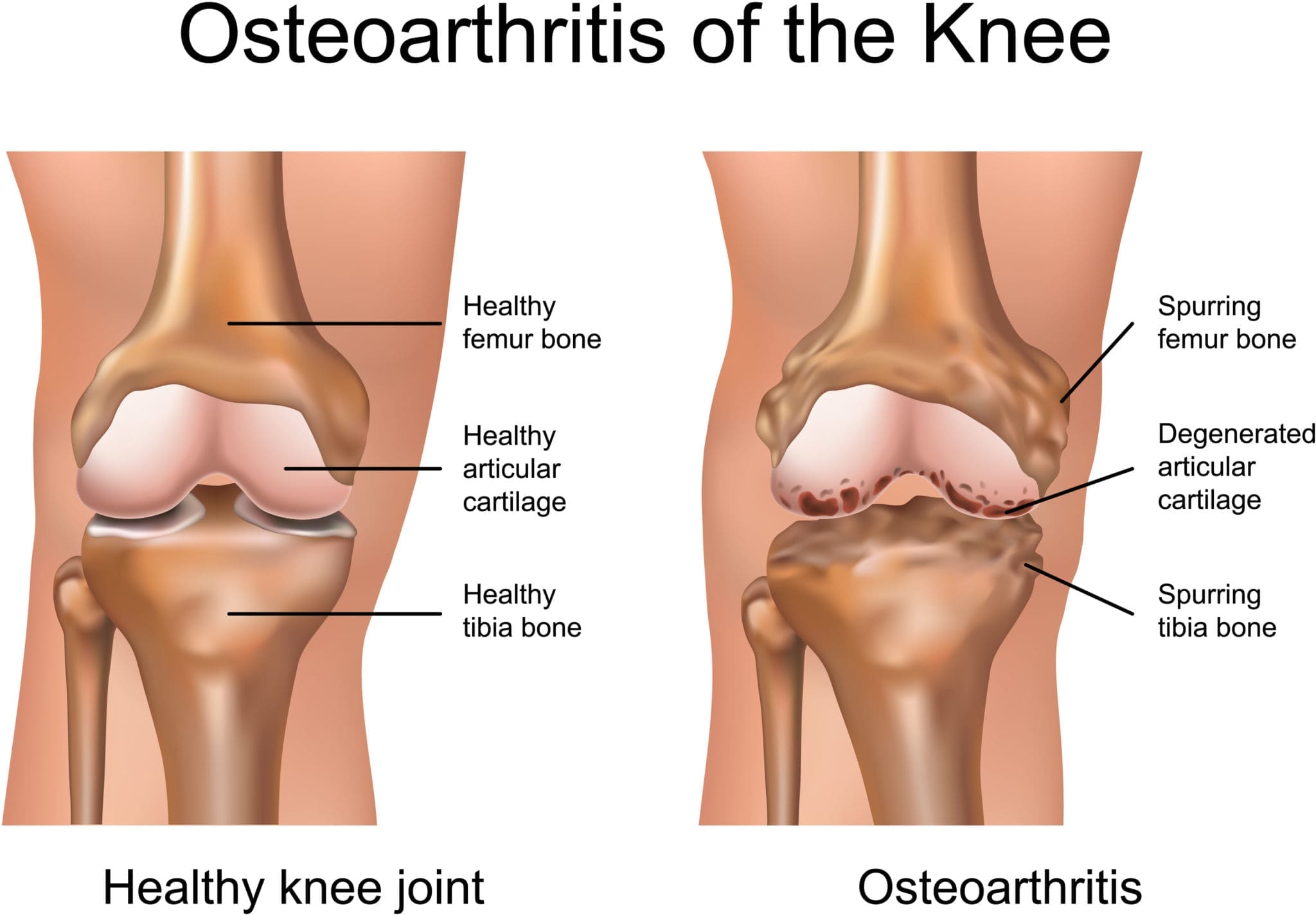
Can individuals with osteoarthritis find the relief they deserve through electroacupuncture to restore knee and hip mobility?
Introduction
The lower extremities provide movement and stability to the body, allowing people to be in motion. The hips, lower back, knees, and feet each have a function to do, and when traumatic issues start to affect the spinal structures, it can cause numerous symptoms to pop up and cause pain-like symptoms. Additionally, degenerative factors are natural to the joints of the lower extremities as many people make repetitive motions to their bodies that lead to the degenerative process. One of the most common degenerative issues that affect the lower extremities is osteoarthritis, which can make many people feel miserable. Today’s article looks at how osteoarthritis affects the lower extremities and how treatments like electroacupuncture reduce inflammation associated with osteoarthritis and restore knee and hip mobility. We talk with certified medical providers who consolidate our patients’ information to understand better how osteoarthritis affects their lower extremities. We also inform and guide patients on how electroacupuncture therapy can help reduce the inflammatory effects of osteoarthritis affecting the hips and knees. We encourage our patients to ask their associated medical providers intricate and important questions about reducing the progression of osteoarthritis through non-surgical treatments. Dr. Jimenez, D.C., includes this information as an academic service. Disclaimer.
Osteoarthritis Affecting The Lower Extremities
Have you been dealing with stiffness in your knees, hips, and lower back in the mornings? Do you feel like you are wobbling a bit too much when walking? Or do you think radiating heat and swelling in your knees? When people experience these inflammatory pain issues in their joints, it is due to osteoarthritis, a degenerative joint disorder affecting the cartilage between the bones and the tissue components surrounding the joint. Osteoarthritis is multifactorial, meaning it can be idiopathic or secondary while influenced by heredity factors. (Bliddal, 2020) The most common places where people experience osteoarthritis are the lower back, hand, hips, and, most commonly, the knees. Some of the major environmental factors that contribute to the development of osteoarthritis include:
Obesity
Age
Repetitive motions
Family history
Injuries
When people are dealing with osteoarthritis, environmental factors can lead to weight overloading on the joints, which results in compression and inflammation. (Nedunchezhiyan et al., 2022)
When inflammation is associated with osteoarthritis, it can cause the joints and surrounding muscle tissues to swell and feel hot to the touch. At the same time, osteoarthritis is one of the leading causes of disability that can become a socio-economic issue for many people. (Yao et al., 2023) This is because many individuals with osteoarthritis have comorbidities that are associated with the effects of inflammatory cytokines, which can cause them to be physically inactive and miserable. (Katz et al., 2021) However, there are various ways to reduce the progression of osteoarthritis and reduce the inflammatory effects on the joints.
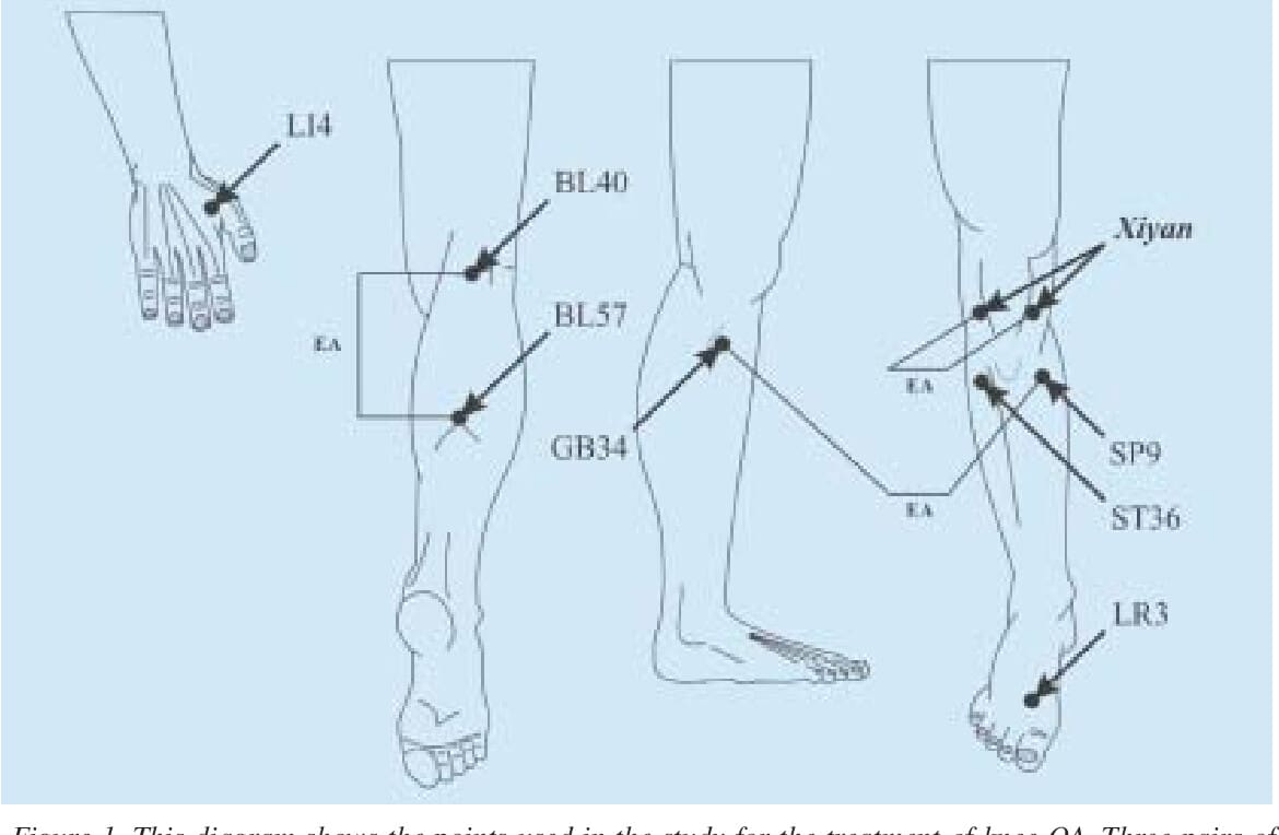
Electroacupuncture Reducing Inflammation Associated With Osteoarthritis
When it comes to inflammation reduction associated with osteoarthritis, many people seek out surgical and non-surgical treatments that can help decrease the progression of this degenerative joint disease. Many people will do aqua therapy to relieve pressure off the joints and improve their mobility. At the same time, others use spinal decompression to create negative pressure on the joint space. However, many people have found that electroacupuncture can help reduce the inflammatory effects of osteoarthritis. Electroacupuncture combines electrical nerve stimulation and acupuncture by highly trained professionals that can help decrease the pain intensity in the joints and provide functionality. (Wu et al., 2020) Additionally, since osteoarthritis is associated with inflammation, electroacupuncture can promote blood circulation and adjustment of muscle tension on the joints, and improve mobility. (Zhang et al., 2023)
Electroacupuncture Restoring Knee & Hip Mobility
Electroacupuncture can help with hip and knee mobility as this non-surgical treatment helps promote pain limitations and muscular atrophy from biomechanical overloading, thus improving cartilage viscoelasticity. (Shi et al., 2020) This allows the joints to retain mobility in the hips, knees, and lower back. When people go through consecutive treatment for osteoporosis, they can recover their muscle strength over time to restore their mobility and reduce the progression of osteoarthritis. (Xu et al., 2020) By doing so, many people can find the relief they are looking for with electroacupuncture, which can enable them to make small changes in their daily routine to ensure they can function throughout the day.
Katz, J. N., Arant, K. R., & Loeser, R. F. (2021). Diagnosis and Treatment of Hip and Knee Osteoarthritis: A Review. JAMA, 325(6), 568-578. https://doi.org/10.1001/jama.2020.22171
Nedunchezhiyan, U., Varughese, I., Sun, A. R., Wu, X., Crawford, R., & Prasadam, I. (2022). Obesity, Inflammation, and Immune System in Osteoarthritis. Front Immunol, 13, 907750. https://doi.org/10.3389/fimmu.2022.907750
Shi, X., Yu, W., Wang, T., Battulga, O., Wang, C., Shu, Q., Yang, X., Liu, C., & Guo, C. (2020). Electroacupuncture alleviates cartilage degradation: Improvement in cartilage biomechanics via pain relief and potentiation of muscle function in a rabbit model of knee osteoarthritis. Biomed Pharmacother, 123, 109724. https://doi.org/10.1016/j.biopha.2019.109724
Wu, S. Y., Lin, C. H., Chang, N. J., Hu, W. L., Hung, Y. C., Tsao, Y., & Kuo, C. A. (2020). Combined effect of laser acupuncture and electroacupuncture in knee osteoarthritis patients: A protocol for a randomized controlled trial. Medicine (Baltimore), 99(12), e19541. https://doi.org/10.1097/MD.0000000000019541
Xu, H., Kang, B., Li, Y., Xie, J., Sun, S., Zhong, S., Gao, C., Xu, X., Zhao, C., Qiu, G., & Xiao, L. (2020). Using electroacupuncture to recover muscle strength in patients with knee osteoarthritis after total knee arthroplasty: a study protocol for a double-blinded, randomized, and placebo-controlled trial. Trials, 21(1), 705. https://doi.org/10.1186/s13063-020-04601-x
Yao, Q., Wu, X., Tao, C., Gong, W., Chen, M., Qu, M., Zhong, Y., He, T., Chen, S., & Xiao, G. (2023). Osteoarthritis: pathogenic signaling pathways and therapeutic targets. Signal Transduct Target Ther, 8(1), 56. https://doi.org/10.1038/s41392-023-01330-w
Zhang, W., Zhang, L., Yang, S., Wen, B., Chen, J., & Chang, J. (2023). Electroacupuncture ameliorates knee osteoarthritis in rats via inhibiting NLRP3 inflammasome and reducing pyroptosis. Mol Pain, 19, 17448069221147792. https://doi.org/10.1177/17448069221147792
IFM's Find A Practitioner tool is the largest referral network in Functional Medicine, created to help patients locate Functional Medicine practitioners anywhere in the world. IFM Certified Practitioners are listed first in the search results, given their extensive education in Functional Medicine

 Causes