Can understanding the planes of motion help individuals adjust fitness training to maximize fitness for physical and sports performance and reduce the risk of injury?
Planes of Motion
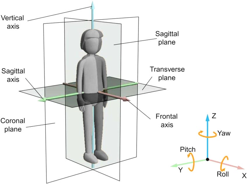
The body’s planes of motion are the sagittal, frontal, and transverse planes, which divide it into left and right, front and back, and top and bottom halves. The body moves in different dimensions during daily work, house chores, and physical activity/exercises. The movements in each plane correspond to forward/backward, side-to-side, and rotational motions. Think of each plane as an imaginary line or a pane of glass that divides the body into opposing segments when standing in the anatomical position. (National Academy of Sports Medicine, 2024)
Sagittal plane -Divides the body into right and left sides.
Frontal plane – Divides the body into front and back.
Transverse plane – Divides the body into top and bottom sections.
To determine the plane of motion of a particular movement, consider how the movement would interact with the imaginary lines or plates. When a movement runs parallel to the imaginary line, the movement is occurring in that plane of motion. For example, when going upstairs, the forward and upward movement at the hip, knee, and ankle occurs primarily in the sagittal plane because that movement runs parallel to the imaginary line that divides the body into right and left sides. Frontal plane movements occur while you walk up the stairs and reach for the handrail. The movement is in the frontal plane because the lateral hand reach runs parallel to the line, dissecting the body into front and back sections. If you turn around to look behind, the rotational movement occurs in the transverse plane because your upper torso runs parallel to the line, dissecting the body into an upper and lower section. Individual movements at any joint in the body can occur in a single plane or multiple planes. Complex movements usually happen in several planes of motion concurrently.
Sagittal Plane
Movement in the sagittal plane generally happens in front or behind. This is the most familiar plane of motion because many typical day-to-day activities happen within arm’s reach in front. Walking, texting, or computer work involves movement primarily in the sagittal plane. Several eating mechanics occur in the sagittal plane. Movements include:
Flexion – A bending movement that decreases the angle at a joint.
Extension – An extending movement that increases the angle at a joint.
Hyperextension – Extending the angle at a joint beyond neutral.
Dorsiflexion – Bending at the ankle so the top of the foot moves toward the shin.
Plantarflexion – Pushing the foot down and away from the body.
Many strength-training exercises in the sagittal plane include biceps curls, forward or reverse lunges, squats, vertical jumping, running, downward dog, and yoga chair poses.
Frontal Plane
The frontal plane divides the body into front/anterior and back/posterior sections. Frontal plane movements are lateral or side-to-side and include:
Abduction – Moving the body or a limb laterally and away from the body’s midline.
Adduction – Moving the body or a limb towards the body’s midline.
Elevation – Moving the shoulder blades up.
Depression – Moving the shoulder blades down.
Eversion – Rolling the foot towards the inside/medial side.
Inversion – Rolling the foot towards the outside/lateral side.
Frontal plane movements are less common than sagittal movements. For example, individuals walk forward more than side to side or reach for something in front rather than directly to the side. Frontal plane movements in fitness include side lunges, lateral shoulder raises, and side shuffles, and in yoga poses, standing side bends and the triangle.
Transverse Plane
The transverse plane divides the body into upper/superior and lower/inferior sections. Transverse plane movements generally involve rotation. Movement in this plane is less common. Exercise injuries most often occur during transverse/rotational movements. (National Academy of Sports Medicine, 2024) Movements include:
Rotation – Moving the torso or a limb around its vertical axis.
Pronation – Rotating the forearm or foot to a palm-side or foot-side down position.
Supination – Rotating the forearm or foot to a palm-side or foot-side-up position.
Horizontal Abduction – Moving the upper arm away from the body’s midline when elevated to 90 degrees.
Horizontal Adduction – Moving the upper arm towards the body’s midline when elevated to 90 degrees.
Typical everyday activities in the frontal plane include turning the head to look behind or turning a doorknob. Exercises in the transverse plane include hitting a golf ball, swinging a baseball bat, or performing a seated twist.
Training Within the Planes of Motion Benefits
Training in all three planes can help with movement in several ways, providing greater ease in life and sports.
Prepares Body for Daily Tasks
Many traditional strength-training programs focus on training one muscle at a time, often in a single plane of motion. For example, weight lifters might do bicep curls to primarily work the biceps in the sagittal plane, a chest fly exercise to primarily work the pectoral muscles in the transverse plane, or lateral raises to work the shoulders in the frontal plane. However, compound exercises have recently become much more common. Compound movements allow individuals to train several muscle groups simultaneously and in different planes of motion.
In this way, training activities mimic daily living activities. For example, individuals often lift several heavy bags of groceries and turn to open the car or trunk, involving both sagittal and transverse movement. Preparing the body for complex activities with compound exercises allows individuals to perform them more easily throughout the day.
Prepares Body for Sports and Physical Activities
Complex multi-planar movements help prepare for safe and effective physical activity and sports performance (National Academy of Sports Medicine, 2024). Researchers and experts understand that many physical and athletic activities require the body to move in different directions, often quickly and under high stress. Several studies have found that anterior cruciate ligament/ACL injuries are more likely to occur during multi-planar rather than single-planar movements. (Quatman C. E., Quatman-Yates C. C., & Hewett T. E. 2010) Training the body to perform multi-planar movements safely and effectively through exercise can help reduce the risk of injury during daily activities or stressful athletic competitions.
Encourages Variation For Full Body Strengthening
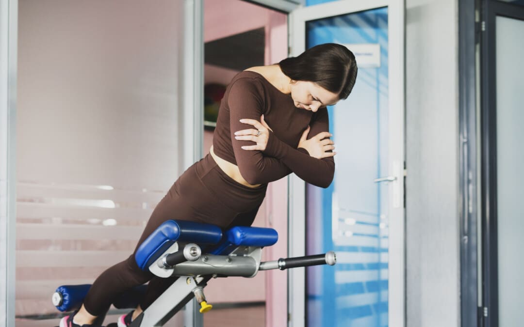
Individuals tend to fall into certain movement patterns, such as repeatedly performing the same fitness activity or exercises. This can cause them to have a favorite plane of motion. One way to break away from the same routine is to include movement from all planes of motion. For example, many abdominal workout machines help train in multiple planes of motion, challenging your body to move in different ways. Dumbbells, kettlebells, TRX straps, and bands allow individuals to move joints freely in various planes of motion and work several muscles.
Runners train primarily in the sagittal plane, even if they cross-train by swimming, cycling, or using cardio machines. For this reason, trainers and coaches often recommend doing some form of yoga or weight training that allows them to move their joints in different ways, including lateral movements or rotation. Even flexibility training should incorporate all three planes of motion. For example, walkers might choose to do a simple calf or hamstring stretch at the end of their workout but may also benefit from a seated spine rotation or a lying hip stretch.
Injury Medical Chiropractic and Functional Medicine Clinic
Understanding the concept and importance of training in the three planes of motion can help improve sports and physical performance and prevent musculoskeletal injuries. Chiropractic care aims to help individuals enhance movement with less pain due to condition, after injury, or surgery. Injury Medical Chiropractic and Functional Medicine Clinic works with primary healthcare providers and specialists to build optimal health and wellness solutions. We focus on what works for you to relieve pain, restore function, prevent injury, and help mitigate issues through adjustments that help the body realign itself. They can also work with other medical professionals to integrate a treatment plan to resolve musculoskeletal problems.
The Difference of Using Custom Foot Orthotics
References
National Academy of Sports Medicine. (2024). Sagittal, Frontal and Transverse Body Planes: Exercises & Movements. NASM. https://blog.nasm.org/exercise-programming/sagittal-frontal-traverse-planes-explained-with-exercises?utm_source=blog&utm_medium=referral&utm_campaign=organic&utm_content=ReasonsToBecomeCES
Quatman, C. E., Quatman-Yates, C. C., & Hewett, T. E. (2010). A ‘plane’ explanation of anterior cruciate ligament injury mechanisms: a systematic review. Sports medicine (Auckland, N.Z.), 40(9), 729–746. https://doi.org/10.2165/11534950-000000000-00000
Can individuals experiencing back pain from various factors incorporate MET (muscle energy techniques) to restore mobility?
Factors That Causes Back Pain
More often than not, many individuals have experienced back pain in various parts of their lives. As one of the leading pains that people have dealt with worldwide, many factors can contribute to the development of back pain, and it can affect different musculoskeletal locations in the upper and lower body quadrants. Back pain has often been associated with the workplace or environmental factors. For back pain, the problem can range from all back sections, affecting the muscles, ligaments, tissues, intervertebral joints, or the bone itself. (Wiberg, 1949) People are constantly in motion, and the muscles can be overstretched and tight over time. When that happens, many people seek treatment to reduce back pain’s overlapping risk profiles and restore mobility. Today’s article focuses on the factors associated with back pain and how non-surgical treatments like MET (muscle energy technique) can help reduce back pain and restore mobility. We discuss with certified medical providers who inform our patients how various environmental factors are correlated with back pain and how it can affect the body. While asking informed questions to our associated medical providers, we advise patients to include various non-surgical treatments like MET to be incorporated to reduce overlapping risk profiles associated with back pain. Dr. Alex Jimenez, D.C., encompasses this information as an academic service. Disclaimer.
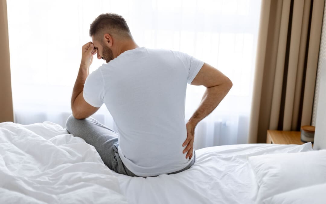
How often do you experience pain in different locations in your back after doing some strenuous activities? Do you feel like you are taking many breaks due to feeling tightness or sharp sensations in your back? Or have you felt that your posture is more hunched than normal? Many of these environmental factors scenarios are correlated with back pain, and it can become an issue over time. Back pain can be in two categories: specific and non-specific, and it can impact a person’s quality of life. Non-specific back pain can be associated with mobility impairment in the different back sections, referred to as radiating pain in the lower extremities or musculoskeletal disorders. (Delitto et al., 2012) This causes issues like repetitive loading to the spine and various pain-like symptoms to the back, causing a disbalance to the individual. (Zemková & Zapletalová, 2021) When it comes to environmental factors correlating with back pain, there are numerous ways for it to be developed and, over time, cause discomfort for the individual, as back pain symptoms vary for everyone.
Sleep
When it comes to the connection between sleep and back pain, these two issues can cause a vicious cycle of disturbed sleep and issues like insomnia. (Van Looveren et al., 2021) Now, when it comes to sleep disturbances and back pain, many individuals may be sleeping with the wrong mattress, causing their bodies to can cause pressure on their joints and spinal discs. This causes in-bed sleep behaviors like movements and postures could lead to health complications like pressure sores, apnea, and painful muscle spasms in the back and lower extremities. (Elnaggar et al., 2023) Luckily, there are various ways to reduce back pain, improve sleep quality, and restore body motion.
Understanding Academic Low Back Pain-Video
What Is MET?
When people come in for back pain treatment, non-surgical therapies can help stretch the overworked and tired back muscles and restore mobility to the body’s upper and lower extremities. One of the non-surgical treatments that pain specialists like chiropractors and massage therapists use is MET therapy or muscle energy technique therapy. MET comprises soft tissue manipulation that uses controlled isometric and isotonic contractions. (Sarkar et al., 2021) This helps the body not only improve the physiological function of the muscles but also decrease pain. MET can also be combined with other therapies to help lengthen short muscles, improve the range of motion from the joints, and increase fluid drainage from the body’s peripheral regions. (Batool et al., 2024)
MET Reducing Back Pain
Regarding MET, reducing back pain is possible as MET can be integrated with physical therapy to improve the disability and functionality of the person with back pain. (Wahyuddin et al., 2020) When people start to incorporate MET and non-surgical treatments as part of their routine for their health and well-being, they will begin to notice that the pain they have been experiencing in their back is diminishing over time. This allows them to be more mindful of their backs and bodies while making small changes to their routine. Sleeping better with a correct mattress, exercising more to stretch and strengthen muscles, eating healthier foods, and relaxing more allow people to be pain-free in their health and wellness journey.
References
Batool, K., Mehmood, M., Jafar, M., & Gull, M. (2024). Comparative efficacy of muscle energy technique and Bowen technique on hamstrings muscle tightness in chronic low back pain patients. Pak J Med Sci, 40(9), 2080-2084. https://doi.org/10.12669/pjms.40.9.8517
Delitto, A., George, S. Z., Van Dillen, L., Whitman, J. M., Sowa, G., Shekelle, P., Denninger, T. R., & Godges, J. J. (2012). Low Back Pain. Journal of Orthopaedic & Sports Physical Therapy, 42(4), A1-A57. https://doi.org/10.2519/jospt.2012.42.4.a1
Elnaggar, O., Arelhi, R., Coenen, F., Hopkinson, A., Mason, L., & Paoletti, P. (2023). An interpretable framework for sleep posture change detection and postural inactivity segmentation using wrist kinematics. Sci Rep, 13(1), 18027. https://doi.org/10.1038/s41598-023-44567-9
Sarkar, M., Goyal, M., & Samuel, A. J. (2021). Comparing the Effectiveness of the Muscle Energy Technique and Kinesiotaping in Mechanical Sacroiliac Joint Dysfunction: A Non-blinded, Two-Group, Pretest-Posttest Randomized Clinical Trial Protocol. Asian Spine Journal, 15(1), 54-63. https://doi.org/10.31616/asj.2019.0300
Van Looveren, E., Bilterys, T., Munneke, W., Cagnie, B., Ickmans, K., Mairesse, O., Malfliet, A., De Baets, L., Nijs, J., Goubert, D., Danneels, L., Moens, M., & Meeus, M. (2021). The Association between Sleep and Chronic Spinal Pain: A Systematic Review from the Last Decade. J Clin Med, 10(17). https://doi.org/10.3390/jcm10173836
Wahyuddin, W., Vongsirinavarat, M., Mekhora, K., Bovonsunthonchai, S., & Adisaipoapun, R. (2020). Immediate effects of muscle energy technique and stabilization exercise in patients with chronic low back pain with suspected facet joint origin: A pilot study. Hong Kong Physiother J, 40(2), 109-119. https://doi.org/10.1142/S1013702520500109
Wiberg, G. (1949). Back pain in relation to the nerve supply of the intervertebral disc. Acta Orthop Scand, 19(2), 211-221, illust. https://doi.org/10.3109/17453674908991094
Zemková, E., & Zapletalová, L. (2021). Back Problems: Pros and Cons of Core Strengthening Exercises as a Part of Athlete Training. International Journal of Environmental Research and Public Health, 18(10), 5400. https://doi.org/10.3390/ijerph18105400
Can performing the hip hinge exercise movement help individuals with lower back pain?
Hip Hinge Exercise
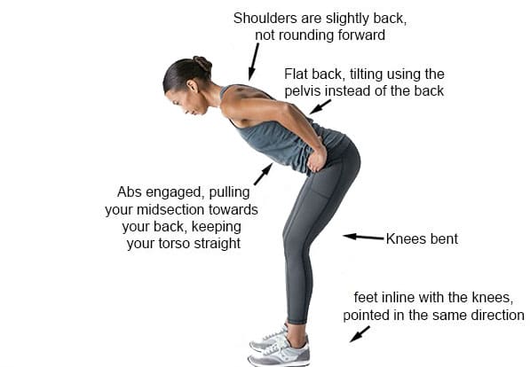
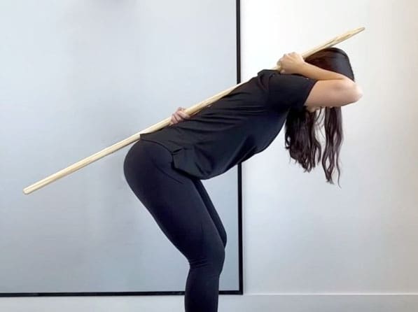
A hip hinge is a controlled movement that involves bending forward from the hips while keeping the spine neutral. The thoracic, lumbar, and pelvis stay neutral while bending forward. The movement comes from the hips, preventing the thoracic and lumbar spine from flexing or rounding. It is a fundamental movement that helps prevent back injuries and strengthens the glutes. It’s used in everyday activities, like picking up objects and sitting down.
The hip hinge exercise targets the posterior chain or back muscles, including the lower back, the glutes, and the hamstrings. It also strengthens the core or abdominal muscles to assist in the movement. When the body hinges at the hips, the bend occurs at the hips, and the spine stays neutral. When the lower back hinges or bends, this causes pain and reduces the range of motion.
Performing the Movement
A wooden dowel, broom handle, or PVC pipe can be used as a guide to help achieve the proper positioning and learn the correct form. Place the dowel or pipe vertically on your back, anchoring it to the head, shoulder blades, and tailbone.
Grasp one end with your right hand in the natural curve of your neck and the other with your left hand in the small of your back. Ensure the dowel touches the back of your head, upper back, and the area where the lower back meets the sacrum. To perform the hip hinge:
Stand with your feet shoulder-width apart
Shift your weight to your heels and
Push your hips back while hinging your torso forward
Keep your chest open and back flat
Slightly bend your knees
Visualize sticking the butt out
The dowel should not lose contact with the three points as you hinge. If it does, the movement is incorrect.
Lower your torso until it’s midway between vertical and parallel to the floor.
Pause when your torso is about 45 degrees
Keep a slight bend in your knees during the downward and upward phases.
Reverse the movement by contracting your glutes and pushing your hips forward and upward to return to the starting position.
Repeat
Benefits
The hip hinge is a fundamental movement pattern that helps the body perform essential tasks such as bending over and picking things up without worry of pain or injury. It’s also required in strength training exercises like the deadlift, kettlebell swing, power clean, and more. The exercise can help strengthen the core, reduce back pain, improve balance, and improve flexion, extension, and trunk rotation. (Michaud F. et al., 2021) Stronger core muscles can increase fitness and athletic performance. (Clark D. R. et al., 2018)
Variations
It is a challenging movement that requires plenty of practice. Individuals who can’t perform it correctly after a few tries may need to modify the movement.
Wall Variation
Using a wall as a guide is an easy way to make the movement easier.
To do this, stand with your back to a wall, about three inches away.
Start hinging at the hips by sticking your butt out touching the wall.
Keep a neutral spine and a flat back.
Once you can do this several times, try stepping out another inch or two and perform the same modified motion. Stick with this until you are away from the wall and can do a full hinge without the wall.
With A Kettlebell
Once you master the basic hinge, you can elevate it using a kettlebell to make this move more difficult.
Start with the kettlebell swing exercise and progress to more challenging moves with the kettlebell.
Common Mistakes
Be aware of common mistakes to keep the move effective and reduce the risk of injury.
Treating the Move Like a Squat
The hip hinge is not the same as a squat.
This is a common misconception. When squatting, the knee joint determines the movement pattern.
But when hip hinging, the movement starts at the hips.
Not Engaging the Core Muscles
This exercise requires core engagement throughout the entire movement.
If these muscles relax, there is an increased risk of dipping the hips during the hinge, which can cause the lower back to dip and cause pain.
Using the Lower Back
Bending or hinging with the lower back rather than letting the hips generate the movement.
Using the wall as a guide can help reduce and eliminate excessive bending at the waist.
Lost Dowel Contact
If the dowel loses contact with one or more set-up positions on the back, the hinge is not being executed correctly.
If your head loses contact with the dowel, the neck is flexing too far forward.
If you lose contact with the sacrum or lower back area, the spine is flexing too much.
If you lose contact with the mid-back, the knees are bending rather than the hips.
Safety
Stop and check your form if you feel back pain during any part of the movement. The movement may need to be modified further or decrease how far the hinge is at the hips. If the pain continues, discontinue the exercise and talk with a doctor or a physical therapist before reattempting the exercise. The dowel is a great tool to help maintain a neutral spine. If you cannot perform the hip hinge while keeping the dowel in contact with the body, you might benefit from working with a personal trainer or physical therapist who can walk you through the steps with the correct form.
Injury Medical Chiropractic and Functional Medicine Clinic
Chiropractic care aims to help individuals improve movement with less pain due to condition, after injury, or surgery. A chiropractic physical therapy team can assess your condition and develop a customized treatment plan to expedite pain relief and improve mobility. Injury Medical Chiropractic and Functional Medicine Clinic works with primary healthcare providers and specialists to build optimal health and wellness solutions. We focus on what works for you to relieve pain, restore function, prevent injury, and help mitigate issues through adjustments that help the body realign itself. They can also work with other medical professionals to integrate a treatment plan to resolve musculoskeletal problems.
Chiropractic: The Secret to Unlocking Mobility
References
Michaud, F., Pérez Soto, M., Lugrís, U., & Cuadrado, J. (2021). Lower Back Injury Prevention and Sensitization of Hip Hinge with Neutral Spine Using Wearable Sensors during Lifting Exercises. Sensors (Basel, Switzerland), 21(16), 5487. https://doi.org/10.3390/s21165487
Clark, D. R., Lambert, M. I., & Hunter, A. M. (2018). Contemporary perspectives of core stability training for dynamic athletic performance: a survey of athletes, coaches, sports science and sports medicine practitioners. Sports medicine – open, 4(1), 32. https://doi.org/10.1186/s40798-018-0150-3
Sleeping with lower back pain and sciatica can be difficult and frustrating. What are ways to get more comfortable sleep?
Sleeping With Lower Back Pain and Sciatica
Various factors can affect sleeping with lower back pain and sciatica, including age, injury and medical history, the mattress (e.g., soft vs. firm mattress), and sleep positions. Unfortunately, there’s no one-cure-all solution for this problem, and depending on the underlying cause/s, they can worsen the pain and cause sleep problems. For example, if a herniated disc places added pressure on the nerves in the lower back, twisting the spine can worsen the lower back pain, and sleeping in a fetal position can exacerbate the nerve pain. (UCF Health, N.D.)
Lower-Back Pain
Low-back pain can be activity-related, intermittent, or constant. (American Association of Neurological Surgeons, 2024) For some, it only occurs occasionally, such as when performing specific movements. For others, it can be excruciating, chronic, and disabling. Low-back pain is unique for everybody and can differ depending on the cause. Some low-back pain symptom descriptions include (Förster M. et al., 2013)
Aching pain deep in the back
Shooting pain flare-ups
Pain caused by slight pressure
Burning
Tingling
Sciatica Not Present
For some, low-back pain may be confined to a specific area known as axial back pain. (Förster M. et al., 2013) The pain may be felt in a band along the lower back and does not radiate down the legs or anywhere else.
Sensory changes radiating down the leg – numbness, burning, and or tingling
Sleep Tools
Consider changing the mattress to improve sleeping with lower back pain and sciatica. For the best spinal support, choose a medium to firm mattress. Pillows, wedges, and other tools can also help improve sleep. (UCF Health, N.D.) It is recommended that individuals who sleep on their backs place a small pillow under their knees to reduce pressure on their lower backs. For those who sleep on their side, placing a pillow between the knees can keep the spine in a neutral/straight position. To relieve lower back pain, consider sleeping in a reclined, angled position with the head and shoulders higher than the hips. This can be accomplished with an adjustable bed or a wedge to prop the body in a regular bed.
Back Sleeping
Easing back pain while sleeping involves maintaining the body in a neutral or straight position. Extending the muscles, tendons, and ligaments too far in any one direction while sleeping with lower back pain and sciatica can cause stiffness, muscle spasms, and pain. Sleeping on the back puts the spine in neutral alignment with the least stress on the neck and back. It evenly distributes body weight to avoid exerting pressure on the joints and prevent backaches. In addition, a supine position allows outstretched ligaments to shrink and recover to their normal positions. (Keck Medicine of USC, 2019)
Chiropractic Assessment
Chiropractic care aims to help individuals improve movement with less pain due to condition, after injury, or surgery. A chiropractic physical therapy team may be best for individuals with acute back, neck, and musculoskeletal pain and discomfort symptoms. A chiropractor can quickly assess your condition and develop a customized treatment plan to expedite pain relief and improve mobility. Injury Medical Chiropractic and Functional Medicine Clinic works with primary healthcare providers and specialists to build optimal health and wellness solutions. We focus on what works for you to relieve pain, restore function, prevent injury, and help mitigate issues through adjustments that help the body realign itself. They can also work with other medical professionals to integrate a treatment plan to resolve musculoskeletal problems.
Sciatica, Causes, Symptoms and Tips
References
UCF Health. (N.D.). The best sleeping position for lower back pain (and the worst). https://ucfhealth.com/our-services/lifestyle-medicine/best-sleeping-position-for-lower-back-pain/
American Association of Neurological Surgeons. (2024). Low back pain. https://www.aans.org/patients/conditions-treatments/low-back-pain/
Förster, M., Mahn, F., Gockel, U., Brosz, M., Freynhagen, R., Tölle, T. R., & Baron, R. (2013). Axial low back pain: one painful area–many perceptions and mechanisms. PloS one, 8(7), e68273. https://doi.org/10.1371/journal.pone.0068273
North American Spine Society. (2020). Evidence-based clinical guidelines for multidisciplinary spine care: Diagnosis and treatment of low back pain. North American Spine Society. https://www.spine.org/Portals/0/assets/downloads/ResearchClinicalCare/Guidelines/LowBackPain.pdf
American Academy of Orthopaedic Surgeons. (2021). Sciatica. https://orthoinfo.aaos.org/en/diseases–conditions/sciatica
Keck Medicine of USC. (2019). The best -and worst – sleep positions for back pain. Keck Medicine of USC Blog. https://www.keckmedicine.org/blog/the-best-and-worst-sleep-positions-for-back-pain/
Is applying ice the best option for individuals who experience an acute back strain injury?
Temperature Treatment
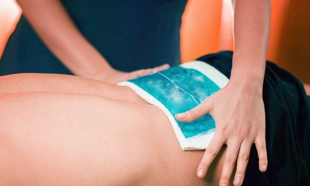
Ice temperature treatment, also known as cryotherapy or cold therapy, is a treatment that uses freezing temperatures to reduce pain and swelling. It can be applied in a variety of ways, including:
Ice packs:
Ice massage
Coolant sprays
Whirlpools
Ice baths
Ice is usually recommended for acute injuries and is a common and simple way to treat pain and swelling. Individuals can buy freezable gel packs or make their own with ice cubes in a plastic bag or towel. Heat therapy tends to be used more with chronic issues involving muscle spasms to increase blood circulation.
How Ice Relieves Pain and Reduces Inflammation
Ice temperature treatment works by:
Narrowing blood vessels slows blood circulation to the injured area and soft tissues and reduces swelling.
Reduced blood flow also helps control excessive swelling.
Ice has a short-term analgesic-numbing effect. The coldness numbs nerve endings, relieving pain symptoms.
Relieving the pain allows the muscles to relax.
Controlling blood circulation helps control pain by reducing the flow of irritating chemicals that can inundate the injury site. These chemicals are a natural and the correct response to inflammation, but the ice keeps them in check to help control pain.
After a Back Injury
For a back strain injury, ice and anti-inflammatory medication like NSAIDs are the first line of treatment during the inflammatory phase, which usually lasts 24 to 72 hours. Because heat can increase inflammation by increasing blood circulation, it is not recommended as an initial treatment. After the first few days, most doctors and pain specialists recommend using ice or heat, depending on the individual’s preference. While researchers continue to investigate the best ways to treat acute injuries, most doctors still recommend ice as the first line of defense for back injuries.
A review of studies evaluated 20 different treatment categories to learn about their safety and effectiveness. (McIntosh G. & Hall H. 2011) Treatments included over-the-counter pain medications, acupuncture, McKenzie exercises, other back exercises, and temperature treatments. Regarding temperature treatment, the review found moderate evidence that using a heat wrap 5 days after the injury could help relieve pain. However, there was not enough evidence to support the effectiveness of any of the temperature treatments, necessitating more research. (McIntosh G. & Hall H. 2011)
Physical Therapy and Activity
The review found that prolonged rest should be avoided, and gentle exercise and a progressive return to physical activity should be encouraged to achieve the best outcomes for pain relief and restoring function. Staying active significantly reduces time off from work and chronic disability for up to 1 year compared to traditional medical treatment. (McIntosh G. & Hall H. 2011) Research also found that introducing physical therapy early on could expedite recovery. Mobility work, targeted exercises, and strengthening exercises have been shown to relieve pain, reduce injury recurrence, and improve overall function. More research supports physical activity and exercise as effective treatment options for acute lower back strains. However, further research regarding temperature treatments is required. (French S. D. et al., 2006) (See Q. Y. et al., 2021)
Injury Medical Chiropractic and Functional Medicine Clinic
It is important to talk with a healthcare provider to determine the cause and extent of the injury to provide individualized patient education regarding treatment. This can include physical therapy, rest, health coaching, medication, and surgery, which may be recommended in certain cases. Overcoming these limitations is possible. Injury Medical Chiropractic and Functional Medicine Clinic works with primary healthcare providers and specialists to develop an optimal health and wellness solution. We focus on what works for you to relieve pain, restore function, prevent injury, and help mitigate the pain through spinal adjustments that help the body realign itself. They can also work with other medical professionals to integrate a treatment plan to resolve musculoskeletal issues.
Beyond the Surface: Understanding the Effects of Personal Injury
References
McIntosh, G., & Hall, H. (2011). Low back pain (acute). BMJ clinical evidence, 2011, 1102.
French, S. D., Cameron, M., Walker, B. F., Reggars, J. W., & Esterman, A. J. (2006). A Cochrane review of superficial heat or cold for low back pain. Spine, 31(9), 998–1006. https://doi.org/10.1097/01.brs.0000214881.10814.64
See, Q. Y., Tan, J. B., & Kumar, D. S. (2021). Acute low back pain: diagnosis and management. Singapore Medical Journal, 62(6), 271–275. https://doi.org/10.11622/smedj.2021086
While some disc herniations don’t cause symptoms, individuals who are overweight with a herniated disc may experience obesity pressure symptoms such as pain, weakness, numbness, or tingling. Can implementing a physical therapy and weight loss treatment program help individuals find relief?
Obesity Pressure
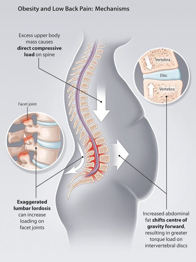
Obesity is one major factor that can contribute to a herniated disc and chronic back pain. When the body has to carry added weight, it can lead to obesity pressure on the intervertebral discs and affect posture and spine position. Researchers have found that other factors, such as inflammation or metabolic changes associated with obesity, can also be involved. (Sheng B. et al., 2017) For individuals who are overweight, weight loss can help resolve a herniated disc combined with physical therapy and can significantly increase herniated disc prevention. (Tokmak M. et al., 2015)
Spine Problems
According to Dr. Alexander Jimenez, owner and head chiropractor at Injury Medical Chiropractic and Functional Medicine Clinic, “When the position of the pelvis and lumbar spine shift out of alignment and become altered, it can profoundly contribute to advanced wearing away of outer fibers in the back region of the discs. These outer fibers house and protect the soft material that cushions and absorbs shock in the spine. Over time, the obesity pressure wear and tear on the fibers can cause chronic pain and microscopic radial tears, leading to a complete rupture.” A rupture causes the soft material to leak, irritate, and inflame surrounding nerve roots. Most herniated discs occur between the sides and back of the vertebra.
Movement Problems
Obesity makes movement difficult, often causing symptoms like shortness of breath and/or early fatigue and exhaustion even with minimal physical activity.
Physical therapy and exercise help relieve obesity and disc herniation.
However, the obesity pressure and herniation pain can make it hard to participate in cardiovascular exercises on a regular basis.
Diagnosis Complications
Obesity can interfere with the diagnosis and treatment of spinal diseases.
This is because weight restrictions and certain imaging tests, like a spinal MRI, can be difficult or impossible to obtain.
A physical examination might not be able to identify signs of nerve compression if an individual is obese. These factors can delay diagnosis.
Disc Position and Posture
Obesity is not the only thing that places pressure on the discs. An individual’s body position significantly influences the health of the shock-absorbing cushions. Sitting generates the most pressure, followed by standing, while lying on your back places the least strain on the discs and, depending on the injury, may help relieve symptoms.
Symptoms depend on the location of the herniation.
The two most common locations are the cervical spine/neck area and the lumbar spine/lower back.
Disc herniations in the neck can affect the arms.
Disc herniations in the lower back affect the buttocks and legs.
Injury Medical Chiropractic and Functional Medicine Clinic
See a healthcare provider if you’re experiencing any of these symptoms. If it is a herniated disc causing symptoms, you might be started on 6 weeks of conservative treatment. This can include physical therapy, rest, health coaching, medication, and surgery, which may be recommended in certain cases. Overcoming these limitations is possible. Injury Medical Chiropractic and Functional Medicine Clinic works with primary healthcare providers and specialists to develop an optimal health and wellness solution. We focus on what works for you to relieve pain, restore function, prevent injury, and help mitigate the pain through spinal adjustments that help the body realign itself. They can also work with other medical professionals to integrate a treatment plan to resolve musculoskeletal issues.
Weight Loss Techniques
References
Sheng, B., Feng, C., Zhang, D., Spitler, H., & Shi, L. (2017). Associations between Obesity and Spinal Diseases: A Medical Expenditure Panel Study Analysis. International journal of environmental research and public health, 14(2), 183. https://doi.org/10.3390/ijerph14020183
Tokmak, M., Altiok, I. B., Guven, M., Aras, A. B., & Cosar, M. (2015). Spontaneous Regression of Lumbar Disc Herniation After Weight Loss: Case Report. Turkish neurosurgery, 25(4), 657–661. https://doi.org/10.5137/1019-5149.JTN.9183-13.1
Can individuals relieve back pain by incorporating core strength training to reduce pain and discomfort in their lower backs?
Introduction
Many individuals worldwide have dealt with back issues that make it difficult to complete any task that they are doing. Many often feel pain and discomfort radiating from the three sections of the back and can radiate from the neck, shoulders, and hips. When these areas of the musculoskeletal system are being affected, it can lead to a life of pain and chronic conditions that cause overlapping risk profiles. At the same time, some causes of back pain often correlate with environmental factors and weak core muscles. When a person is dealing with weak core muscles, they will experience instability and pain when they are mobile, leading to back pain. In today’s article, we look at what the core muscles are, how they are connected to back pain, and how strengthening them can reduce the effects of back pain. We discuss with certified medical providers who inform our patients how strengthening the core muscles can reduce back pain. While asking informed questions to our associated medical providers, we advise patients to incorporate various core strengthening exercises to prevent overlapping risk profiles correlated with back pain. Dr. Alex Jimenez, D.C., encompasses this information as an academic service. Disclaimer.
What Are The Core Muscles?
Do you feel a constant ache or pain in your back after lifting or carrying objects in a hunched position? Do you experience muscle weakness in your torso that you can’t stay in a plank position for a few seconds? Or do you experience radiating pain from your lower back to your leg? The core muscles are a group of muscles wrapped around the torso like a support belt that helps with stability, balance, and protecting the lumbar from injuries. The core muscles are found in the lower body’s front, back, and sides. At the same time, the core muscles can help generate intrabdominal pressure while moving the vertebral column. (Flynn & Vickerton, 2024) In the core muscles, the transverse abdominis muscle or the seatbelt muscle. This muscle works with the inspiratory muscles through elastic loading to evoke transversus expiratory activity when breathing and functioning in motion. (De Troyer et al., 1990)
Core Muscles & Back Pain
The core muscles, especially the transverse abdominis muscles, are often overlooked as many individuals frequently deal with numerous factors that cause low back pain. Common core muscles are associated with back pain because environmental factors can neglect the transverse abdominis muscles. Fatigue in the core muscles can cause repetitive asymmetric loading on the spine, which enhances susceptibility to back pain and other injuries. (Zemkova & Zapletalova, 2021) Since back pain is a multifactorial condition that is one of the leading causes of hospital visits and socio-economic issues, many individuals start to neglect the core muscles over time, causing them to be weak. Some symptoms correlated with weak core muscles include:
When this happens, many people start looking for treatment to reduce their back pain and help strengthen their core muscles.
Discover The Benefits Of Chiropractic Care- Video
Strengthening Core Muscles
Before people reduce their back pain and return to their daily routine, they would have to be assessed by a pain specialist like a chiropractor to assess fully what environmental factors are causing the back pain. After the assessment, a chiropractor can work with a physical therapist to reduce back pain and strengthen the core muscles to prevent back pain from returning. When it comes to core strengthening exercises, they focus on either the deep or superficial muscles of the torso, which might produce different effects on lumbar motion. (Puntumetakul et al., 2021) Additionally, core stability exercises can reduce pain in the back and disability reduction and improve a person’s quality of life. (Kanwal et al., 2021) Engaging the muscles through core strengthening exercises can help achieve optimal strength with twisting and side-bending movements to reduce back pain.
Pay Attention To Engaged Core Muscles
However, individuals need to pay attention to engaging their core muscles while maintaining a neutral spine position for stability and preventing back pain. (Cigdem Karacay et al., 2022) This allows individuals to be consistent with strengthening their core and to commit to short workouts incorporated as part of not only their routine but also as part of their customized treatment plan. When it comes to reducing back pain, it is important to ensure that the core muscles are engaged to help build strength and stability, decrease back pain, and improve functionality. When people start strengthening their core muscles, they will be able to be more mindful about how to present themselves and live healthier lives.
References
Cigdem Karacay, B., Sahbaz, T., Gurtekin, B., Yildiz, S., & Ozcan, E. (2022). Effectiveness of whole-body vibration exercise and core stabilization exercise in chronic non-specific low back pain: A randomized-controlled study. Turk J Phys Med Rehabil, 68(2), 184-194. https://doi.org/10.5606/tftrd.2022.7060
De Troyer, A., Estenne, M., Ninane, V., Van Gansbeke, D., & Gorini, M. (1990). Transversus abdominis muscle function in humans. J Appl Physiol (1985), 68(3), 1010-1016. https://doi.org/10.1152/jappl.1990.68.3.1010
Kanwal, S., Yaqoob, I., Shakil-Ur-Rehman, S., Ghous, M., Ghazal, J., & Namroz, N. (2021). Effects of core muscle stability on low back pain and quality of life in post-menopausal women: A comparative study. J Pak Med Assoc, 71(1(A)), 37-40. https://doi.org/10.47391/JPMA.151
Puntumetakul, R., Saiklang, P., Tapanya, W., Chatprem, T., Kanpittaya, J., Arayawichanon, P., & Boucaut, R. (2021). The Effects of Core Stabilization Exercise with the Abdominal Drawing-in Maneuver Technique versus General Strengthening Exercise on Lumbar Segmental Motion in Patients with Clinical Lumbar Instability: A Randomized Controlled Trial with 12-Month Follow-Up. Int J Environ Res Public Health, 18(15). https://doi.org/10.3390/ijerph18157811
Wattananon, P., Sinsurin, K., & Somprasong, S. (2020). Association between lumbopelvic motion and muscle activation in patients with non-specific low back pain during forward bending task: A cross-sectional study. Hong Kong Physiother J, 40(1), 29-37. https://doi.org/10.1142/S1013702520500043
Zemkova, E., & Zapletalova, L. (2021). Back Problems: Pros and Cons of Core Strengthening Exercises as a Part of Athlete Training. Int J Environ Res Public Health, 18(10). https://doi.org/10.3390/ijerph18105400
IFM's Find A Practitioner tool is the largest referral network in Functional Medicine, created to help patients locate Functional Medicine practitioners anywhere in the world. IFM Certified Practitioners are listed first in the search results, given their extensive education in Functional Medicine