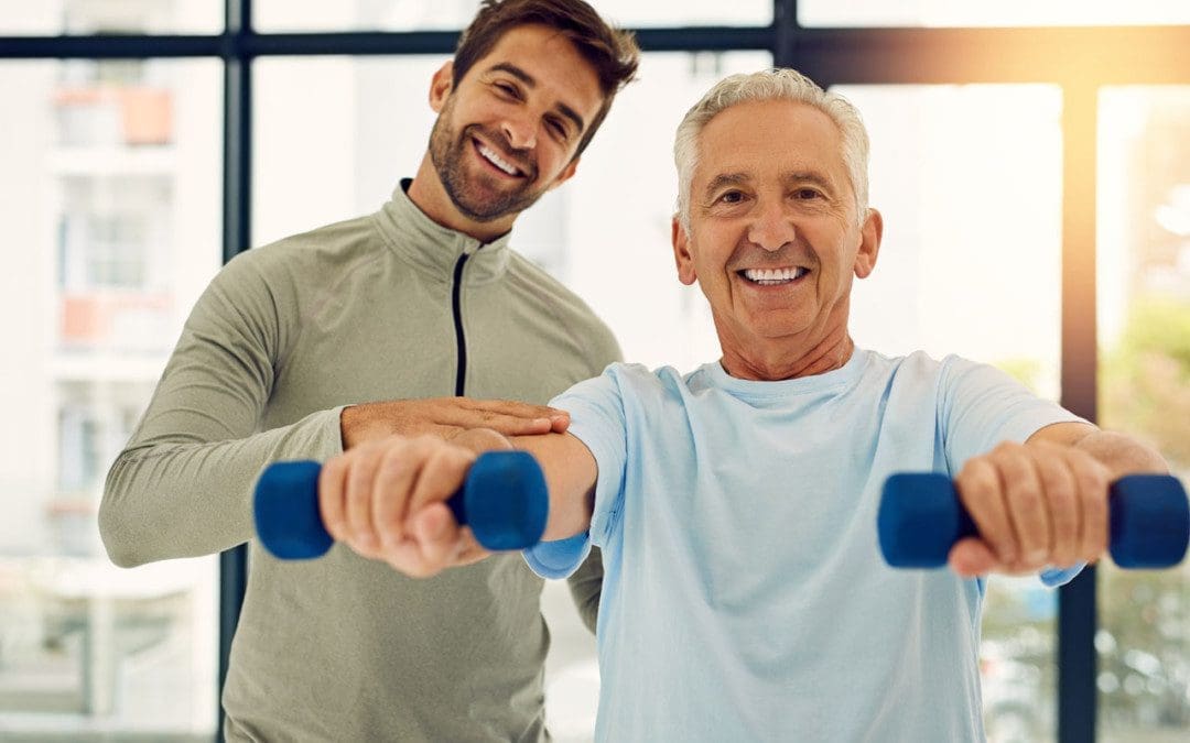
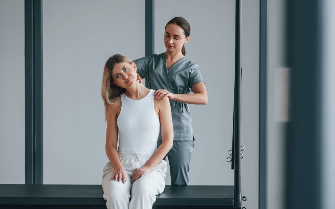
Regular exercise and physical activity help with cardiovascular health, improved mood, better management of chronic conditions, and can help digestion. For individuals with any GI distress or inflammatory bowel disease that has caused digestive enzyme deficiencies, exercise, and physical movement have been found to provide digestive aid. Here we look at activities to help digestion.
Exercises To Help Digestion
When exercising the body, the cardiac output/volume of blood the heart pumps every minute increases as the demand for oxygenated blood throughout the body increases, particularly in the working muscles. During exercise, the same increase in blood circulation happens within the digestive system’s muscle groups. The blood flow to digestive organs causes peristalsis, which is involuntary constriction and relaxation of the muscles in the digestive tract. This process helps move food efficiently through the gastrointestinal tract. Exercise supports the growth of beneficial gut bacteria to maintain a healthy digestive system.
Exercise helps relieves stress which means lower amounts of cortisol.
Research has found that elevated cortisol levels are associated with compromised digestive function.
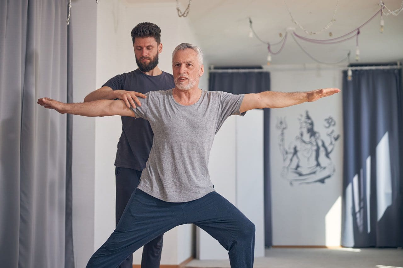
Tai chi has been shown to improve immune function and inflammation of the gut and is a helpful tool for maintaining homeostasis/gut balance.
This is a form of moderate-intensity exercise and meditative practice.
The emphasis is on slow controlled movements and deep breathing.
This makes it recommended for individuals looking to improve digestion and those with gastrointestinal conditions.
Pilates
Pilates is the practice that targets abdominal muscles and utilizes diaphragmatic breathing to help individuals perform a series of movements to strengthen and elongate the body’s muscles.
Individuals who perform this exercise often report relief from bloating and gas.
Whether new to exercise or returning, a plan can help you get there. Meeting with a fitness trainer or sports chiropractor is a great place to begin if you have limited knowledge about what works best for your body and schedule.
A certified trainer can help guide you toward an achievable program that focuses on gut health.
Individuals with a GI disorder should talk with their doctors before starting a new exercise plan.
This does not mean you can’t do intense exercises like running; you’ll want to work with a doctor to set up a program that doesn’t cause flare-ups.
Aim for roughly three hours of moderate-intensity weekly exercise to support a healthy digestive system.
Sit less and move more.
Do at least two or more muscle-strengthening activities of moderate intensity every week.
Cherpak, Christine E. “Mindful Eating: A Review Of How The Stress-Digestion-Mindfulness Triad May Modulate And Improve Gastrointestinal And Digestive Function.” Integrative medicine (Encinitas, Calif.) vol. 18,4 (2019): 48-53.
Drouin, Jacqueline S et al. “Comparisons between Manual Lymph Drainage, Abdominal Massage, and Electrical Stimulation on Functional Constipation Outcomes: A Randomized, Controlled Trial.” International Journal of environmental research and public health vol. 17,11 3924. June 1. 2020, doi:10.3390/ijerph17113924
Hamasaki, Hidetaka. “Exercise and gut microbiota: clinical implications for the feasibility of Tai Chi.” Journal of integrative medicine vol. 15,4 (2017): 270-281. doi:10.1016/S2095-4964(17)60342-X
Joyner, Michael J, and Darren P Casey. “Regulation of increased blood flow (hyperemia) to muscles during exercise: a hierarchy of competing physiological needs.” Physiological Reviews vol. 95,2 (2015): 549-601. doi:10.1152/physrev.00035.2013
LeBouef T, Yaker Z, Whited L. Physiology, Autonomic Nervous System. [Updated 2023 May 1]. In: StatPearls [Internet]. Treasure Island (FL): StatPearls Publishing; 2023 Jan-. Available from: https://www.ncbi.nlm.nih.gov/books/NBK538516/
Singhal, Rashi, and Yatrik M Shah. “Oxygen battle in the gut: Hypoxia and hypoxia-inducible factors in metabolic and inflammatory responses in the intestine.” The Journal of biological chemistry vol. 295,30 (2020): 10493-10505. doi:10.1074/jbc.REV120.011188
van Wijck, Kim, et al. “Physiology and pathophysiology of splanchnic hypoperfusion and intestinal injury during exercise: strategies for evaluation and prevention.” American Journal of Physiology. Gastrointestinal and liver physiology vol. 303,2 (2012): G155-68. doi:10.1152/ajpgi.00066.2012
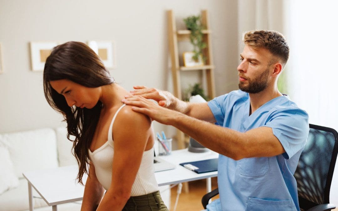
Body stiffness is common, especially as the body ages. Stiffness can result from intense work, lack of physical exercise, or specific conditions. The reasons vary from individual to individual. Some feel stiff when they wake up, while others become stiff after stopping physical activity. For others, stiffness can result from practicing unhealthy postures, intense workouts, or something new that the body is beginning to get used to. There are several ways to prevent and treat stiffness, no matter the cause, including targeted physical movements, posture corrections, body decompression, chiropractic realignment, stretches, and therapeutic massage.
Body Stiffness
Knowing the cause of body stiffness and how to relieve it can help prevent and treat the condition so the body can function better. It’s vital to see a health care professional immediately if stiffness results from an injury, accompanied by pain, it does not go away with home treatments, or if an insect bite or infection could be the cause.
Individuals should speak to a healthcare professional for frequent stiffness that interferes with their quality of life.
Most of the time, stiffness can be treated at home and reduced through preventative measures.
Stay active but not too hard until the body gets used to the activity.
Various relief methods include a warm bath, massage shower, or self-massage.
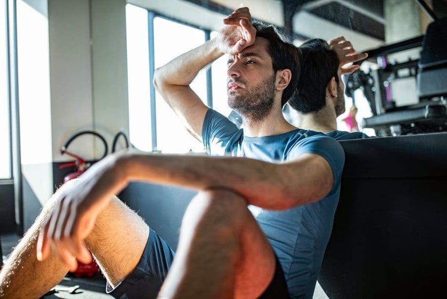
Intense Work or Exercise
The muscles incur tiny tears when exercising or performing heavy labor, especially when the body is not used to the intensity or duration.
These tears are normal and help build larger and stronger muscles.
Individuals may feel stiff and sore for 24-72 hours as the body repairs itself.
Inflammation surrounding the joints/synovial fluid after heavy activity or repetitive movements is another cause.
Inactivity
Moving around generates the synovial fluid that lubricates the joints.
When body movement stops, like going to sleep or long periods spent sitting, working, or watching tv, fluid production slows down, resulting in body stiffness.
The lack of fluid after movement can make the body feel stiff when returning to activity.
Unhealthy Posture
The body can become stiff and sore when routinely holding the body in a way that strains the muscles, tendons, and ligaments.
Sitting or standing incorrectly from an unhealthy workstation setup or postural habits contributes to stiffness and musculoskeletal problems.
Medical Conditions
Medical conditions can cause stiffness like rheumatoid arthritis, Lyme disease, thyroid disease, strains and sprains, and low vitamin D levels.
See medical attention if you suspect any medical causes are behind the body stiffness.
Prevention
Depending on the reason behind body stiffness, there are ways to prevent it.
Warm-Up
Warming up before any physical activity loosens up the muscles before fully engaging.
Soreness will present and is part of the muscle repair process.
Properly warming up can help the repair go faster.
Mobility and Flexibility Breaks
Taking breaks from inactivity by getting up and moving around, walking, or performing mobility movements could increase the secretions of joint fluid, prevent stiffness, and relieve the effects of poor postural habits you may have been making.
Set a timer to break up periods of inactivity and move around.
Get up for 5 minutes every hour to move the muscles and get the blood flowing.
Stay Aware of Posture and Form
Postural awareness can help prevent muscle strain that leads to stiffness.
Adjusting the workspace and posture can help prevent stiffness.
The posterior chain: head, neck, torso, and legs are aligned with the feet flat on the floor and back supported.
Stay Active
Maintaining muscle movement maintains blood circulation, which can help reduce stiffness.
Exercise helps reduce inflammation, increases synovial fluid production, and helps strengthen the muscles.
Active Recovery
Participating in active recovery can help bring blood flow to the muscles and prevent inflammation.
Light cardiovascular activities include swimming, cycling, walking, or bodyweight movements.
Anti-Inflammatory Nutrition
Anti-inflammation nutrition like the Mediterranean diet, which includes healthy fats, plenty of fruits and vegetables, lean proteins, seafood, and whole grains, can help reduce aches and stiffness.
Getting enough vitamin D can reduce stiffness.
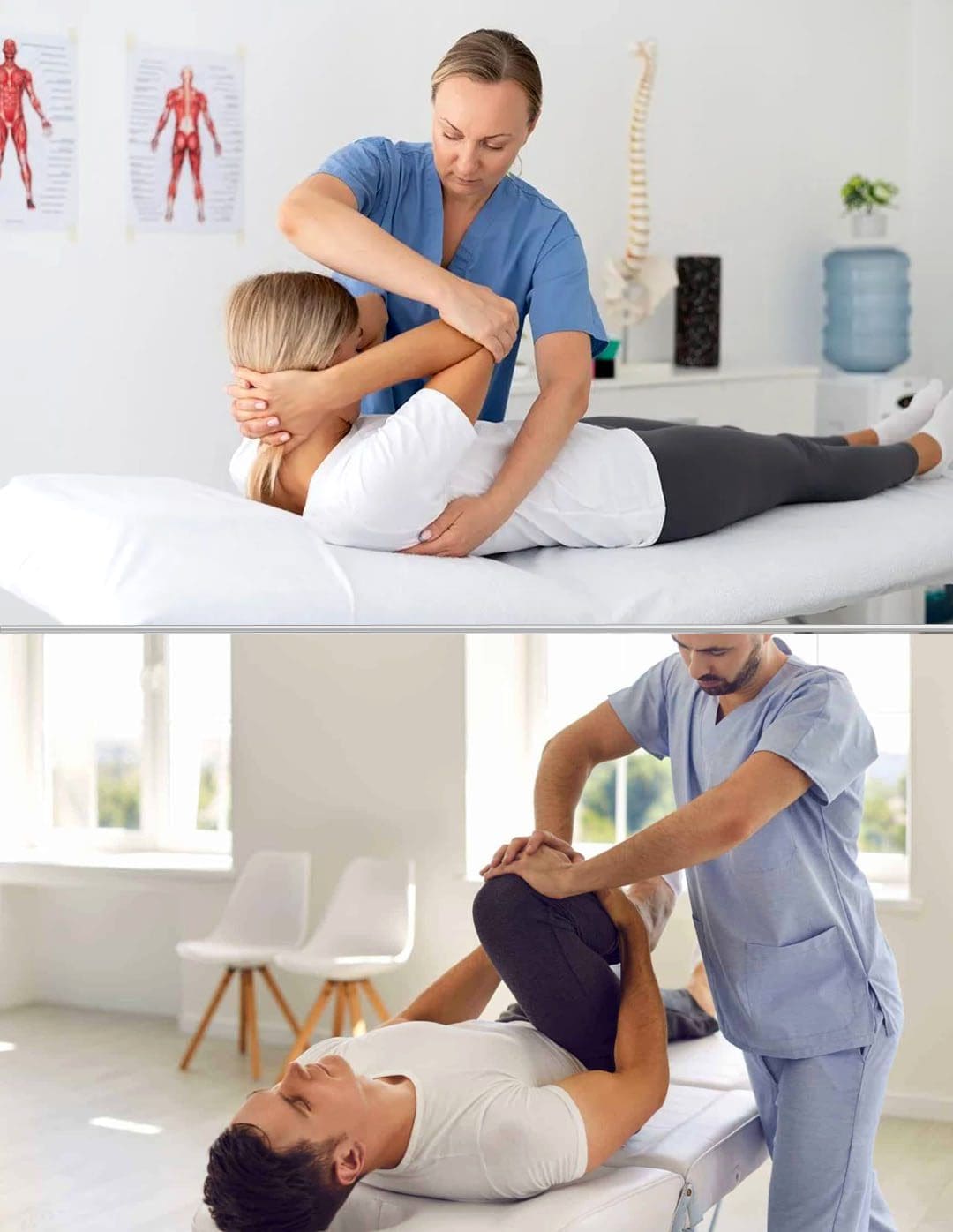
Chiropractic Flexibility Restoration
Chiropractic adjustments, decompression, MET, and therapeutic massage techniques can relieve muscle soreness and stiffness and restore body function. The chiropractic team will evaluate the individual, diagnose the cause/s, and develop a personalized treatment plan. The team will provide posture training, stretching the body, using a percussive massager or foam roller to break tight, stiff muscles and release adhesions of tissues.
Enhance Your Lifestyle
References
Mailey, Emily L et al. “Comparing the effects of two different break strategies on occupational sedentary behavior in a real-world setting: A randomized trial.” Preventive medicine reports vol. 4 423-8. 9 Aug. 2016, doi:10.1016/j.pmedr.2016.08.010
Schleip, Robert, and Werner Klingler. “Active contractile properties of fascia.” Clinical Anatomy (New York, N.Y.) vol. 32,7 (2019): 891-895. doi:10.1002/ca.23391
Shimoyama, Daisuke, et al. “Reliability of shoulder muscle stiffness measurement using strain ultrasound elastography and an acoustic coupler.” Journal of medical ultrasonics (2001) vol. 48,1 (2021): 91-96. doi:10.1007/s10396-020-01056-0
Trube, Niclas, et al. “How muscle stiffness affects human body model behavior.” Biomedical engineering online vol. 20,1 53. 2 Jun. 2021, doi:10.1186/s12938-021-00876-6
Weerapong, Pornratshanee, et al. “The mechanisms of massage and effects on performance, muscle recovery, and injury prevention.” Sports medicine (Auckland, N.Z.) vol. 35,3 (2005): 235-56. doi:10.2165/00007256-200535030-00004
When the body shifts out of homeostasis or when something in the body is out of balance, the body sweats. Sweating is a process known as perspiration that releases salt-based fluids from the body’s sweat glands to help the body stay cool and regulate body temperature. Sweat is commonly found under the arms, on the feet, and on the palms of the hands. Body temperature, outdoor temperature, or emotional state changes can cause sweating.
Sweating
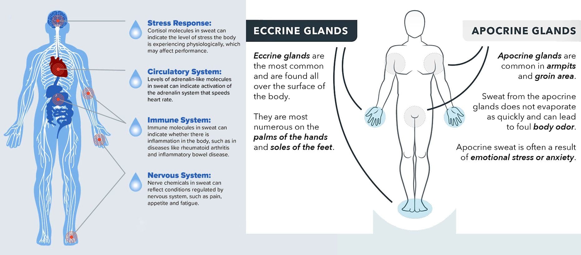
An individual has around 2-4 million sweat glands, which begin to become fully active during puberty. There are two types of sweat glands: eccrine and apocrine. The most common areas of sweating include:
Face.
Armpits.
Palms of the hands.
Soles of the feet.
Sweating in normal amounts is an essential bodily process.
Not sweating enough or sweating too much can cause problems.
Sweat is mostly water but contains small amounts of salt.
Sweat also contains electrolytes and minerals – including potassium, chloride, magnesium, zinc, copper, proteins, urea, and ammonia.
Electrolyte levels need to be replenished after heavy sweating.
Causes
Sweating is normal. However, a variety of causes can stimulate increased sweating.
High Temperature
Elevated body temperature.
Elevated outdoor temperature.
Are the primary cause of increased sweating.
Emotions and stress
Emotions and conditions can also make the body break out in a heavy sweat.
Emotional stress
Anxiety
Anger
Fear
Embarrassment
Foods
Sweating may be a response to certain foods. This type of sweat is known as gustatory sweating, which can be caused by:
Spicy foods
Caffeinated drinks – like soda, coffee, and tea.
Alcoholic beverages.
Medications
Illness and Medications
Sweating may be caused by medication use and certain illnesses:
Individuals can become dehydrated and have an increased risk of heatstroke.
Chiropractic Adjustments
The nervous system coordinates and oversees all functions of the body. Some can be consciously controlled, and others are automatic. The autonomic nervous system– ANS regulates involuntary processes, including blood pressure, heart rate, digestion, respiration, gland function, sweating, etc. The ANS is divided into the sympathetic and parasympathetic systems.
The sympathetic nervous system – when activated, creates a state of elevated activity and attention or the fight or flight response.
This process increases blood pressure and heart rate, preparing the body to respond to various stressors.
The parasympathetic nervous system promotes resting and digesting processes that lower heart rate and blood pressure.
The parasympathetic calms the body.
Chiropractic adjustments have been known to affect the autonomic nervous system. This is achieved by increasing parasympathetic activity/relaxation and down-shifting the sympathetic/fight or flight response and inflammation. A chiropractic adjustment can remove subluxations, which cause interferences in the nervous system. Chiropractic restores and improves the brain and body system communication.
Thoracic Spine Pain
References
Baker, Lindsay B. “Physiology of sweat gland function: The roles of sweating and sweat composition in human health.” Temperature (Austin, Tex.) vol. 6,3 211-259. 17 Jul. 2019, doi:10.1080/23328940.2019.1632145
Cabanac, M. “Temperature regulation.” Annual Review of Physiology vol. 37 (1975): 415-39. doi:10.1146/annurev.ph.37.030175.002215
Cui, Chang-Yi, and David Schlessinger. “Eccrine sweat gland development and sweat secretion.” Experimental dermatology vol. 24,9 (2015): 644-50. doi:10.1111/exd.12773
Kiani, Aysha Karim, et al. “Neurobiological basis of chiropractic manipulative treatment of the spine in the care of major depression.” Acta bio-medica : Atenei Parmensis vol. 91,13-S e2020006. 9 Nov. 2020, doi:10.23750/abm.v91i13-S.10536
McCutcheon, L J, and R J Geor. “Sweating. Fluid and ion losses and replacement.” The Veterinary clinics of North America. Equine practice vol. 14,1 (1998): 75-95. doi:10.1016/s0749-0739(17)30213-4
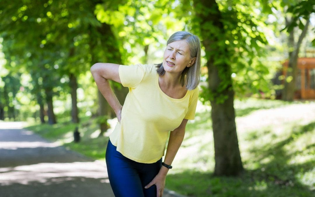
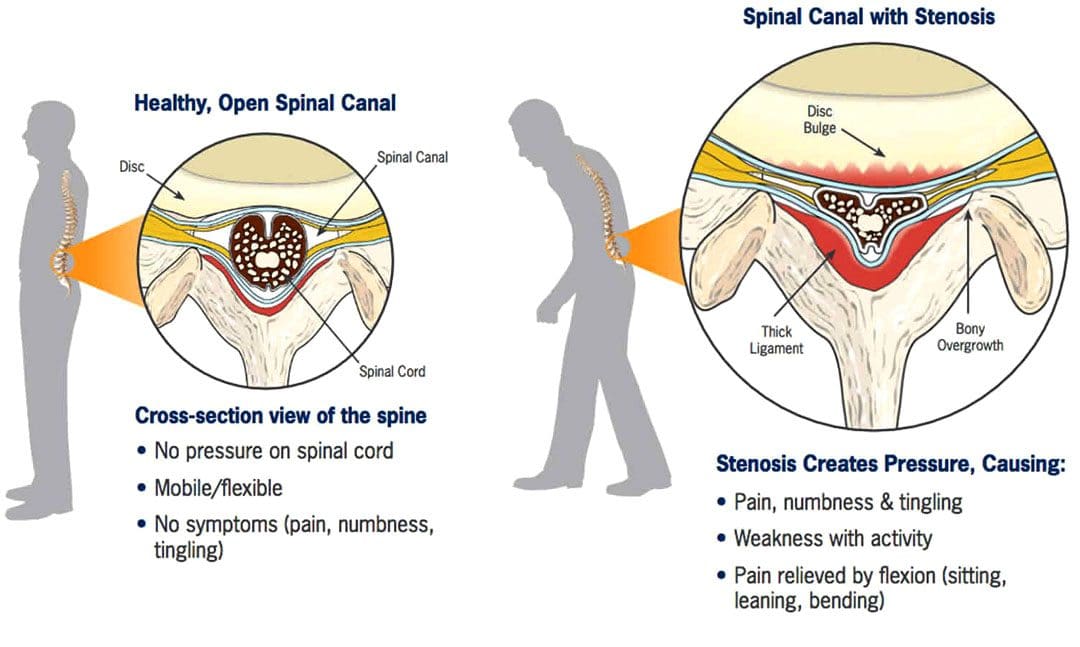
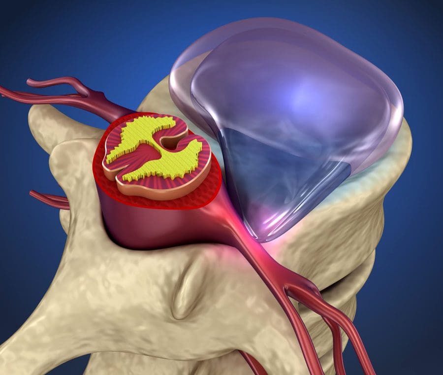
Spinal Stenosis Walking Issues:Stenosis means a narrowing. Spinal stenosis can happen in any spine region, but the neck and lower back are the most common locations. The spinal canal becomes narrower and can cause the nerves to become compressed, pinched, and irritated and can extend from the lumbar spine through the hips, buttocks, legs, and feet. Individuals with lumbar spinal stenosis may have difficulty walking caused by sensations of discomfort like numbness, electrical shocks, and pain, requiring the need to lean forward to relieve pressure and symptoms. Additionally, symptoms are likely to worsen the longer the walk. Chiropractic treatment can treat spinal stenosis because it corrects and re-aligns the spine, thus reducing pressure on the spinal cord, joints, and nerve roots.
Spinal Stenosis Walking Issues
The spine is made up of interlocking vertebrae. The regions are cervical, thoracic, lumbar, and sacral bones with a foramen opening. These openings form the protective tunnel/spinal canal surrounding the spinal cord. The spinal cord is a group of nerves that run through the tunnel. The narrowing suffocates the nerves supplying the lower extremities that can influence walking activity.
Symptoms
There may be no symptoms with early lumbar spinal stenosis. Most individuals develop symptoms gradually and may begin to notice them while walking or standing. These can include:
Lower back pressure sensations when standing upright or walking.
Leg numbness, tingling, weakness, burning, and/or cramping.
Muscle weakness.
Persistent pain in the back, hips, buttocks, or legs while walking.
Difficulty lifting the top part of the foot – known as drop foot.
Loss of sensation in the feet.
A weak foot that drops/slaps down when walking.
Loss of sexual ability.
In more serious cases, severe numbness, bladder problems, and inability to stand.
Individuals begin to lean forward when symptoms start, bringing relief by reducing the pressure on the nerves. However, constantly leaning forward leads to other posture and health problems.
Diagnosis
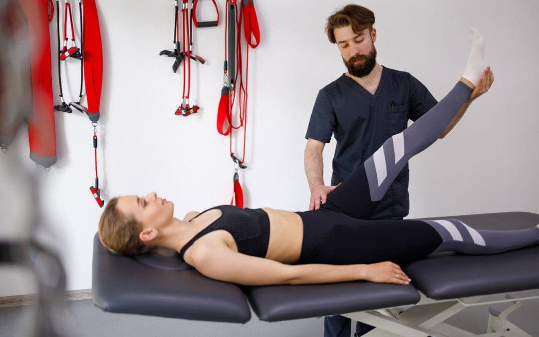
A doctor or chiropractor will ask questions about symptoms and medical history and perform a complete physical examination to diagnose lumbar spinal stenosis. During the physical examination, a healthcare provider will look for signs, such as loss of sensation, weakness, and abnormal reflexes.
Tests:
X-rays of the lumbar spine may show bone growths called spurs that push on spinal nerves and/or narrowing of the spinal canal.
Imaging tests – A CT or MRI scan can provide a detailed look at the spinal canal and nerve structures.
Other studies include – bone scans, myelogram, which is a CT scan that uses a color dye, and EMG, which is an electrical test of muscle activity.
Chiropractic Treatment
Chiropractic care combined with physical therapy is a tried-and-true treatment for spinal stenosis. A chiropractic treatment plan can include targeted and passive exercise programs. Targeted exercises involve strengthening the core and back muscles. Passive treatments include hot and cold therapy, massage, decompression, and electrical stimulation. The objective of chiropractic therapy is to:
Strengthen muscles in the core and legs
Correct posture and body mechanics.
Improve mobility.
Maintain ability to perform day-to-day activities.
Recommend stretches.
Educate on how to keep the spine and back muscles safe.
Train on using devices like a back brace, cane, or walker properly.
Advise about shoe inserts and splints.
Suggest work and home environment modifications, such as ergonomics and cushions.
Chiropractic Relief
References
Conway, Justin, et al. “Walking assessment in people with lumbar spinal stenosis: capacity, performance, and self-report measures.” The spine journal: official North American Spine Society journal vol. 11,9 (2011): 816-23. doi:10.1016/j.spinee.2010.10.019
Lurie, Jon, and Christy Tomkins-Lane. “Management of lumbar spinal stenosis.” BMJ (Clinical research ed.) vol. 352 h6234. 4 Jan. 2016, doi:10.1136/bmj.h6234
Macedo, Luciana Gazzi, et al. “Physical therapy interventions for degenerative lumbar spinal stenosis: a systematic review.” Physical therapy vol. 93,12 (2013): 1646-60. doi:10.2522/ptj.20120379
Tomkins-Lane, Christy C et al. “Predictors of walking performance and walking capacity in people with lumbar spinal stenosis, low back pain, and asymptomatic controls.” Archives of physical medicine and rehabilitation vol. 93,4 (2012): 647-53. doi:10.1016/j.apmr.2011.09.023
Herniated disc injuries and the time it takes to heal depend on the injury’s cause, the severity, and where it occurred along the spine. Symptoms can last a few days to months. Chiropractic treatment, massage therapy, and decompression realign the spine and return the disc to its correct position. Still, the herniated disc signs it is returning to normal can take time as the rest of the spine and body adjust to the realignment.
Herniated Disc Signs It Is Returning To Normal
Most cases take a few weeks with healing time depending on health conditions, physical activity level, and age. However, in severe cases, a herniated disc can take up to several months to fully heal, but discomfort symptoms usually resolve sooner.
Expectations From a Healing Disc
Resting the spine and taking it easy after the injury is recommended.
Too much rest is not recommended as it can cause muscle stiffness.
While the herniated disc is healing, a primary doctor may prescribe anti-inflammatories or muscle relaxants to help ease discomfort.
A chiropractor and/or physical therapist can teach exercises and stretches to relieve pressure on nerves, loosen tight muscles, and improve circulation.
Signs The Herniated Disc Is Healing
Most herniated discs cause significant pain in the back and neck from the nervous system, causing some of the muscles of the low back or neck to spasm to protect the area from further damage.
Usually, the muscle spasms relax within the first days of the injury.
After spinal decompression, neurological symptoms like the sharp, shooting pain down a nerve in the arm or leg are the first symptoms to go away.
Then muscle weakness along the path of the nerve goes away.
Numbness in the extremities can linger around longer.
Length of Time
The wear and tear of adult spinal discs, combined with unhealthy posture habits, job occupation, previous injuries, etc., decrease blood circulation.
This is why it can take some time to heal completely, as the entire blood supply needs to reset to optimal circulation.
Nerve compression causing aches and pain sensations down the nerves can also take time.
Regular Activity
Returning to regular activities depends on the individual’s case and condition. It is essential not to overdo things that can cause excessive loading of the spine before the disc has fully healed, which increases the risk of re-herniation and other injuries.
Inactivity can slow the healing process and cause inflammation.
Patients are encouraged to return to activities that generate gentle motion to stimulate the stabilizing muscles to function properly and increase blood circulation to the injured area.
Individuals are recommended to:
Learn posture improvement when walking, sitting, standing, and sleeping.
Adjust sleep patterns.
Incorporate anti-inflammatory nutrition during the healing process.
This provides a mechanical and biological environment that eventually becomes a personalized exercise physical therapy program.
DOC Spinal Decompression
References
Díez Ulloa, Máximo Alberto. “Role of Microangiogenensis in Disc Herniation Healing.” Journal of investigative surgery: the official journal of the Academy of Surgical Research vol. 34,6 (2021): 685. doi:10.1080/08941939.2019.1682725
Factors that influence recovery: Mayo Clinic. February 8, 2022. “Herniated disk.” https://www.mayoclinic.org/diseases-conditions/herniated-disk/symptoms-causes/syc-20354095
Factors that influence recovery: NHS. March 22, 2021. “Slipped Disc.” https://www.nhs.uk/conditions/slipped-disc/
How to speed up healing time: American Academy of Orthopaedic Surgeons. January 2022. “Herniated Disk in the Lower Back” https://orthoinfo.aaos.org/en/diseases–conditions/herniated-disk-in-the-lower-back/
Keramat, Keramat Ullah, and Aisling Gaughran. “Safe physiotherapy interventions in large cervical disc herniations.” BMJ case reports vol. 2012 bcr2012006864. 18 Aug. 2012, doi:10.1136/bcr-2012-006864
Stoll, T et al. “Physiotherapie bei lumbaler Diskushernie” [Physiotherapy in lumbar disc herniation ]. Therapeutische Umschau. Revue therapeutique vol. 58,8 (2001): 487-92. doi:10.1024/0040-5930.58.8.487
Swartz, Karin R, and Gregory R Trost. “Recurrent lumbar disc herniation.” Neurosurgical focus vol. 15,3 E10. 15 Sep. 2003, doi:10.3171/foc.2003.15.3.10
Nerves control muscle fibers. Muscle twitching is an involuntary contraction of the muscle fibers. When individuals play sports/work out vigorously or for a long time, they may experience muscle twitching and can often see and/or feel the twitches happening. The most worked-out muscles are likely to twitch, which includes the biceps, thighs, and calves, but twitches can occur in any muscle. Chiropractic care, massage therapy, and functional medicine can help relax the muscles, improve circulation, restore function, and train individuals to prevent future episodes.
Muscle Twitching
A muscle twitch often occurs after intense physical activity or a hard workout because the muscle or muscles have been overworked, and there is hyper-excitability of the nerve/s that makes the muscle/s continue to contract.
A muscle twitch that can be seen is called fasciculation.
A muscle twitch that cannot be seen is called fibrillation.
If there is pain or the twitching is prolonged, it is a muscle spasm.
Causes
The most common causes include the following:
Intense exercise and rigorous physical activity build up lactic acid in the muscles.
Dehydration is a very common factor for shaky muscles.
Vitamin D and calcium deficiencies could cause muscle spasms in the hand, calves, and eyelids.
Using caffeinated products to increase physical performance.
Not enough or a lack of healthy sleep.
Anxiety or stress.
Certain medications like estrogen and corticosteroids.
Nicotine and tobacco use.
Physical Activity/Exercise
Intense exercise and physical activity can cause muscle fatigue.
Muscle fatigue triggers twitching and cramping in overworked muscle fibers.
Electrolytes play a role in muscle contraction.
Electrolyte loss and imbalances within muscle fibers through sweating can lead to twitching.
Dehydration
Muscle mass comprises 75% water.
Water carries nutrients and minerals to muscles to support function.
Not being properly hydrated can cause twitching and cramping.
Vitamin D Deficiency
Nerves need vitamin D to relay messages to and from the brain to the body’s muscles.
A vitamin D deficiency can cause muscle weakness and twitching.
Lack of sleep can affect neurotransmitter function.
A common site of fasciculation tiredness occurs in the eyelids.
Anxiety and Stress
Experiencing psychological stress or high anxiety levels can cause excess muscle tension.
This can lead to muscle twitching.
Muscle fasciculation caused by stress can occur anywhere in the body.
Certain Medications
Certain medications can lead to involuntary muscle twitching.
The reaction can be a side effect due to interactions with other medications.
Individuals should discuss side effects and medication interactions with their doctor when taking a new medication.
Chiropractic Care
Chiropractors are experts on the musculoskeletal system and have many techniques to treat muscle fasciculation and spasms. It often depends on the cause/s, and specific treatment varies on a case-by-case basis. Common chiropractic treatments include:
Massage therapy
Heat and ice therapy
Manual manipulation
Joint adjustments
Ultrasound
Stretches to keep the muscles flexible
Exercises to strengthen the muscles
Nutritional recommendations
Fasciculation
References
Bergeron, Michael F.. Muscle Cramps during Exercise-Is It Fatigue or Electrolyte Deficit?. Current Sports Medicine Reports July 2008 – Volume 7 – Issue 4 – p S50-S55 doi: 10.1249/JSR.0b013e31817f476a
Gragossian A, Bashir K, Friede R. Hypomagnesemia. [Updated 2022 May 15]. In: StatPearls [Internet]. Treasure Island (F.L.): StatPearls Publishing; 2022 Jan-. Available from: https://www.ncbi.nlm.nih.gov/books/NBK500003/
Küçükali, Cem Ismail, et al. “Peripheral nerve hyperexcitability syndromes.” Reviews in the neurosciences vol. 26,2 (2015): 239-51. doi:10.1515/revneuro-2014-0066
Maughan, Ronald J, and Susan M Shirreffs. “Muscle Cramping During Exercise: Causes, Solutions, and Questions Remaining.” Sports medicine (Auckland, N.Z.) vol. 49, Suppl 2 (2019): 115-124. doi:10.1007/s40279-019-01162-1
Miller, Kevin C et al. “Exercise-associated muscle cramps: causes, treatment, and prevention.” Sports health vol. 2,4 (2010): 279-83. doi:10.1177/1941738109357299
Riebl, Shaun K, and Brenda M Davy. “The Hydration Equation: Update on Water Balance and Cognitive Performance.” ACSM’s health & fitness journal vol. 17,6 (2013): 21-28. doi:10.1249/FIT.0b013e3182a9570f
Sports exercise headaches are exertion headaches that involve pain during or immediately after sports, exercise, or some physical activity. They come on quickly but can last a few minutes, hours, or days. Activities associated with exercise headaches include running, weightlifting, tennis, swimming, and rowing. Chiropractic, massage, decompression, and traction therapies can realign the body and relax the muscles allowing for optimal circulation and certain strategies to help prevent future episodes. Usually, there is no underlying disease or disorder, but it is recommended to talk to a healthcare provider to make sure.
Sports Exercise Headaches
When individuals exert their bodies intensely, they need added blood and oxygen, particularly with activities that involve tightening/tensing the abdominal muscles or increasing chest pressure. Doctors and scientists believe an exertional headache occurs when intense physical activity causes the veins and arteries to expand to circulate more blood. The expansion and increased blood circulation generate pressure in the skull that can cause pain.
Alternate Triggers
Exercising is not the only cause; other physical activities that can trigger an exertion headache include:
Sneezing
Coughing
Straining to use the bathroom
Sexual intercourse
Lifting or moving a heavy object
Symptoms
Symptoms of a sports exercise headache include:
Neck stiffness or pain
Pain on one or both sides of the head
Pulsating pain discomfort
Throbbing pain discomfort
Shoulder tightness, discomfort, and/or pain
Sometimes individuals report the headache can feel like a migraine that could include:
Vision problems like blind spots
Nausea
Vomiting
Light sensitivity
Most exercise headaches last five to 48 hours and can continue for three to six months.
Diagnosis
An underlying disease or disorder does not cause most exertional headaches. However, individuals experiencing severe or frequent headaches should consult their doctor or a healthcare provider. Tests will be ordered to rule out possible causes that include:
MRI will take computer-generated images of the brain.
A spinal tap/lumbar puncture takes a sample of fluid from the spine for testing.
If there is no underlying cause found, the medical provider can diagnose exertion headaches if there have been at least two headaches that:
Were caused by exercise or physical activity.
Started during or after the physical activity.
Lasted less than 48 hours.
Chiropractic Treatment
According to the American Chiropractic Association, spinal adjustments are an effective headache treatment option. This includes migraines, tension headaches, or sports exercise headaches. Using the targeted approaches, chiropractic restores the body’s natural alignment to improve function and alleviate stress on the nervous system. This allows the body to operate at optimal levels reducing muscle stress and muscle tension.
DOC Decompression Table
References
American Migraine Foundation. Secondary Headaches. (https://americanmigrainefoundation.org/resource-library/secondary-headaches/) Accessed 11/17/2021.
Evans, Randolph W. “Sports and Headaches.” Headache vol. 58,3 (2018): 426-437. doi:10.1111/head.13263
International Headache Society. HIS Classification ICHD-3. (https://ichd-3.org/other-primary-headache-disorders/4-2-primary-exercise-headache/) Accessed 11/17/2021.
McCrory, P. “Headaches and exercise.” Sports medicine (Auckland, N.Z.) vol. 30,3 (2000): 221-9. doi:10.2165/00007256-200030030-00006
National Headache Foundation. Exertional Headaches. (https://headaches.org/2007/10/25/exertional-headaches/) Accessed 11/17/2021.
Ramadan, Nabih M. “Sports-related headache.” Current pain and headache reports vol. 8,4 (2004): 301-5. doi:10.1007/s11916-004-0012-1
Trotta K, Hyde J. Exercise-induced headaches: prevention, management, and treatment. (https://www.uspharmacist.com/article/exerciseinduced-headaches-prevention-management-and-treatment) U.S. Pharm. 2017;42(1):33-36. Accessed 11/17/2021.
IFM's Find A Practitioner tool is the largest referral network in Functional Medicine, created to help patients locate Functional Medicine practitioners anywhere in the world. IFM Certified Practitioners are listed first in the search results, given their extensive education in Functional Medicine