Of course, I’m prone to injuries and Dr. Alex Jimenez has been helping me. I’ve known him for about six years and every time something comes up, either it’s a small injury or a major one, he’s always been there and he’s always helped me get back to my feet to start playing sports again really fast.
Madison Hill
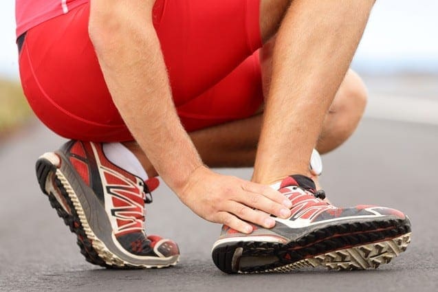
Ankle pain refers to any type of pain or discomfort in the ankle. This pain could generally be due to an injury, such as a sprain, or due to another health issue. As stated by the National University of Health Sciences, or NUHS, an ankle sprain is one of the most frequent causes of foot pain, making up 85 percent of all ankle injuries. A sprain occurs when the ligaments tear or are overstretched.
Most ankle sprains are lateral sprains, which occur when the foot rolls, causing the ankle to twist toward the ground. This action rips or stretches the ligaments, which connect two bones or cartilages and holds a joint together. A sprained ankle often swells and lumps for a temporary amount of time. However, it might take a couple of weeks to get a severe injury like this to�heal completely.
Once healed, the sprained ankle is occasionally permanently weaker and less stable compared to the other ankle. According to a paper released by the American Academy of Family Physicians, or AAFP, the highest risk for ankle sprains includes a previous ankle sprain. Although, ankle sprains are not the only cause of foot pain. Below, we will discuss several common causes of foot and ankle pain as well as their treatment.
Causes of Ankle and Foot Pain
The ankle is a hinge joint formed by the assembly of three bones: the tibia, the fibula, and the talus. The bony knobs on both sides are called the malleoli. Overall, the ankle is an intricate structure. These constructions provide support for walking and standing. Also, stability is provided by the ligaments on the surface of the ankle. Additionally, some tendons also attach to the muscles of the ankle.
Ankle pain may be brought on by various ailments, such as sprain, strain, arthritis, gout, and tendinitis, among others. These kinds of injuries can occur on both sides of the joint. There can be pain and discomfort as well as swelling. A sprain is considered to be the most frequent cause of foot pain. As�mentioned above, a sprain is generally caused when the ankle rolls or twists so the ankle moves toward the ground, tearing or overstretching the ligaments of the ankle that hold the bones together.
An x-ray is typically done to rule out a fracture. The remedy for an ankle strain or sprain generally includes restricting the total amount of weight-bearing on the ankle, getting rest and applying ice. Drugs and/or medications can reduce symptoms. Chiropractic care can also help diagnose and treat ankle sprains and strains. Ankle and foot pain may also be due to:
Arthritis, specifically osteoarthritis,
Gout
Tendinitis
Nerve injury or disease, such as sciatica
Blocked blood vessels
Infection from the joint
While ankle strains and sprains are the most common form of foot pain, arthritis can also frequently lead to ankle pain. Arthritis is the inflammation of the joints, although multiple kinds of arthritis may lead to pain in the joints. Foot pain can be caused by three common forms of arthritis: osteoarthritis, rheumatoid arthritis, and post-traumatic arthritis.
Osteoarthritis is a degenerative condition where the cartilage slowly begins to wear away. Osteoarthritis�causes the natural wear and tear of the joints associated with age. Older adults are more inclined to develop osteoarthritis. In most cases, an individual’s pain and discomfort, including swelling and�stiffness, among other symptoms may worsen over time.
Rheumatoid arthritis is a chronic autoimmune disease. This health issue may severely impact the foot and ankle joints. With rheumatoid arthritis, the human body’s immune cells attack the synovium covering the foot joints. Joint deformity is common with rheumatoid arthritis. A fungal or bacterial infection causes septic arthritis. If the septic arthritis is among the ankle regions, this may result in foot pain.
Following an injury, post-traumatic arthritis can develop from trauma or damage to the ankle or foot. Previous fractures and dislocations are the most common ailments that may lead to post-traumatic arthritis. Like gout, which we will discuss further below, the joints begin to wear away, although it may take several years for this to happen after the injury.
Gout occurs when uric acid accumulates in the human body. This higher than average concentration of uric acid, which is generally a by-product of the human body’s normal breakdown of older cells, can deposit crystals in the joints, causing sharp pain. Pseudogout is a similar illness where calcium deposits build up in the joints. Indicators of gout and pseudogout include soreness, swelling, and redness.
Tendinitis is a swelling of the tendon. In the ankle, it may frequently involve the anterior tibial tendon or the Achilles tendon. Tendinitis can result from an overuse injury or disorders like rheumatoid arthritis and ankylosing spondylitis. All types of tendinitis trigger pain, inflammation, and tenderness. Drugs and/or medications, applying ice and immobilizing the region are often the first line of treatment for tendinitis. Chiropractic care can also be helpful in the treatment of tendinitis. Casting may be required if the patient’s tendinitis is severe or advanced.
�
Foot pain can commonly occur due to ankle injuries. In the United States alone, approximately 2 million acute ankle sprains occur every year, one of the most prevalent causes of ankle pain. Chiropractic care is a popular alternative treatment option which can help treat a variety of health issues, including foot and ankle pain.
Dr. Alex Jimenez D.C., C.C.S.T.
Chiropractic Care for Foot and Ankle Pain
Chiropractors utilize a mixture of treatment techniques and methods to ease ankle and foot pain. Chiropractic care is a safe and effective, alternative treatment option which focuses on the diagnosis, treatment, and prevention of a variety of injuries and conditions associated with the musculoskeletal and nervous system, including foot and ankle pain.
Soft tissue and joint mobilizations are done to restore proper mechanics and muscle activation. Manual therapy may be used to improve the mobility of the ankle and foot along with reducing pain. Furthermore, a chiropractor may recommend a series of lifestyle modifications to help promote a faster recovery process. Exercises are targeted to the areas that were affected. Balance training might also be implemented.
Some treatment modalities that chiropractors utilize to treat injuries to the foot and ankle include ultrasound, electrical stimulation, heat and ice treatment, and massage. These treatment methods increase circulation to enhance recovery, decrease inflammation, reduce pain and improve mobility. When you visit a healthcare professional, a full evaluation is done, goals are discussed along with an individualized treatment program which is intended to target your specific treatment requirements.
Home Treatment for Ankle and Foot Pain
For immediate at-home treatment of foot and ankle pain, the RICE system is generally recommended. The RICE treatment includes:
Rest: Avoid putting weight on the ankle. Try to move as little as possible for the first couple of days. If you have to walk or run, consider using a cane or crutches.
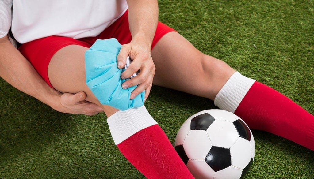
Ice: Begin by putting a bag of ice in your ankle for a minimum of 20 minutes at a time. Repeat this three to five times every day for three days. This�treatment helps decrease pain. Give yourself about 90 minutes between sessions.
Compression: Wrap your injured foot with an elastic bandage, such as an ACE bandage. Don’t wrap it too tightly to where your feet turn blue or your ankle becomes numb.
Elevation: Whenever possible, keep your ankle raised over heart level on a pile of pillows or another type of support arrangement to promote healing.
It’s possible to take over-the-counter drugs and/or medications, such as acetaminophen or ibuprofen, to relieve swelling and pain, however, these are often only offer temporary relief from the symptoms. Make sure to talk to a certified and qualified healthcare professional regarding any home treatment options to prevent further injury and symptoms.
If�you are suffering from foot pain or ankle pain, do not delay anymore. Chiropractors can help patients who suffer from foot, and ankle pain and they can help you, too. The scope of our information is limited to chiropractic as well as to spinal injuries and conditions. To discuss the subject matter, please feel free to ask Dr. Jimenez or contact us at�915-850-0900�.
Curated by Dr. Alex Jimenez
Additional Topics: Acute Back Pain
Back pain�is one of the most prevalent causes of disability and missed days at work worldwide. Back pain is attributed�to the second most common reason for doctor office visits, outnumbered only by upper-respiratory infections. Approximately 80 percent of the population will experience back pain at least once throughout their life. The spine is a complex structure made up of bones, joints, ligaments, and muscles, among other soft tissues. Because of this, injuries and/or aggravated conditions, such as�herniated discs, can eventually lead to symptoms of back pain. Sports injuries or automobile accident injuries are often the most frequent cause of back pain, however, sometimes the simplest of movements can have painful results. Fortunately, alternative treatment options, such as chiropractic care, can help ease back pain through the use of spinal adjustments and manual manipulations, ultimately improving pain relief.
It’s been great, my turf toe has been getting a lot better. Actually, I didn’t see a doctor for about 4 months and it just kept getting worse. But when I started seeing Dr. Jimenez, it just, little by little it’s been starting to get better. It feels a lot better when I practice and stuff like that. So, it’s getting better. – Vincent Garcia
There’s always a particular risk for athletes when it comes to sports-related injuries, or sports injuries, particularly when participating in physical activities. Moreover, contact sports, such as soccer, baseball, football and basketball, tend to have higher injury rates than any other sport.
Twice as many men suffer sports-related injuries in contrast with females as a consequence of the types of sports that they most commonly engage in. Whether you’re a seasoned and experienced athlete or you simply play as a weekend warrior, there’s always a chance of experiencing a sports injury. Below, we will discuss several of the most common types of sports injuries, or sports-related injuries.
Common Sports Injuries
Sprains and strains are the most common sports injuries. Sprains are medically defined as injuries to the ligaments, or the strong bands which connect bones to the joints. Overly stretching these ligaments beyond their natural range can ultimately damage or even tear them.
Strains are medically referred to as injuries to the muscle fibers or tendons, which connect muscles to bones. Strains are known as “pulled muscles” for a reason, overly stretching or overuse of a muscle can cause tears in the muscle fibers or tendons.
�Think of ligaments and muscle-tendon units like springs,� explained Dr. William Roberts, MD, sports medicine physician at the University of Minnesota and spokesman for the American College of Sports Medicine. �The tissue lengthens with stress and returns to its normal length, unless it is pulled too far out of its normal range.� Additionally, sports injuries can result in a variety of other health issues.
Patellofemoral Syndrome
Accidents in sports which can harm an athlete generally are inclined to be knee injuries. Patellofemoral syndrome could be caused by a slide or fall onto the knees. This type of sports injury involves swelling, inflammation and an imbalance of the knee at its groove. Strengthening exercises and stretching can help provide flexibility and mobility to the muscles. Apart from strengthening exercises and stretches, a doctor of chiropractic, or chiropractor, may utilize therapeutic techniques for this specific injury.
Concussion
A blow to the head could lead to a concussion. Concussions are a serious type of sports injury and these should never be disregarded. Symptoms indicating a possible head injury may include nausea, vomiting, confusion, headache, and slurred speech. Any athlete who incurs a concussion must seek immediate medical attention. Chiropractic care can help with several of the symptoms, such as headaches, related to a concussion.
ACL Tear
The anterior cruciate ligament, or the ACL, is a fundamental ligament found in the knee. An ACL tear can be caused due to a sudden change in directions or coming to a sudden stop when playing sports or during exercise and physical activities. There’s typically swelling, inflammation and uncertainty in movement working with an ACL tear. Chiropractic care can assist with the recovery process of an ACL tear, particularly through physical therapeutics and rehabilitation programs.
Hip Flexor Strain
The hip flexor muscles are all located in the upper front area of the thigh. Sprinting, running slopes and sudden movements could lead to a hip flexor strain. There can be pain and discomfort together with swelling and inflammation in the region surrounding the thigh. Stretching and range of motion exercises employing a doctor of chiropractic, or chiropractor, can help aid with recovery. A chiropractor will work closely with a patient to determine the best treatment approach for their sports injuries.
Shin Splints
With shin splints, there’s usually pain and other painful symptoms in the lower leg, particularly along the tibia. Shin splints are the most common type of sports injuries among runners or running athletes. Ice and cold therapy can help reduce swelling and inflammation on the site. Moreover, runners or running athletes can prevent suffering shin splints by purchasing a good pair of shoes with proper arch support. The right equipment can always promote a safe participation in sports and physical activities.
Sciatica
Sciatica is back pain which radiates down the back of the leg and into the foot. This collection of symptoms is often seen in cyclists and athletes who perform a lot of backwards turning and swinging sports like tennis and golf. Sciatica, or sciatic nerve pain, may be caused by a pinched or compressed nerve, frequently due to a bulging or herniated disc. Chiropractic care is a well-known, alternative treatment option which can help alleviate sciatica, or sciatic nerve pain, symptoms.
Shoulder Injury
Shoulder injuries in sports commonly range from dislocations and misalignments to strains and sprains of the shoulder tendons and ligaments. Because the shoulder is frequently referred to as a weak joint, it is often vulnerable to suffering harm from sports injuries during exercise and physical activities, aside from the athlete’s specific sport. Ice and cold therapy as well as chiropractic care and rehabilitation can help ease the symptoms associated with shoulder injuries.
Tennis or Golf Elbow
This issue is known as an overuse sports injury. Repetitive actions inflame the forearm and wrist. Ice and cold therapy as well as rest normally helps with the symptoms, but stretching and strengthening exercises recommended by a chiropractor can also help.
Groin Pull
Additionally known as a groin strain, the groin muscles can get strained with quick side-to-side movements when engaging in exercises and physical activities. Stretching and strengthening exercises can help with the recovery process in this case as well.
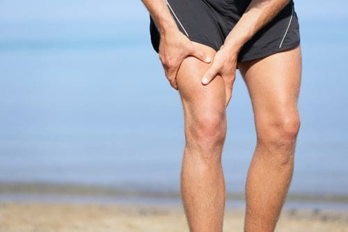
Hamstring Strain
The hamstring muscles can be found in the back of the thigh. When athletes fail to stretch or exercise accordingly before engaging in their specific sports, it can cause this muscle to be pulled. If the symptoms of this condition continue over a couple of weeks, a chiropractor, or doctor of chiropractic, can help provide the necessary treatment through the use of other treatment approaches, such as ultrasound, among others, to help encourage the natural healing of the muscle and improve symptoms.
Dr. Alex Jimenez’s Insight
Although many common sports injuries are often beyond our control, athletes can engage in stretches and exercises before participating in their specific physical activities to help prevent a sports injury. Every workout should start with a gentle warm-up to prevent most of these sports injuries. It’s important for athletes to be mindful of the amount of pressure they exert on their bodies in order for them to avoid suffering sports injuries.
If you’ve suffered a sports injury, make sure to seek immediate medical attention from a qualified and experienced healthcare professional. Many skilled sports medicine doctors are dedicated to sports medicine and also focus on providing rehabilitation determined by the performance requirements of athletes. Healthcare professionals will design a treatment plan targeted to your sports injuries.
Get back in the game with the guidance of qualified and experienced healthcare professionals in sports injuries. Contact us and make sure to schedule a consultation.�The scope of our information is limited to chiropractic as well as to spinal injuries and conditions. To discuss the subject matter, please feel free to ask Dr. Jimenez or contact us at�915-850-0900�.
Curated by Dr. Alex Jimenez
Additional Topics: Acute Back Pain
Back pain is one of the most prevalent causes for disability and missed days at work worldwide. As a matter of fact, back pain has been attributed as the second most common reason for doctor office visits, outnumbered only by upper-respiratory infections. Approximately 80 percent of the population will experience some type of back pain at least once throughout their life. The spine is a complex structure made up of bones, joints, ligaments and muscles, among other soft tissues. Because of this, injuries and/or aggravated conditions, such as herniated discs, can eventually lead to symptoms of back pain. Sports injuries or automobile accident injuries are often the most frequent cause of back pain, however, sometimes the simplest of movements can have painful results. Fortunately, alternative treatment options, such as chiropractic care, can help ease back pain through the use of spinal adjustments and manual manipulations, ultimately improving pain relief.
Running Shoes: Feet are important. By the time the typical American reaches the age of 50, they will have walked 75,000 miles.
Runners put even more miles on their feet, and stress. Your feet are your foundation. A problem with your feet can throw your entire body out of balance. That is why when it comes to running shoes, it is important to find the right type. This guide will help you find the running shoes that are right for you.
Running Shoes
Before You Shop
Know the type of runner you are.
Different types of running require different features in shoes.
Some questions to consider:
Do you run or jog?
What surface do you run on � asphalt, treadmill, or trails?
A larger person will not move and run the same way a thin, wiry person does. An overweight person will put more stress on their feet � and shoes.
Know your running style.
The way you run, the motion of your stride and how your foot strikes the ground has great bearing on the type of running shoe you need. When your foot comes in contact with the ground, what hits first? Does the inside of your forefoot hit first? The center of your heel? The outside of your heel? Where your foot first hits is where you really want the cushion.
Know what injuries you may have sustained from running.
Plantar fasciitis, shin splints, tendonitis, and blisters are a few common injuries can be reversed or improved when you wear running shoes that fit properly.
Know the type of arch you have.
Whether you supinate (foot rolls to the outside) or pronate (foot rolls to the inside) is determined, at least in part, by the shape of your arch. While supinators are rare, quite a few people over pronate. This can be the source of injuries due to overuse.
When You Shop
Give it the 360-degree test.
When people try on shoes they typically check for fit in the toe box, but look no further than that. When you try on running shoes, you do need to make sure you have adequate space in the toe box, but you also need to check that your entire foot fits on the shoe�s platform.
Give your foot enough space.
The upper should have enough room but should not be loose. It shouldn�t squeeze your foot either though. It should fit well with no pinching or binding.
Shop later in the day.
Throughout the day your feet swell. When you run they also swell so when you shop for shoes, going when your feet are the largest will help ensure that you get the most accurate and more comfortable fit possible.
Bring your old running shoes along when you shop.
Having your old shoes with you when you shop will help the sales person determine what kind of running shoe you need. They can look at the wear on the shoe to see your running patterns and help you find a shoe that works best for you.
Get your foot measured.
As you age your feet actually change; they can expand or flatten. Don�t every assume your shoe size, get your foot measured every time. A comfortable fit is dependent upon wearing the right size shoe. You also need to keep in mind that shoe sizes may differ from brand to brand.
Dress for the run.
When you are shopping for a new pair of running shoes, dress as you would when you run. Don�t show up wearing flip flops or when you are dressed for the office. Definitely don�t show up without socks.
Forget the latest trend or what�s fashionable; think functionality.
There are plenty of sharp looking shoes, but that doesn�t mean they are the right running shoe for you. Go for fit and functionality first and fashion second.
Take them for a test drive.
Once you have settled on a pair or two, try them both on and try them out. Many stores that specialize in running shoes have a treadmill or area where runners can try their shoes. That is the only way you can tell for shoe if the shoe is right for you.
IT Iliotibial band syndrome is a very common injury among runners. If it is diagnosed early and treatment commences immediately the chances of it becoming a chronic condition are reduced. It responds very well to chiropractic since it involves the pelvis and related muscles. When pelvic mechanics are not functioning properly the muscle don�t work efficiently which hinders flexibility and mobility. This can lead to tight muscles which may inhibit motion and cause pain. Chiropractic adjustments have been proven to help with the condition.
What Is The Iliotibial Band?
The Iliotibial Band, or fasciae latae, is the outer casing of muscle that extends along the outer thigh, from the top of the hip to the outside of the knee. IT Iliotibial band syndrome occurs when that casing becomes thickened. It is flexed or tight when you stand; it is what keeps your let straight, allowing the larger thigh muscle to rest.
There are two primary muscles that are involved in iliotibial band syndrome, the buttock muscle, or gluteus maximus, and the tensor fasciae latae muscles. Sometimes Iliotibial Band Syndrome is referred to Tensor Fasciae Latae Syndrome and the two terms can be used interchangeably.
IT Iliotibial Band Syndrome Defined
As the iliotibial band thickens it pulls in the area where it connects to the knee. This results in knee pain due to the application of too much pressure on the bursa. The bursa then becomes swollen, inflamed, and painful. During activity, such as running on an incline, the glutes are heavily involved.
The other end of the iliotibial band is inserted at the glutes so as the band tightens from this activity, it can trigger iliotibial band syndrome pain. Repeated activity further aggravates it, as does running on tight indoor tracks or uneven roads as well as having collapsed arches or running it inferior or worn out running shoes.
Symptoms Of Iliotibial Band Syndrome
There are several symptoms that can be used to diagnose iliotibial band syndrome. Lateral knee pain (pain on the outside of the knee) is a primary symptom and often used as a key diagnostic tool. Few conditions involve lateral knee pain. Other symptoms include:
Pain that worsens after running, particularly after running on an incline, climbing stairs, or climbing hills
There may not be any pain until you do something that aggravates it like climbing a hill.
The pain may not begin until you are mid-way through a run.
The pain can be intense and debilitating.
It can accompany a snapping hip, which occurs when the muscles that cross the outer hip may click or snap while running or walking.
The pain may be present along the lateral thigh without incorporating the knee, but it is only in very rare instances that it is concentrated on the gluteal or hip muscles.
Iliotibial band syndrome is often attributed to over training. This can mean suddenly increasing hill repeats or doubling your mileage.
Treatments For IT Iliotibial Band Syndrome
If your iliotibial band syndrome is caused by a problem with pelvic function, relieving the pain from the condition can be difficult. Stretching is not likely to bring relief � and if it does it won�t last long. If the pain from iliotibial band syndrome lasts for more than two weeks even if you are only stretching, your regular exercise routine, and ice and you don�t see much improvement, a chiropractor can help.
Even if the pain is located in the knee, the problem could originate in the pelvis. A chiropractor can assess your condition, check to see that your pelvis is functioning properly. If it isn�t, spinal adjustments and other chiropractic treatments can bring the body back into alignment and make the pelvis more functional.
Chiropractic Clinic Extra: Sport Injury Treatments
Feeling back pain, being unable to perform daily tasks, workout and play sports can be frustrating for anyone. The debilitating symptoms can drive individuals to seek fast relief. But, while a person�s only concern maybe only to fix the day�s pain, fixing the root/cause of the problem is far better in the long run and�can be easily achieved from chiropractic treatment. After receiving a single adjustment, many people especially athletes can expect an increase in their range of motion and less pain. Regardless of the reasons for seeking chiropractic treatment, one question always crosses people�s minds, how often should one see a chiropractor?
The answer to that question depends on the individual�s goals. Generally, spinal complications are not the result of a single day�s activities but tend to occur gradually over a period of time. Many spine conditions and injuries result in symptoms that may intermittently increase and decrease over several years, causing constant, nagging pain or sharp, extreme pain due to wear and tear type of injuries that the body is no longer able to heal on its own.
Chiropractic Treatment Sports Injury
Healing requires time and patience, a person also needs to be aware of what caused the complications in the first place. Suddenly stopping strict exercise routines or gaining weight in a certain amount of time can create an accelerated aging process on the joints.
If an individual�s goals are solely focused on alleviating the pain resulting from one time, then it won�t take much time to heal. Generally, receiving adjustments 2-3 times per week for several weeks can ease pain and decrease other symptoms. But, if a person is seeking to relieve the symptoms associated with an underlying condition or injury, or if a person is seeking to correct an improper posture or a mechanical dysfunction, the process could be much longer. This healing process often may require about 2-3 months of regular adjustments.
Despite completing treatment and successfully alleviating any symptoms, it is recommended to continue chiropractic adjustments on a regular basis. What is considered a regular basis for adjustments? Getting adjusted at least once a week by a chiropractor can help maintain a person�s overall health and can prevent small problems from becoming greater issues. For a greater majority of individuals, especially those who sit most of the day, it�s recommended to maintain an adjustment schedule every week or two. A chiropractor will explain what is the right schedule.
By Dr. Alex Jimenez
Chiropractic Clinic Extra: Sport Injury Treatments
Sports Injuries: Sandra Rubio has been working with Dr. Alex Jimenez for about 6 years. By caring for patients on a regular basis, Sandra has learned how essential and effective chiropractic care can be. Sandra describes how Dr. Alex Jimenez provides patients with a better way of healing themselves naturally, without the use of drugs/medications and surgery. The trust between Dr. Jimenez and the patient establishes a positive treatment outcome for many athletes with sports injuries as well as patients with other types of injuries and/or conditions through chiropractic care. Sandra Rubio expresses that Dr. Alex Jimenez is a safe non surgical choice for sports injuries.
Sports Injuries
Share Free Ebook
Sprains and strains are some of the most common type of sports injuries frequently reported by the average athlete. Sprains are injuries that affect the ligaments, tough bands which connect bones to the joints. Abrupt stretching of the ligaments beyond their natural range can deform or tear them. Strains are injuries that affect the muscle fibers or tendons, which function by anchoring muscles to bones. While most sports injuries are mild or moderate in nature, seeking immediate medical attention can help these heal faster in order for the athlete to be able to return-to-play quicker. A variety of treatment options, including chiropractic care can help treat sports injuries.
If you have enjoyed this video and/or we have helped you in any way please feel free to subscribe and share us.
Thank You & God Bless.
Dr. Alex Jimenez DC, C.C.S.T
There is no doubt that football is a rough sport. At times it can be downright brutal, especially on the body. The sport sees a myriad of injuries, some serious, including head and neck, ankle, knee, and spinal. Chiropractic care is quickly becoming a popular, viable method of not only treating injuries, but also for managing injury related pain and even injury prevention. There are several significant benefits that football players can glean from chiropractic care.
A survey exploring the prevalence of chiropractic care in the National Football League (NFL) showed that of the team trainers surveyed, 77 percent responded that they have referred players to a chiropractor for treat or evaluation. At that time, in 2002, 31 percent of the NFL teams have a team chiropractor on staff. Now all 32 of the NFL teams utilize chiropractic care for their players and staff.
College teams are also finding that chiropractic care can help their players. Virginia Tech has a chiropractor who routinely treats the players, helping them with injury recovery and physical health. The doctor has an office in the sports complex and the trainers are able to schedule appointments for the athletes.
The Benefits Of Chiropractic Treatment For Football Players
Improved Mobility
Chiropractic manipulative treatment (CMT), aka the chiropractic adjustment, is one of the primary chiropractic therapies in sports medicine. It helps to increase flexibility and minimize or eliminate pain in movement. CMT is often used by football players as an injury prevention measure. It has also been shown to improve athletic performance.
Solid Body Maintenance
The brutality of football leads to frequent injuries. However, soreness and stiffness are even more commonplace. Many players use chiropractic care to ease the general pain that comes from overexertion and rough play. They may not be injured, per se, but are experiencing the normal pain that comes with the territory when one is an athlete. Chiropractic care keeps the body working as it should at optimal athletic performance.
Pain Relief
Chiropractic care has long been recognized as an effective pain management tool and now athletes are discovering the benefits of chiropractic therapy as well. It has been shown to relieve headaches, particularly those stemming from head and neck injuries. It also relieves shoulder pain and pain from ankle and knee injuries such as sprains.
Injury Prevention
There is a direct correlation between athletes who regularly use chiropractic care and a decline in sports injuries. Those who use the treatment experience better flexibility and mobility which has been linked to injury prevention. Additionally, chiropractic care helps to increase strength and by making the body stronger it is more resistant to injury.
Increase Strength & Endurance
Chiropractic care has been linked with increased muscular strength. While it is often thought of as a way to relieve pain, it has been shown to effectively promote strength in the muscles after only a few sessions. This can help players avoid injury by combining the increase in strength with increased mobility. This also works to promote endurance and stamina.
Sports Hernia Relief
A significant amount of athletes experience groin pain due to injury. Athletic pubalgia is one of the most common causes of injury related groin pain. Also known as sports hernia, athletic pubalgia can cause significant pain. Chiropractic care has been shown to relieve the discomfort within 8 weeks when combined with rehabilitative exercises.
Chiropractic care plays a very important role in injury prevention, whole body care, and pain relief for athletes in one of the roughest sports in the world. These benefits can also be translated to other sports where injury is a frequent occurrence.
IFM's Find A Practitioner tool is the largest referral network in Functional Medicine, created to help patients locate Functional Medicine practitioners anywhere in the world. IFM Certified Practitioners are listed first in the search results, given their extensive education in Functional Medicine


 Symptoms Of Iliotibial Band Syndrome