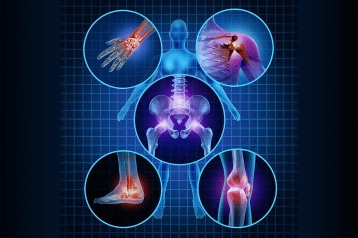
How Integrative Sports Chiropractic Speeds Injury Recovery at the Mechanical and Cellular Levels
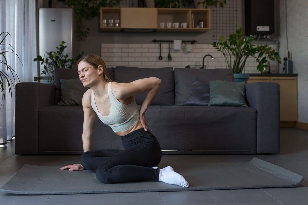
Athletes push their bodies hard. A sudden twist, a hard landing, or repeated stress can leave them with sprains, disc problems, tendon issues, or lingering pain. Traditional rest and pain pills often only quiet the symptoms. Integrative sports chiropractic takes a different path. It treats the injury at both the mechanical level—how bones, joints, and soft tissues line up and move—and the cellular level—how cells repair, reduce swelling, and rebuild tissue.
This approach combines spinal decompression, precise chiropractic adjustments, MLS laser therapy, shockwave therapy, and peptide support. Together, they create a non-invasive plan that moves athletes from simply managing pain toward faster, more complete tissue regeneration.
Why Mechanical and Cellular Care Matter Together
Injuries create two problems at once. Mechanically, a disc may bulge and press on a nerve, or joints may shift out of place. Soft tissues tighten or form scar tissue, limiting motion. At the cellular level, inflammation increases, blood flow decreases, and cells lack the energy and building blocks needed to heal effectively.
Treating only one side leaves the other unfinished. Aligning the spine without calming inflammation or feeding the cells often leads to slow progress or return of symptoms. Addressing cells without fixing alignment allows the mechanical problem to keep stressing the tissues. Integrative care solves both.
Spinal Decompression: Creating Space and Delivering Nutrients
Spinal decompression uses a specialized table that gently and controllably stretches the spine. This creates a mild negative pressure inside the disc. The pressure pulls bulging or herniated material away from nerves and draws nutrient-rich fluid back into the disc.
Discs have a limited blood supply, so they depend on this fluid exchange for oxygen and nutrients. When decompression restores that flow, the disc environment becomes more conducive to repair. Athletes with lower back or neck disc issues often experience reduced nerve pressure and improved mobility after a series of sessions. The treatment is comfortable and requires no downtime.
Chiropractic adjustments carefully restore normal position and motion to the spine and joints. When vertebrae sit correctly, nerve pathways stay open, and muscles no longer work overtime to compensate. Adjustments also help maintain the space created by decompression so the disc does not quickly return to its previous stressed state.
For sports injuries, proper alignment improves how force travels through the body during running, jumping, or throwing. This reduces secondary strain on tendons and ligaments. The combination of decompression and adjustments addresses the mechanical foundation of the injury.
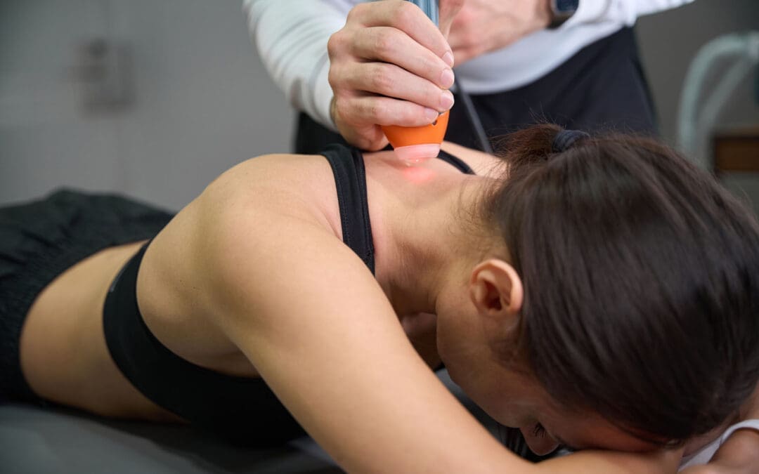
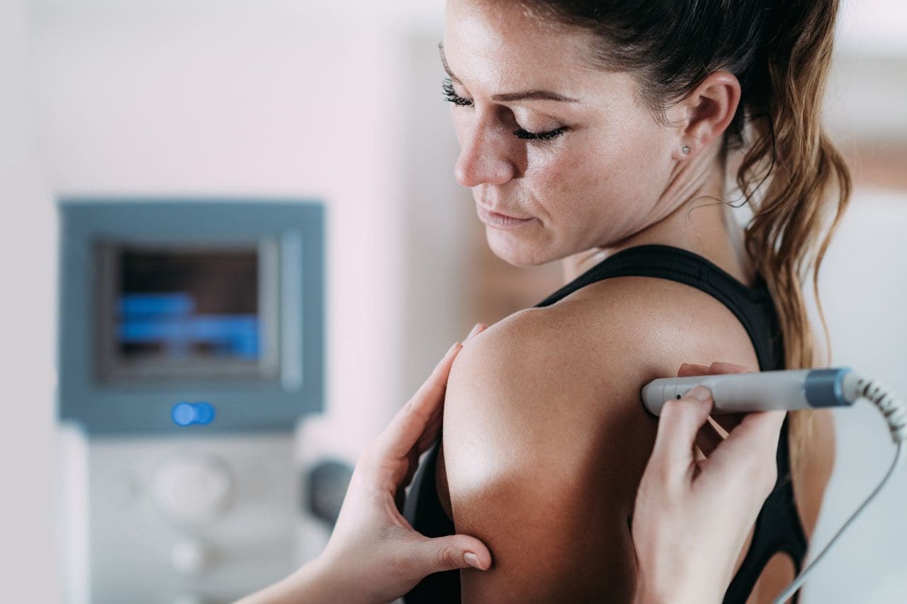
MLS Laser Therapy: Energizing Cells to Heal
MLS laser therapy delivers specific, synchronized wavelengths of light deep into tissue. One wavelength helps calm inflammation. The other supports pain relief and cellular activity. The light energy reaches the mitochondria inside cells and boosts ATP production, the main energy source cells use for repair.
Higher ATP levels allow cells to work more efficiently. Swelling decreases, blood flow improves, and collagen production rises. Sessions are short and painless. Athletes often notice reduced soreness and faster return of function when laser is added to mechanical care. Research and clinical use show that combining laser with chiropractic shortens recovery time compared with either method alone.
Shockwave Therapy: Breaking Scar Tissue and Restarting Healing
Shockwave therapy sends focused acoustic waves into injured soft tissue. These waves increase local blood flow, break down dense scar tissue and calcifications, and release growth factors that restart stalled healing.
Chronic tendon problems—such as tennis elbow, Achilles issues, or rotator-cuff irritation—respond especially well. The waves create a controlled micro-stimulus that tells the body to rebuild stronger tissue rather than leave restrictive scar behind. When used with adjustments, shockwave improves the way muscles and tendons glide over newly aligned joints.
Certain peptides act as signaling molecules that encourage the body’s own repair systems. Peptides such as BPC-157 and TB-500 have been studied for their ability to support blood-vessel growth, modulate inflammation, and promote organized collagen deposition in tendons, ligaments, and muscle.
In the setting of disc or joint injury, these signals can help create a more favorable environment for tissue recovery. They work alongside the mechanical and energy-based therapies rather than replacing them. Clinical protocols often include peptides as part of a broader regenerative plan under medical oversight. Evidence is strongest in preclinical models and growing in human musculoskeletal use, with careful patient selection and monitoring.
How the Full Combination Accelerates Recovery
A typical integrated plan might begin with evaluation of posture, joint motion, nerve function, and soft-tissue quality. Spinal decompression and adjustments restore mechanical balance. Laser and shockwave then address inflammation and scar tissue at the cellular and tissue levels. Peptides supply additional regenerative signals.
The result is more than pain relief. Blood flow improves, cells receive energy and nutrients, restrictive scars soften, and new tissue forms under better biomechanical conditions. Athletes often progress faster through rehabilitation exercises because the underlying tissues are better prepared to handle load.
Key advantages include:
Non-invasive nature with little or no downtime
Simultaneous treatment of structure and cellular function
Reduced need for long-term medication or invasive procedures
Support for both acute sports injuries and chronic overuse problems
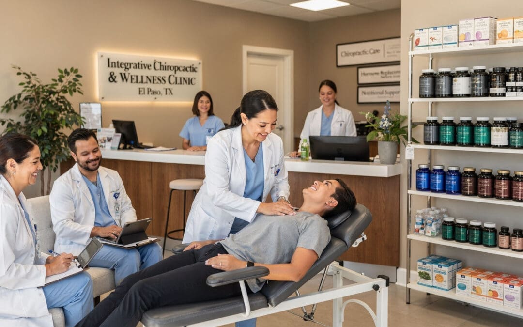
Multidisciplinary Care at Injury Medical Clinic PA in El Paso
In El Paso, Texas, Injury Medical Clinic PA brings these therapies together under one coordinated team. Dr. Alexander Jimenez, DC, APRN, FNP-BC, CCST, CFMP, IFMCP, ATN, provides chiropractic and functional medicine care focused on sports injuries, personal injury recovery, and whole-person restoration. His clinical observations, detailed on dralexjimenez.com and his professional profile, emphasize identifying root mechanical and metabolic contributors to pain rather than masking symptoms. He notes that many athletes and active patients improve when alignment, soft-tissue health, nutrition, and cellular support are addressed together.
Medical direction and collaborative oversight are provided by Dr. Maria Guadalupe Cardenas, MD. She is board-certified in internal medicine (NPI #1164426749, Texas MD License #J2933) and brings more than 40 years of experience as an internist. As Medical Director and Collaborative Physician, Dr. Cardenas works alongside Dr. Jimenez to ensure medical safety, appropriate diagnostic correlation, and integrated planning. This MD–DC partnership is common in modern integrative and injury-focused clinics. It allows seamless blending of chiropractic adjustments, spinal decompression, laser and shockwave therapies, functional-medicine strategies, personal-injury documentation, and regenerative options such as peptides—all under physician collaboration.
The clinic’s multidisciplinary model also incorporates rehabilitation exercises, nutritional support, and careful monitoring so that mechanical corrections and cellular therapies reinforce one another. Patients receive coordinated care that respects both the structural demands of sport and the biological processes of healing.
Moving from Symptom Control to Active Tissue Regeneration
Symptom management keeps pain low enough for daily life. Active tissue regeneration aims higher: it restores the quality and resilience of injured structures, enabling athletes to return to training with confidence. Spinal decompression and adjustments handle the mechanical framework. MLS laser supplies cellular energy. Shockwave clears barriers of scar tissue. Peptides amplify the body’s repair signals. When these tools are used in sequence under experienced clinical guidance, recovery becomes more efficient and complete.
Athletes who once faced months of limited activity often regain function sooner and with less residual restriction. The same principles apply to active people of all ages who want durable results without surgery.
Integrative sports chiropractic offers a clear path: fix the structure, feed the cells, clear the obstacles, and support the body’s own regenerative capacity. The combination delivers a practical, non-invasive route back to performance from injury.
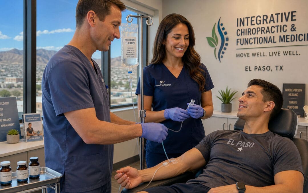
IV Infusion Therapy Benefits for Athletes: Faster Recovery After Tough Workouts and Events
After a long race, intense game, or heavy training week, your body can feel completely drained. You might feel exhausted, sore, thirsty, and slow to bounce back. Drinking water and eating nourishing food help a lot, but sometimes your stomach feels upset, or you need faster help to restore fluids and nutrients to your system. That is where IV infusion therapy can step in as a helpful tool.
IV infusion therapy puts fluids, electrolytes, vitamins, and other nutrients straight into your bloodstream through a small needle in your arm. This method provides your body with nearly 100 percent absorption because it bypasses the digestive system entirely. In sports, it serves as a targeted way to fix real problems like low fluid levels or nutrient shortages after intense effort. It is not a magic shortcut for healthy athletes who can eat and drink normally. Instead, it acts as a clinical support when your body is depleted and needs quick replenishment to recover and prepare for the next challenge.
Many athletes use this approach to feel better faster so they can return to training or competition with more energy and less downtime.
What IV Therapy Actually Does for Athletes
IV therapy delivers a mixture of saline or similar fluids, along with vitamins and minerals, directly into your bloodstream. This helps replace what you lose from heavy sweating, hard breathing, and muscle work. The process usually takes 30 to 60 minutes while you rest comfortably.
The main goals include restoring fluid balance, easing muscle fatigue, supporting energy production inside your cells, and calming inflammation that builds up during tough sessions. When done properly under medical guidance, it can shorten the time you feel wiped out after big efforts.
Rapid Rehydration When Oral Fluids Are Not Enough
During long endurance events or intense training camps, you can lose a large amount of water and important salts, such as sodium and potassium, through sweat. This drops your blood volume and can leave you feeling weak or dizzy. If you also have stomach upset or nausea, drinking large amounts of fluid becomes hard or even impossible.
IV therapy solves this by sending fluids and electrolytes straight into your circulation. Your body absorbs them right away instead of waiting for your gut to process them. This method works especially well when high-intensity exercise has already pulled blood away from your stomach to your working muscles, slowing normal digestion. Athletes often notice they feel rehydrated and more stable much quicker than with sports drinks alone.
Bypassing Digestion for Better Nutrient Delivery
Your digestive system sometimes struggles after very hard workouts. Blood flow shifts to your muscles, and gut movement can slow down. Oral supplements or drinks may not absorb well in these moments.
IV infusions avoid that problem completely. The nutrients go directly into your blood and reach your cells fast. This means depleted muscles and organs get what they need without delay. The result is faster support for repair and energy restoration than waiting for your stomach to do the work.
Reducing Inflammation and Muscle Soreness
Hard exercise causes minor damage to muscle fibers and produces additional free radicals that induce oxidative stress. This leads to delayed-onset muscle soreness (DOMS), which can make the next day or two feel stiff and painful.
Certain ingredients in athletic IV drips help fight this. Amino acids such as glutamine and arginine support muscle repair and calm inflammation. Antioxidants like vitamin C and glutathione help clear waste products and protect cells from extra stress. Many athletes report less lingering soreness and faster return to comfortable movement when these supports are added at the right time.
Supporting Cellular Energy and Recovery
Inside your cells are tiny structures called mitochondria that turn nutrients into usable energy. After intense training, these powerhouses can become stressed or less efficient. IV formulas often include magnesium, B-complex vitamins, vitamin B12, and NAD+ to give them direct support.
Magnesium helps muscles relax and prevents cramps while keeping your heart rhythm steady. B vitamins assist in turning food into energy at the cellular level. NAD+ aids in repairing small cell damage and keeping energy production running smoothly. Together, these nutrients help your body handle the repair work from training sessions more effectively.
Common Nutrients in Athletic IV Fluids and Their Roles
Here are some of the key ingredients often used and why they matter for active people:
Magnesium: Helps tight muscles relax, reduces cramp risk, and supports steady heart rhythm during and after exercise.
B-Complex Vitamins and B12: Aid everyday cell metabolism and energy creation so you feel less drained.
Amino Acids (such as Glutamine): Encourage protein building in muscles and help repair the small tears that come from hard training.
Vitamin C and Zinc: Act as antioxidants to fight free radicals created during workouts and support your immune system when training stress is high.
NAD+: Supports cell repair, DNA maintenance, and efficient energy production inside the mitochondria.
These are chosen based on what your body typically loses or uses up during demanding activity.
Important Anti-Doping Rules Every Competitive Athlete Must Know
If you compete at a level where drug testing happens, you need to understand the rules set by the World Anti-Doping Agency (WADA) and the U.S. Anti-Doping Agency (USADA). IV infusions or injections that total more than 100 milliliters in any 12-hour period are prohibited both in and out of competition. This limit applies even if the fluid contains only permitted substances, such as vitamins or saline.
Exceptions exist mainly for true medical needs:
Treatment inside a hospital or during emergency transport to a hospital.
Care given as part of surgery or certain diagnostic tests.
Urgent medical situations handled in a hospital-linked urgent care setting.
Three main reasons explain the restriction:
Large fluid volumes can temporarily increase blood plasma levels, which may improve heart and circulation performance for a short time.
IVs can sometimes interfere with how labs detect other banned substances in urine samples.
Quick changes in blood volume and values can affect the Athlete Biological Passport system that tracks an athlete’s blood markers over time.
Most everyday recovery IVs given in wellness clinics, hotel rooms, or non-hospital settings fall under the prohibited category if they exceed the volume limit. Always check with your sport’s governing body or a knowledgeable medical professional before considering any IV treatment if you are a tested athlete. In true emergencies, get medical care first and handle paperwork afterward.
IV Therapy Works Best as Part of a Bigger Recovery Plan
IV infusion therapy gives fast support when your body is low on fluids or nutrients. However, it works best alongside the basics: consistent quality sleep, proper daily fueling with whole foods, steady oral hydration, and smart training loads. Experts note that in most situations, drinking fluids and eating balanced meals remain the preferred and sufficient methods. IV therapy shines as an extra option during extreme events, multi-day competitions, or when stomach issues block normal intake.
Integrative Care That Supports Athletes in El Paso, Texas
Athletes looking for well-rounded support often benefit from clinics that combine different types of care under one roof. In El Paso, Texas, Injury Medical Clinic PA offers this kind of integrated approach. Dr. Alexander Jimenez, DC, APRN, FNP-BC, CFMP, IFMCP, ATN, CCST, brings extensive experience in chiropractic and functional medicine, helping people recover from injuries and improve performance. He works closely with Dr. Maria Guadalupe Cardenas, MD, a board-certified internal medicine physician with more than 40 years of experience. She serves as Medical Director and Collaborative Physician, providing medical oversight for the team.
This setup allows chiropractic care for spine alignment, nervous system health, and mobility to work together with medical direction for therapies that may include IV infusions when appropriate. The clinic also emphasizes functional medicine to address root causes of fatigue or slow recovery, personal injury care, and structured rehabilitation programs. Clinical observations from Dr. Jimenez highlight that athletes recover better when care addresses the whole person—alignment, inflammation levels, nutrient delivery, and nervous system balance—rather than isolated symptoms. When IV therapy fits into a personalized plan, having an experienced internal medicine physician’s oversight helps ensure safety and proper use in accordance with the rules.
Many patients appreciate this team model because it combines hands-on therapies with advanced supportive options in a single coordinated setting.
Final Thoughts on Using IV Therapy Wisely
IV infusion therapy can help athletes rehydrate quickly, deliver key nutrients fast, ease inflammation, and support cellular energy after demanding efforts. IV therapy serves as a useful clinical tool when your body is truly depleted and oral methods fall short. At the same time, it is not a replacement for daily healthy habits or a way around anti-doping regulations.
If you train hard and sometimes struggle with recovery, speak with a qualified healthcare provider who understands the demands of sports and local regulations. They can help decide whether this option makes sense for your specific situation and guide you safely. When used thoughtfully as part of a complete plan, IV therapy can help you get back to feeling and performing at your best.
IV Infusion Therapy: How It Delivers Vitamins and Nutrients Straight to Your Body
IV infusion therapy puts vitamins, minerals, and fluids directly into your bloodstream. This bypasses the digestive tract, so your body can use more of the nutrients more quickly and fully. Clinics often use it to support immune function, fix dehydration, ease chronic fatigue, and correct nutritional shortfalls that oral supplements sometimes cannot fix well.
Many people feel run down, foggy, or slow to recover because their gut does not absorb everything from food or pills. IV therapy changes that by sending the mixture straight into circulation through a small tube placed in the arm. The result is higher amounts of nutrients reaching your cells faster than you can usually get from eating or swallowing capsules.
How Intravenous Therapy Works
Intravenous (IV) therapy uses a sterile mix of vitamins, minerals, and amino acids. A trained professional inserts a thin catheter into a vein, usually in the arm or hand. The liquid then drips in over 30 to 60 minutes while you rest in a comfortable chair.
Because it bypasses the stomach and intestines, the body absorbs nearly 100 percent of the nutrients. Oral supplements often lose a large portion during digestion. IV delivery avoids that loss and gives a rapid boost when someone needs quick rehydration or higher nutrient levels.
Why People Choose IV Infusion Therapy
Clinics report several common reasons patients try this therapy. Here are the main ones explained simply:
Fast hydration and electrolyte balance — After illness, intense workouts, travel, or long days, fluids and minerals go straight in to restore balance quickly.
More steady energy — B vitamins, magnesium, and other nutrients help cells produce energy. Many people notice less afternoon drag and better focus.
Immune support — High amounts of vitamin C, zinc, and antioxidants can give the body’s defense system extra help during cold and flu season or times of stress.
Recovery from physical stress — Athletes, active workers, and people healing from injuries often use it to supply building blocks for tissue repair and to reduce downtime.
Filling nutrition gaps — When digestion is off due to stress, medications, or long-term conditions, IV can deliver what the gut is missing.
These effects happen because the nutrients reach cells directly. Still, results vary from person to person. What works well for one individual may feel different for another.
IV Therapy Inside an Integrative Care Team
When an integrative chiropractic and functional medicine clinic offers IV therapy, patients gain extra layers of support. The approach focuses on three important ideas: personalized and data-driven treatment, a comprehensive care team, and a root-cause focus.
The team reviews lab work, health history, symptoms, and lifestyle before recommending a formula. They do not use a one-size-fits-all drip. Instead, they match the mix to what the person actually needs. This data-driven step helps avoid unnecessary or poorly matched nutrients.
A full care team means different experts work together. Chiropractic care addresses spinal alignment and nerve function. Functional medicine explores gut health, inflammation, and lifestyle factors. Medical oversight adds safety checks and the ability to handle more complex health pictures. Rehabilitation and personal injury support fit in when someone is recovering from accidents or ongoing pain.
It is crucial to consult a qualified healthcare professional to ensure the treatment aligns with your unique health profile and objectives, as individual needs and responses to IV therapies can vary.
How One El Paso Clinic Combines These Services
At Injury Medical Clinic PA in El Paso, Texas, this team model is in action every day. Dr. Alexander Jimenez, DC, APRN, FNP-BC, CFMP, IFMCP, ATN, CCST, brings decades of experience in chiropractic care and advanced functional and integrative approaches. He works closely with Dr. Maria Guadalupe Cardenas, MD, a board-certified internist (NPI #1164426749, Texas MD License #J2933) with more than 40 years of experience.
Dr. Cardenas serves as Medical Director and Collaborative Physician. Her role provides medical direction and oversight for procedures such as IV infusions. This partnership is common in integrative or injury-focused clinics: the chiropractor handles structural and nervous system care, while the medical doctor ensures the safe, appropriate use of advanced therapies.
Patients receive coordinated care. Someone coming in after a car accident might receive chiropractic adjustments for whiplash, rehabilitation exercises, and, when appropriate, IV nutrients to support healing and energy. The medical oversight helps the team monitor interactions, select safe doses, and track lab results when needed. Dr. Jimenez has observed in his clinical work that patients with lingering fatigue, slow recovery, or chronic discomfort after injuries often respond better when nutrition and hydration are optimized alongside hands-on treatments.
This multidisciplinary setup allows the clinic to address the whole person rather than isolated symptoms. Chiropractic improves movement and nerve signaling. Functional medicine targets underlying drivers like inflammation or gut issues. IV therapy provides rapid nutritional support when oral intake is insufficient. Personal injury and rehabilitation services tie everything together, helping patients return to daily life with less pain and greater function.
What a Typical Session Looks Like
Most visits follow a clear, comfortable flow:
You meet with a provider to review your health history, current symptoms, and any recent labs.
The team selects or customizes a nutrient formula based on your goals.
A small catheter is placed in your arm (most people feel only a quick pinch).
You relax for 30–60 minutes while the solution drips in. Many people read, listen to music, or nap.
The catheter is removed, and you receive simple aftercare instructions, such as drinking extra water and resting as needed.
The whole process is designed to be low-stress. Clinics with proper medical oversight keep emergency supplies and trained staff on hand.
Safety and Smart Choices
IV therapy is generally well tolerated when performed by licensed professionals in a clinical setting. Mild side effects can include temporary bruising or soreness at the insertion site. More serious risks, such as infection or nutrient overload, are rare but possible, which is why medical supervision matters.
Experts note that while many people report feeling better, high-quality studies on broad wellness benefits for otherwise healthy individuals are still limited. IV therapy works best as one tool inside a larger plan that includes good nutrition, movement, sleep, and treatment of any underlying conditions. It is not a replacement for a healthy lifestyle or prescribed medical care.
People with certain conditions (kidney disease, heart issues, or specific medication regimens) should always check with their doctor first. In a clinic like the one described, the collaborative MD-NP team helps screen for these factors before any drip begins.
Putting It All Together
IV infusion therapy gives your body a direct route for vitamins, minerals, and fluids when you need fast, high-level support. By skipping digestion, it delivers higher usable amounts in less time. In an integrative setting that includes chiropractic care, functional medicine, rehabilitation, and strong medical oversight, it becomes part of a broader strategy aimed at addressing root causes and achieving lasting improvement.
Whether you are dealing with everyday fatigue, recovering from physical stress, or simply want to optimize how you feel, the key is to work with qualified professionals who personalize their approach. Clinics that combine these services under proper medical direction, such as the team model in El Paso, demonstrate how different therapies can support one another for better overall results.
Talk with your healthcare provider to see if IV infusion therapy fits your health picture. When used thoughtfully, it can be a helpful step on the path to feeling stronger, recovering faster, and supporting your body’s natural ability to heal and perform.
Regenerative Therapies Combined with Chiropractic Care Offer New Hope for Sports and Auto Accident Injuries in El Paso
Many people in El Paso deal with ongoing pain and limited movement after sports injuries or car accidents. Simple rest or basic physical therapy often helps at first, but sometimes healing stalls. Tissues stay inflamed, joints feel stiff, and daily life or sports become difficult again. When that happens, more people look for advanced options that work with the body instead of just covering up symptoms.
Regenerative therapies and integrative chiropractic care team up to tackle these tough problems. They focus on real repair at the tissue level while also fixing how the body moves. This combined approach helps many patients get back to feeling better and moving easier without jumping straight to surgery.
Why Standard Treatments Sometimes Fall Short
Injuries from sports collisions or car crashes often damage more than one area. Muscles tear, ligaments stretch, tendons become inflamed, and spinal discs or joints become irritated. Swelling and scar tissue can block normal blood flow and healing signals.
Physical therapy and rest build strength and reduce pain for many people. Yet when progress plateaus, underlying tissue damage or poor joint alignment may still be holding back recovery. That is when patients often seek care that actively supports the body’s repair systems instead of only managing symptoms.
What Regenerative Therapies Actually Do
Regenerative medicine uses materials from your body to kick-start healing. These treatments deliver growth factors and helpful cells directly to the damaged area. The goal is to lower inflammation, encourage new tissue growth, and improve long-term function.
Three main options stand out for musculoskeletal and spinal injuries:
PRP (platelet-rich plasma) comes from a small sample of your blood. The blood is spun in a machine to concentrate platelets, which carry natural growth factors. Doctors inject this concentrated solution into tendons, ligaments, joints, or around nerves. The growth factors signal cells to repair and rebuild.
PFP (platelet-fibrin products) uses protein concentrates from your blood. These capture growth factors and create a stronger, longer-lasting healing signal for tissues that have not responded well to simpler treatments.
MFAT (microfragmented adipose tissue) takes a small amount of your own fat tissue, processes it into tiny fragments, and injects it. The fat contains supportive cells and signaling factors that cushion joints and help repair cartilage, tendons, and soft tissues.
These are called orthobiologics because they come from your biology. They carry a low risk of allergic reactions or rejection since they use your materials.
Epidural injections sometimes join the plan for spine-related pain and nerve irritation. Under careful medical guidance, they reduce inflammation around spinal nerves while the regenerative injections work to repair deeper tissue.
How Chiropractic Care Completes the Picture
Injections alone help tissues heal, but they do not fix how the bones, joints, and muscles line up or move. That is where chiropractic adjustments come in. Gentle, precise realignments improve joint mobility, ease muscle tension, and restore better posture and movement patterns.
When regenerative injections and chiropractic care happen together, the results often last longer. The injections create a better healing environment inside the tissues. The adjustments keep the joints moving correctly so that new tissue forms properly and does not get stressed again. This partnership addresses both the biology of repair and the mechanics of the body.
The Strength of a True Multidisciplinary Team
Patients get the best results when they receive care from a well-established integrative and functional medicine clinic that brings different experts together under one roof. At Injury Medical Clinic PA in El Paso, Texas, the team combines advanced regenerative procedures with chiropractic expertise, functional medicine, rehabilitation, and personal injury support.
Dr. Alexander Jimenez, DC, APRN, FNP-BC, CFMP, IFMCP, ATN, CCST, leads the clinical approach. With decades of experience as a chiropractor and additional training as a board-certified family nurse practitioner, he focuses on whole-person recovery. His clinical observations show that patients with sports trauma or old auto accident injuries often improve when care targets both tissue repair and nervous system function. He uses detailed exams, imaging, and personalized plans that include regenerative injections, adjustments, rehabilitation, and lifestyle support.
Dr. Maria Guadalupe Cardenas, MD, a board-certified internal medicine physician with over 40 years of experience (NPI #1164426749, Texas MD License #J2933), serves as Medical Director and Collaborative Physician. She provides medical oversight for procedures, ensures safety and compliance, manages complex health factors, and brings an internal medicine perspective to every case. This collaboration means patients receive both expert spinal and musculoskeletal care from Dr. Jimenez and broad medical direction from Dr. Cardenas.
This setup is common in high-quality integrative injury clinics. The MD handles medical aspects and procedure oversight while the chiropractor and nurse practitioner team deliver hands-on treatment and functional strategies. Everyone works from the same records and goals, so care stays coordinated and thorough.
Clear Benefits Patients Notice
People who choose this combined path often report several practical improvements:
Noticeable drops in pain and swelling without relying only on medications
Better tissue repair that supports longer-lasting results
Improved joint movement and daily function
Faster return to work, sports, or normal activities when healing had stalled
Lower chance of needing more invasive procedures later
Thorough documentation that helps with insurance and legal needs after personal injury cases
Because the treatments use your own biological materials, side effects stay minimal for most people. Soreness at the injection site usually fades within a few days.
The functional medicine side of care looks at nutrition, inflammation levels, sleep, and stress. These factors influence how well tissues heal. Addressing them alongside the injections and adjustments gives the body every advantage.
What a Typical Care Journey Looks Like
Most patients start with a full evaluation that includes history, physical exam, and any needed imaging. The team identifies exactly which tissues need help and whether alignment issues are slowing progress.
Next comes a customized plan. This may include one or more regenerative injections (PRP, PFP, or MFAT), chiropractic adjustments over several weeks, guided rehabilitation exercises, and supportive therapies such as shockwave treatment when appropriate. Follow-up visits track progress and adjust the plan as tissues respond.
Many people begin to feel meaningful relief within weeks, with continued improvement over the next few months as repair progresses. The team stays involved through the entire process.
Who Benefits Most from This Approach
This type of care often helps adults dealing with:
Lingering pain after sports collisions or overuse injuries
Whiplash, back strain, or nerve irritation from car accidents
Old injuries that never fully settled
Joint or tendon problems that limit activity
It works especially well when conventional treatments have already been tried, and progress has slowed. The focus stays on restoring real function rather than temporary relief.
Moving Forward with Confidence
Healing from serious injuries takes time and the right tools. Regenerative therapies give tissues the biological signals they need. Integrative chiropractic care helps the body use those new repairs by improving movement and alignment. When both occur within a coordinated team that includes medical direction, functional medicine, and personal injury expertise, patients often regain greater comfort and capability than they expected.
If you or someone you know in the El Paso area continues to struggle after sports trauma or an auto accident, consider learning more about these combined options. A thorough evaluation at a clinic experienced in both regenerative procedures and chiropractic care can show whether this path fits your situation. Many people find it opens the door to meaningful, lasting improvement.
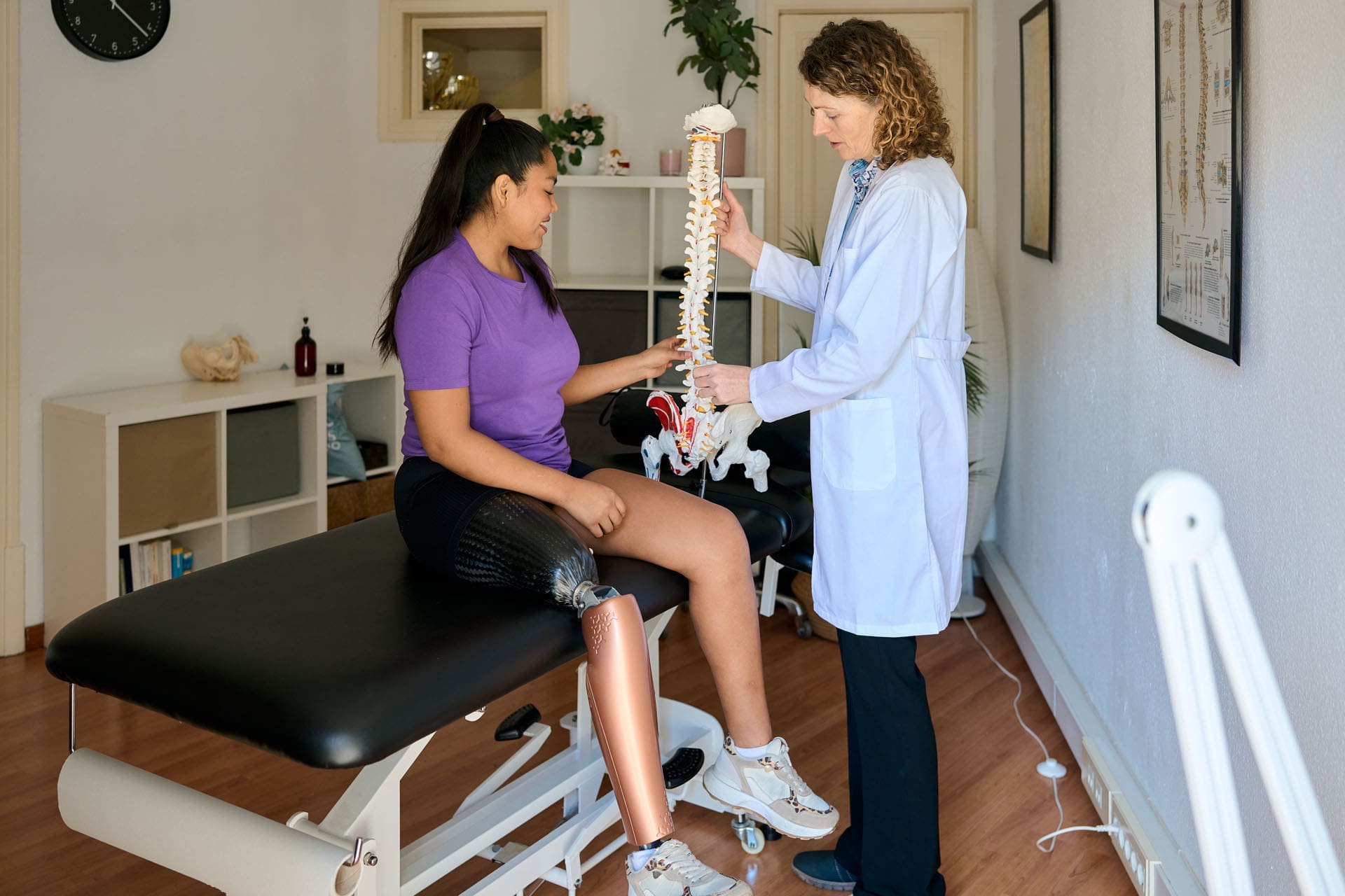
Joint Pain Relief Through Regenerative Chiropractic
Abstract
In this educational post, I, Dr. Alexander Jimenez, DC, APRN, FNP-BC, CFMP, IFMCP, ATN, CCST, guide you through a practical, evidence-based approach to shoulder and knee care using integrative chiropractic methods, functional rehabilitation, ultrasound-guided procedures, and regenerative strategies. You will learn how we identify pain generators and biomechanical contributors, why we select specific manual therapies and corrective exercises, and how we safely use ultrasound to guide injections into targeted tissues. I also introduce our multidisciplinary team, led medically by Dr. Maria Guadalupe Cardenas, MD (Board Certified in Internal Medicine) (NPI #1164426749, Texas MD License #J2933), who serves as Medical Director and Collaborative Physician at Injury Medical Clinic PA (Mission Plaza Injury Medical Clinic) in El Paso, Texas. We show how chiropractic care, internal medicine oversight, functional medicine, personal injury care, rehab, and physical therapy combine to restore function and reduce pain, while keeping hormones and medications in the background for elpasobackclinic.com’s audience. Finally, I translate complex anatomy and physiology into clear, actionable steps and provide citations with linked references so you can explore the research behind each decision.
Chiropractic And Internal Medicine Collaboration In El Paso, Texas
At Injury Medical Clinic PA (Mission Plaza Injury Medical Clinic) in El Paso, Texas, our multidisciplinary model is designed for precision diagnostics, safe care, and sustainable outcomes.
Medical direction: Dr. Maria Guadalupe Cardenas, MD (Internal Medicine), brings over 40 years of clinical experience, ensuring medical safety, bi-directional care coordination, and evidence-based protocols across complex cases.
Chiropractic integration: I lead integrative chiropractic care, combining spinal biomechanics, regional joint assessment, soft-tissue methods, and functional rehabilitation targeted to the patient’s presentation.
Functional medicine lens: We prioritize nutrition, sleep, stress physiology, and metabolic health as supportive pillars for tissue healing, while minimizing reliance on hormones or medications unless medically indicated.
Physical therapy emphasis: Coordinated mobility, stability, motor control, and return-to-function plans are sequenced with chiropractic adjustments and soft-tissue care, including sports-specific and work-injury progressions.
Personal injury workflows: For PI cases, we document thoroughly, use validated outcome measures, and align care with imaging, guided procedures, and gradual load progressions to restore confidence and capacity.
Why This Integrative Model Matters
Safety first: Internal medicine oversight reduces procedural risk and guides comorbidity management.
Precision: Ultrasound-guided interventions and biomechanical assessments target the right tissue at the right dose.
Durability: Chiropractic care, physical therapy, and functional medicine together produce longer-lasting outcomes by addressing root causes.
Patient-centered: We build stepwise care pathways, educate patients, and align expectations to reduce fear and improve adherence.
Shoulder Pain: Anatomy, Biomechanics, And Why It Hurts
The shoulder is a dynamic, multi-planar joint system in which the glenohumeral joint, acromioclavicular (AC) joint, scapulothoracic articulation, and sternoclavicular joint must synchronize to ensure smooth function. The rotator cuff—supraspinatus, infraspinatus, teres minor, and subscapularis—stabilizes the humeral head to prevent excessive superior or anterior translation during elevation.
Key physiology driving pain:
Tendinopathy: Repetitive load and poor scapular control foster collagen disorganization, neovascularization, and nociceptive sensitization within cuff tendons, especially the supraspinatus footprint on the greater tuberosity.
Subacromial space mechanics: Limited thoracic extension or scapular upward rotation narrows the subacromial space, increasing bursal and tendinous stress.
AC joint degeneration: Microinstability and load transfer through the clavicle result in capsular irritation, osteophytes, and localized pain with cross-body movements.
Biceps-labral interface: The long head of the biceps traverses the bicipital groove and contributes to anterior shoulder pain when overloaded or in SLAP variants.
Neurovascular proximity: The neurovascular bundle in the anterior shoulder region requires meticulous mapping during procedures to avoid iatrogenic injury.
What I Look For During A Real Patient Encounter
Drawing from my clinical experience:
Visual and palpatory cues: I watch for asymmetry, protective guarding, and painful arcs. Palpation maps tenderness over the supraspinatus footprint, AC joint, subscapularis, and bicipital groove.
Functional patterns: I analyze bird-dog, superman, and scapular setting drills to identify deficits in anti-extension control and rotator cuff endurance. These tests help me see how trunk stability informs shoulder mechanics.
Ultrasound landmarks: I trace the humeral head, articular cartilage, supraspinatus footprint, subacromial bursa, AC joint, and biceps tendon sheath, maintaining a safe distance from neurovascular structures.
Load tolerance: I progress from low-load tasks to higher-load regions (e.g., triceps or deep cuff work), carefully managing patient expectations and discomfort.
Integrative Chiropractic Approach To Shoulder Care
Our shoulder pathway prioritizes chiropractic and physical therapy methods:
Thoracic mobility and rib mechanics
Why: Thoracic extension and rib mobility enable scapular upward rotation and posterior tilt, reducing impingement risk.
Methods: Thoracic spine manipulation and mobilization to improve segmental motion; breathing retraining for costovertebral rhythm.
Evidence: Manual therapy to the cervical-thoracic junction can reduce shoulder pain and improve function through regional interdependence (Domenech-Garcia et al., 2011).
Scapular motor control
Why: Proper serratus anterior and lower trapezius activation improves humeral head centering, decreasing superior migration under load.
Methods: Wall slides with lift-off, prone Y/T/W, serratus punches, anti-shrug carries to re-pattern scapular mechanics.
Evidence: Scapular-focused intervention enhances pain and function in shoulder disorders (Kibler et al., 2013).
Rotator cuff capacity building
Why: The cuff stabilizes micro-movements. Progressive isometrics and eccentrics remodel tendon integrity.
Methods: Isometric external rotation, eccentric abduction, side-lying ER, full-can holds; later closed-chain perturbations.
Evidence: Eccentric loading promotes tendon remodeling and reduces pain in tendinopathies (Rio et al., 2015).
Soft-tissue and fascia
Why: Myofascial restrictions elevate local shear and neural input.
Methods: Instrument-assisted soft-tissue mobilization, percussion, cupping, and nerve glides where appropriate.
Evidence: Soft-tissue approaches can modulate pain, improve ROM, and support exercise tolerance (Cheatham et al., 2015).
Patient education and pacing
Why: Expectation management reduces threat perception and enhances adherence.
Methods: Transparent planning, explaining why each step is chosen and how measurable progress is tracked.
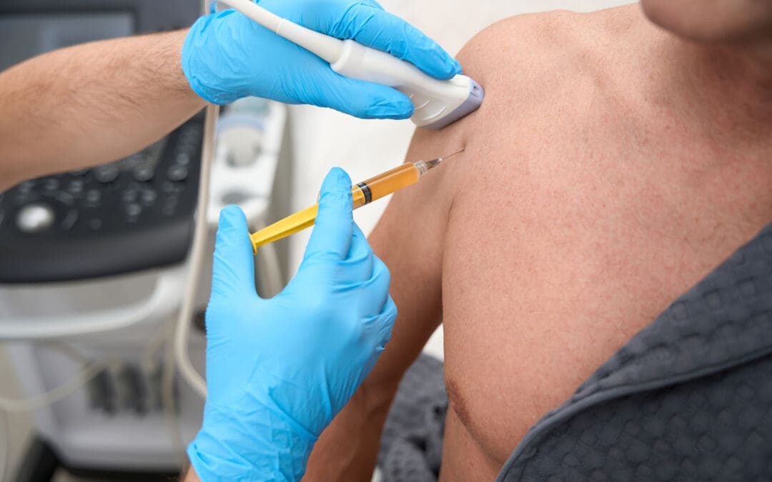
Ultrasound-Guided Shoulder Procedures: What We Do And Why
When indicated, we use ultrasound to guide precise injections. While this post emphasizes chiropractic and physical therapy, understanding our interventional choices clarifies our iterative care model.
Subacromial bursa, supraspinatus footprint, and AC joint
Why: Pain may originate from bursitis, partial-thickness supraspinatus lesions, or AC joint capsular irritation. Ultrasound guidance ensures in-plane or out-of-plane needle control, keeping the needle away from neurovascular structures.
Technique: Identify bright cortical bone under the footprint; visualize bursal fluid and capsule integrity. Use small aliquots and reassess spread, avoiding intratendinous trauma unless intentionally performing a tendon fenestration or PRP in tendinopathic zones.
Evidence: Ultrasound-guided shoulder injections improve accuracy compared with landmark techniques and can more precisely target pathologic pain generators (Sibbitt et al., 2011).
Biceps tendon sheath
Why: Anterior shoulder pain often involves the long head of biceps. Sheath injection—distinct from intratendinous injection—reduces irritability and allows rehab to progress.
Technique: Map the groove, maintain longitudinal needle trajectory, and confirm spread along the sheath without tendon violation.
AC joint microvolume injection
Why: Small-volume injections can modulate capsular irritability. Cross-body adduction reproduction of pain is a clinical cue.
Technique: Orient to the joint cleft, avoid over-distention, and recheck cross-body ROM post-procedure.
Our Procedure Safety And Team Coordination
Pre-procedure planning: We plan labs, imaging, and rehab scheduling in advance. My nurse and lab tech process any biologics as needed, while I maintain room-side focus on mapping and safety.
Minimal staff burden: Our care flow allows other team members to handle follow-ups, therapy sessions, and patient education while I perform the procedure efficiently.
Internal medicine oversight: Dr. Cardenas reviews risk factors, comorbidities, contraindications, and post-procedural monitoring when warranted.
Rehabilitation Sequencing After Shoulder Interventions
We deliberately move from low-threat to higher-load tasks:
Start with what hurts least: Early sessions prioritize thoracic mobility, scapular setting, and isometric cuff work at angles that do not provoke pain.
Gradual load introduction: As irritability recedes, we add eccentrics, closed-chain stabilization, and overhead progressions using tempo, isometric holds, and pause reps.
Return-to-sport or work tasks: We simulate reach, lift, carry, and press patterns relevant to the patient’s goals, using pain-guided progression and rate of perceived exertion to keep tissues within safe adaptive ranges.
Knee Care: Integrative Chiropractic And Physical Therapy Emphasis
The knee often presents with MCL strain, medial meniscal involvement, and synovial irritability—themes echoed in the transcript. Our approach blends chiropractic, PT, and when appropriate, ultrasound guidance.
Knee Biomechanics And Physiology
Load transmission: The knee depends on hip control and ankle mobility for shock absorption and alignment. Poor hip abduction and external rotation strength elevate medial compartment stress.
Meniscal physiology: Menisci distribute load and contribute to joint stability. Intra-meniscal degeneration and synovial inflammation can perpetuate pain and mechanical symptoms.
MCL healing: The MCL typically responds to graded load and frontal-plane stability training. Excess valgus strain irritates healing tissue.
Chiropractic And PT Integration For The Knee
Pelvic and lumbar alignment
Why: Pelvic tilt and lumbar rotation alter femoral tracking and tibial alignment under dynamic load.
Methods: Lumbopelvic adjustments, hip mobilizations, and gluteal activation to normalize kinetic chain input.
Motor control and strength
Why: Stable knees require hip abductors, external rotators, hamstrings, and quadriceps working in harmony.
Methods: Side-steps with bands, split-squat isometrics, Spanish squats, hamstring bridges, and tempo squats to train tolerance and tissue remodeling.
Tendon and fascia support
Why: Tendinopathic tissues benefit from eccentric and isometric loading; fascia responds to improved glide and hydration.
Methods: Patellar tendon isometrics, eccentric decline squats as tolerated, and soft-tissue mobilization to quadriceps and adductors.
Progressive return to function
Why: Sequenced progressions reduce flare-ups and build confidence.
Methods: Low-impact conditioning, step-down drills, landings, and multi-directional gait under supervision.
Ultrasound-Guided Knee Procedures When Indicated
Intra-articular injections
Why: Targeted delivery to the joint space supports modulation of synovial irritation.
Technique: Short-axis or long-axis guidance to visualize needle entry and avoid neurovascular structures.
MCL and medial meniscus region
Why: Pain generators can localize to the MCL or posteromedial meniscus. High-precision mapping reduces the risk of non-target injections.
Technique: In-plane approach along the MCL with careful hydrodissection when necessary; avoid intrameniscal violation unless using a specialist technique aligned with current evidence.
Clinical Observations From Dr. Alex Jimenez
From practice patterns noted across my work at elpasobackclinic.com and shared on my LinkedIn profile, several themes consistently emerge:
Patients thrive when care is sequenced, explained, and measured. Clear progress markers—ROM, strength, pain thresholds—reduce anxiety and improve outcomes.
The shoulder and knee respond best when the spine and hip are addressed concurrently. Regional interdependence is not academic—it is observable daily in the clinic.
Education and expectation management are as therapeutic as manual care. When patients understand why a technique is used, adherence and results improve.
Small-aliquot injections with ultrasound guidance allow real-time adjustments based on tissue spread and patient feedback, enhancing comfort and safety.
We emphasize movement literacy, teaching patients how to maintain neutral positions, breathe, and move through ranges of motion without provoking symptoms.
How Our Team Coordinates Care
Intake and triage: Medical review by Dr. Cardenas for complex histories; chiropractic exam and movement analysis by me; imaging decisions based on need.
Plan creation: A written plan outlines manual therapy, exercise progression, imaging, procedural options, and follow-up cadence.
Execution: Therapy staff handles laser, shockwave, and exercise coaching; I manage manual and chiropractic care, as well as any ultrasound-guided procedures, as appropriate.
Reassessment: We use validated outcome scales, ROM, strength testing, and return-to-function checkpoints to iterate the plan.
Communication: Patients receive clear instructions on post-session expectations and a simple home exercise sequence.
Why We Prioritize Chiropractic and Physical Therapy for elpasobackclinic.com
For our web audience and community, practical hands-on care, exercise therapy, and movement education are the cornerstones of recovery. While medications and hormones are part of comprehensive medical practice, we keep them in the background here, emphasizing:
The power of adjustments to restore joint motion and relieve nociception.
The value of targeted strengthening and motor control to protect tissues.
The role of patient-guided progression to boost independence and long-term resilience.
Safety, Dosing, And Patient Comfort
Dosing matters: Whether we are adjusting, mobilizing, loading a tendon, or injecting, we dose according to irritability, stage of healing, and patient goals.
Comfort strategies: We start with low-pain tasks, use paced breathing, and deploy brief micro-breaks to maintain composure in procedures.
Monitoring: Signs of over-irritation (escalation of night pain, heat, swelling) prompt plan adjustments or a medical review.
Putting It All Together: An Easy-To-Follow Care Journey
Step 1: Assessment
Detailed history, movement analysis, palpation, and ultrasound mapping when indicated.
Step 2: Early Care
Thoracic and cervical-thoracic mobilization, scapular setting, isometric cuff work; knee lumbopelvic alignment, hip strength foundations.
Step 3: Load And Control
Eccentrics, closed-chain drills, perturbation training, and gait re-education.
Step 4: Targeted Procedures If Needed
Ultrasound-guided bursa, AC joint, or intra-articular knee injections based on clear indications, with medical oversight.
Step 5: Return To Function
Task-specific progressions, confidence building, and preventive strategies.
Evidence-Based References That Inform Our Practice
We continually incorporate high-quality research into decisions:
Ultrasound guidance improves injection accuracy and patient outcomes in shoulder pathology (Sibbitt et al., 2011).
Scapular-focused programs and regional interdependence considerations enhance the effectiveness of shoulder rehabilitation (Kibler et al., 2013).
Eccentric and isometric loading strategies reduce tendinopathy pain and remodel tissue (Rio et al., 2015).
Myofascial techniques can improve pain and functional outcomes, supporting active rehabilitation (Cheatham et al., 2015).
Practical Takeaways For Patients
Movement is medicine: Consistency beats intensity early on.
Pain-guided progression: Minor discomfort is normal; escalating night pain or swelling means you should check in with us.
Whole-system support: Sleep, nutrition, and stress management help tissues heal and adapt.
Team-based care: Chiropractic, physical therapy, and medical oversight ensure your pathway is safe, precise, and personalized.
How To Get Help
If you are in El Paso or nearby and dealing with shoulder or knee pain, our team can create a clear, step-by-step plan designed for your goals. We will explain why we select each technique, how it fits your stage of healing, and how we measure progress so you can return to life with confidence.
References
Domenech-Garcia, V., Palsson, T. S., Boudreau, S. A., & Arendt-Nielsen, L. (2011). Upper cervical and upper thoracic manipulation in patients with shoulder pain: A randomized clinical trial. Journal of Orthopaedic & Sports Physical Therapy. https://www.jospt.org/doi/10.2519/jospt.2011.3579
Kibler, W. B., Sciascia, A., & Wilkes, T. (2013). Scapular dyskinesis and its relation to shoulder pain. Journal of the American Academy of Orthopaedic Surgeons. https://journals.lww.com/jaaos/Abstract/2013/06000/Scapular_Dyskinesis_and_Its_Relation_to_Shoulder.3.aspx
Rio, E., Kidgell, D., Purdam, C., Gaida, J., Moseley, L. G., & Cook, J. (2015). Isometric exercise for pain relief in tendinopathy: Mechanisms and implications. British Journal of Sports Medicine. https://bjsm.bmj.com/content/49/10/645
Sibbitt, W. L., Band, P. A., Kettwich, S. C., et al. (2011). Does ultrasound-guided injection improve outcomes for shoulder pain? A randomized controlled trial. Journal of Rheumatology. https://www.jrheum.org/content/38/9/1917
Cheatham, S. W., Kolber, M. J., & Cain, M. (2015). Instrument-assisted soft tissue mobilization: A systematic review. Journal of the Canadian Chiropractic Association. https://www.ncbi.nlm.nih.gov/pmc/articles/PMC4566596/
Unlocking Cellular Healing: The Power of Advanced Laser Therapy in Integrative Care
Abstract
As a clinician with a diverse background spanning chiropractic, advanced practice nursing, and functional medicine, my primary goal is to offer patients the most effective, evidence-based treatments available. In this educational post, I will take you on a journey into the world of Multiwave Locked System (MLS) Laser Therapy, a cutting-edge technology that is transforming how we manage pain and inflammation. We will explore the science behind this therapy, moving beyond surface-level explanations to understand its profound effects on cellular biology, including its impact on mitochondria and the inflammatory cascade. I will share insights from leading researchers and demonstrate how we apply this technology in clinical settings, particularly for conditions such as low back pain and joint issues. Furthermore, I will explain how MLS Laser Therapy integrates seamlessly into a comprehensive care model like ours at Injury Medical Clinic, where we combine chiropractic adjustments, physical rehabilitation, and advanced medical oversight from our Medical Director, Dr. Maria Guadalupe Cardenas, MD, to optimize patient outcomes. This post will detail specific treatment protocols, the importance of energy density, and how this therapy can augment other regenerative treatments, such as Platelet-Rich Plasma (PRP), offering a multifaceted approach to true healing.
A New Frontier in Healing at Injury Medical Clinic
Hello, I’m Dr. Alex Jimenez. With my credentials as a Doctor of Chiropractic (DC) and Advanced Practice Registered Nurse (APRN), and my certifications in functional and integrative medicine (CFMP, IFMCP), my passion has always been to bridge gaps between healing disciplines. At Injury Medical Clinic PA, we have built a practice on this very principle: a truly integrative approach to patient wellness.
A cornerstone of our collaborative model is my partnership with Dr. Maria Guadalupe Cardenas, MD. Dr. Cardenas is Board Certified in Internal Medicine and serves as our esteemed Medical Director and Collaborative Physician. With over 40 years of invaluable experience, she provides essential medical oversight, ensuring our patients receive safe, comprehensive, and well-rounded care. This multidisciplinary structure allows us to blend the best of chiropractic and physical rehabilitation with the diagnostic and medical expertise of internal medicine. Our team works in synergy, designing treatment plans that address not just the symptoms but the underlying physiological dysfunction. Whether a patient is recovering from a personal injury, managing a chronic condition, or seeking to optimize their overall health, our integrated team provides a holistic, evidence-based pathway to recovery.
Navigating Low Back Pain with MLS Laser Therapy
One of the most common ailments we see is chronic low back pain. Today, we have a patient, John, who is experiencing persistent joint pain and stiffness in his lumbar spine, specifically around the L4-L5 facet joints, with some discomfort radiating down his right side. This is a classic presentation that responds exceptionally well to a targeted, multimodal approach.
For John, we are utilizing the M6 Robotic MLS Laser. The first priority is always patient comfort. When using a robotic system, it’s critical that the patient remains still, as the laser is programmed to treat a precise area. We position the patient face down to allow direct access to the skin over the lumbar spine, as the laser energy must be delivered without the barrier of clothing.
The Clinical Multimodal Approach: More Than Just the “Spot of Pain”
Once John is comfortable, we begin the setup. The robotic laser interface is remarkably sophisticated yet user-friendly.
Targeting the Ailment: I select the “Joint Pain and Stiffness” protocol for the back.
Centering the Treatment: I zero out the X and Y axes on the control panel. This temporarily stops the robotic arm’s movement, allowing me to manually position the guiding red light directly over the primary source of John’s discomfort—the L4-L5 region he indicated.
Expanding the Field: This is where our clinical multimodal approach comes into play. Instead of just treating the single spot of pain, I expand the treatment area using the X and Y controls. This creates a larger therapeutic field that covers not only the symptomatic facet joints but also the surrounding connective tissue, muscles, and nerve roots. We aren’t just chasing pain; we are treating the entire functional unit to address the source of the dysfunction and support the interconnected biological systems.
The laser head is positioned at a precise distance from the skin—about six inches—using a provided ruler. This is crucial because the MLS laser beam is collimated, meaning the light rays are parallel. The focal point is engineered to be most effective at this distance, ensuring the therapeutic energy penetrates deep into the tissues rather than dissipating at the surface.
The Science of Healing: How MLS Laser Therapy Works
With the treatment underway—an eight-minute session for John’s low back—let’s dive into what’s happening at a cellular level. It’s common for patients to ask if they will feel anything. Most feel nothing at all, though some may notice a gentle warmth or tingling. This lack of intense heat is a hallmark of the MLS system’s advanced design.
The device combines two specific wavelengths of light: an 808-nanometer (nm) continuous-wave and a 905-nanometer (nm) pulsed-wave.
The 808 nm wavelength works more superficially to reduce inflammation and edema. It enhances blood circulation to the area, which helps clear out inflammatory byproducts and deliver oxygen and nutrients.
The 905 nm wavelength, delivered in powerful, short pulses, penetrates much deeper, reaching tissues such as muscle, nerve, and even the joint capsule. This pulsed energy is what provides the powerful analgesic (pain-relieving) effect.
These two wavelengths are synchronized, creating the patented “MLS pulse.” This enables delivery of very high peak power (up to 50 watts) in extremely short bursts (nanoseconds). This high-intensity “punch” of energy stimulates the cells without generating heat. A period of rest follows each pulse, allowing the tissue to absorb the energy efficiently. If a laser produces significant heat at the skin’s surface, it often means the energy isn’t being absorbed properly by the target tissues. The MLS system maintains tissue temperature at a constant level, ensuring optimal therapeutic delivery.
Seeing the Invisible: A Window into the Treatment
A fascinating demonstration of this technology involves using a smartphone camera. While the red aiming light is visible to the naked eye, the therapeutic infrared laser light is not. However, a camera’s sensor can detect it. If you were to look at John’s back through a phone camera during treatment, you would see a distinct triangle of light—this is the 808 nm wavelength at work, covering a significant area and illustrating how comprehensively we are treating the region.
Energy Density: The Key to Effective Dosing
A critical concept in laser therapy is energy density, measured in joules per centimeter squared (J/cm²). This is more important than the total number of joules delivered. Think of it like watering a plant: you need to provide the right amount of water for the pot’s size. Too little has no effect; too much drowns it. Similarly, our goal is to deliver a precise dose of light energy to the target tissue.
The World Association for Laser Therapy (WALT) and a large body of research support an optimal therapeutic window of 4-10 J/cm².
For John’s condition, the protocol is set to deliver approximately 6 J/cm². The laser’s software automatically calculates the treatment time required to achieve this density over the selected area. If I were to make the treatment area smaller or larger, the software would instantly recalibrate the time to ensure the correct dose is delivered.
This concept also relates to the Arndt-Schultz Law, a pharmacological principle stating that low doses stimulate, moderate doses inhibit, and high doses are toxic. With laser therapy, if you “overcook” an area with too much energy, you risk a bioinhibitory effect, in which the treatment becomes less effective or even counterproductive. The body’s cells can only absorb so much energy at once. This is why our protocols focus on precise energy density and, if more treatment is needed, we target different areas (e.g., an anterior and posterior approach for a knee) rather than just increasing the time on one spot.
Integrating Modalities for Superior Results
While the robotic laser treats the broader lumbar region, I can simultaneously use a handheld MLS laser applicator. This handpiece allows for more focused treatment on specific points, such as trigger points or “knots” in the muscle. I often use the “cooked meat” versus “raw meat” analogy that a physical therapist once taught me. Healthy, relaxed muscle feels like raw meat, while a tight, knotted trigger point feels firm, like cooked meat. The handheld applicator is perfect for treating these punctual spots.
The robot and the handpiece operate on two separate channels, allowing us to perform this dual treatment. This is a perfect example of our integrative philosophy in action:
Chiropractic Care: Before or after the laser session, I can perform specific chiropractic adjustments to restore proper motion to the L4-L5 facet joints and relieve mechanical stress.
Physical Rehabilitation: Our team can guide John through exercises to strengthen his core musculature and improve spinal stability.
MLS Laser Therapy: The laser works at the cellular level to reduce pain and inflammation that may be hindering his ability to engage in rehabilitation, thereby accelerating healing.
This combination addresses the structural, functional, and biochemical aspects of his condition simultaneously.
Advanced Applications: Augmenting Regenerative Medicine
The conversation around healing is increasingly turning toward orthobiologics, such as Platelet-Rich Plasma (PRP) injections. This is where MLS Laser Therapy shows even more remarkable potential. A common question arises: if PRP induces a beneficial pro-inflammatory phase to kickstart healing, won’t an anti-inflammatory laser treatment counteract it?
The answer is no. In fact, the laser augments the process. The data and our clinical observations show that using laser therapy in conjunction with PRP can improve outcomes by an estimated 15-20%.
Here is the progressive protocol we often recommend:
Pre-Injection Priming (2-3 treatments): In the weeks leading up to the PRP injection, we use the laser to “prepare the soil.” These sessions are designed to increase local blood circulation, reduce baseline chronic inflammation, and optimize the cellular environment, making the tissue more receptive to the growth factors in the PRP.
Day of Injection (1 treatment): A treatment on the day of the procedure can further enhance the effects.
Post-Injection Support (6+ treatments): Following the injection, a series of laser treatments helps manage pain and supports the regenerative cascade initiated by the PRP. The laser enhances mitochondrial function, which is critical for providing the cellular energy (ATP) needed for tissue repair.
The Cascade of Healing: From Acute Relief to Chronic Repair
How does a single modality address both acute pain and chronic conditions? The effects occur in a cascade.
Immediate Effect (Acute Phase): The initial pain relief often comes from the laser’s effect on small, unmyelinated nerve fibers (C-fibers) that transmit pain signals. The energy can temporarily block these signals, providing rapid relief. This is the analgesic effect.
Subsequent Effect (Inflammatory Modulation): Over the next few hours and days, the anti-inflammatory effect takes hold. The laser energy modulates the immune response, reducing pro-inflammatory cytokines and promoting the resolution of inflammation and edema.
Long-Term Effect (Biostimulation and Chronic Repair): With a series of treatments, we get to the core of cellular repair. Light energy is absorbed by cytochrome c oxidase in the mitochondria, the powerhouses of our cells. This significantly increases ATP (adenosine triphosphate) production, the body’s primary energy currency. This surge in available energy fuels all cellular repair processes, from protein synthesis to cell replication, promoting true, long-term tissue healing.
This mitochondrial boost is especially relevant in today’s world, where many common medications, such as statins, can impair mitochondrial function. By enhancing mitochondrial biogenesis and efficiency, laser therapy can help overcome these hurdles and optimize the body’s innate healing capacity. This is why we also discuss nutritional and lifestyle factors—such as CoQ10 supplementation to support mitochondrial function—as part of a truly comprehensive functional medicine approach.
Treatment Frequency and The Cumulative Effect
Healing is a process, not an event. The effects of MLS Laser Therapy are cumulative. We recommend a series of treatments to achieve lasting results.
Acute Conditions: Typically, a course of 6 treatments is effective.
Chronic Conditions: A more intensive course of 12 treatments is often needed.
Ideally, treatments are scheduled close together (e.g., Monday, Wednesday, Friday) to build therapeutic momentum. It is crucial for patients to complete the full course. Many start feeling significantly better after just 3-4 sessions and are tempted to stop. However, completing the entire protocol ensures deeper cellular repair, leading to more durable outcomes.
At Injury Medical Clinic, our mission is to empower your body’s own ability to heal. By integrating the best of chiropractic, medical oversight, and groundbreaking technologies like MLS Laser Therapy, we offer a path to recovery that is not only faster but also more complete.
World Association for Laser Therapy. (n.d.). WALT Recommended Treatment Doses for LLLT. WALT. Retrieved from https://waltza.co.za/wp-content/uploads/2012/08/Dose_table_780-860nm_for_Low_Level_Laser_Therapy_WALT-2010.pdf
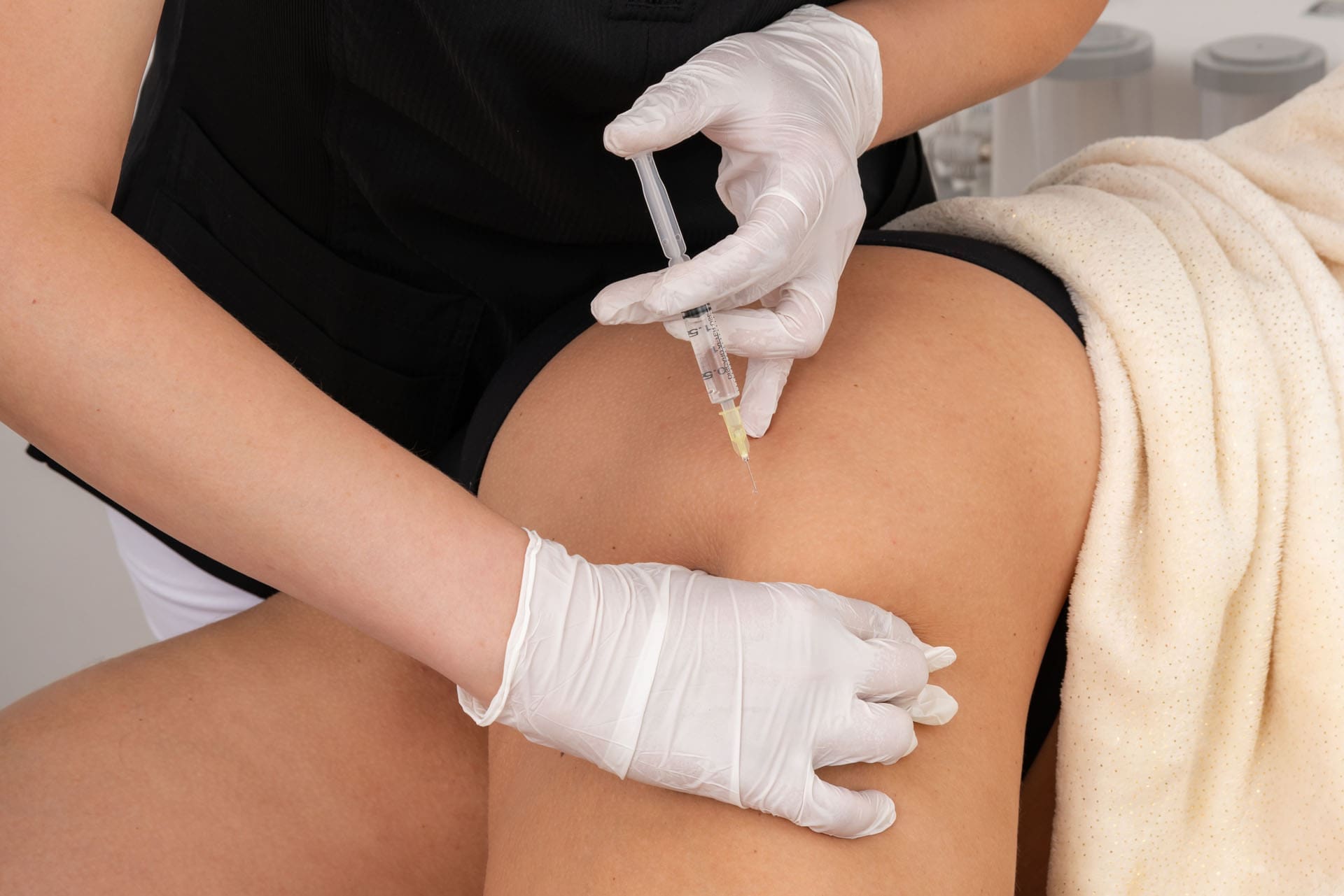
The Power of Precision: Platelet-Rich Plasma for Spine and Injury Recovery
Abstract
Welcome to our educational journey into the world of regenerative medicine, with a focus on Platelet-Rich Plasma (PRP) therapy. As a clinician dedicated to integrative and evidence-based care, I am thrilled to share insights from the forefront of musculoskeletal treatment. This post will demystify PRP, exploring what it is, how it’s prepared, and, most importantly, the critical role of dosage in achieving successful clinical outcomes. We will examine groundbreaking research revealing how the precise concentration and number of platelets can dramatically influence healing, particularly in conditions such as osteoarthritis and tendon injuries. We’ll also discuss the importance of ultrasound guidance for accurate delivery and how integrative chiropractic care and structured rehabilitation are essential partners to PRP therapy, creating a comprehensive strategy that not only alleviates pain but also fosters true, lasting tissue regeneration. Join me as we uncover how this powerful biologic treatment is changing the landscape of healing.
What Exactly Is Platelet-Rich Plasma (PRP)?
Many of us may have a distant memory from our early science education about platelets. We often think of them simply as the components in our blood that help form clots when we get a cut. While that is true, it’s only a small part of their incredible story.
Platelets are small, anucleated (meaning they don’t have a nucleus) cell fragments that are absolute powerhouses of healing. Each one is packed with hundreds of proteins called growth factors and cytokines. These are signaling molecules that act as the body’s own project managers for tissue repair. When an injury occurs, platelets rush to the scene not just to plug the leak but to orchestrate a complex, coordinated healing cascade. They call in other cells, direct the removal of damaged tissue, and stimulate the growth of new, healthy cells.
Given their central role in healing, it’s logical to ask: what if we could concentrate these powerful healing factors and deliver them directly to the site of chronic injury, such as a worn-out knee joint or a nagging tendon tear? That is the fundamental concept behind Platelet-Rich Plasma (PRP) therapy.
From Your Blood to a Healing Solution
The process of creating PRP is elegant in its simplicity.
Blood Draw: It all begins with a simple blood draw from your arm, much like a standard lab test. The amount of blood drawn can vary depending on the specific system used and the therapeutic dose we are aiming to achieve—a concept we will explore in detail.
Centrifugation: This blood is then placed in a sterile, closed-system kit and spun in a specialized centrifuge. The spinning process uses centrifugal force to separate the blood into its different components based on their density.
Separation and Concentration: The heavier red blood cells sink to the bottom. The lighter, platelet-poor plasma rises to the top. In the middle, a thin, precious layer forms known as the “buffy coat.” This layer, along with a portion of the adjacent plasma, is where the vast majority of platelets and a population of white blood cells are concentrated. This is the Platelet-Rich Plasma.
This final product is a small volume of plasma containing a significantly higher concentration of platelets—and their associated growth factors—than in your normal circulating blood.
Not All PRP Is Created Equal: The Critical Importance of Dose
One of the most significant advancements in the field of regenerative medicine has been the realization that PRP is not a one-size-fits-all treatment. To think of it effectively, we must approach it as a biologic drug. As with any medication, there is a therapeutic dose—the specific amount needed to produce the desired clinical effect. An amount below this threshold will be sub-therapeutic and likely ineffective, while an excessive amount could potentially hinder the healing process.
The Problem of Variability
For years, the results of PRP studies were inconsistent, leaving both clinicians and patients confused. Why did it work so well in some cases and not in others? Pioneering researchers like James Clayton, D. Patrick, and their team in Australia began to uncover the answer. They analyzed five different commercial PRP preparation systems and found staggering variability in the final product. The platelet count, white blood cell count, and final volume were all over the map.
Imagine seeing the PRP prepared from the same patient’s blood using four different systems. You would see four different “products” of varying colors and cellular compositions. This lack of standardization was a major hurdle. Early studies often failed to report the specific platelet dose injected, making it impossible to compare results or understand what truly worked.
Thanks to the meticulous work of researchers like Peter Everts and Scott Rodeo, we are now beginning to decode the dose-response relationship for specific conditions. A landmark 2018 study analyzed numerous PRP studies for soft tissue applications. When they plotted the results based on the total number of platelets injected, a clear pattern emerged.
Studies using a low dose of PRP, typically under 3 billion platelets, were overwhelmingly negative. They showed little to no benefit over a placebo.
Studies using a higher dose, generally above 3.5 billion platelets, were overwhelmingly positive.
This suggests a distinct therapeutic threshold for soft tissue and tendon healing. For instance, in my clinical observations at El Paso Back Clinic, treating conditions like tennis elbow (lateral epicondylitis) or plantar fasciitis with an insufficient platelet dose often yields disappointing results. However, when we ensure the delivered dose is within that therapeutic range of 3.5 to 5 billion platelets or higher, we see a much more robust and consistent healing response. The body needs a sufficient signal to switch from chronic degeneration to active regeneration, and the dose provides that signal. We also know that a patient’s age can impact the required dose, with older patients often benefiting from a higher starting concentration to achieve the same therapeutic effect.
Perhaps the most compelling evidence for PRP dosing comes from the treatment of knee osteoarthritis (OA). Knee OA is a condition I see daily, and it can be profoundly debilitating for patients. For years, the primary non-surgical options were limited.
The famous RESTORE trial, published in JAMA, initially concluded that PRP was ineffective for knee OA. However, a deeper dive into their methodology reveals a critical flaw: they used a low-dose PRP system that delivered only 1.6 billion platelets per injection. Based on what we now know about the dose-response curve, this was a sub-therapeutic dose, destined to fail. While the study was beautifully executed, we learned a valuable lesson from its negative result—it helped define the lower boundary of what doesn’t work.
In stark contrast, a study by van der Weegen used a high-dose PRP preparation that delivered approximately 10 billion platelets in a single injection. The results were remarkable. Patients not only experienced significant improvements in pain and function compared to hyaluronic acid or saline injections, but MRI scans also suggested a disease-modifying effect. The progression of cartilage loss actually slowed down in the PRP group. This was a groundbreaking finding, suggesting that with the right dose, PRP might do more than just manage symptoms—it could potentially alter the course of the disease.
Based on the current body of evidence, the therapeutic target for treating knee OA appears to be 5 to 10 billion platelets per injection. Calculating and delivering this precise dose is paramount to achieving the kind of outcomes our patients deserve.
The Role of Chiropractic Care and Guided Injections in Maximizing PRP Success
Achieving a successful outcome with PRP involves more than just getting the dose right. It requires a holistic, integrative approach that addresses the entire patient and the mechanics of their injury. This is where chiropractic care, physical therapy, and advanced injection techniques become indispensable partners.
Precision Matters: The Necessity of Ultrasound Guidance
Growth factors in PRP work by forming a bioactive scaffold that stimulates local cells. For this to happen, the PRP must be delivered with pinpoint accuracy directly into the site of injury—be it a tear within a tendon, the space within a joint, or an area of damaged cartilage. If the injection is off by even a few millimeters, the therapeutic benefit can be lost entirely.
This is why ultrasound guidance is not a luxury; it is the standard of care for regenerative injections. Using real-time ultrasound imaging, I can visualize the needle’s path and confirm its placement directly in the target tissue. This ensures that the powerful biologic product we’ve carefully prepared is delivered precisely where it’s needed most, maximizing the potential for a successful healing response. Injecting “blind” is simply not an acceptable approach when the goal is true tissue regeneration.
The Foundational Role of Integrative Chiropractic and Rehabilitation
At El Paso Back Clinic, we view PRP not as a standalone “magic bullet” but as a catalyst within a comprehensive treatment plan. A chronically injured joint or tendon doesn’t exist in a vacuum. It is almost always accompanied by biomechanical dysfunction, muscle imbalances, poor movement patterns, and joint restrictions. Injecting PRP into a dysfunctional environment without addressing these underlying root causes is like planting a seed in barren soil.
This is the crucial role of integrative chiropractic care.
Restoring Biomechanics: Before and after a PRP procedure, we focus on correcting biomechanical faults. Through specific chiropractic adjustments, we restore proper joint mobility, particularly in the spine, pelvis, and extremity joints related to the injury. This ensures that forces are distributed evenly across the kinetic chain, taking undue stress off the healing tissue.
Addressing the Kinetic Chain: An arthritic knee, for instance, is often linked to problems in the hip, ankle, or even the lower back. Our comprehensive assessment identifies these related dysfunctions. By treating the entire kinetic chain, we create a stable and supportive environment for the PRP to work effectively.
Targeted Rehabilitation: A structured physical therapy and rehabilitation program is essential. The initial goal post-injection is to protect the healing tissue. This is followed by a progressive program designed to:
Improve Flexibility and Range of Motion.
Strengthen Supporting Musculature.
Retrain Neuromuscular Control and Proprioception (your body’s sense of its position in space).
This rehabilitation phase translates the biological healing initiated by PRP into functional, long-lasting improvement. It teaches the body to use the newly repaired tissue properly and helps prevent reinjury. The healing process stimulated by PRP takes time—often three to six months or more to see the full benefit. A patient, supportive, and well-structured rehabilitation plan is the bridge to that successful long-term outcome.
By combining a precisely dosed and accurately delivered PRP injection with expert chiropractic care and targeted physical therapy, we create a powerful synergy. We are not just chasing symptoms; we are correcting dysfunction, stimulating a biological repair process, and rebuilding a foundation for durable health and function.
IFM's Find A Practitioner tool is the largest referral network in Functional Medicine, created to help patients locate Functional Medicine practitioners anywhere in the world. IFM Certified Practitioners are listed first in the search results, given their extensive education in Functional Medicine