Back Clinic Imaging & Diagnostics Team. Dr. Alex Jimenez works with top-rated diagnosticians and imaging specialists. In our association, imaging specialists provide fast, courteous, and top-quality results. In collaboration with our offices, we provide the quality of service our patients’ mandate and deserve. Diagnostic Outpatient Imaging (DOI) is a state-of-the-art Radiology center in El Paso, TX. It is the only center of its kind in El Paso, owned and operated by a Radiologist.
This means when you come to DOI for a radiologic exam, every detail, from the design of the rooms, the choice of the equipment, the hand-picked technologists, and the software which runs the office, is carefully chosen or designed by the Radiologist and not by an accountant. Our market niche is one center of excellence. Our values related to patient care are: We believe in treating patients the way we would treat our family and we will do our best to ensure that you have a good experience at our clinic.
Diagnosis of the diseases of the abdomen can be classified into:
Abnormalities of the gastrointestinal tract (esophagus, stomach, small & large bowel, and the appendix)
Abnormalities of the accessory digestive organs (Hepatobiliary & pancreatic disorders)
Abnormalities of the genitourinary & reproductive organs
Abnormalities of the abdominal wall and major vessels
This presentation aims to provide the most basic understanding of general diagnostic imaging approach and appropriate clinical management of patients with the most common diseases of the abdomen
Imaging modalities used during an investigation of the abdominal complaints:
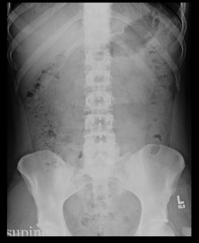
AP abdomen (KUB) and upright CXR
Abdominal CT scanning (with oral and I.V. contrast and w/o contrast)
Upper and Lower GI Barium studies
Ultrasonography
MRI (most used as Liver MRI)
MRI enterography & enteroclysis
MRI rectum
Endoscopic Retrograde Cholangiopancreatography (ERCP)- mostly hepatobiliary and pancreatic ductal pathology
Nuclear imaging
Why Order An Abdominal X-ray?
Include a preliminary evaluation of bowel gas in an emergent setting. For example, a negative study in a low probability patient may obviate the need for a CT or other invasive procedures
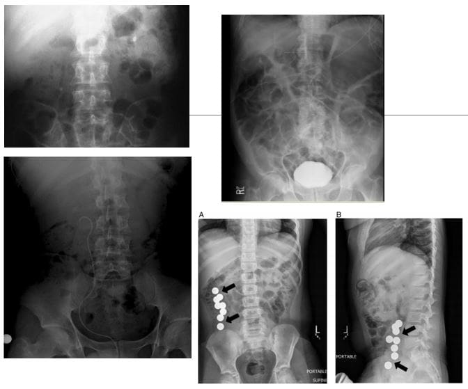
Evaluation of radiopaque tubes, lines, and radiopaque foreign bodies
Post-procedural evaluation intraperitoneal/retroperitoneal free gas
Monitoring the amount of bowel gas and resolution of postoperative (adynamic) ileus
Monitoring the passage of contrast through the bowel
Colonic transit studies
Monitoring renal calculi
What to Note on AP Abdomen: Supine vs. Upright vs. Decubitus
Free Air (pneumoperitoneum)
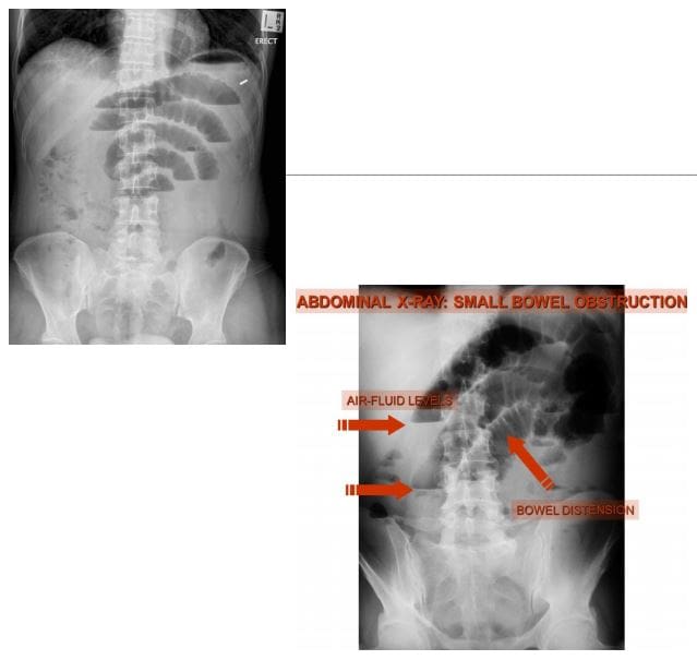
Bowel obstruction: Dilated loops: SBO vs LBO (3-6-9 rule) SB-upper limit-3-cm, LB-upper limit-6-cm, Caecum-upper limit-9-cm. Note loss of haustra, note dilation (presence) of valvule conivente (plica semilunaris) in SBO
SBO: note different heights air-fluid levels on upright film step ladder� appearance, typical of SBO
Note paucity of rectal/colonic gas (evacuated) in SBO
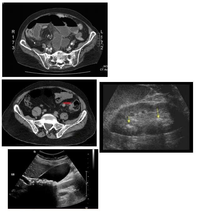
Abdominal CT scanning -modality of choice during the investigation of acute and chronic abdominal complaints especially in adults. For example, abdominal malignancy can be successfully diagnosed and staged providing clinical information for care planning
Abdominal, renal and pelvic ultrasound can be performed to help the diagnosis of appendicitis (esp. in children), acute & chronic vascular pathology, hepatobiliary abnormalities, obstetric and gynecological pathology
Use of ionizing radiation (x-rays & CT) should be minimized in children and other vulnerable groups.
Diagnostic Imaging of Major Diseases of the Gastrointestinal System
1) Esophageal disorders
2) Gastric carcinoma
3) Gluten Sensitive Enteropathy
4) Inflammatory Bowel Disease
5) Pancreatic ductal adenocarcinoma
6) Colorectal carcinoma
7) Acute Appendicitis
8) Small bowel obstruction
9) Volvulus
Esophageal disorders
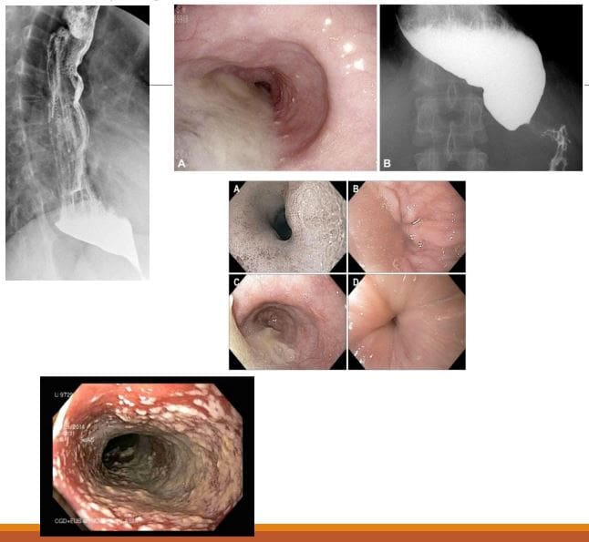
Achalasia (primary achalasia): failure of organized esophageal peristalsis d/t impaired relaxation of the lower esophageal sphincter (LOS) with marked dilatation of the esophagus and food stasis. Obstruction of the distal esophagus (often due to tumor) has been termed “secondary achalasia” or “pseudoachalasia.� Peristalsis in the distal smooth muscle segment of the esophagus may be lost due to an abnormality of Auerbach plexus (responsible for smooth muscle relaxation). Vagus neurons can also be affected
Primary: 30 -70s, M: F equal
Chagas disease (Trypanosoma Cruzi infection) with the destruction of the Myenteric plexus neurons of the GI system (megacolon & esophagus)
However, the heart is the M/C affected organ
Clinically: Dysphagia for both solids and liquids, in comparison to dysphagia for solids only in cases of esophageal carcinoma. Chest pain and regurgitation. M/C mid esophageal squamous cell carcinoma in approximately 5% due to chronic irritation of the mucosa by stasis of food and secretions. Aspiration pneumonia may develop. Candida esophagitis
Imaging: �Bird -beak� on upper GI barium swallow, dilated esophagus, loss of peristalsis. An endoscopic exam is crucial.
Rx: difficult. Calcium channel blockers (short -term).Pneumatic dilatation, effective in 85% of patients with 3 -5% risk of bleeding/perforation. Botulinum toxin injection lasts only approx. 12 months per treatment. May scar the submucosa leading to increased risk of perforation during subsequent myotomy. Surgical myotomy (Heller myotomy)
10 -30% of patients develop gastroesophageal reflux (GERD)
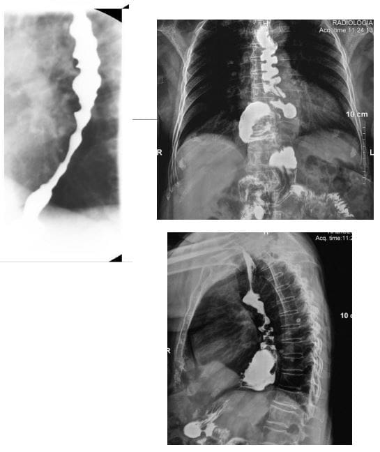
Presbyesophagus: used to describe the manifestations of degenerating motor function in the aging esophagus >80-y.o. Due to interruption of the reflex arc with decreased sensitivity to distension and alteration in peristalsis.
Patients may complain of dysphagia or chest pain, but most are asymptomatic
Diffuse/distal esophageal spasm (DES) is a motility disorder of the esophagus that may appear as a corkscrew or rosary bead esophagus on barium swallow.
2% of non-cardiac chest pain
Manometry is the gold-standard diagnostic test.
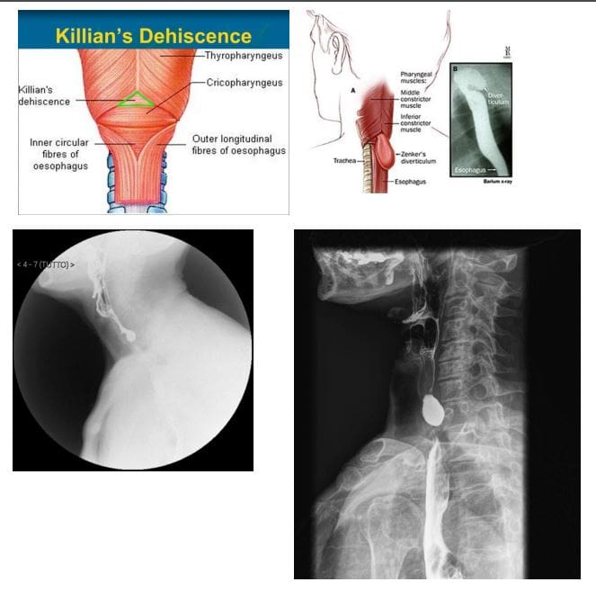
Zenker diverticulum (ZD) aka pharyngeal pouch
An outpouching at the level of the hypopharynx, just proximal to the upper esophageal sphincter, known as the Killian dehiscence or Killian triangle
Patients are 60-80 y.o and present with dysphagia, regurgitation, halitosis, globus sensation
May complicate with aspiration and pulmonary abnormalities
Patients may accumulate medications
ZD- is a pseudodiverticulum or pulsion diverticulum resulting from herniation of the submucosa through the Killian dehiscence, forming a sac where food and other contents may accumulate.
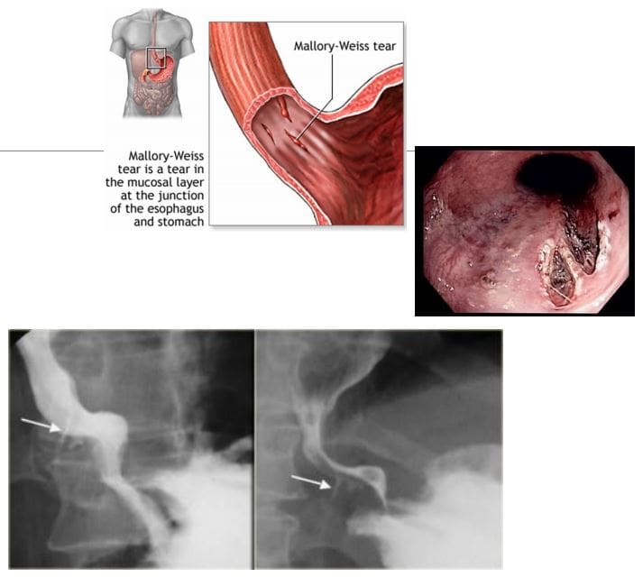
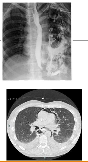
Mallory-Weiss syndrome refers to mucosal and submucosal tears of the distal oesophageal venous plexus associated with violent retching/vomiting and projection of gastric contents against the lower esophagus. Alcoholics are at particular risk. Cases present with painless hematemesis. Treatment is typically supportive.
Dx: imaging plays little role, but contrast esophagram may demonstrate some mucosal tears filled by contrast (bottom right image). CT scanning may help to exclude other causes of upper GI bleeding
Boerhaave syndrome: esophageal rupture secondary to forceful vomiting
Mechanisms involve forceful expulsion of gastric contents especially with large undigested foods when esophagus forcefully contracts against closed glottis with 90% occurring along left posterolateral wall
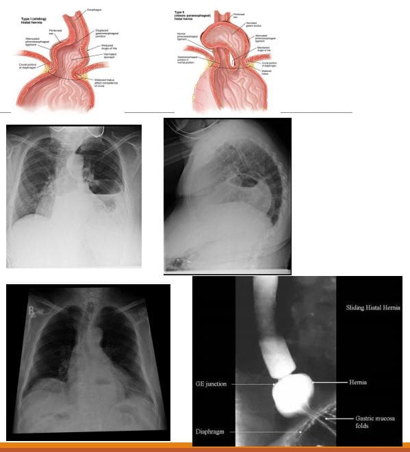
Hiatus hernias (HH): herniation of abdominal contents through the esophageal hiatus of the diaphragm into the thoracic cavity.
Many patients with HH are asymptomatic, and it is an incidental finding. However, symptoms may include epigastric/chest pain, postprandial fullness, nausea and vomiting
Sometimes HH is considered synonymous with gastro-oesophageal reflux disease (GORD), but there is a poor correlation between the two conditions!
2-types: sliding hiatus hernia 90% & rolling (paraoesophageal) hernia 10%. The latter may strangulate leading to ischemia and complications.
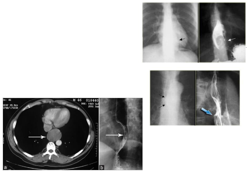
Esophageal Leiomyoma is the M/C benign esophageal neoplasm. It is often large but yet non-obstructive. Gastrointestinal stromal tumors (GIST) are the least common in the esophagus. Should be differentiated from Esophageal carcinomas.
Imaging: contrast esophagram, upper GI barium swallow, CT scanning. Gastroesophagoscopy is the Dx method of choice.
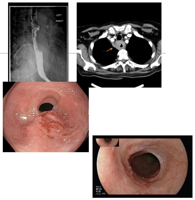
Esophageal carcinoma: presented with increasing dysphagia, initially to solids and progressing to liquids with obstruction in more advanced cases
<1% of all cancers and 4-10% of all GI malignancies. There is recognized male preponderance with the squamous cell subtype due to smoking and alcohol. Barrett esophagus and adenocarcinoma
M: F 4:1. Black individuals are more susceptible than White individuals 2:1. Poor prognosis!
A barium swallow can be sensitive in identifying esophageal mass. Gastroesophagoscopy (endoscopy) confirms the diagnosis with tissue biopsy
Overall the most common malignancy is 2ndary gastric fundal carcinoma invading distal esophagus
Squamous cell is typically found in the mid esophagus, Adenocarcinoma in the distal region
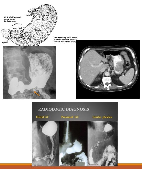
Gastric carcinoma: primary malignancy of gastric epithelium. Rare before the age of 40. The median age at diagnosis in the United States is 70 years for males and 74 years for females. Japan, South Korea, Chile, and Eastern European countries have one of the highest rates of stomach cancer in the world. Stomach cancer rates are declining worldwide. Gastric cancer is the 5th causes of cancer-related death. Association with Helicobacter pylori infection 60- 80%, but only 2% population with H. Pyloris develop Stomach cancer. 8-10% have an inherited familial component.
Gastric Lymphoma is also linked to H. Pyloris infection. Gastrointestinal Stromal Cell Tumour or GIST is another neoplasm affecting the stomach
Clinically: No symptoms when it is superficial and potentially curable. Up to 50% of patients may have non-specific GI complaints. Patients may present with anorexia and weight loss (95%) as well as vague abdominal pain. Nausea, vomiting, and early satiety d/t obstruction may occur with bulky tumors or infiltrative lesions that impair stomach distension.
Prognosis: Most gastric cancers diagnosed late and may reveal local invasion with regional adenopathy, liver, and mesenteric spread. A 5-year survival rate of 20% or less. In Japan and S. Korea, early screening programmes increased survival to 60%
Imaging: Barium upper GI study, CT scanning. Endoscopic examination is the method of choice for the diagnosis. On imaging, Gastric cancer may appear as an exophytic (polypoid) mass or Fungative type, Ulcerative or Infiltrative/diffuse type (Linitis Plastica). CT scanning is important to evaluate local invasion (nodes, mesentery, liver, etc.)
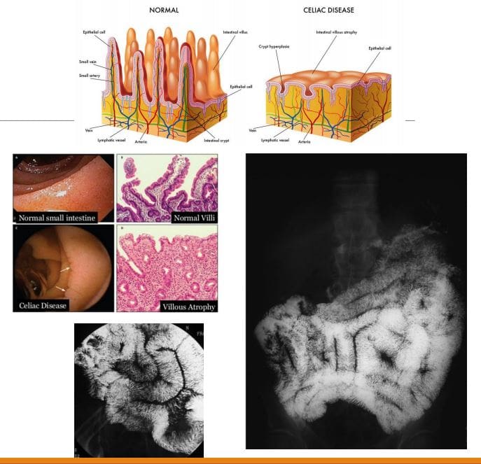
Celiac disease aka non-tropical sprue aka Gluten-sensitive enteropathy: A T-cell mediated autoimmune chronic gluten-induced mucosal damage resulting in loss of villi in the proximal small bowel and gastrointestinal malabsorption (i.e., sprue). Considered in some cases of iron deficiency anemia of undetermined cause. Common in Caucasians (1 in 200) but rare in Asian and black individuals. Two peaks: a small cluster in early childhood. Typically in 3rd and 4th decades of life.
Clinically: Abdominal pain is the m/c symptom, malabsorption of nutrients/vitamins: IDA and guaiac-positive stools, diarrhea, constipation, steatorrhea, weight loss, osteoporosis/osteomalacia, dermatitis herpetiformis. Increased association with T-cell lymphoma, Increased association with esophageal squamous cell carcinoma, SBO
Dx: Upper GI endoscopy with multiple duodenal biopsies is considered a diagnostic standard for celiac disease. Histology reveals T-cell infiltration and lymphoplasmacytosis, Villi atrophy, Crypts hyperplasia, Submucosa, and Serosa are spared. Rx: elimination of gluten-containing products
Imaging: Not required for Dx but on Barium swallow fluoroscopy: mucosal atrophy and obliteration of mucosal folds (advanced cases only). SB dilation is the most typical finding. Nodularity of the duodenum (bubbly duodenum). Reversal of jejunal and ileal mucosal folds:
�The jejunum looks like ileum, the ileum looks like the jejunum, and the duodenum looks like hell.�
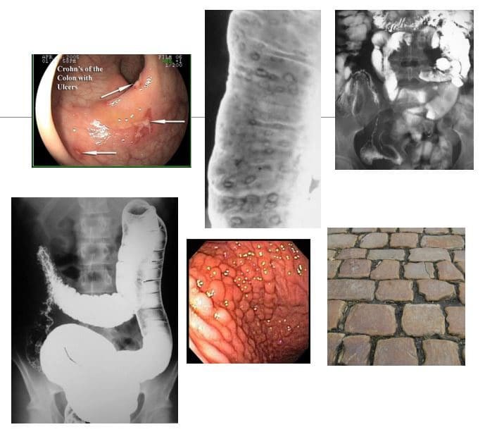
CD: chronic relapsing-remitting autoimmune inflammation that affects any part of the GI tract from the mouth to the anus but at onset most typically involves the terminal ileum. M/C presentation: abdominal pain/cramping and diarrhea. Path: granulomata formation that unlike UC is transmural, potentially leading to strictures. Areas affected by the inflammation are typically patchy
Complications are numerous: malabsorption of nutrients/vitamins (anemia, osteoporosis, developmental delay in children, susceptibility to GI malignancy, bowel obstruction, fistula formation, extra-abdominal manifestations: uveitis, arthritis, AS, erythema nodosum and others. 10- 20% may require abdominal surgery after 10-years of CD usually for strictures, fistiluzation, BO.
Dx: clinical, CBC, CMP, CRP, ESR, serological tests: DDx of IBD: anti-Saccharomyces cerevisiae antibodies (ASCA), perinuclear antineutrophil cytoplasmic antibody (p-ANCA) histologically or in serum. Fecal Calprotectin test helps to DDx IBS and evaluate response to treatment, disease activity/relapses.
Dx of choice: endoscopy, ileoscopy, and multiple biopsies may reveal endoscopic and histological changes. Video capsule endoscopy (VCE), Imaging may help with Dx of complications. Rx: immunomodulatory drugs, complementary medicine, diet, probiotics, operative. No cure but the aim is to induce remission, control symptoms and prevent/treat complications
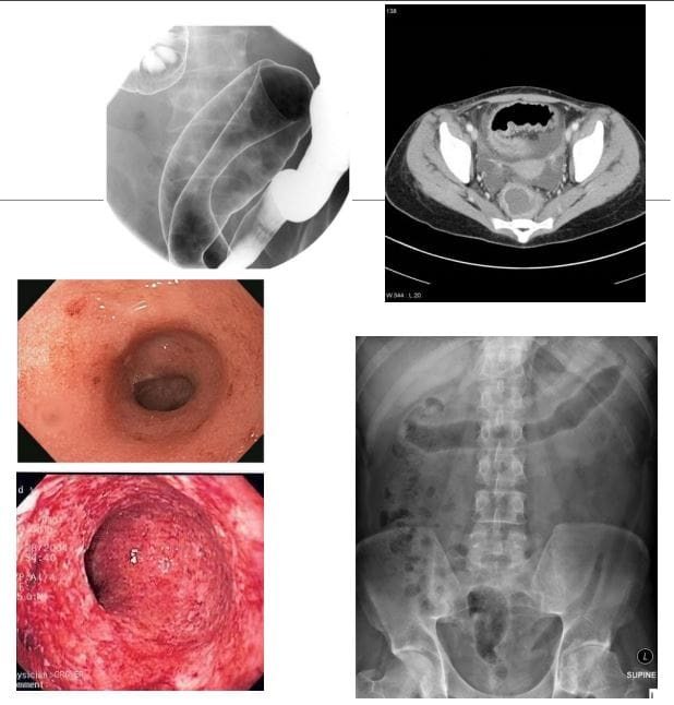
Imaging Dx: KUB to DDx SBO, Barium enema (single and double contrast), small bowel follow through. Findings: skip lesions, aphthous/deep ulcerations, fistula/sinus tracts, String sign, creeping fat pushed loops of LB, cobblestone appearance d/t fissures/ulcers pushing mucosa, CT scanning with oral and IV contrast.
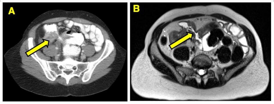
Imaging from a Crohn’s patient who had a small bowel resection for obstruction.
(B) MRE of the same area shows a fibrostenotic stricture
UC: characteristically involves only the colon but backwash ileitis may develop. Onset is typically at 15-40s and is more prevalent in males, but the onset after the age of 50 is also common. More common in North America and Europe (hygiene hypothesis). Etiology: A combination of environmental, genetic and gut microbiome changes are involved. Smoking and early appendectomy tend to show a negative association with UC, unlike in CD considered some of the risk factors.
Clinical Features: Rectal bleeding (common), diarrhea, rectal mucous discharge, tenesmus (occasionally), lower abdominal pain and severe dehydration from purulent rectal discharge (in severe cases, especially in the elderly), fulminant colitis and toxic megacolon can be fetal but are rare complications. Pathology: No granulomata. Ulcerations affect mucosa and submucosa. Pseudopolyps present as elevated spared mucosa.
An initial process always affects the rectum and remain a local disease (proctitis) in (25%). 30% Proximal disease extension may occur. UC may present as left-sided (55%) and pancolitis (10%). Majority of cases are mild to moderate
Imaging: not required for Dx but barium enema may reveal ulcerations, thumbprinting, in advanced cases loss of haustra and narrowing of the colon producing �lead-pipe colon.� CT scanning may help with Dx seen as mucosal thickening detected only in moderate and severe cases. CT may help with of Dx of complications. Plain film image reveals �lead-pipe colon� and sacroiliitis as Enteropathic arthritis (AS)
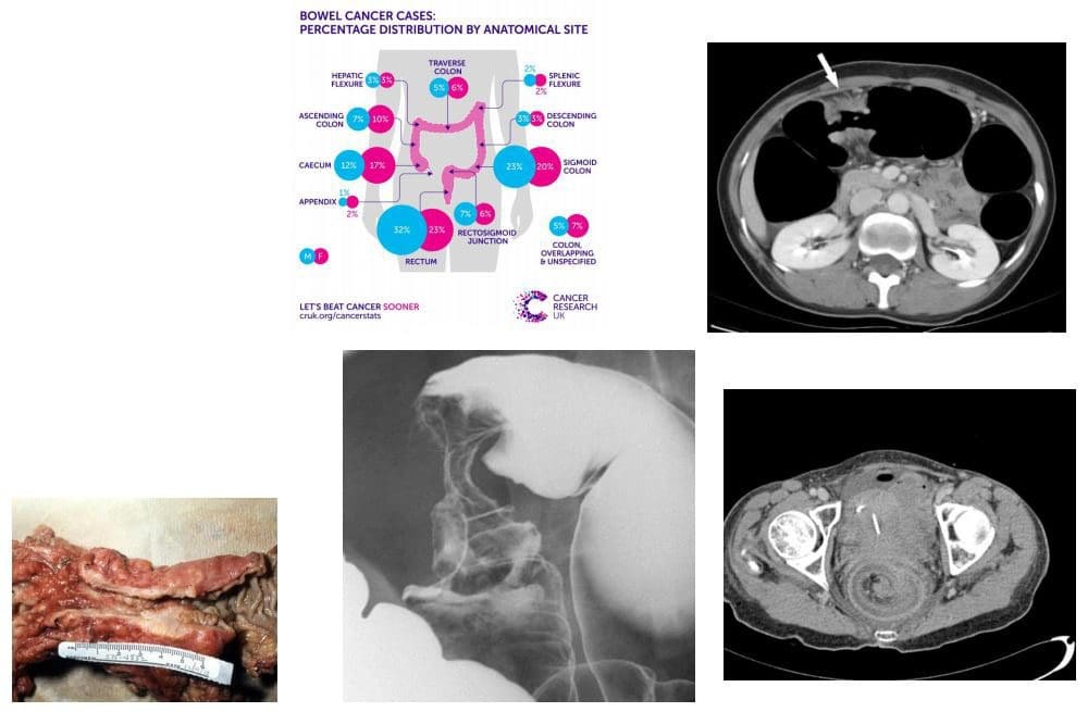
Colorectal carcinoma (CRC) m/c cancer of the GI tract and the 2nd most frequent malignancy in adults. Dx: endoscopy and biopsy. CT is the modalities most frequently used for staging. Surgical resection may be curative although the five-year survival rate is 40- 50% depending on staging. Risk factors: low fiber and high fat and animal protein diet, obesity (especially in men), chronic ulcerative colitis. Colonic adenomas (polyps). Familial adenomatous polyposis syndromes (Gardener syndrome) and Lynch syndrome as non-familial polyposis.
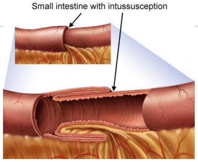
Clinically: insidious onset with altered bowel habits, fresh blood or melena, iron deficiency anemia from chronic occult blood loss especially in the right-sided tumors. Bowel obstruction, intussusception, heavy bleeding and metastatic disease especially to the Liver may be initial presentation. Path: 98% are adenocarcinomas, arise from pre-existing colonic adenomas (neoplastic polyps) with malignant transformation. The five-year survival rate is 40-50%, with stage at operation the single most important factor affecting prognosis. M/C rectosigmoid tumors (55%),
N.B. Some adenocarcinomas esp. mucinous types typically presented late and usually carry poor prognosis due to late presentation and mucin secretion and local/distant spread
Imaging: Barium enema is sensitivities for polyps >1 cm, single contrast: 77-94%, double contrast: 82-98%. Colonoscopy is a modality of choice for prevention, detection, and identification of colorectal carcinoma. Contrast-enhanced CT scanning is used for staging and prognosis evaluation of mets.
Screening: colonoscopy: men 50 y.o.-10-years if normal, 5-years if polypectomy, FOB, 1st degree relative with CA begin surveillance at 40 y.o
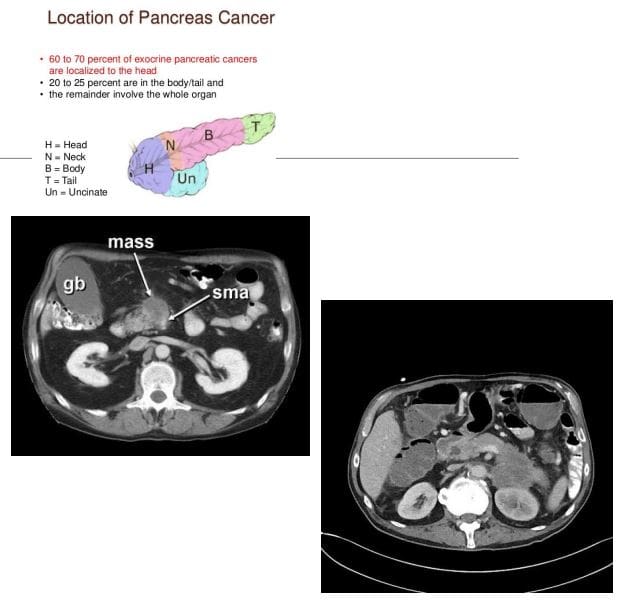
Pancreatic Cancer: ductal epithelial adenocarcinoma (90%), very poor prognosis with high mortality. 3rd M/C abdominal cancer. Colon is #1, stomach #2. Pancreatic cancer accounts for 22% of all deaths due to gastrointestinal malignancy, and 5% of all cancer deaths. 80% of cases in 60+. Cigarette smoking is the strongest environmental risk factor, a diet rich in animal fats and protein. Obesity. Family history. M/C detected in the head and uncinate process.
Dx: CT scanning is crucial. Invasion of Superior Mesenteric Artery (SMA) indicates unresectable disease. 90% of pancreatic adenocarcinomas are unresectable at Dx. Most patients die within 1-year of Dx. Clinically: painless jaundice, abd. Pain, Courvoisier�s gallbladder: painless jaundice and enlarged gallbladder, Trousseau�s syndrome: migratory thrombophlebitis, new onset diabetes mellitus, regional and distant metastasis.
CT Dx: pancreatic mass with strong desmoplastic reaction, poor enhancement, and slightly lower attenuation compared to the adjacent normal gland, SMA invasion.
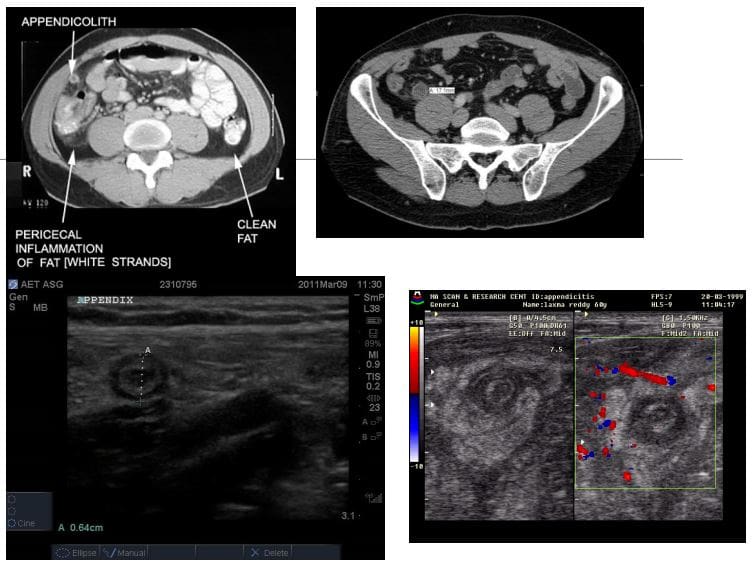
Appendicitis: very common condition in general radiology practice and is a major cause of abdominal surgery in young patients
CT is the most sensitive modality to detect appendicitis
Ultrasound should be employed in younger patients and children
KUB Radiographs should play no role in the diagnosis of appendicitis
On imaging, appendicitis reveals inflamed appendix with wall thickening, enlargement, and periappendiceal fat stranding. Similar findings of wall thickening and enlargement are noted on US. Typical �target sign� is noted on short axis US probe position.
If the appendix is retro-caecal than US may fail to provide accurate Dx and CT scanning may be required
Rx: operative to avoid complications
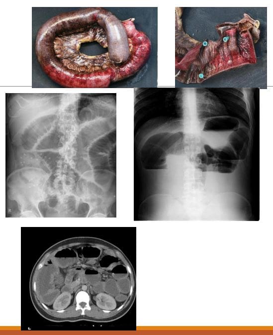
Small bowel obstruction (SBO)-80% of all mechanical intestinal obstruction; the remaining 20% result from large bowel obstruction. It has a mortality rate of 5.5%
M/C cause: any Hx of previous abdominal surgery and adhesions
Classical presentation is constipation, increasing abdominal distension with nausea and vomiting
Radiographs are only 50% sensitive for SBO
CT will demonstrate the cause of SBO in 80% of cases
There are variable criteria for maximal small bowel obstruction, but 3.5 cm is a conservative estimate of dilated bowel
On Abd x-ray: supine vs. upright. Dilated bowel, stretched valvulae conivente (mucosal folds), alternative air-fluid levels �step ladder.� Absent gas in the rectum/colon
Rx: operative as �acute abdomen.�
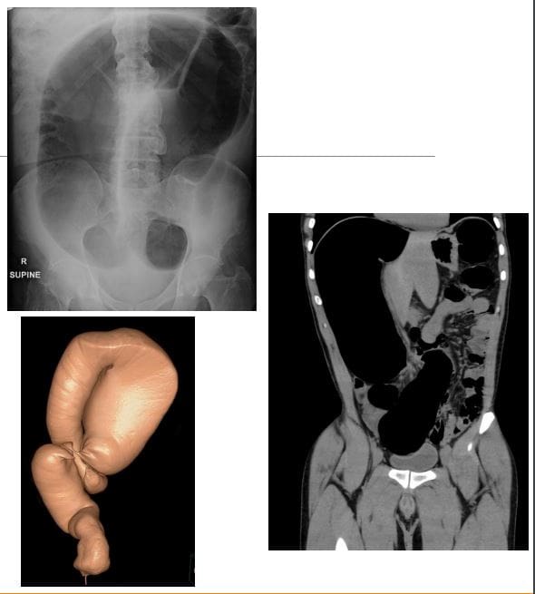
Volvulus-m/c in the Sigmoid colon esp. in elderly. The main reason: chronic constipation with redundant sigmoid twisting on sigmoid mesocolon. Leads to Large bowel obstruction (LBO). Other common causes: a colon tumor. Sigmoid vs. Caecum volvulus
Clinically: signs of LBO with constipation, abdominal bloating, pain, nausea, and vomiting. Onset may be acute or chronic
Radiographically: loss of haustra in the LB, LB distension (>6-cm), �coffee bean sign� next slide, the lower end of volvulus points to the pelvis
N.B: Rule of thumb for dilated bowel should be 3-6-9 where 3-cm SB, 6-cm LB & 9-cm Coecum
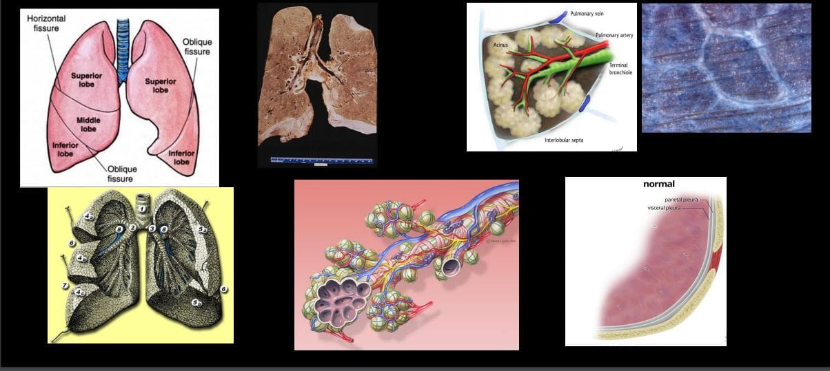
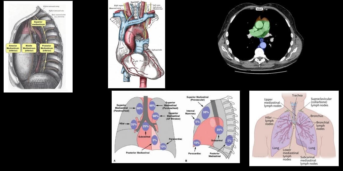
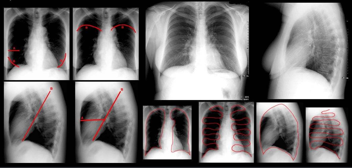
Note generations of the tracheal-bronchial tree, lobes, segments, and fissures. Note secondary pulmonary lobule (1.5-2-cm)-the basic functional unit of lungs observed on HRCT. Note important structural organization of the alveolar spaces with communications in between (pores of Kohn & canals of Lambert) that permit air drift and by the same mechanism allow exudative or transudative fluid to spread through the lung and stopped at the fissure. Note the anatomy of the pleura: parietal that is a part of the endothoracic fascia and the visceral that forms a lung edge � pleural space in between.
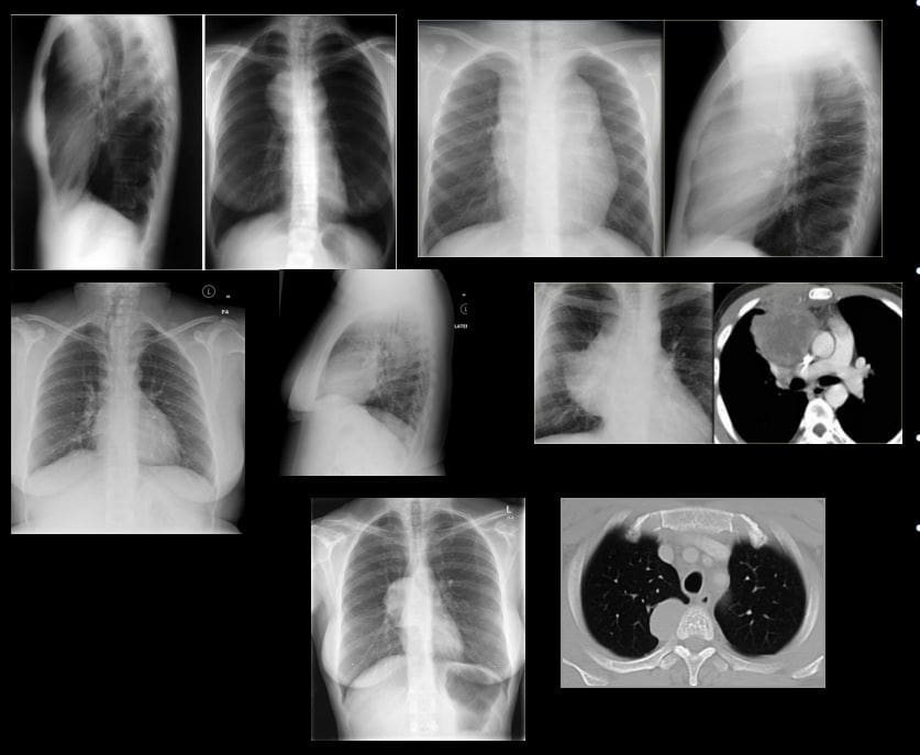
Mediastinum: surrounded by the pleura and the lung. Accommodates major structures contains numerous lymph nodes (see diagramme showing mediastinal nodes and their involvement in Lymphoma
General Approach to Investigating Chest Complaints
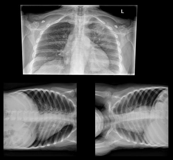
Decubitus views right and left: help to evaluate subtle pleural effusion, pneumothorax and other pathology
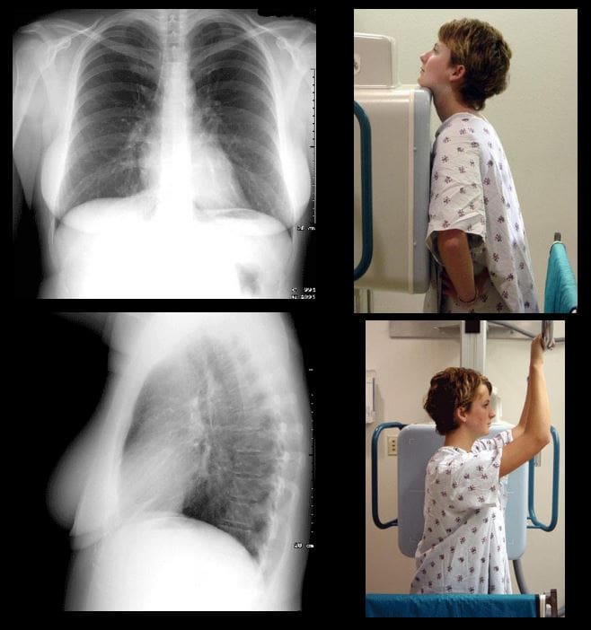
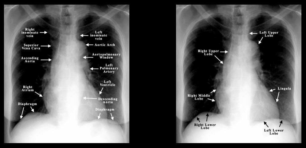
Normal CXR PA & Lateral views. Ensure good exposure: T-spine discs and vessels through the heart are visualized on PA view. Count 9-10 right posterior ribs to confirm adequate inspiratory effort. Begin a thorough survey using the following approach: Are There Many Lung Lesions A-abdomen/diaphragm, T-thorax wall, M-mediastinum, L-lungs individually, Lungs-both. Develop a good search pattern
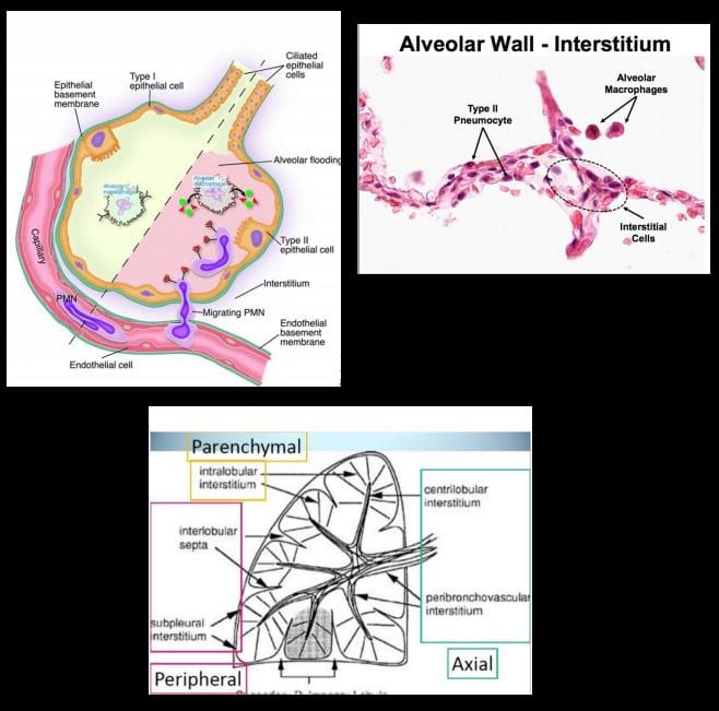
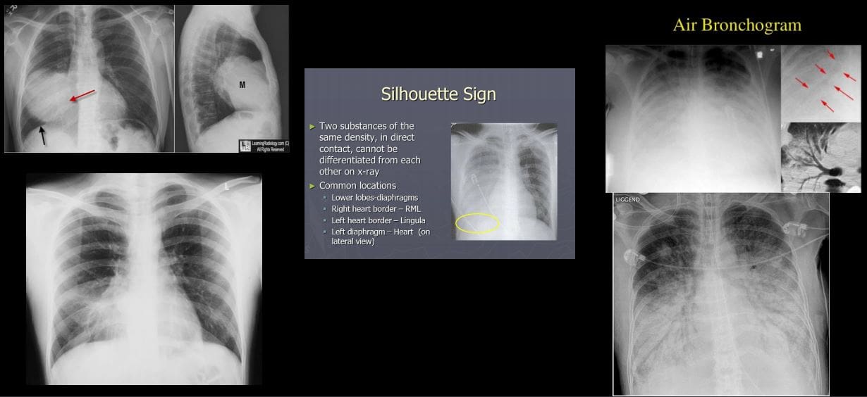
1) Airspace disease aka alveolar lung disease? Filling of the lung’s alveoli, acini and subsequently the entire lobe with fluid or substance of any composition (blood, pus, water, proteinaceous material or even cells) Radiographically: lobar or segmental distribution, airspace nodules may be noted, tendency to coalesce, air bronchograms and silhouette sign present. Batwing (butterfly) distribution noted as in (CHF). Rapidly changing over time, i.e., increase or decrease (days)
2) Interstitial disease: infiltration of pulmonary interstitium (alveoli septum, lung parenchyma, vessel walls, etc.) by for example by viruses, small bacteria, protozoans. Also infiltration by cells such as inflammatory/malignant cells (e.g., lymphocytes) Presented as an accentuation of lung interstitium with a reticular, nodular, mixed reticulonodular pattern. Different etiologies: inflammatory autoimmune diseases, fibrosing lung disease, occupational lung disease, viral/mycoplasma infection, TB, sarcoidosis lymphoma/leukemia and many other.
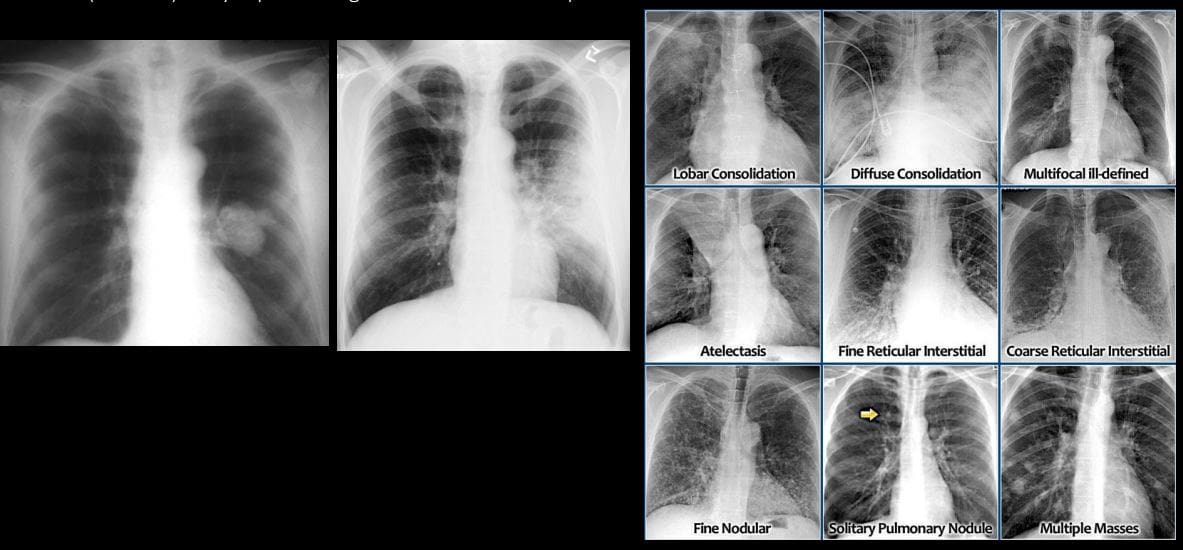
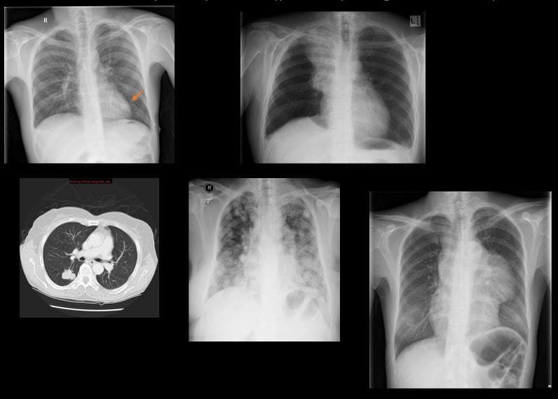
Recognizing different patterns of pulmonary disease can help with DDx. Mass vs. Consolidation (left). Note different patterns of pulmonary disease: airspace disease as lobar consolidation indicative of pneumonia, diffuse consolidation indicative of pulmonary edema. Atelectasis (collapse and volume loss). Interstitial patterns of pulmonary disease: reticular, nodular or mixed. SPN vs. Multiple focal consolidations (nodules) likely representing mets infiltrates vs. septic infiltrates
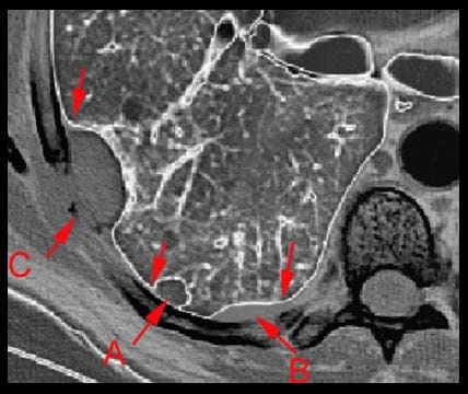
A = intraparenchymal
B = pleural
C = extrapleura
Recognize important location of chest lesions
Important signs: Silhouette sign: help with localization and DDx. Example: Bottom left image: radiopacity in the right lung, where is it located? Right MM because the right heart border that is adjacent to right middle lobe is not seen (silhouetted) Air bronchograms: air containing bronchi/bronchioles surrounded by fluid
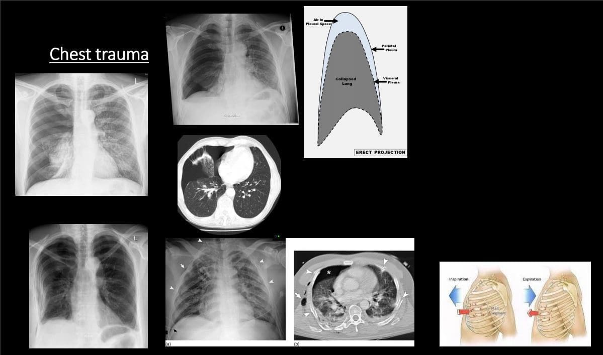
Chest Trauma
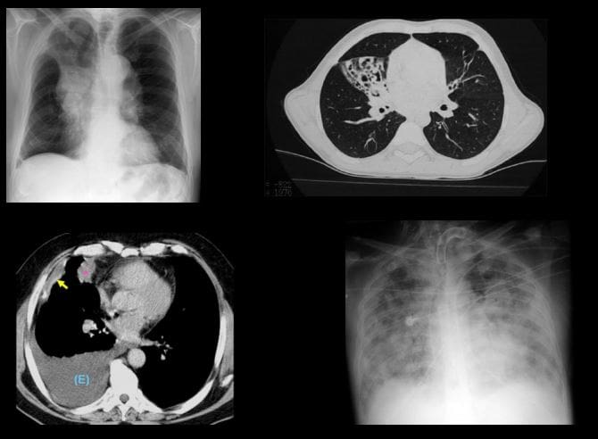
Pneumothorax (PTX): air (gas) in the pleural space. Many causes. Complications:
Tension PTX: continuous increase of air in the pleural space that rapidly compresses mediastinum and lung rapidly reducing venous return to the heart. It can be fatal if not treated rapidly
Spontaneous PTX: primary (young adults (30 -40) especially tall, thin men. Additional causes: Marfan�s syndrome, EDS, Homocystinuria, a – 1 -antitrypsin deficiency. Secondary: older pts with parenchymal disease: neoplasms, abscess, emphysema, lung fibrosis and honeycombing, catamenial PTX d/t endometriosis and others.
CXR: note visceral pleural line aka lung edge. An Absence of pulmonary tissue/vessels beyond the visceral pleural line. Subtle pneumothorax can be missed. On erect position, air rises and PTX should be sought at the top.
Rib fractures: v.common. Traumatic or pathological (e.g., mets, MM) Rib series x – rays are not very useful because CXR and/or CT scanning are more important to evaluate posttraumatic PTX (bottom left) lung laceration and another major path
Infection
Pneumonia: bacterial vs. viral or fungal or in the immunocompromised host (e.g., Cryptococcus in HIV/AIDS) Pulmonary TB
Pneumonia: community-acquired vs. hospital-acquired. Typical bacterial pneumonia or Lobar (non-segmental) pneumonia with purulent material filling the alveoli and spreading to the entire lobe. M/C organismStreptococcus Pneumonia or the Pneumococcus
Others: (Staph, Pseudomonas, Klebsiella esp. in alcoholics potentially leading to necroSIS/lung gangrene) Mycoplasma (20-30s) aka walking pneumonia, etc.
Clinically: a productive cough, fever, pleuritic chest pain sometimes hemoptysis.
CXR: confluent airspace opacity confined to the entire lobe. Air bronchograms. Silhouette sign help with location.
Viral: Influenza, VZV, HSV, EBV, RSV, etc. presents as interstitial lung disease that can be bilateral. May lead to respiratory compromise
Atypical pneumonia and Fungal Pneumonia: Mycoplasma, Legionnaire’s disease, and some fungal/Cryptococcus pneumonia may present with interstitial lung disease.
Pulmonary abscess: an infectious collection of purulent material in the lungs that often necrotizes. May lead to significant pulmonary and system complications/life-threatening.
On CXR or CT: round collection with thick borders and central necrosis containing air-fluid level. DDx from empyema that distorts the lung and pleural-based
Rx: antibiotics, antifungal, antiviral agents.
Pneumonia needs to be followed up with repeat CXR to ensure complete resolution
Lack of radiographic improvement of pneumonia may represent declined immunity, antibiotic resistance, underlying lung carcinoma or other complicating factors
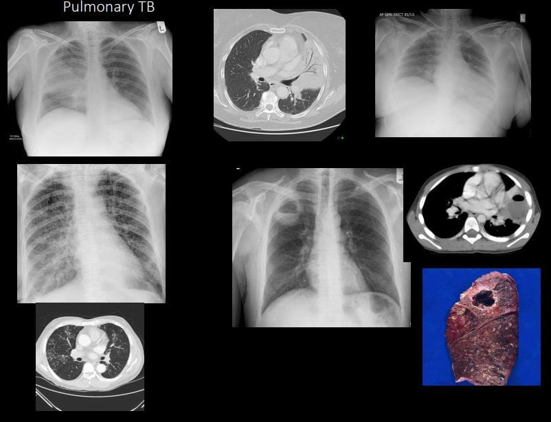
Pulmonary TB
Common infection worldwide (3rd world countries). 1 in 3 persons worldwide is affected by TB. TB is caused by Mycobacterium TB or Mycobacterium Bovis. Intracellular bacillus. Macrophage plays a key role.
Primary Pulmonary TB & Post-primary TB. Requires repeated exposure through inhalation. In most immunocompetent hosts, the active infection does not develop
TB presents as 1) cleared by the host, 2) suppressed into Latent Tuberculosis Infection (LTBI) 3) cause active disease TB. Patients with LTBI are not spreading TB.
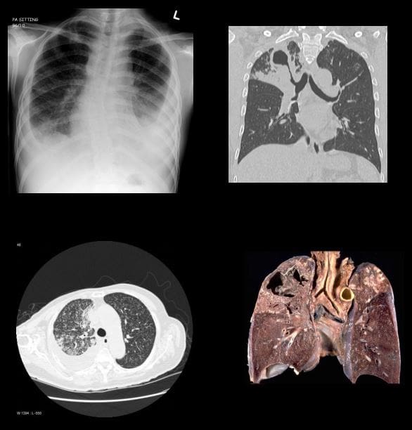
Imaging: CXR, HRCT. Primary TB: pulmonary airspace consolidation (60%) lower lobes, lymphadenopathy (95%- hilar & paratracheal), pleural effusion (10%). The Spread of primary TB most likely in immunocompromised and children.
Milliary TB: pulmonary and system complication dissemination that can be fatal
Post-primary (secondary) or reactivation infection: Mostly in the Apices and posterior segments of the upper lobes )high PO2), 40%-cavitating lesions, patchy or confluent airspace disease, fibrocalcific. Latent features: nodal calcifications.
Dx: Acid-fast bacilli (AFB) smear and culture (sputum). HIV serology in all patients with TB and unknown HIV status
Rx: 4-drug regimen: isoniazid, rifampin, pyrazinamide, and either ethambutol or streptomycin.
Pulmonary Neoplasms (primary lung cancer vs. pulmonary metastasis)
Lung cancer: m/c cancer in men and 6th most frequent cancer in women. Strong association with carcinogens inhalation. Clinically: late discovery, depending on the location of the tumor. Pathology (types): Small cell (SCC) vs. Non-small cell carcinoma
Small cell: (20%) develops from neuroendocrine aka Kultchitsky cell, thus may secrete biologically active substances presenting with paraneoplastic syndrome. Typically located centrally (95%) at or near the mainstem/lobar bronchus. Most show poor prognosis and unresectable.
Non-small cell: Lung adenocarcinoma (40%) (M/C lung cancer), M/C in women and non-smokers. Others: Squamous cell (may present with cavitating lesion), Large cell and some others
Plain film (CXR): new or enlarged focal lesion, widened mediastinum suggestive of lymph node involvement, pleural effusion, atelectasis, and consolidation. SPN-may represents potential lung cancer especially if it contains irregular borders, feeding vessels, thick wall, in the upper lungs. Multiple lung nodules are likely to represent metastasis.
Best Modality: HRCT with contrast.
Other chest neoplasms: Lymphoma is v. common in the chest especially in mediastinal and internal mammary notes.
Overall M/C pulmonary neoplasms are a metastasis. Some tumors show a higher predilection for lung mets, e.g., Melanoma, but any cancer can metastasize to the lungs. Some mets referred as �Cannonball� metastasis
Rx: radiation, chemotherapy, resection
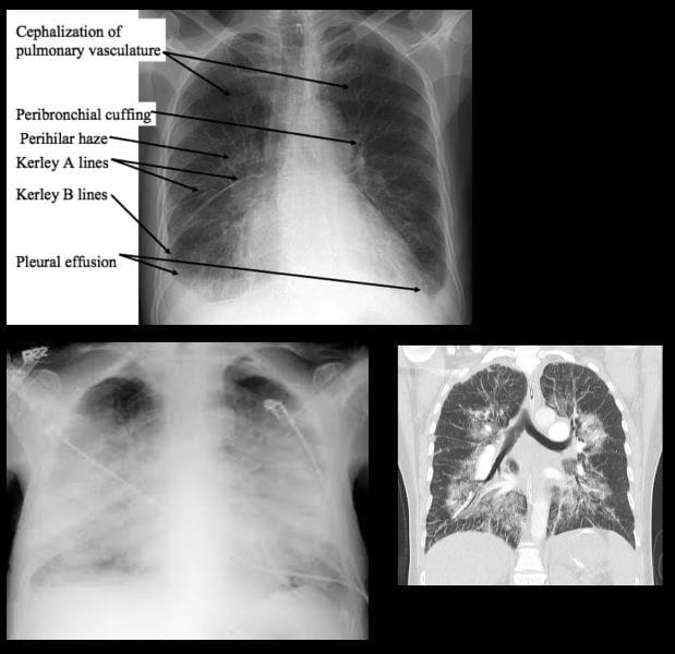
Pulmonary edema: a general term defines abnormal fluid accumulation outside vascular structures. Broadly divided into Cardiogenic (e.g., CHF, mitral regurgitation) and Non-cardiogenic with a multitude of causes (e.g., fluid overload, post-transfusion, neurological causes, ARDS, near drowning/asphyxiation, heroin overdose, and others)
Causes: increased in Hydrostatic pressure vs. decreased in oncotic pressure.
Imaging: CXR and CT: 2-types Interstitial and Alveolar flooding. Imaging presentation depends on stages
In CHF: Stage 1: redistribution of vascular flow (10- 18-mm Hg) noted as �cephalization� of the pulmonary vasculature. Stage 2: Interstitial edema (18-25-mm Hg) Interstitial edema: peribronchial cuffing, Kerley lines (lymphatics filled with fluid) A, B, C lines. Stage 3: Alveolar edema: airspace disease: patchy consolidations developing into diffuse airspace disease: Batwing edema, air bronchograms
Rx: 3 main goals: Initial O2 to keep O2 at 90% saturation
Next: (1) reduction of pulmonary venous return (preload reduction), (2) reduction of systemic vascular resistance (afterload reduction), and (3) inotropic support. Treat underlying causes (e.g., CHF)
Lung atelectasis: incomplete expansion of pulmonary parenchyma. The term “collapsed lung” is typically reserved for when the entire lung is collapsed
1) Resorptive (obstructive) atelectasis occurs as a result of complete obstruction of an airway (e.g. tumor, inhaled objects, etc.)
2) Passive (relaxation) atelectasis occurs when contact between the parietal and visceral pleura is disrupted (pleural effusion & pneumothorax)
3) Compressive atelectasis occurs as a result of any thoracic space-occupying lesion compressing the lung and forcing air out of the alveoli
4) Cicatricial atelectasis: occur as a result of scarring or fibrosis that reduces lung expansion as in granulomatous disease, necrotizing pneumonia, and radiation fibrosis
5) Adhesive lung atelectasis occurs from surfactant deficiency and alveolar collapse
6) Plate-like or discoid often developed after following general anesthesia
7) Imaging features: lung collapse, migration of lung fissures, deviation of the mediastinum, rising of the diaphragm, hyperinflation of adjacent unaffected lung
Mediastinum: pathology can be divided into those that result in a focal mass or those that result in diffuse disease involving the mediastinum. Additionally, air may track into the mediastinum in pneumomediastinum. Knowledge of mediastinal anatomy helps the Dx.
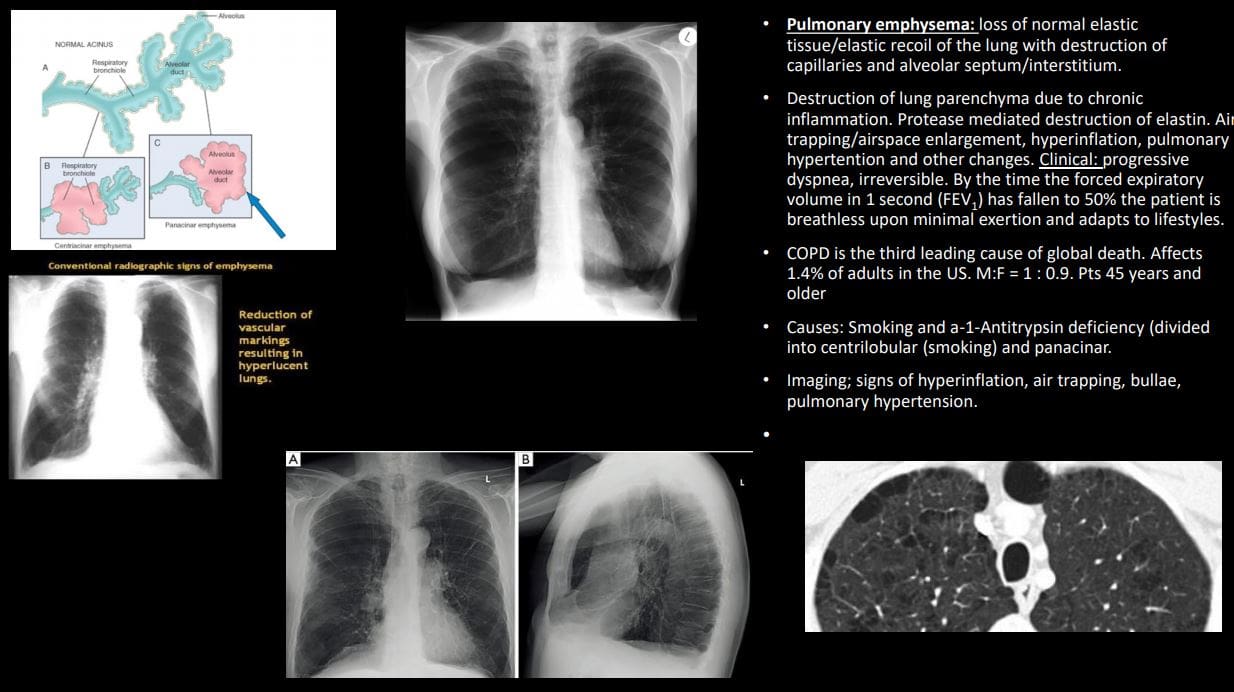
Pulmonary emphysema: loss of normal elastic tissue/elastic recoil of the lung with the destruction of capillaries and alveolar septum/interstitium.
Destruction of lung parenchyma due to chronic inflammation. Protease-mediated destruction of elastin. Air trapping/airspace enlargement, hyperinflation, pulmonary hypertension, and other changes. Clinical: progressive dyspnea, irreversible. By the time the forced expiratory volume in 1 second (FEV1 ) has fallen to 50% the patient is breathless upon minimal exertion and adapts to lifestyles.
COPD is the third leading cause of global death. Affects 1.4% of adults in the US. M:F = 1 : 0.9. Pts 45 years and older
Causes: Smoking and a-1-Antitrypsin deficiency (divided into centrilobular (smoking) and panacinar.
Imaging; signs of hyperinflation, air trapping, bullae, pulmonary hypertension.
SKULL FX: COMMON IN THE SETTINGS OF HEAD INJURIES. SKULL FX OFTEN POINT TO OTHER COMPLICATING FACTORS: INTRA-CRANIALHEMORRHAGING, CLOSED TRAUMATIC BRAIN INJURY AND OTHER SERIOUS COMPLICATIONS
SKULL X-RAYS ARE VIRTUALLY OBSOLETE IN EVALUATING HEAD INJURY. CT SCANNING W/O CONTRAST IS THE MOST IMPORTANT INITIAL STEP IN EVALUATION OF ACUTE HEAD TRAUMA. MRI HASA POOR ABILITY TO REVEAL SKULL FRACTURES, AND NOT TYPICALLY USED FOR AN INITIAL DX OF ACUTE HEAD TRAUMA.
SKULL FX ARE IDENTIFIED AS FXS OF SKULL VAULT, SKULL BASE AND FACIAL SKELETON EACH ASSOCIATED WITH SPECIFIC FEATURES AND HELP TO PREDICT COMPLICATIONS.
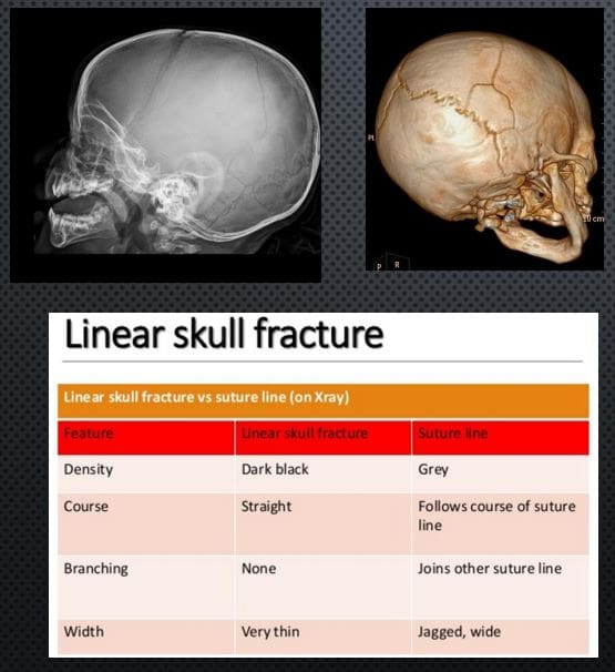
LINEAR SKULL FX: SKULL VAULT. M/C FX. CT SCANNING IS THE KEY TO EVALUATE ARTERIALEXTRADURAL HEMORRHAGING
X-RAY DDX: SUTURES VS. LINEAR SKULL FX. FX IS THINNER, �BLACKER� I.E. MORE LUCENT, CROSSESSUTURES,�AND VASCULAR GROOVES, LACKSSERRATIONS
RX: IF NO INTRACRANIAL BLEEDS THAT NO TREATMENT. NEUROSURGICAL CARE IF BLEEDSDETECTED BY CT SCANNING
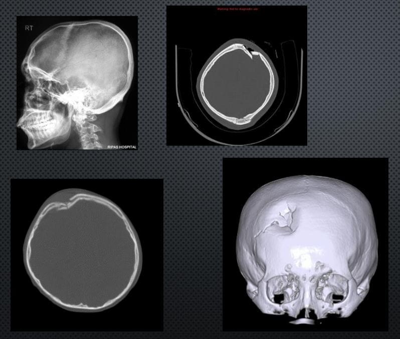
DEPRESSED SKULL FX: 75% IN THE VAULT. CAN BE DEADLY. CONSIDERED AN OPEN FX. MOST CASES NEED NEUROSURGICALEXPLORATION ESPECIALLY IFFRAGMENTS DEPRESSED >1-CM.COMPLICATIONS: VASCULAR INJURY/HEMATOMAS, PNEUMOCEPHALUS, MENINGITIS, TBI, CSF LEAK, BRAIN HERNIATION ETC.
IMAGING: CT SCANNING W/O CONTRAST
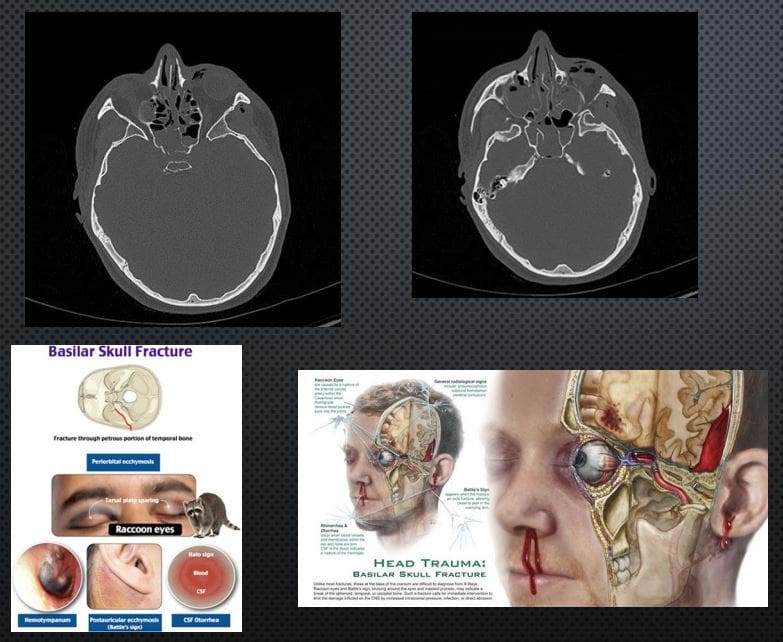
BASILAR SKULL FX: CAN BE DEADLY. OFTEN ALONG OTHER MAJOR HEAD TRAUMA OF THE VAULT AND FACIALSKELETON, OFTEN WITH TBI AND MAJORINTRACRANIAL HEMORRHAGING. OFTEN OCCUR AS �HEADBAND� EFFECT OF IMPACT AND MECHANICAL TENSION THROUGH THE OCCIPUT AND TEMPORAL BONES THROUGH SPHENOID AND OTHER BASE OF SKULL BONES. CLINICALLY: RACCOON EYES, BATTEL SIGN, CSFRHINO/OTORRHEA.
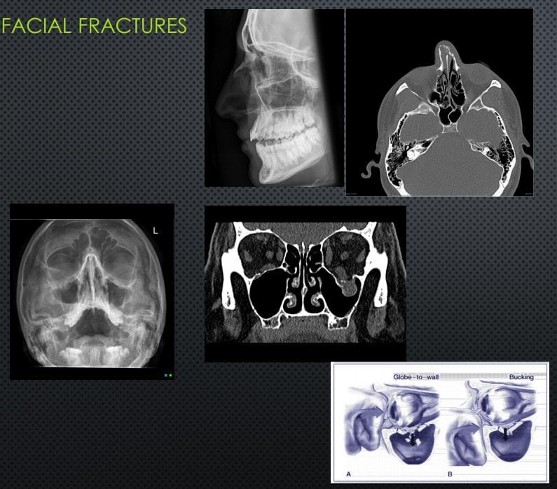
Facial Fractures
NASAL BONES FX: 45% OF ALLFACEFXM/C IMPACT IS LATERAL(FIST BLOW ETC.) IF UNDISPLACEDNO TREATMENT, IF DISPLACED MAY COMPLICATE AIR FLOW AND RESPIRATORY PASSAGE, MAY BE ASSOCIATED WITH OTHER FACIAL/SKULL INJURY. X-RAYS 80%SENSITIVE, FOLLOWED BY CT INCOMPLEX INJURIES.
ORBITAL BLOW OUT FX: COMMONORBITAL INJURY D/T IMPACT ON THE GLOBE AND/OR ORBITAL BONE. FX OF ORBITAL FLOOR INTOMAXILLARY SINUS VS. MEDIAL WALL INTO ETHMOID SINUS. COMPLICATIONS: ENTRAPPEDINFERIOR RECTUS M, PROLAPSEORBITAL FAT,�AND SOFT TISSUES, HEMORRHAGING AND OPTIC NERVE DAMAGE. RX: CONCERNS OF GLOBE INJURY ARE IMPORTANT, GENERALLY TREATEDCONSERVATIVELY IF NO COMPLICATIONS PRESENT
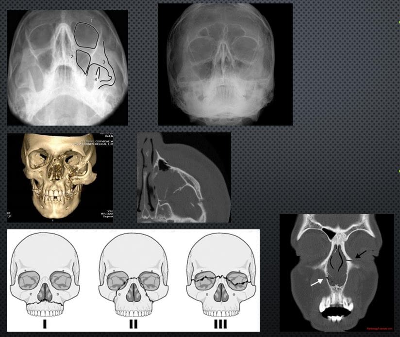
TRIPOD FX: 2ND M/C FACIAL FX#AFTER NASAL (40% OF MIDFACEFX) 3-POINT FX-ZYGOMATICARCH, ORBITAL PROCESS OF ZYGOMATIC BONE & SIDE OF MAXILLARY SINUS WALL, MAXILLARY PROCESS OF ZYGOMATIC BONE.COMPLICATED BY NERVE INJURY, TEMPORALIS M DAMAGE ETC. CT SCANNING IS MORE INFORMATIVE THAT X-RAYS (WATER�S VIEW).
LEFORT FX: SERIOUS FX ALWAYS INVOLVES PTERYGOID PLATES, POTENTIALLY SEPARATINGMIDFACE AND ALVEOLAR PROCESS WITH TEETH FROM THE SKULL. CONCERNS: AIRWAYS, HEMOSTASIS, NERVE INJURIES. CT SCANNING IS REQUIRED. POTENTIAL RISK OF BASILAR SKULL FX
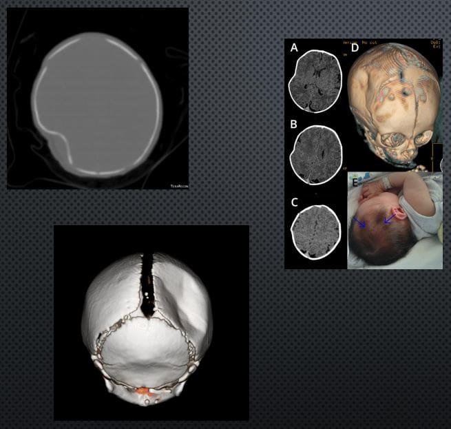
PING-PONG FX:�EXCLUSIVELY IN INFANTS. AN INCOMPLETE FX D/T FOCALDEPRESSION: FORCEPS DELIVERY, DIFFICULT LABOUR ETC. FOCALTRABECULAR MICROFRACTURIINGLEAVING DEPRESSION RESEMBLING APING-PONG. DX IS MAINLY CLINICALSEEN AS FOCAL DEFECT �DEPRESSION� IN THE SKULL. TYPICALLYNEUROLOGICALLY INTACT. CT MAY HELP IF BRAIN INJURY IS SUSPECTED. RX: OBSERVATIONAL VS. SURGICAL IN COMPLICATED INJURIES. SPONTANEOUSREMODELING HAS BEEN REPORTED
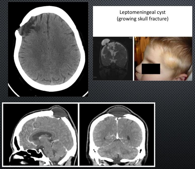
LEPTOMENINGEAL CYST (GROWING SKULL FX)- ARE AN ENLARGING SKULL FRACTURE THAT DEVELOPS ADJACENT TO POSTTRAUMATIC ENCEPHALOMALACIA
IT IS NOT A CYST, BUT AN EXTENSION OF THEENCEPHALOMALACIA THAT SEEN A FEW MONTHS POST-TRAUMA WITH PREVIOUS SKULL FX FOLLOWEDBY HERNIATION OF THE MENINGES AND ADJACENTBRAIN WITH PULSATIONS OF THE CSF. CT IS BEST ATDX THIS PATHOLOGY. INDICATES: GROWING FX AND ADJACENT ENCEPHALOMALACIA AS FOCALHYPOATTENUATING LESION.
DDX: INFILTRATING CELLS/METS/OTHER NEOPLASMSINTO SUTURES, EG, INFECTION ETC.
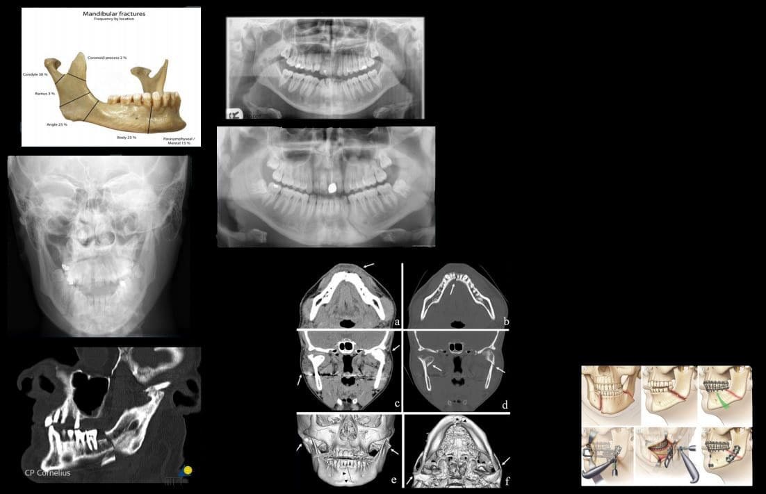
MANDIBULAR FXS: COMMON. POTENTIALLYCONSIDERED AN OPEN FX D/T INTRA-ORALEXTENSION. 40% FOCAL BREAK DESPITEMANDIBLE BEING A RING. DIRECT IMPACT(ASSAULT) M/C MECHANISM
PATHOLOGICAL FX D/T BONE NEOPLASMS, INFECTION ETC. IATROGENIC DURING ORAL SURGERY (TOOTH EXTRACTION)
IMAGING: MANDIBLE X-RAYS, PANOREX, CT SCANNING ESP. IN CASES OF ASSOCIATEDFACE/HEAD TRAUMA
COMPLICATIONS: AIRWAY OBSTRUCTION, HEMOSTASIS IS A MAJOR CONSIDERATION, DAMAGE TO MANDIBULAR N, OSTEOMYELITIS/CELLULITIS AND POTENTIAL SPREAD THROUGH FLOOR OF THE MOUTH (LUDWIGANGINA) AND NECK FASCIAL SOFT TISSUES INTOMEDIASTINUM. CANNOT BE NEGLECTED D/T HIGH MORTALITY RATES.
RX: CONSERVATIVE VS. OPERATIVE
Acute Intracranial Hemorrhage
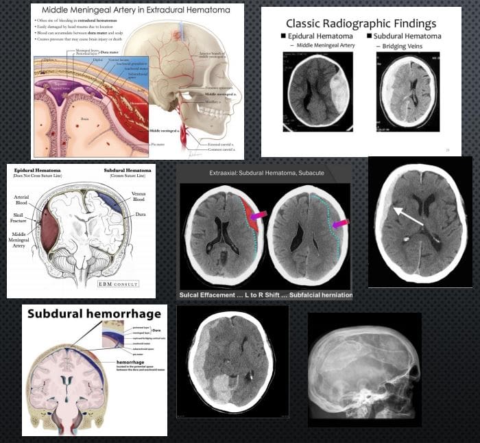
EPI AKA EXTRADURAL: (EDH) TRAUMATIC RAPTURE OF MENINGEAL ARTERIES (MMA CLASSIC) WITH RAPIDLY FORMING HEMATOMA BETWEEN THE INNER SKULL AND OUTER DURA. CT SCANNING IS THE KEY TO DX: PRESENTS AS �LENTIFORM� I.E. BICONVEX COLLECTION OF ACUTE (HYPERDENSE) BLOOD THAT DOES NOT CROSSSUTURES AND HELPS WITH DDX OF A SUBDURAL HEMATOMA. CLINICALLY: HA, LUCID EPISODE INITIALLY AND DETERIORATING IN A FEW HOURS.COMPLICATIONS: BRAIN HERNIATION, CN PALSY. O/A GOOD PROGNOSIS IF QUICKLY EVACUATED.
SUBDURAL HEMATOMA (SDH): RAPTURE OF BRIDGINGVEINS BETWEEN INNER DURA AND THE ARACHNOID.SLOW BUT PROGRESSIVE BLEED. MAY PARTICULARLYAFFECT THE VERY YOUNG AND ELDERLY AND IN ALL AGES (MVA, FALLS ETC.) MAY DEVELOP IN �SHAKEN BABY SYNDROME�. DX MAY BE DELAYED AND WORSEN THE PROGNOSIS WITH HIGH FATALITIES. IN ELDERLY HEAD TRAUMA MAY BE MINOR OR NOT RECALLED. EARLYIMAGING WITH CT IS CRUCIAL. PRESENTS AS CRESCENTSHAPEDCOLLECTION THAT CAN CROSS SUTURES BUT STOPPED AT DURAL REFLECTIONS. DIFFERENTATTENUATION ON CT D/T DIFFERENT STAGES OF BLOODDECOMPOSITION: ACUTE, SUBACUTE,�AND CHRONIC.MAY FORM A CHRONIC COLLECTION-CYSTICHYGROMA. CLINICALLY: VARIABLE PRESENTATION, 45-60% PRESENT WITH SEVERELY DEPRESSED CNS STATUS, PUPILLARY INEQUALITY. OFTEN WITH INITIAL BRAIN CONTUSION, THEN A LUCID EPISODE BEFORE SEVERELYDETERIORATING. IN 30% CASES OF FATAL BRAIN INJURY PATIENTS HAD SDH. RX: URGENT NEUROSURGICAL.
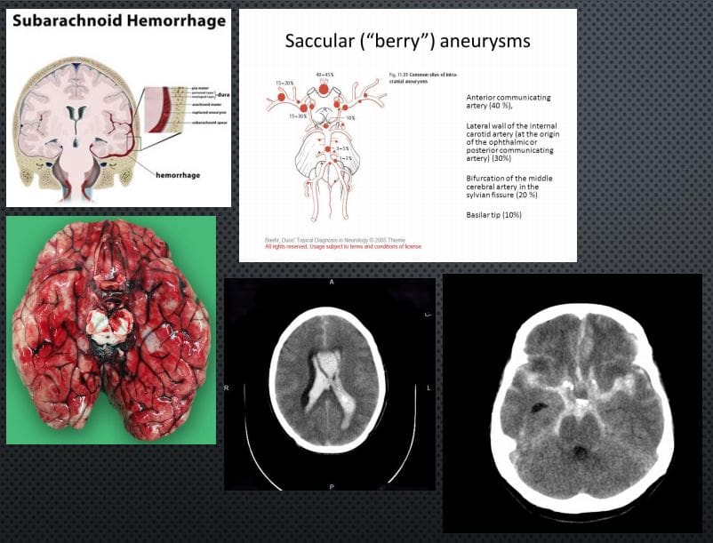
SUBARACHNOID HEMORRHAGE (SAH): BLOOD IN THE SUB-ARACHNOID SPACE AS THE RESULT OF TRAUMATIC OR NON-TRAUMATIC ETIOLOGY: BERRY ANEURYSMS AROUND CIRCLE OF WILLIS.SAH 3% OF STROKES, 5% OF FETAL STROKES.CLINICALLY: PRESENTS AS A �THUNDERCLAP HEADACHE� DESCRIBED AS A �WORST HA INLIFE�. PT COLLAPSES MAY OR MAY NOT REGAIN CONSCIOUSNESS. PATHOGY: DIFFUSE BLOOD INSA SPACE 1)SUPRASELLAR CISTERN WITH DIFFUSE PERIPHERAL EXTENSION, 2)�PERIMESENCEPHALIC, 3) BASAL CISTERNS. BLOOD LEAKED INTO SA SPACE UNDERARTERIAL PRESSURE INDUCES GLOBAL INCREASE IN INTRACRANIAL PRESSURE, ACUTE GLOBAL ISCHEMIA WORSENED BY VASOSPASM AND OTHER CHANGES.
DX: IMAGING: URGENT CT SCANNING W/O CONTRAST, CT ANGIOGRAPHY MAY HELP TO RULE OUT 99% OF SAH. LUMBAR PUNCTUREMAY HELP IN DELAYED PRESENTATION. AFTER INITIAL DX: MR ANGIOGRAPHY HELPS TO FIND THE CAUSE AND OTHER IMPORTANT FEATURES
IMAGING FEATURES: ACUTE BLOOD IS HYPERDENSE ON CT. FOUND IN DIFFERENTCYSTERNS: PERIMESENCEPHALIC, SUPRASELLA, BASAL, VENTRICLES,
RX: INTRAVENOUS ANTIHYPERTENSIVE MEDS, OSMOTIC AGENTS (MANNITOL) TO DECREASEICP. NEUROSURGICAL CLIPPING AND OTHER APPROACHES.
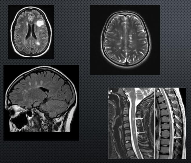
CNS Neoplasms: Benign vs. Malignant
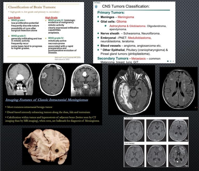
BRAIN TUMORS REPRESENT 2% OF ALL CANCERS. ONE THIRD ARE MALIGNANT, OF WHICH METASTATIC BRAIN LESIONS ARE THE MOST COMMON
CLINICALLY PRESENT WITH LOCAL CNS ABNORMALITIES, INCREASED ICP, INTRACEREBRAL BLEEDING ETC. FAMILIALSYNDROMES: VON-HIPPEL-LANDAU, TUBEROUS SCLEROSIS, TURCOT SYNDROME, NF1 & NF2 INCREASE THE RISK. IN CHILDREN: M/C ASTROCYTOMAS, EPENDYMOMAS, PNETNEOPLASMS (E.G. MEDULLOBLASTOMA) ETC. DX: BASED ON WHO CLASSIFICATION.
IMAGING IS CRUCIAL: INITIAL SYMPTOMS MAY PRESENT AS SEIZURE, ICP SIGNS HA. EVALUATED BY CT AND MRI WITH IV GADOLINIUM.
IMAGING DETERMINES: INTRA-AXIAL VS. EXTRA-AXIALNEOPLASMS. METS FROM PRIMARY BRAIN NEOPLASMS MAYO CCUR VIA CSF AND LOCAL VESSELS INVASION
NOTE AXIAL CT SLICE OF MENINGIOMA WITH AVIDCONTRAST ENHANCEMENT.
AXIAL MRI ON FLAIR PULSE SEQUENCE REVEALED EXTENSIVE NEOPLASM AND MARKED CYTOTOXIC EDEMA OF THE BRAIN PARENCHYMA CHARACTERISTIC OF GRADE IV GLIOMA (GBM) WITH VERY POOR PROGNOSIS. ABOVE FAR RIGHT IMAGE: AXIAL MRI FLAIR: BRAIN METASTASIS FROM BREAST CANCER. MELANOMA IS COMMONLY METASTASIZESTO THE BRAIN (SEE PATH SPECIMEN) MRI CAN BE DIAGNOSTIC D/T HIGH SIGNAL ON T1 AND CONTRAST ENHANCEMENT.
RX: NEUROSURGICAL, RADIATION, CHEMOTHERAPY,�IMMUNOTHERAPY TECHNIQUES ARE EMERGING
Most healthcare providers use x-rays as a diagnostic tool to treat a variety of patient complaints, including chiropractors. They can help doctors identify the source of a problem or if there is something more going on. X-rays can also help chiropractors determine the best course of action for treatment. To understand more, let’s take a closer look at what they are and how they’re used in most chiropractic offices.
What are x-rays?
An x-ray is a very vigorous form of electromagnetic radiation that is similar to radio waves, ultraviolet radiation, microwaves, or visible light that is used to view the internal composition of a person or thing. A beam is focused on a specific part of a person�s body, such as the back, it produces a digital image of the skeletal structure.
The beam passes easily through skin and other soft tissues�but is unable to pass through bone and teeth. Soft tissue that is denser, such as organs, ligaments, and muscles, will be visible�but will be captured in shades of gray. Areas like the bowel or lungs appear on the film as black.
The use of chiropractic x-rays
Chiropractic x-rays provide vital information that can affect how the chiropractor chooses to treat a patient. In some cases, chiropractic care or spinal manipulation may not be an appropriate course of action at that time, and the patient may be started on a different, gentler therapy.
Other times, it can show the chiropractor how to best proceed in treating the patient. In short, patients can receive better, more well-rounded care which can better facilitate their healing and pain management.
Identify a condition or symptom, such as a spinal tumor or lesion�that would provide a medical reason that a specific course of care should not be done.
Obtain important biomechanical information that can aid in guiding treatment.
To stay apprised and maintain a record of a patient�s degenerative process.
Aid in identifying anomalies in the spine and joints that can affect treatment.
Allows patients to understand their condition and treatment plan better, allowing them to take ownership of the process and be more involved in their therapy and healing.
What does a chiropractor look for on an x-ray film?
When a chiropractor takes an x-ray of a patient, they are looking for things in several particular areas. The first thing they check is to make sure that there are no dislocations, fractures, cancer, infections, tumors, or other potentially dangerous conditions.
They then look for disk height and other signs of disk degeneration, bone density, bone spurs, joint spaces, and alignment. This allows them to identify conditions like scoliosis and other conditions that may require specific forms of treatment.
Many chiropractors prefer that the patient is in a weight-bearing position when taking spinal x-rays. This differs from the majority of medical facilities which have the patient lie down.
The advantage of weight-bearing x-rays as a diagnostic tool�is that it allows for measuring, i.e., leg length deficiency, scoliosis, and the narrowing of joint space. It can also show that certain bones, such as the tibia and fibula, are separating which can be an indication of a torn tendon or problem with the joint. A non-weight bearing x-ray cannot provide the same perspective,�and vital clues to a patient�s condition may be missed.
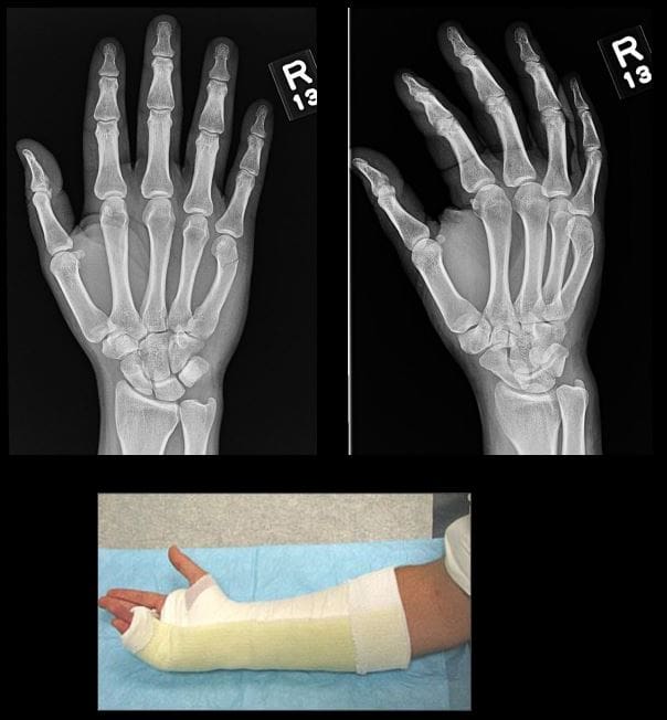
In all cases, Orthopedic hand surgical referral is required
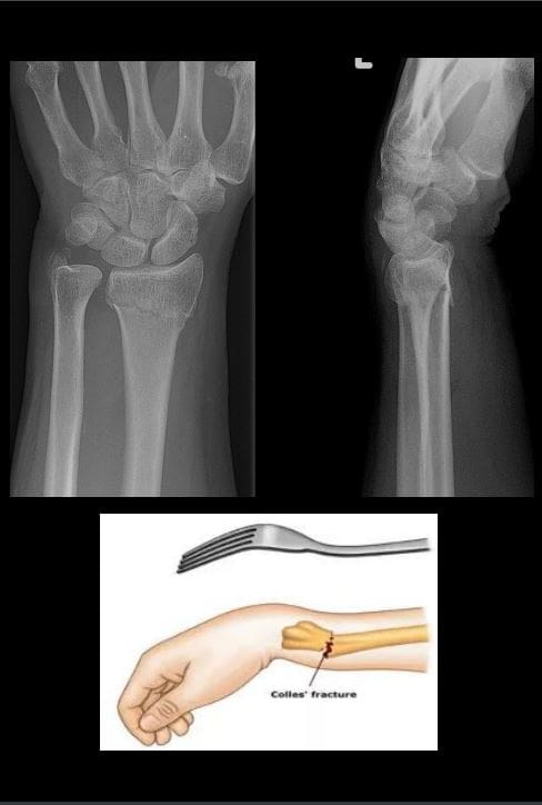
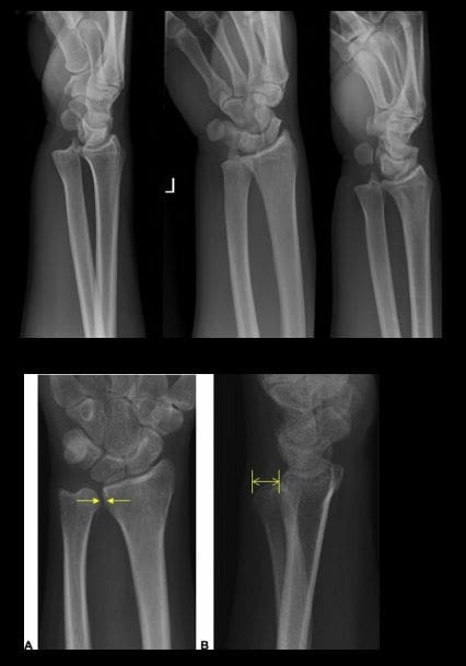
Colles fx: m/c d/t FOOSH+pronation. m/c inOSP/elder women. Rare in men and if occurs need DEXA to avoid hip Fx etc. Young pts: high-energy trauma. Typically extra-articular.50%-cases show Ulna styloid (US) Fx.
Imaging: x-rad is sufficient, CT in complex Fx, MRI helps with ligament tears and TFC.
Rx: if extra-articular and <5-mm distal radius shortening and <5-degree dorsal angulation closed reduction+casting is sufficient. ORIF in complex cases.
�Image Dx: distal rad impaction/shortening,dorsal angulation of distal fragment, carefully examine if intra-articular extension, 50% US Fx
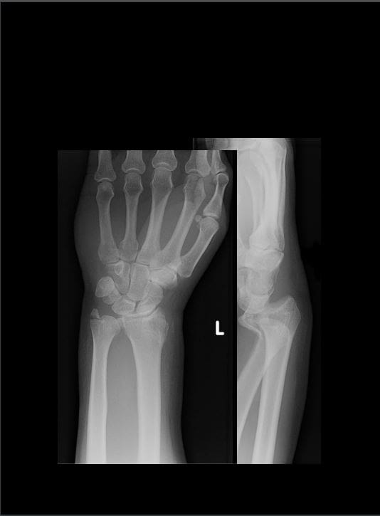
Smith Fx: Goyrand in French literature. Considered as reversed Colles, otherwise almost identical, I.e., 85% extra-articular, 50%US Fx, OSP/elderly women, young pts-high-energy trauma. Differences: mechanismFOOSHwith flexed wrist thus m. Less frequent.
Imaging steps: (see Colles Fx) C
Complications: similar to Colles Fx
Rad Dx: 85% extra-articular with volar(anterior) angulation of the distal fragment,radial shortening. Carefully examine cortical breach suspecting intra-articular extension that can be named as Smith type 2 or Reversed Barton Fx (next)
Rx: similar approach as in Colles.
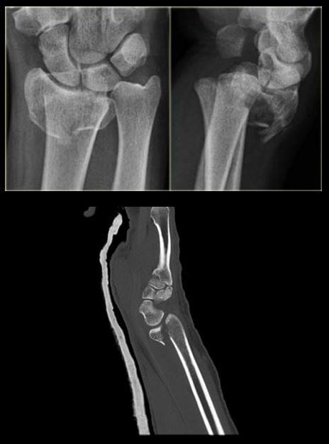
Barton fx: FOOSH, impaction of distal radius similar to Colles but the Fx line extends from the dorsal radial aspect into radiocarpal joint resulting with dorsal slip/dislocation of the carpus.
Imaging: 1st sept x-radiography often with CTto examine intra-articular Fx extension and operative planning
Rad Dx: distal radius Fx extending from dorsal into the radiocarpal joint with a variable degree of displacement, the proximal slip of the carpus
If Fx line extends from the volar aspect into the wrist joint named Reversed Barton aka Smith type 2 (above bottom image)
Complications: similar to all distal radius Fx
Rx: operative with ORIF
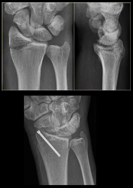
Chauffeur’s/backfire Fx aka Hutchinson Fx: intra-articular Fx of Radial styloid. The name derives from the time when the car had to be started with a hand crank that could backfire inducing wrist dorsiflexion and radial deviation.
Imaging: x-radiography is sufficient. CT may be helpful if Fx not readily shown by x-rays.
Rx: operative with percutaneous lagscrewin all cases d/t intra-articularnature
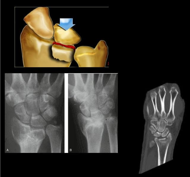
Die-Punch Fx: impaction Fx by the Lunate bone into distal articularLunate fossa of the Radius. IntraarticularFx. Derives its name from a technique to shape (impress) a hole in industrial machining “die-punch.”FOOSH injury.
Imaging: 1st step x-rays, may be equivocal d/t subtle depression of the Lunate fossa then CT scanning is most informative.
Rad Dx: impacted lunate fossa region with intra-articular Fx extension. This can present as a comminuted Fxarticular Fx of the Distal Radius.
Rx: operative d/t intra-articular Fx
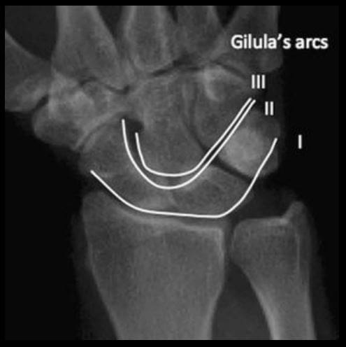
Construct arcs of Gilula when evaluating carpal injuries. An Important step required to avoid missing subtle changes in carpal alignment and cortical continuity
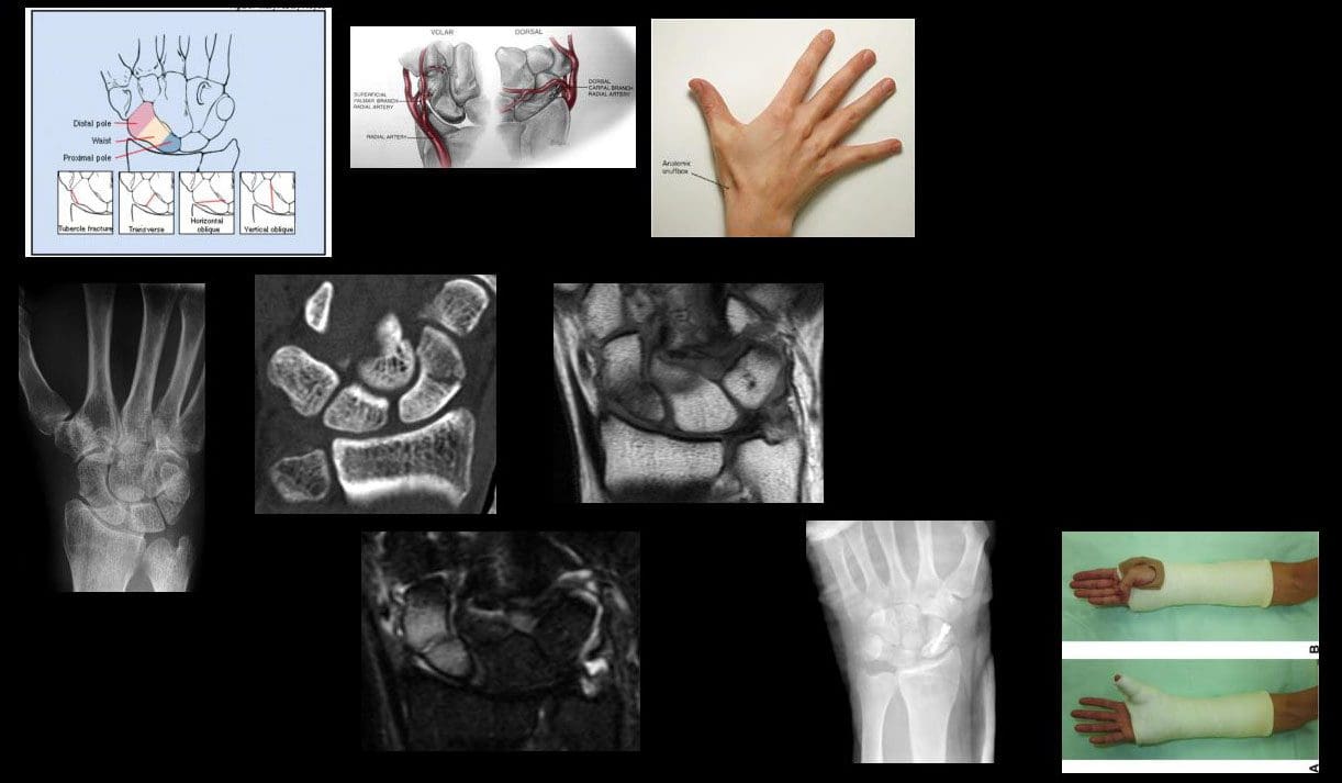
Scaphoid bone Fx: m/c Fx carpal bone. D/tFOOSH wrist extended radially deviated. Location of Fx is most important to prognosis: Waist-m/c location (70%). May have 70-100%chance of AVN. Proximal pole Fx: 20-30% with a high risk of non-union. Distal pole-10%shows better prognosis. Distal pole Fx is m/c in children. Key clinical sign; pain in the snuffbox.
Imaging: 1st step-x-radiography but 15-20%missed d/t occult Fx. Special views required. Thus MRI is the most sensitive and specific for early occult Fx. Bone scintigraphy has98/100% specificity & sensitivity esp. 2-3 days after the onset. Key rad. Dx: Fx line if evident, displacement and obscuration of scaphoid(navicular) fat pad, examine for scapholunate dissociation. If proximal bone appears sclerotic-AVN occurred. MRI: low on T1 & high on T2/STIR/FSPD d/t bone edema, a�low signalFx line can be noted.
Rx: Spica cast should be applied if clinically suspected even w/o x-ray findings. For waistFx-cast for 3-mo for prox pole 5-mo immobilization. ORIF or percutaneous pinning with a Herbert screw.
Scapholunate Ligaments Dissociation
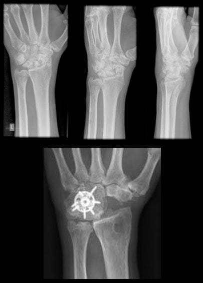
SNAC wrist: scaphoid non-union advanced collapse. Often d/t non-union and dissociation of scapholunate ligaments (SLL)with progressive radiocarpal and intercarpalDJD. The Proximal scaphoid fragment is attached to Lunate with distal dissociating and rotating�signet ring� sign on x-rays.
SNAC wrist may often result in DISI
Rx: progressive DJD may lead to four-corner arthrodesis
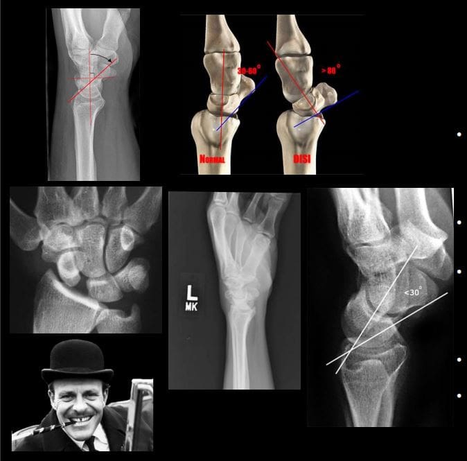
Scapholunate advanced collapse (SLAC wrist): SLLdissociation with progressive intercarpal and radiocarpal DJD and volar or dorsal carpal displacement (DISI & VISI). Causes: trauma, CPPD, DJD, Kienboch disease (AVN of Lunate), Preiserdisease (AVN of Scaphoid).
SLL dissociation will lead to Dorsal or VolarIntercarlate aka Intercarpal Segmental Instability (DISIor VISI).
Rad Dx: Dx underlying cause. X-rays demonstrate dorsal or volar angulation of the Lunate with increased or decreased scapholunate angle on the lateral view. On frontal view: Terry Thomas sign or widening of scapholunate distance 3-4-mm as the upper limit of normal.
�MRI may help with ligament evaluation and pre-surgical planning
Rx: often operative with late DJD. Four-corner arthrodesis
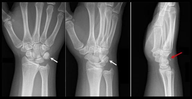
Triquetrum Fx: 2nd m/c carpal bone Fx. M/C dorsal aspect is avulsed by the�tough Dorsal radiocarpal ligament. Cause: FOOSH.
Imaging: x-radiography wrist series is sufficient. Best revealed on the lateral view as an avulsed bone fragment adjacent to the dorsum of the Triquetrum. CT may help if radiographically equivocal.
Rx: conservative care
Complications: rare, may persist as pain on the dorsum of the wrist
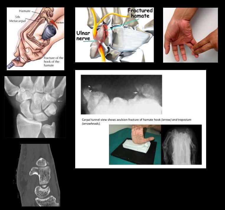
Hook of the Hamate Fx: m/c occurs in batting sports (cricket, baseball, hockey, impact by a golf club, etc.) 2% of carpusFx.
Imaging: x-radiography may fail to detect an Fx unless “carpal tunnel view” is used. CT may help if x-rays unrewarding.
Clinically: pain, positive pull test, weak, painful grip. Deep ulnar n. Branch may be affected within the�Guyon canal.
Rx: usually non-operative, but chronic non-union may require excision.
DDx: bipartite hamate
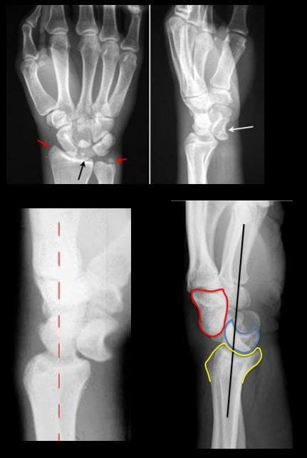
�Lunate vs. Perilunate dislocation: Lunate is m/c dislocated carpal bone. Overall infrequent carpal injury. However, often missed!
Occurs with FOOSH and wrist extended and ulnar deviated. Imaging: 1st step x-rays. Ifunrewarding or require more complex injury evaluation CT scanning.
Key Rad DDx: DDx Lunate from perilunate dislocation. Lunate dislocation: lunate lost its contact with distal radius �spilled teacup� on the lateral. Perilunate dislocation: Lunate maintains its contact with distal radius despite the Capitatedorsally dislocated. Lunate dislocation is additionally helped to identify a �pie sign� d/t Lunate overlapping the Capitate
Rx: emergency reduction and operative repair of torn ligaments
Metacarpal & Phalangeal Injuries
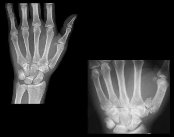
Bennett Fx: intra-articular but noncomminuted impact-type Fx of the base of 1st MC bone of the thumb. X-radiography is sufficient.
Rad Dx: characteristic triangular fragment of bone on the ulnar aspect of the 1st MCbase, often with radial subluxation of the remaining radial aspect of the 1st MC
Complications: DJD, non-union, etc.
Rx: prone to instability/non-union requiring an operative care
Rolando Fx: aka comminuted Bennett with Y or T-configuration. More complex injury. It is unstable requiring operative care
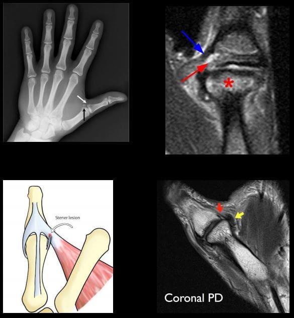
Gamekeeper thumb: traditionally described as a chronic tear of the�ulnar (medial) collateral ligament at 1stMCP in English Gamekeepers’ who performed neck twisting/killing of small game. An acute injury may also be named as Skier’s thumb. This injury can be ligamentous w/o a fracture and an avulsion injury at the 1st proximal phalanx base.
Complication: Stener lesion or displacement of torn ligament over Adductor pollicis muscle that cannot heal w/o surgical repair. MRI Dx is required.
Avoid thumb stress views that can induce a Stenerlesion
Imaging: x-radiography followed by MRI to Dx Stenerlesion. MSK US can be used if MRI is unavailable.
�Stener lesion on MRI & MSUS: ulnar collateral stump is more superficial to Adductor pollicis aponeurosis and appears like a low signal mass-like stump forming so-called “yo-yo on the string sign” reported both on MRI and MSK US.
Rx: often operative
Boxer Fx: m/c MC Fx. An extra-articular usually non-comminuted or minimal comminuted Fx through m/c the 5th and sometimes the 4th MCneck-head junction (occasionally through the shaft) resulting in volar head angulation. Mechanism: direct impact as in clenched fist punching hard surface (e.g., facial bones/wall punching) hence 95% in young males.
Imaging: x-radiography hand series is sufficient
Rad Dx: Fx line transverse or oblique through MCneck with volar head angulation. Evaluate the degree of displacement, critical to report.
N.B. If the same mechanism fractures the 2ndand 3d MC in the same anatomic area, it may require operative care.
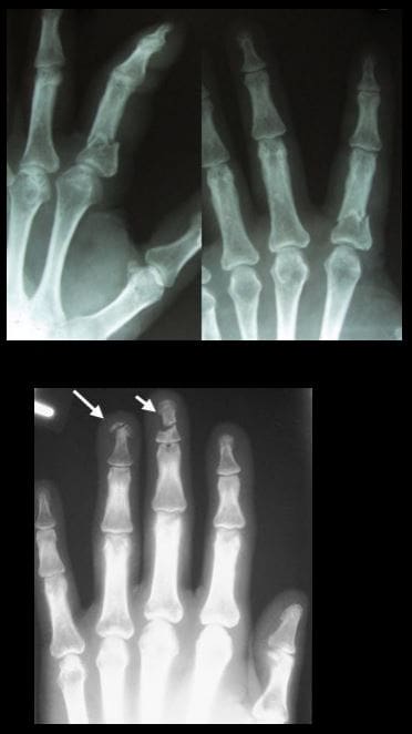
Phalangeal hand Fx: m/c skeleton Fx (10% of all Fx). Sports and industrial injuries dominate
Imaging: x-radiography with hand series or PA/lateral finger views will suffice
Rad Dx: if prox phalanx Fx, distal fragment is angled volarly with prox fragment dorsally. Distal phalanx may be angled dorsally. Key observation: nail bed injury, which considered an open Fx with a�risk of infection.
Rx: if <10-degree angulation-buddy-taping with motion rehab. CRPP vs. ORIF can be considered in complex cases-Orthopedic hand surgeon referral
Complication: loss of motion, necrosis, infection.May result with amputation
For additional common injuries: PIP is m/c dislocated joint. Mullet (Baseball) finger, Jersey finger and other injury refer to:
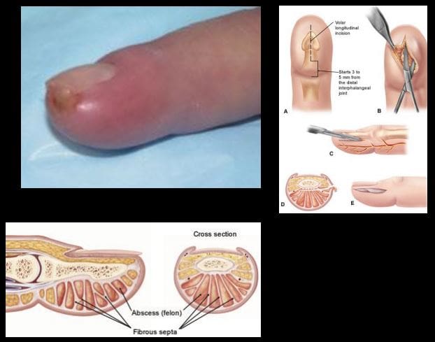
Felon: septic infection of the fingertip pulp typically with Staph.Aureus. Causes: needle prick(diabetics), paronychia, nail splinters, etc. m/c in index and thumb, presenting with pain, swelling, etc.
D/t specific pulp anatomy theinfection>swelling leads to pulp compartment syndrome-pressure and necrosis.
Rx: operative with incision distal to DIP, irrigation/debridement
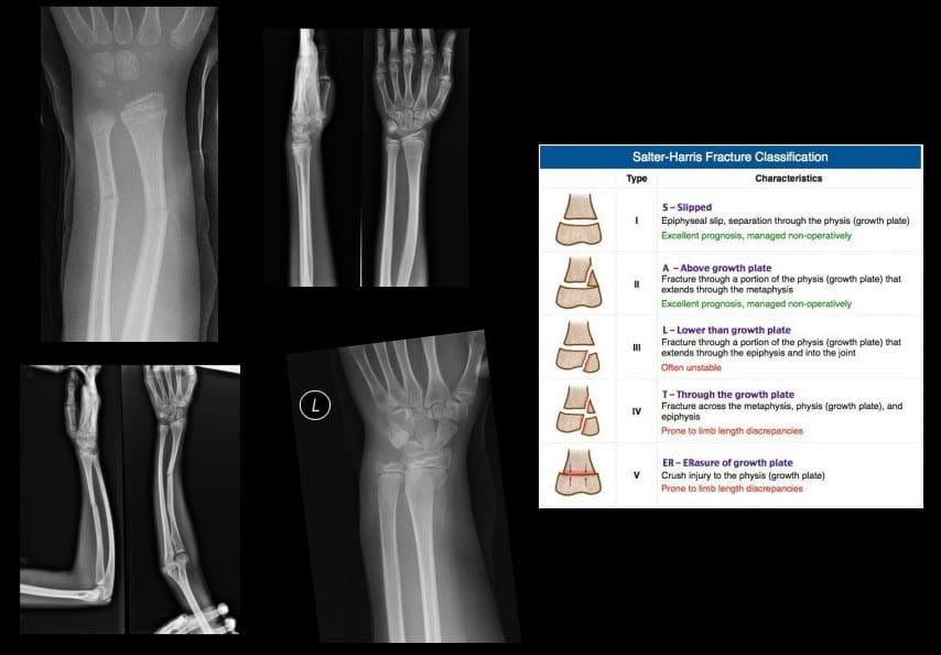
Pediatric Wrist Injury
Incomplete Fx: Greenstick Fx, Torus (Buckle)Fx, Bowing (Plastic) deformity/Fx. D/t FOSHe.g. fell off the monkey bar. m/c affects <10-years-old.
Rx: usually non-operative (closed reduction and casting)
Distal Radioulnar Joint (DRUJ) Instability-common injury following trauma as in FOOSHwith wrist hyperextension and rotation and disruption of DRUJ ligaments and TFCcomplex. Avulsion of ulnar styloid with the�dorsal or volar displacement of distal ulnar should be noted.
Imaging steps: x-rays initially, MRI may identify ligaments and TFC damage, MSKUScan help with ligaments tearing.
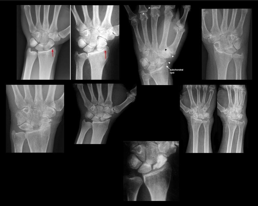
Wrist DJD-typically is secondary to trauma, scapholunate dissociation, SLAC, SNAC wrist, CPPD, Keinboch or Preiser Disease and others.
May lead to major functional loss
Imaging: typically presents as radiocarpal JSL, subchondral sclerosis,osteophytosis, subcortical cysts, and loose bodies. Typically additional induces intercarpal degeneration and particularly Tri-scaphe joint.
MRI may be helpful with early recognition of scapholunate dissociation, Lunate/Navicular AVN.
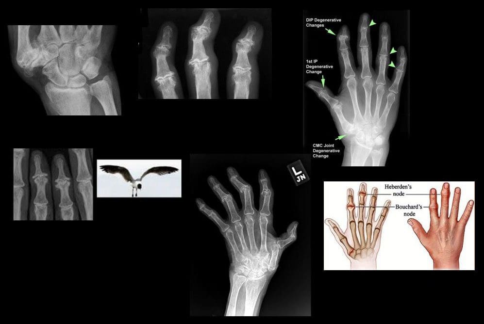
If isolated MCP OA noted considerCPPD & Hemochromatosis (Hook-like osteophytes)
Clinically:
Mid-age females
Typically painless except 1st CMC OA
DIPs-Heberden nodes, PIPs-Bouchard nodes
Erosive OA (occasionally called�inflammatory OA�)
A Spectrum of OA but producing central proximal erosions at DIPs and PIPsresulting with very characteristic �gullwing� appearance. No systemic inflammation (no CRP, RF, Anti-CCP Ab)typically in middle-aged/elderly females, like Hand OA, often seen in families
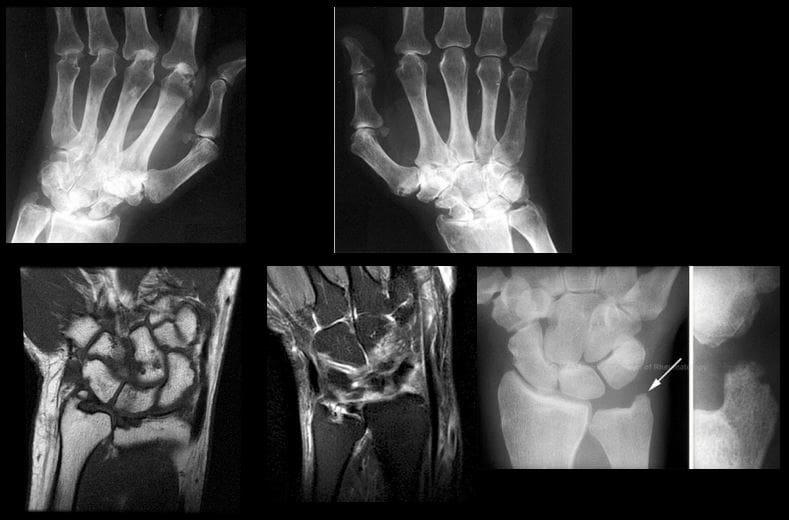
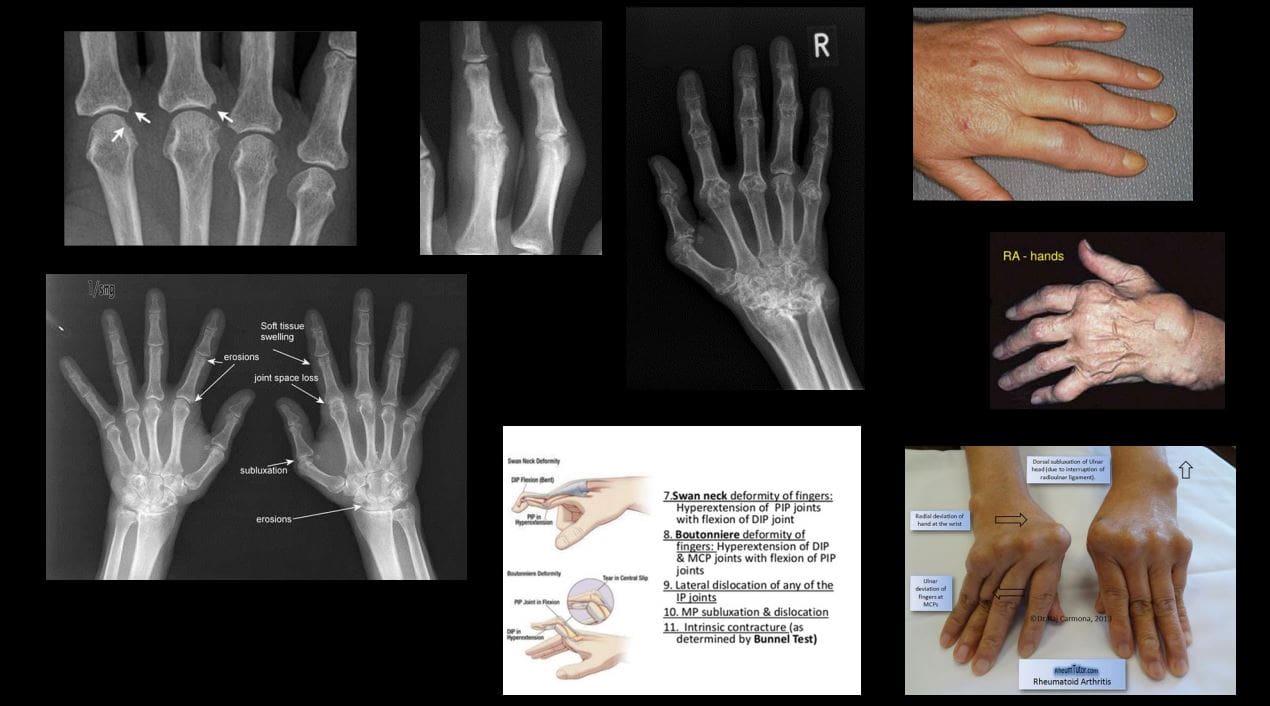
Rheumatoid Arthritis
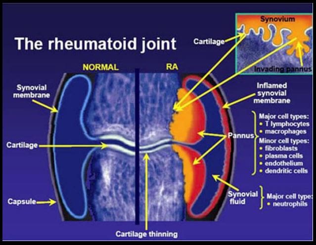
Rheumatoid Arthritis (RA)-chronic systemic inflammatory disease of unknown etiology, targeting synovial joints, tendons with multiple systemic involvement (lung, CVS, Ocular, Skin, etc.) Pathology: Tcell>Macrophage/APC>mediatedautoimmune process resulting in pannus formation and gradual destruction of ST, cartilage, bone,�and other tissues. 3% FemalesVS.1% Males. Environmental triggers: infection, trauma, smoking,�and others in a genetically susceptible individual. 20-30%may be disabled after 10-years.
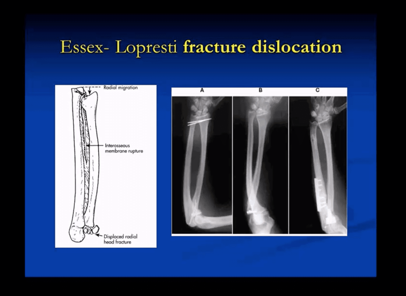
In adults: Radial head Fx is the m/c (33%) and accounts for 1.5-4% of all fractures. Etiology: FOOSH with forearm pronated. Associated injuries: elbow collateral ligaments tears. EssexLoprestiFx with interosseous membrane tearing and dislocation of the Distal Radio-Ulnar Joint(DRUJ)
Terrible triad: of the Radial head Fx, elbow dislocation and Coronoid process Fx (typically avulsed by the Brachialis M)
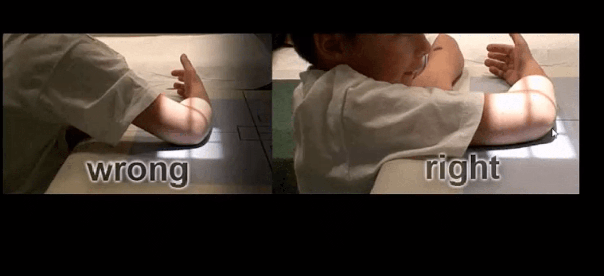
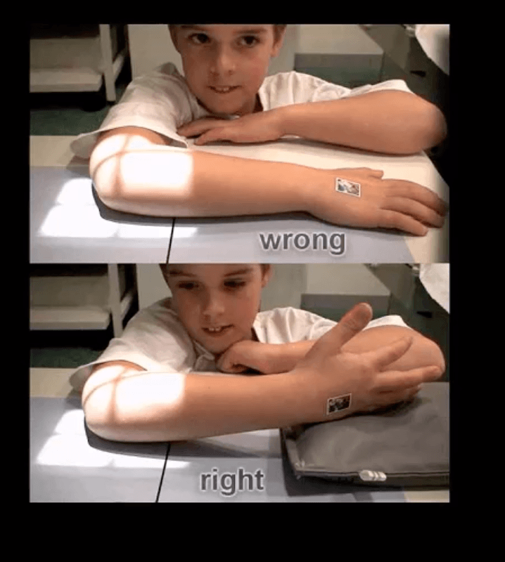
Imaging: 1st step is x-radiography with elbow series, CT scanning may help in complex cases, MRIif ligamentous injury.
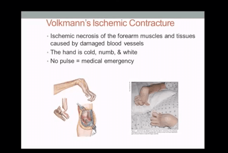
In children: Supracondylar Fx of the distal humerus accounts for 90% of acute trauma. It is always d/t accidental trauma with FOOSH and elbow extended, rarely <5% with flexed elbow. MostSupracondylar Fx occur in children <10 y.o. Males>Females. Complications: malunion in cubitus varus aka Gunstock deformity, vascular injury and acute ischemic compartment syndrome with Volkmann contracture
Imaging: 1st step x-radiography can be sufficient. CT occasionally used in complex cases.
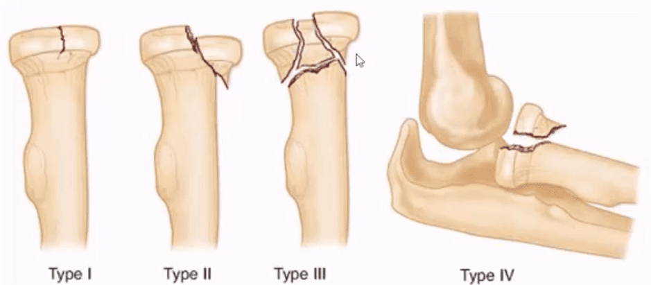
Radial head (RH) Fx: Mason classification helps to determine the degree of complexity and mode of treatment
Type 1- undisplaced is the m/c and stable contained by ligaments. On radiographs can be very subtle and evaluation of abnormal elbow fat pads is critical and often the only diagnostic clue
Type 2- displaced by 2-mm or > with rotational block
Type 3- comminuted >2-3 fragments and
Type4 is presented with RH fx, posterior elbow dislocation and sometimes Coronoid process fracture often d/t Brachialis M avulsion
Rx: Type 1 managed non-operatively by immobilization and movement rehab. Type 2- ORIF if rotational block. Type 3 and 4, ORIF and RH resection or RH arthroplasty
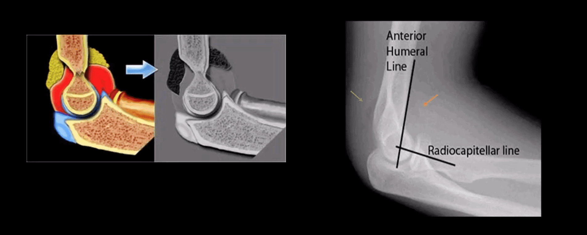
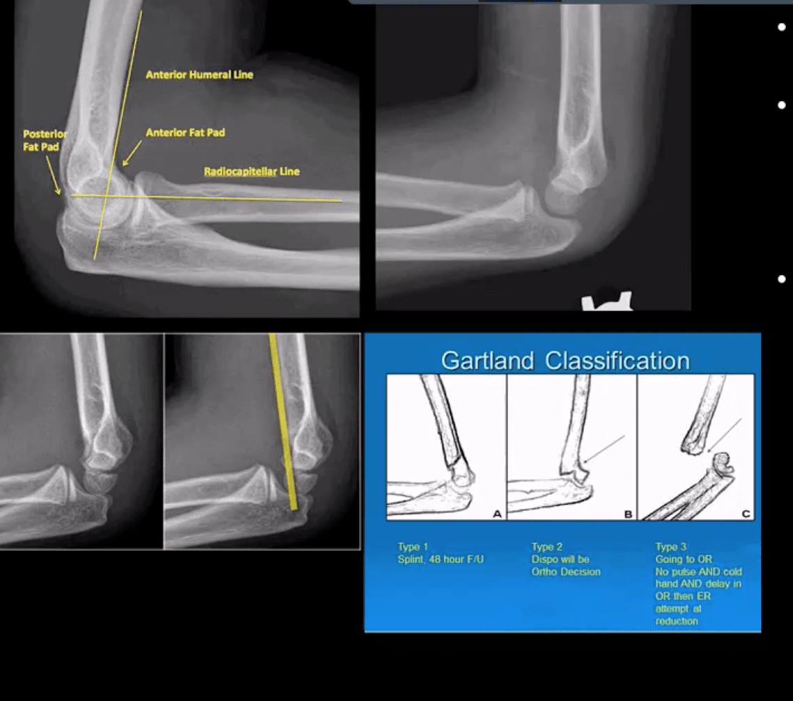
Note abnormally displaced anterior fat pad (orange arrow) and the emergence of the posterior fat pad (green arrow) that is usually deep in the olecranon fossa and not seen unless acute hemarthrosis or other effusiondevelopsFat pad signs are most reliable indicators of intra-articular elbow Fx
Mason type 1 RH Fx can be v. subtle and missed. Radiographic search should involve a�close evaluation of positive fat pad signs. Note anterior fat pad displacement aka Sail sign and the presence of the post fat pad d/t acute bleed
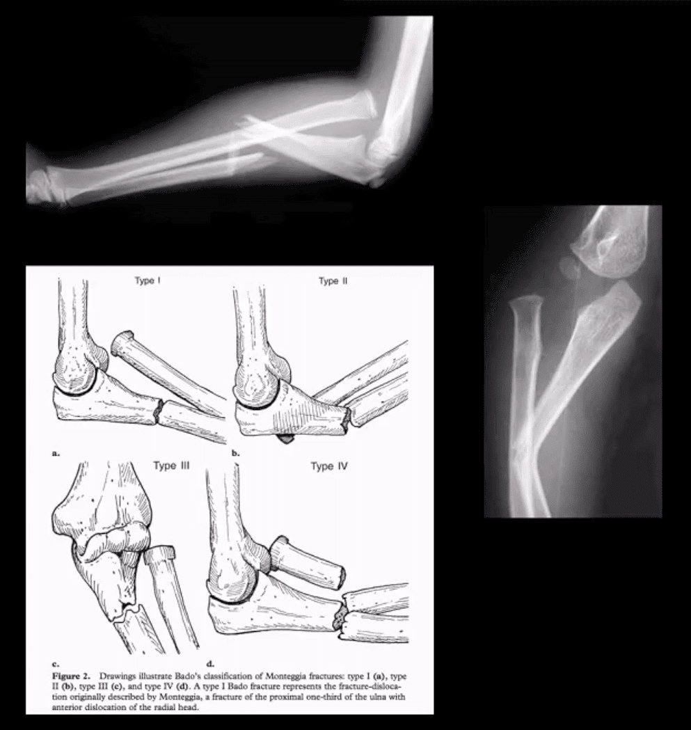
Monteggia fracture-dislocations: prox 1/3ulnar shaft Fx. with concomitant dislocation of PRUJ (radial head). FOOSH injury. Children4-12 y.o. Infrequent in adults.
X-rays readily reveal ulnar Fx, but radial head dislocation may be subtle and occasionally missed. This is a serious injury leading to elbow disability if Dx delayed 2-3 weeks or left untreated. X-rays are typically sufficient:Rx: casting vs. operative.
Supracondylar Fx: this is the M/C elbow Fx in children.
Especially, the un-displaced types 1(top right) is difficult to Dx. Abnormality of “fat pads” and anterior humeral line and radiocapitella line disturbance are often most reliable
Type 3 carries a particularly high risk for Volkmann contracture (vascular ischemic-necrosis of the anterior forearm muscle compartment
Elbow complaints in a young athlete
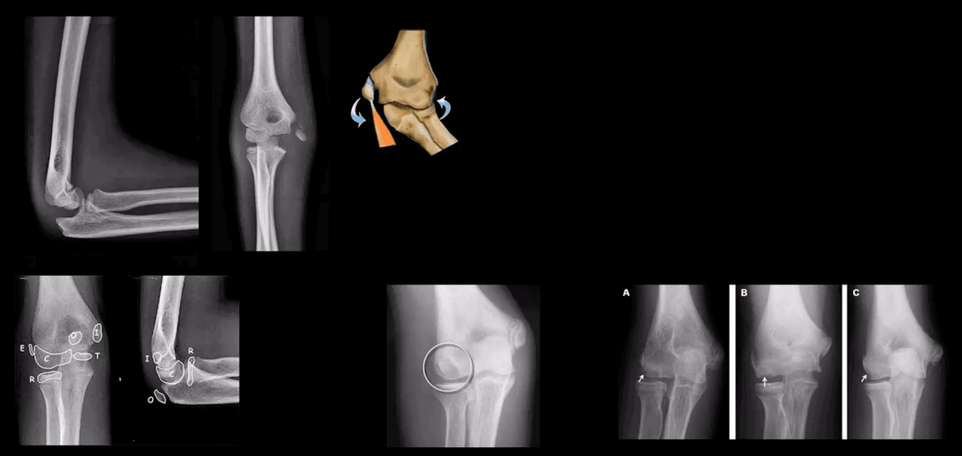
Epicondyle Fx: common pediatric injury, about 10%.Essentially an avulsion Fx and a MUCL tear. Medial epicondyle is m/c Fx. FOOSH is the m/c mechanism.M>F. If minimally displaced or undisplaced can be treated with casting esp. in non-dominant arm. If displaced as in this case, require ORIF.
Medial epicondyle avulsive Fx in a young baseball pitcher was coined a �little league elbow� in the 60sand now should be avoided to avoid confusion
OCD of the Capitellum is a common athletic injury induced by repeated compression/flexion. OCD must be DDx from Panner�s disease or osteochondritis typically presented in younger patients
Difficulty in diagnosis may stem�from multipleapophysis about the elbow (see CRITOE)
Imaging: 1st step: x-rays followed by MRI and MRarthrogramme if indicated.
CT may help with complex injury evaluation. MRI and MSKUS may help with a�ligament injury.
Elbow Arthritis
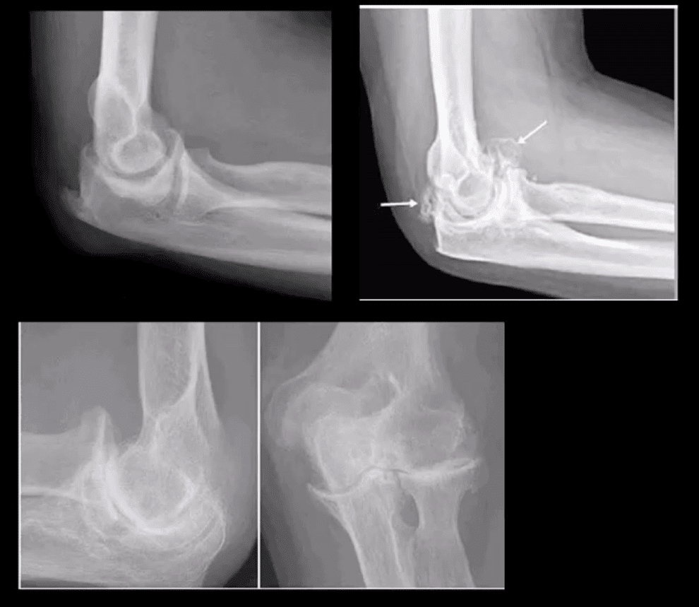
DJD of the elbow is uncommon and typically 2nd to trauma, occupation, CPPD, OCD of theCapitellum or other pathology. Clinically: pain, reduced ROM esp. in dominant arm, deterioration of ADL. Loss of terminal flexion and extension. 50% develop Ulnarcompressive neuropathy. Rx: conservative,arthroscopic debridement/osteophytes removal, capsular release. In older patients and not active patients Total Elbow Arthroplasty (TEA) can be used
Imaging: x-radiography is sufficient, CT helps with pre-operative planning
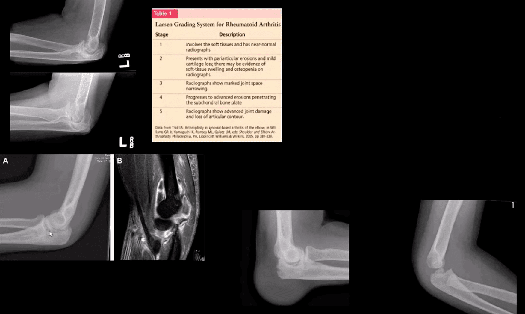
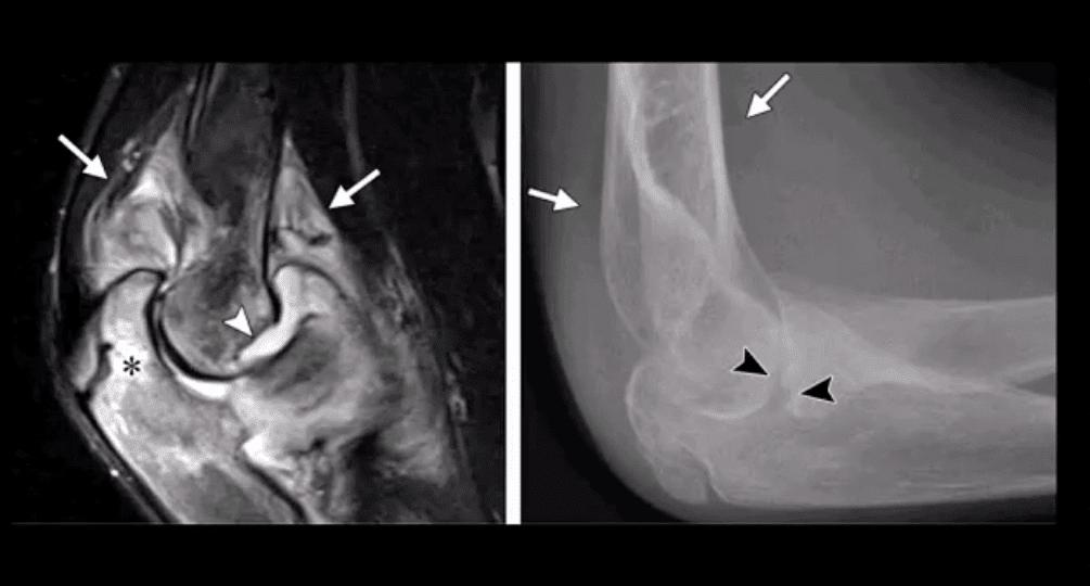
Inflammatory Arthritis: RA of the elbow is frequent (20-50%) and destructive d/t synovitis, pannus, bone/cartilage,�and ligamentous destruction/laxity. Clinically: begins after the onset of hands symptoms with, symmetrical swelling, pain, reduced ROM, flexion contracture. Presence of rheumatoid nodules can be noted along the olecranon and posterior forearm. Rx: DMARD, operative tendons repair.
Imaging: x-radiography with early non-specific effusion (fat pads),later: erosions, symmetric JSL, osteopenia. MSK US helps early Dx. MRI reveals synovitis; bone edema correlates with pre-erosive x-ray findings, synovial enhancement on FS T1+C.
Gouty Arthritis: may affect the elbow but less than in the lower extremity. Olecranon bursitis causing a �rising sun sign� on x-rays with or w/o bone erosions. Aspiration and polarised microscopy revealing needle-shaped negatively birefringent monosodium urate crystals. Rx: colchicine, other meds.
Septic Arthritis: consider in people with diabetes, IV drug users, concurrent RA, patients with active TB, gonococcal in young adults. Clinically presents as monoarthritis with or w/o constitutional signs. X-ray: poor detection in early stages. US may show effusion and high Doppler.MRI: effusion, osseous edema. Bone scintigraphy can help as well. Labs: CBC, ESR, CRP. Diagnostic arthrocentesis with gram staining and culture are crucial. Rx: Prompt IV antibiotics
Juvenile Idiopathic Arthritis (JIA) considered M/C chronic disease of childhood and preceded IBD infrequency. Dx is clinical and imaging: Criteria: Joint pain and swelling in a child 0-16-years for 6-weeks or longer. Many forms exist�M/C pauciarticular(oligoarticular) 40%, F>M, associated with ocular involvement (iridocyclitis) and potential blindness. Polyarticular and Systemic forms.
Elbow is frequently affected along with the knee, wrists,�and hands,�especially in polyarticular dz.
Labs: ESR/CRP RF-VE in most cases
Imaging: early x-ray features are non-specific. Later: osseous erosion, destruction of joint cartilage, overgrowth of articular epiphyses, early closure of physis. Delayed features: 2nd DJD, joint ankyloses.DDx: hemophilic arthropathy. Cervical radiographs are crucial.
Rx: DMARD, conservative care
Miscellaneous pathologies
Supracondylar process: 2% of the population. Described by Sir JohnStruthers in 1854. Fibrous band(Ligament of Struthers) may lead to compression of the Median N. DDx fromOsteochondroma that typically points away from the joint
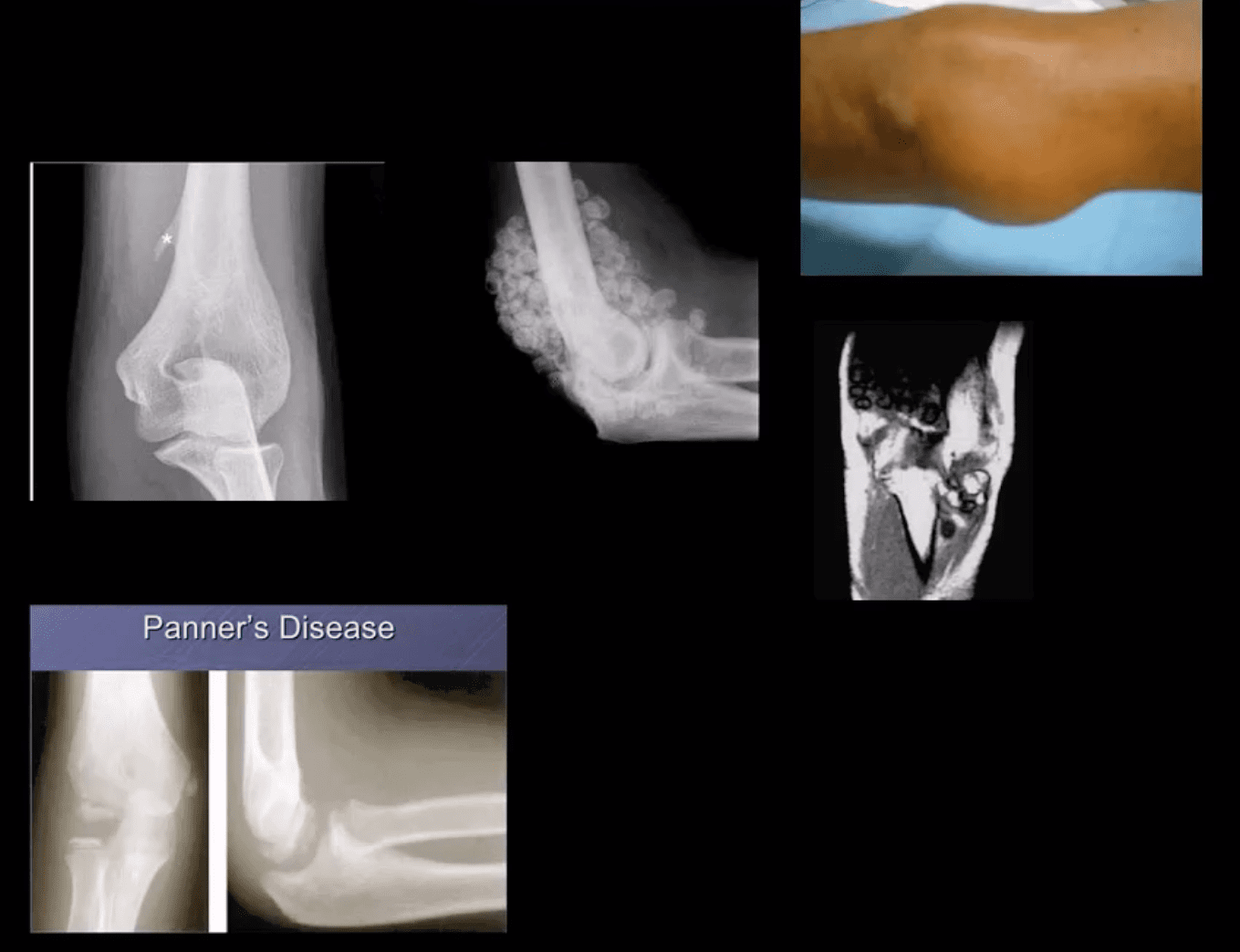
Primary synovial chondrometaplasia�(Reichel Syndrome): abnormalmetaplasia of synovial cells shedding cartilage into joint potentially causing DJD, extrinsic bone erosion, synovitis, nerve compressions etc. Removedoperatively. Imaging: multiple osseocartilaginous loose bodies of relatively equal sizes in the joint cavityDDx with DJD and 2ndosteochondromatosis. MRI-low signal onT1 and T2 with potential joint effusion. Ina tight joint like the elbow may present with large joint distention.�
Panner�s Disease: osteochondrosis of theCapitellum typically in 5-10 y.o. young athlete DDX from OCD of Capitellum(discussed) that occurs in teenagers.Clinically: pain on activity. Recovery occurs in most cases by spontaneous healing. Imaging: x-rays reveal sclerosis and slight fragmentation of theCapitellum w/o loose body. MRI: low T1and high T2 signal in the entireCapitellum.
Myositis Ossificance:
Soft Tissue & Bone Neoplasms about the Elbow
Lipoma: intramuscular, subcutaneous. Most common soft tissue neoplasms. Composed of fat but a substantial number may undergo fat necrosis-calcification-fibrosis. Typically remains benign. Occasionally difficult to DDx from a well-differentiated liposarcoma. Imaging: x radiography: radiolucent lesion well-circumscribed with or w/o calcification. US and MRI are important. On MRIT1high, T2 low SI.
Hemangioma: benign vascular lesion, often composed of multiple vascular channels. Capillary vs. cavernous. More common in children, but found in any age. May often form phleboliths (calcification). Imaging: x-rays reveal soft tissue mass containing phleboliths. MRI: T1-high or variable signal. T2-high signal in areas of slow flow. �bag of worms� sign. Biopsy best avoided. Rx: difficult: local excision vs. embolization vs. observation. High recurrence.
Peripheral Nerve sheath tumor (PNST): benign vs.malignant. Greater incidence in NF1 with a higher risk of malignant PNST. Benign PNST: Schwannoma vs.Neurofibroma. Spinal vs. peripheral nerves. Histology: Schwann cells interspersed with fibroblast and vessels.Clinically: pts in 20s and 30s, palpable mass with or w/o local pressure. Imaging: MRI: T1: split-fat sign, T2: target sign. T1+C enhancement
Soft Tissue Sarcomas: MFH, Synovial sarcoma,(discussed), Liposarcoma (more frequent in the retroperitoneum) Dx: MRI. Clinically: Dx is delayed d/t painless enlarging mass often ignored. Clinically palpable mass deserves MRI examination, US may be helpful. Biopsy confirms Dx.
Malignant bone Neoplasms: Children: OSA, Ewing�s sarcoma (discussed) Adults: Mets, Myeloma (discussed)
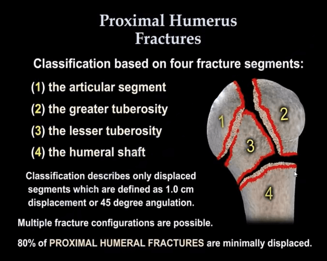
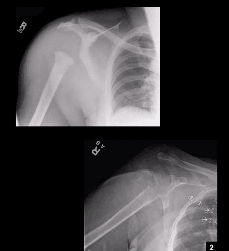
Proximal humeral Fx account for 4-6% of all Fxs. Osteoporotic (OSP) Fx in >60 y.o associated with minimal trauma with F: M 2:1 ratio. In young patients, acute high energy trauma predominates.
Complications: AVN humeral head, Axillary N paralysis.
Neer Classification: considers fractures along 4-anatomical lines with or w/o displacement >1-cm & 45-degree angulation
One part Neer Fx- no displacement or very minimal <1-cm/45-degree. Can affect 1-4 lines and M/C at greater tuberosity. 80% of proximal humeral Fx are one-part Neer.
Two-part Fx: 1-part is displaced >1-cm/45-degrees. m/c involves the surgical neck
Three-part Fx: 2-parts are displaced >1-cm/45-degrees.
Four-part Fx: all 4-parts can be displaced. Uncommon <1%
Imaging: 1st step-radiography, CT may be used in more complex cases. Orthopedic referral
Management: Neer one-part Fx is treated with Sling Immobilisation and progressive rehab
The vast majority of Fx in the elderly are treated non-operatively
Younger patients (40-65) may occasionally require hemiarthroplasty if 3 or 4-part Neer Fx present. Greater risk of AVN
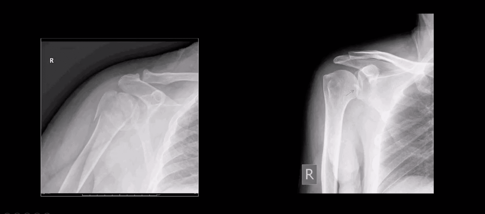
Proximal Humerus Fractures
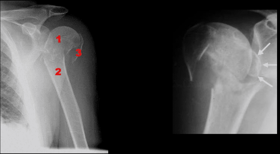
Note: Left image: Fx involving the anatomical neck and the greater tuberosity with minimal displacement <1-cm/45-degree thus Dx as one-part Fx. Right image: Small avulsion Fx of the greater tuberosity with significant displacement (>45-degrees & 1-cm) thus Dx as two-part Fx
Note: three-part Neer Fx (left) and four-part Neer Fx (right)> Management: operative in most cases in younger (40-65) patients
Refers to complete separation of the humerus from scapula glenoid. In 20-40s M: F 9:1 ratio, in60-80S M: F 3:1
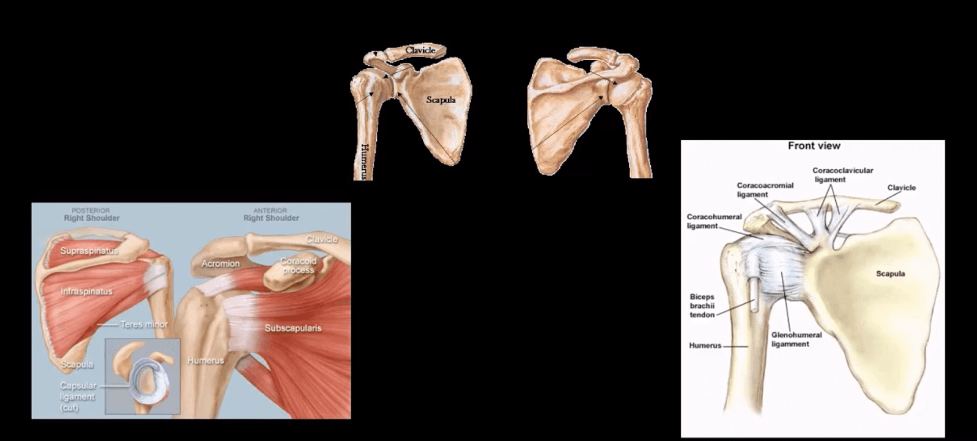
Anatomy: Shoulder stability is sacrificed for mobility, and overall GHJD is the m/c among large joints in the body
Protective falls (e.g., FOOSH) and MVA are m/c causes. GHJ is most vulnerable in abduction, extension and external rotation. Anatomical factors: shallow glenoid, laxed ant-inferior capsule and GH ligaments. GHJD will induce severe tearing of major GHJ restraints. Associated osseous and labral injuries are common and may lead to chronic instability, DJD,�and functional changes
3-types: Anterior GHJD (95%)
Posterior GHJD (4%) especially associated with epileptic seizures, electrocution and can occur b/l
Inferior GHJD aka Laxatio Erecta (<1%) associated with severe trauma
Clinically: AGHJD presents with severe pain, the arm is externally rotated and adducted, severe limitation of movement. GHJD may persist as chronic dislocation.
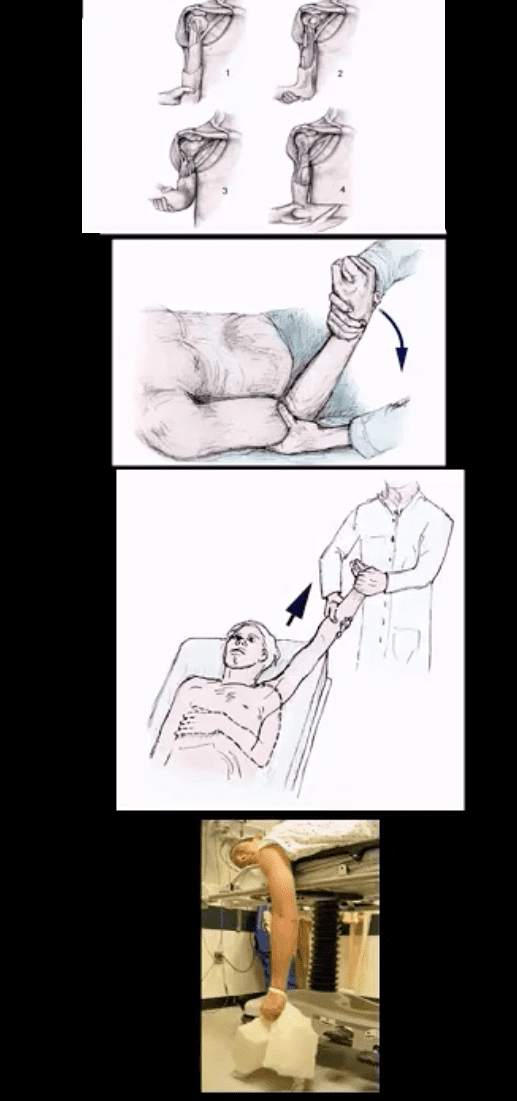
Management: prompt reduction in ED under anesthesia or heavy sedation with Kocher technique top image (not used), External rotation method (middle) or Milch technique (can be used w/o anesthesia) and a few other methods. Delay in reduction correlates with greater risk of immediate and long-term�complications
Diagnostic Imaging Approach
Shoulder series x-radiography is sufficient. Additional Imaging with CT scanning and MRI may be helpful to Dx osseous, cartilage, labral/ligaments pathology
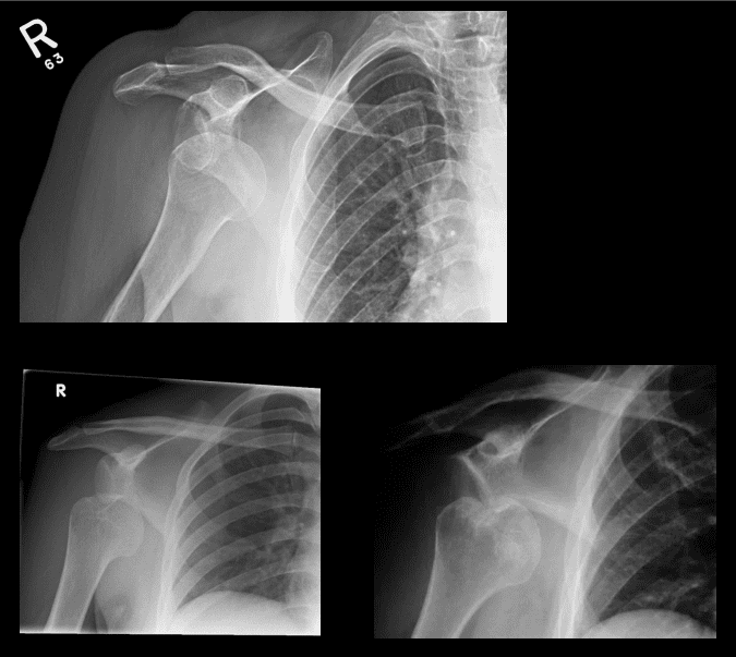
Anterior GHJD (95%). Subcoracoid position(top right) of the humerus is the m/c
Anterior GHJD may also occur as subglenoid(bottom left)and infrequently as subclavicular
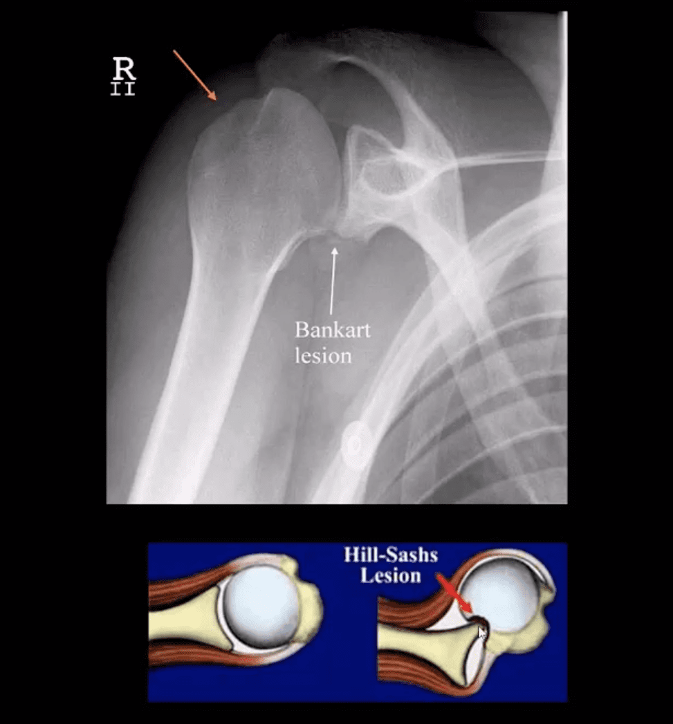
Key to radiographic search is to evaluate associated Bankart and Hill-Sachs injuries
Bankart Lesion
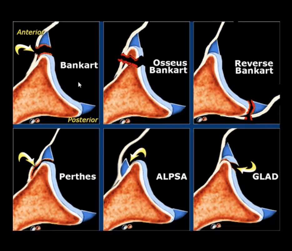
Occurs during anterior GHJD d/t impaction of the head into anterior-inferior glenoid. Variations exist (see next slide). BonyBankart can be seen on x-rays. So-called soft tissue Bankart requires MRI. Cartilage (soft)Bankart is the m/c.
Hill-Sachs aka Hatchet deformity (arrow postreduction)occurs during the same mechanism as Bankart, i.e., compression and impaction of posterolateral aspect of the head against the glenoid producing wedge-shape Fx. Hill-Sachs lesion may predispose to recurrent/chronic GHJD.
Bankart lesion may heal, but operative suture anchors are needed sometimes
CT arthrogram and MRI may be helpful
Types of Bankart Lesion
Note different types of Bankart lesion. Onlyosseous Bankart can be seen radiographically. Soft tissue Bankart requires MRI with and without intra-articular gadolinium(arthrogram).
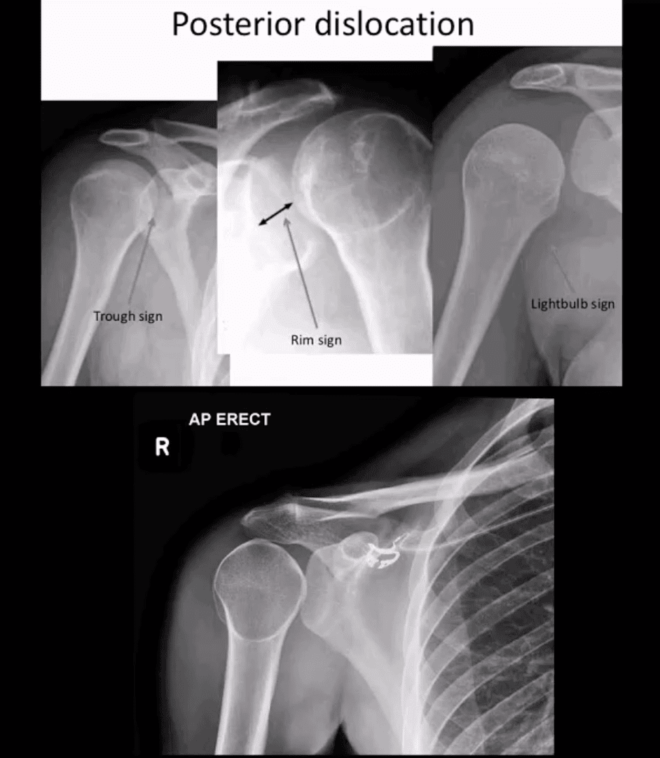
Posterior Dislocation
Note: posterior GHJD with its characteristic signs:
Trough sign aka reverse Hill-Sachs. Occurs d/t anterolateral head impaction Fx
Rim sign: only occurs in the PGHJD d/t posterior position of the head and anterior glenoid-to humeral head distance 6-mm or greater
Light-bulb sign: d/t acute internal rotation of the humerus (head)
Inferior GHJD
Inferior GHJD aka Laxatio Erecta
Severe hyperabduction and inferior displacement of the humerus. Greater chances of severe neurovascular injury and acromial Fx
The dislocated arm is hyperabducted and fixed with the elbow flexed and the arm above the head
ACJ Dislocation (ACJD)
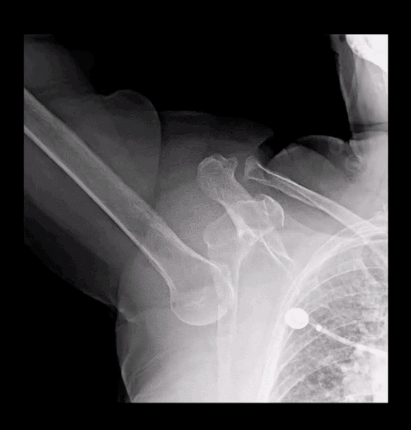
ACJD: common injury, 9% of shoulder girdle injuries esp. in male athletes by a direct blow
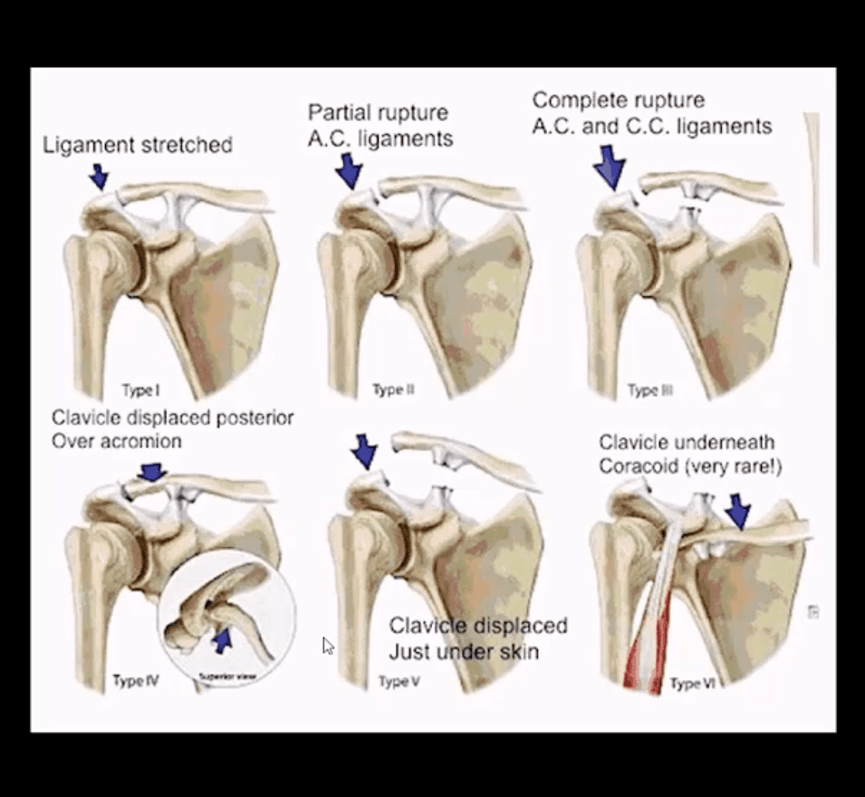
Rockwood classification (left) evaluates tearing of AC and CC ligaments and regional muscles
Type1, 2, 3 among the m/c
Type 1: sprain of ACL w/o tearing
Type 2: tear of ACL and sprain of CCL
Type 3: tear of AC & CCL. The clavicle is elevated above the acromion. If <2-cm good results with conservative Rx.
Imaging: x-radiography with b/l ACJ views with and w/o weights to compare both ACJs. In complex cases CT scanning esp. if Fx is considered
Management: Type 3 (>2-cm) & Types 4-6Operative
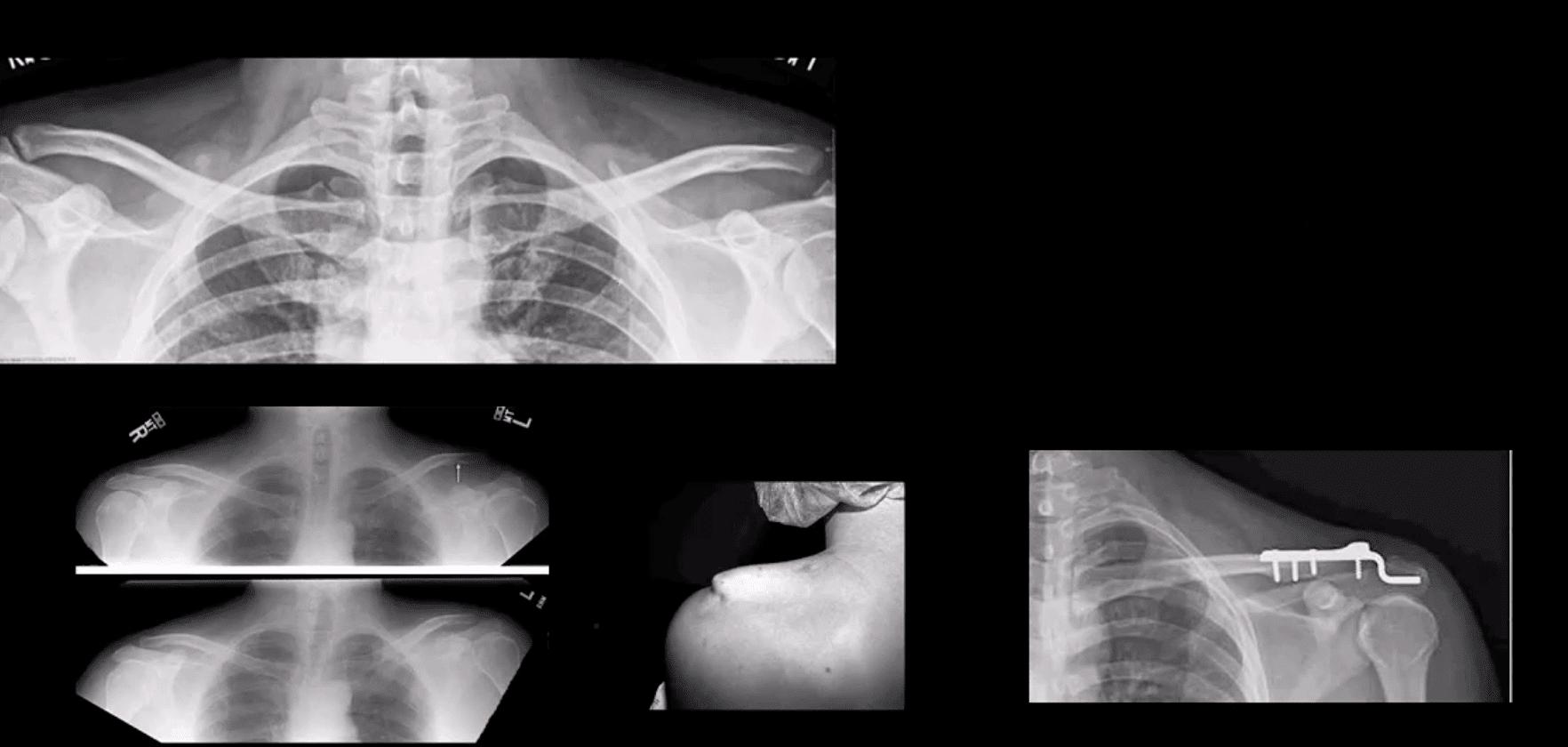
Type 3 ACJ Separation
Type 3 ACJ separation (top left)
More significant ACJD (bottom images) with clinical sign of acromion under the skin and resultant ORIF
Rotator Cuff Muscles (RCM) Pathology
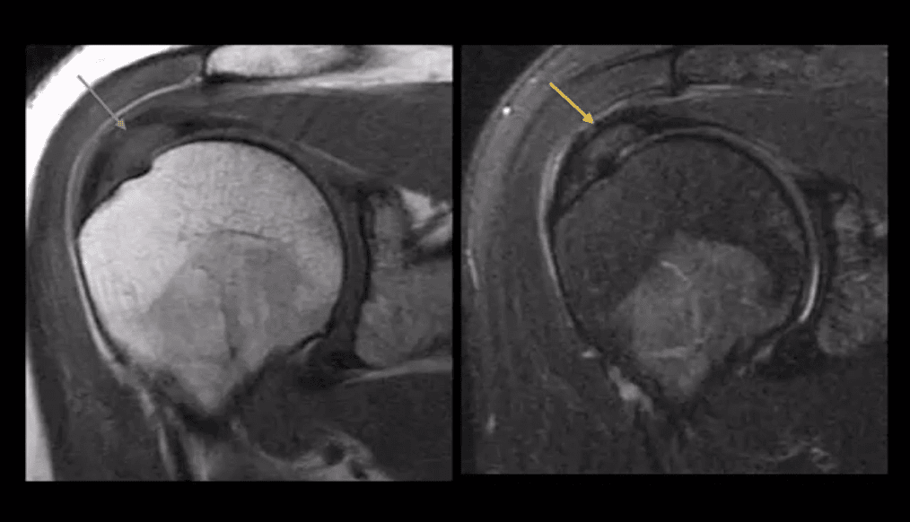
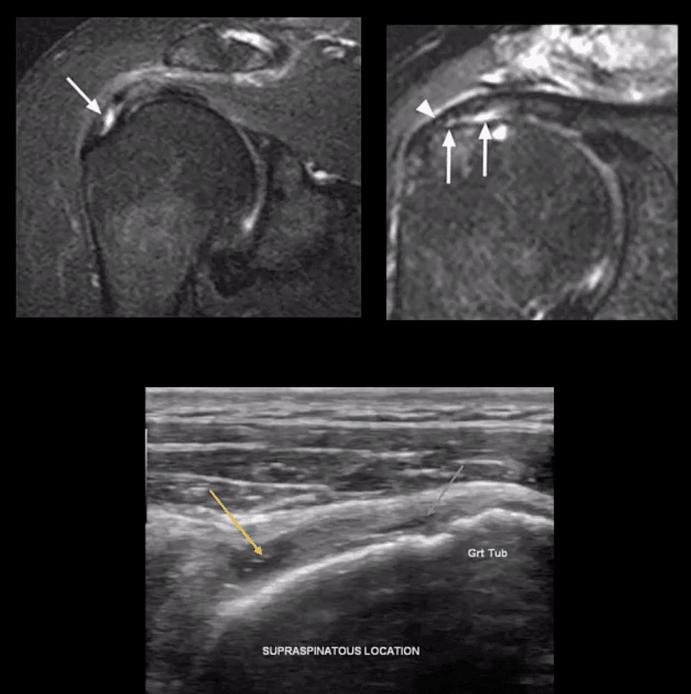
RCM tendinopathy: collagenous degeneration of RCM particularly Supraspinatus M. tendon(SSMT) d/t overuse/degeneration-micro tearing with collagenous replacement. Impingement syndrome is a 2nd extrinsic cause. Presented clinically as pain and limited ROM
Imaging Dx: MSK US can be as accurate as MRI and better in some cases d/t dynamic evaluation v. cost effective
Key MRI clue is thickened inhomogeneous SSMTwith increased signal on all pulse sequences d/t fatty degeneration and inflammation (left images: T1 & T2 FS)
MSKUS findings: thickening of the SSMTsubstance with a change�in normal echogenicity.MSKUS is good to DDx with SSMT tears. US advantages are that it allows dynamic evaluation of painful structures
Partial tear of SSMT: partial (incomplete) tear ofSSMT may occur at the bursal and articular surface or interstitial, i.e., intra-substance/noncommunicating. Etiology: sub-acromial impingement, acute strain, and chronic microtrauma tendinosis
Clinically: pain on abd and flexion, impingement tests, Hawkins-Kennedy tests, etc. Pearls: partial tears can be more painful than complete tears
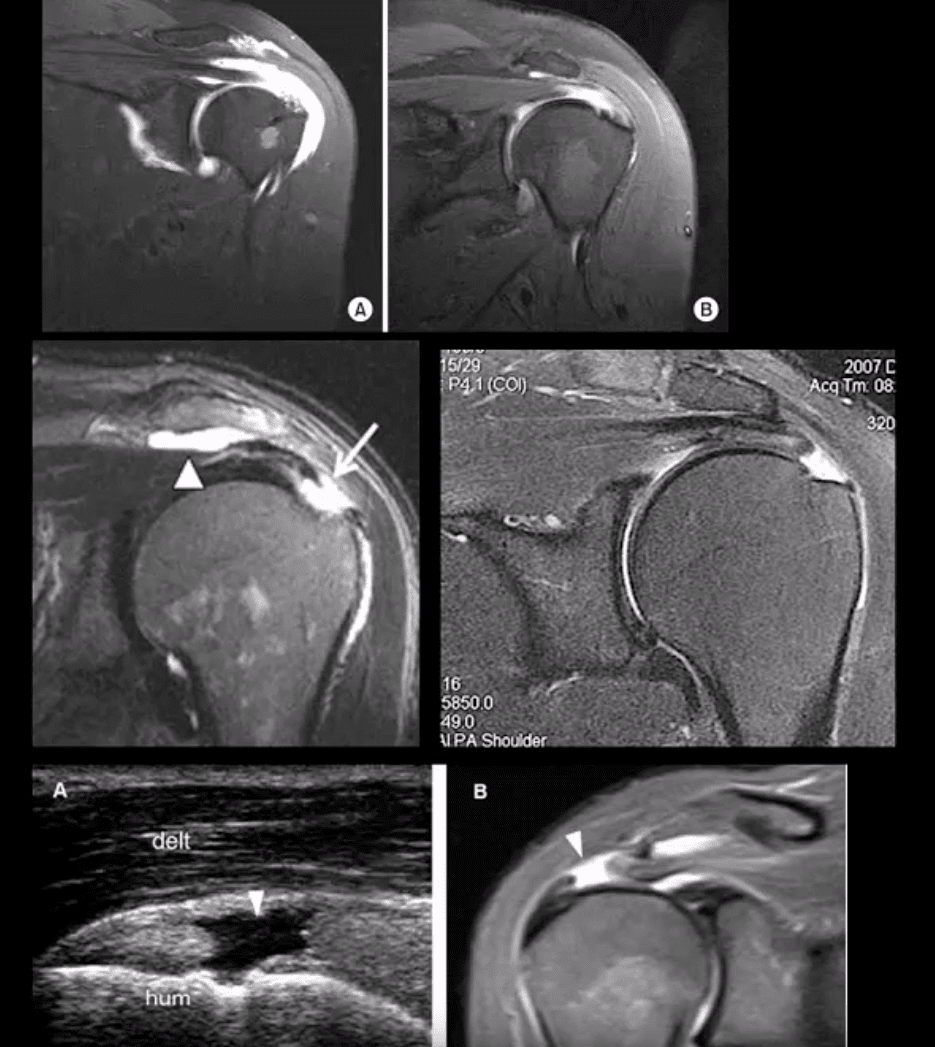
Imaging Dx: MSKUS is as good as MRI (N.B.some studies indicated MSKUS is more superior to MRI). Key MRI findings: gap/incomplete tear of SSMT filled with joint fluid +/- granulation tissue
MSKUS: decreased echogenicity of SSMT, thinning and partial tearing filled with fluid(anechoic areas arrows). Lost convexity of tendon bursal or articular interface.
Full Thickness SSMT (rot cuff) tear: degeneration/tearing of rot cuff. 2nd to impingement by Hooked acromion, overhead overuse or acute trauma. 7-25% of shoulder pain in the general population. Clinically: pain on impingement tests.
Imaging Dx: MSKUS is as good as MRI.Limitations: poor Dx of labral pathology. Key USDx: focal tendon interruption, an anechoic gap (fluid filled), hypoechoic tendon, tendon retraction, uncovered cartilage sign (bottom left, A: US B: MRI)
MRI: key Dx: insertional tear extending through entire SSMT crescent, retraction with fatty degeneration of SSMT and the muscle. If retraction is at 12 o�clock or greater (top images), it may not be anchored operatively
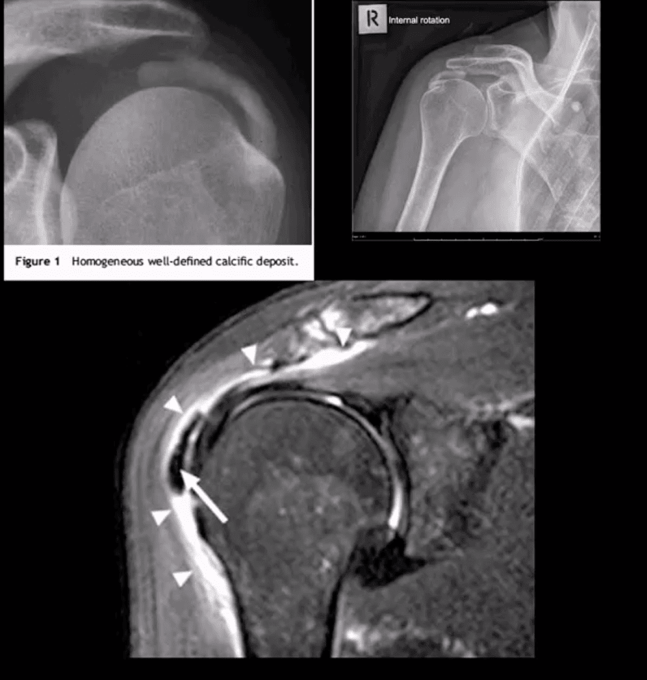
Rotator Cuff (RTC) Calcific Tendinitis: usually d/t calcium HADD crystals. Middle-aged women are most affected. Ranges from asymptomatic imaging finding to severe destructive arthropathy or Milwaukee shoulder(infrequent)
HADD has 3-pathological phases: formation resting-resorption.Mild-to-moderate pain esp. in resting phase.
Imaging: x-radiography: homogenous ovoid mineralization within RTCMT, m/c in SSMT. MRI: ovoid/globular decreased signal on all pulse sequences often with surrounding edema (bottom left)
Rx: self-resolution occurs. Advanced cases: operative aspiration etc.
Superior Labrum Anterior to Posterior (SLAP) Lesions/Tears
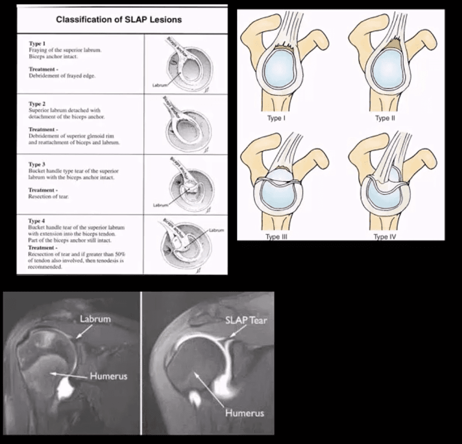
SLAP tears: FOOSH and throwing sports or chronic shoulder instability aka Multidirectional shoulder instability (in 20%). Type 1-9 exist but the M/C areType 1-4
In all 4-types superior labrum is affected with or w/oLHBMT anchor tear (see pictures). Clinically: pain, limitation of AROM with active compression tests, typically non-specific findings mimicking RTCpathology
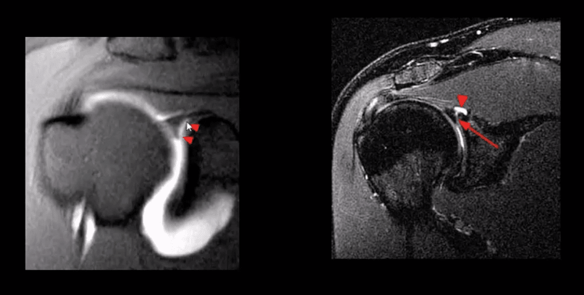
Imaging is crucial: best imaging is MRI arthrography. Key signs: hyperintense linear fluid signal within superior labrum +/- extending along the LHBT on fat-suppressed fluid sensitive imaging and FS T1 arthrogram. Best observed on coronal slices.
Rx: small tears may heal, but unstable tears require operative care.
Key DDx: anatomical variants like Buford complex andSub-labral foramen
SLAP tear with a paralabral cyst (bottom right)
Normal variant DDx: sub labral foramen(bottom left) note: MR arthrography with contrast undercutting the labrum but w/o extending posteriorly to the LHBT
Shoulder Arthritis
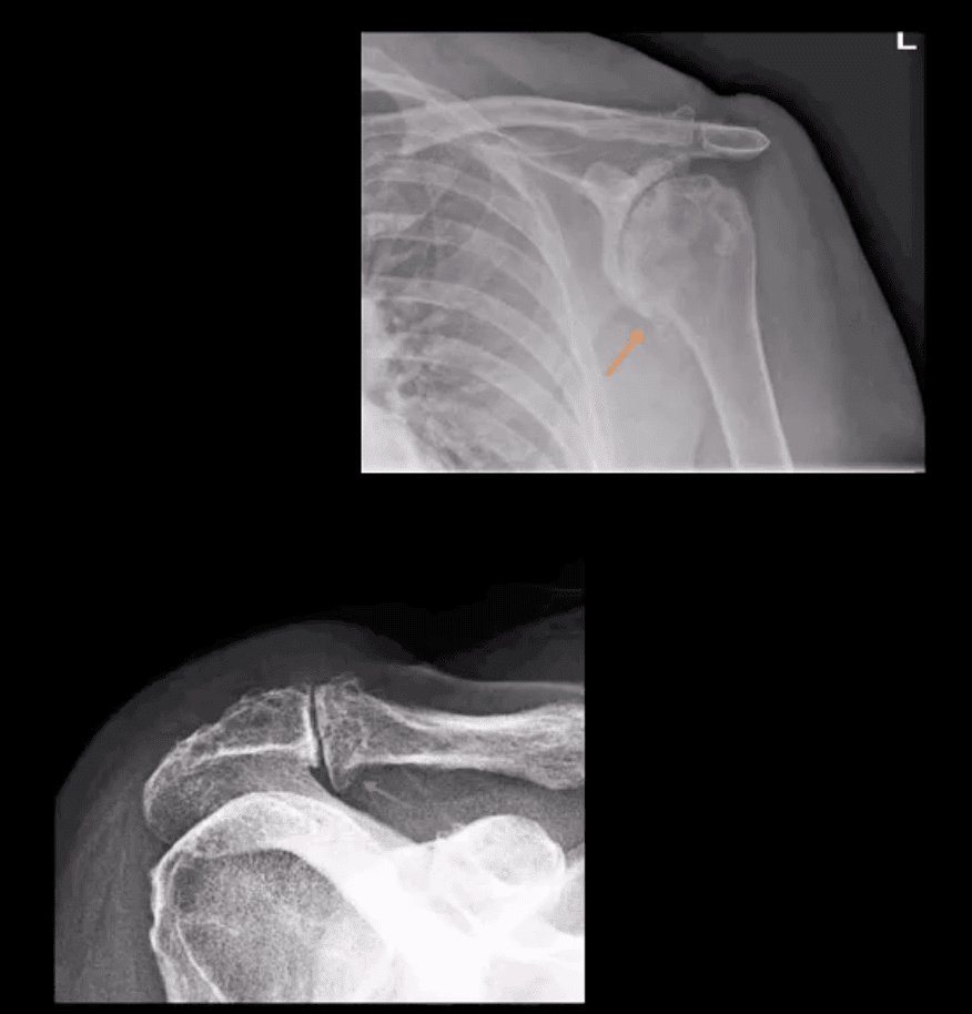
GHJ DJD: usually associated with a 2nd cause: trauma, instability, AVN, CPPD, etc. Presented with pain, crepitus and decreased ROM/function. Associated RTC disease may be present. Imaging; x-radiography is sufficient and provides grading/care planning.Major findings: joint narrowing, osteophytosis esp. at the inferior-medial head (orange arrow), subchondral sclerosis/cysts. Often noted superior head migration d/t RTC disease.
ACJ OA: common and typically primary with aging. Presents with ACJ loss and osteophytes. Osteophytes along the undersurface of the ACJ �keel osteophytes�(blue arrow) may lead to RTC muscle tear. Regional bursitis is other clinical feature of ACJ arthrosis.
Management: usually conservative depending on clinical signs/symptoms
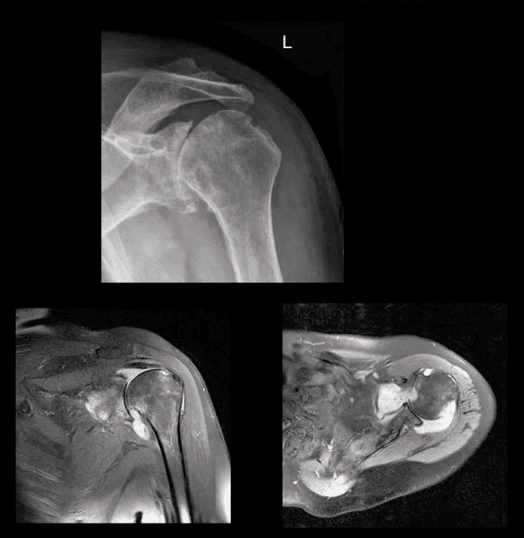
Rheumatoid Arthritis GHJ: RA is a multisystem inflammatory disease affecting multiple joints lined by the synovium. GHJ RA is common (m/c large joints in RA knees/shoulders). Clinically: pain, limited ROM and instability, muscle weakness/wasting. Hands, feet,�and wrists are m/c affected. Imaging: x-radiography reveals periarticular erosions, uniform joint space loss, juxta-articular osteoporosis, subluxations,�and soft tissue swelling. MRI can help detect�commonly associated RTC tearing and instability. Early changes can be detected by MSKUS esp. with power Doppler use indicating hyperemia/inflammation.
Note: L shoulder x-ray revealing cartilage destruction and symmetrical joint loss, multiple erosions, and likely loss of RTCM support with superior head migration, ST effusion present.
Note: PDFS coronal and axial MRI slices of GHJ RA indicating marked inflammatory joint effusion, bone erosion/edema, synovial pannus formation and likely tear in RTC m. Management: Rheumatological referral and pharmacotherapy with DMARD. Operative care asRTCM repair. 10% of patients are disabled d/t RA
Neuropathic Osteoarthropathy aka Charcot’s shoulder: d/t neurovascular and neural periarticular damage. Multiple causes exist.M/c develops in diabetics in midfoot. Shoulder Charcot is m/c in Syringomyelia (25%), trauma paralysis, MS, etc. Dx: clinical(50% pain/swelling 50% painless destruction). Imaging is crucial. X-radiography is sufficient in well-established cases, but early Dx is challenging. MRI may help with early Dx and delayed complications. Rad Dx: Shoulder Charcot is m/c presented as atrophic type destructive arthropathy with humeral head appearing as if surgically amputated along with intra-articular debris, density, distention, dislocation, and other key features
Septic Shoulder: shoulder is the 3rd m/c followingknee>hips. Patients at risk: diabetics, RA pts, immunocompromised, I.V. drug users, indwelling catheters, etc. Routes: hematogenous (m/c), direct inoculation (iatrogenic, trauma etc.) adjacent spread(e.g. OM). Staph. Aureus (>50%) m/c.
Clinically: joint pain and dec. ROM, fever 60% only, toxemia, inc. ESR/CRP. Dx: imaging and joint aspiration/culture. RadDx: early x-rays often unremarkable except ST effusion/fat planes obscuration, joint widening. Later7-12 days patchy osteopenia, moth-eaten/permeating bone resorption, articular destruction, joint narrowing. May progress to severe joint destruction and ankyloses. Early Dx & I.V. antibiotics are crucial even before culture. Operative irrigation and joint drainage in some cases. Complications are possible esp. if Rx is delayed. MSKUS with needle aspiration may help. Note: (top image) non-traumatic joint widening with inferolateral head displacement d/t septic A dx: by needle aspiration Staph. Aures.
Ischemic Osteonecrosis
Ischemic Osteonecrosis of the humeral head may occur d/t trauma (Neer four-part Fx), Steroids, Lupus, Sickle cell, Alcoholism, Diabetes,�and many other conditions. Imaging is crucial: MRI detects earliest changes as intraosseous edema. X-ray features are late, presented as a collapse of subchondral bone with sclerosis �snow cap� sign, fragmentation, and progressive severe DJD
Management: orthopedic referral, core decompression in early cases, hemiarthroplasty in moderate and total arthroplasty in severe cases.
Shoulder Neoplasms
In adults >40, bone Mets d/t lung, breast, renal cell, thyroid CA & prostate are the m/c causes. Clinically: may mimic pain resemblingRTC/joint changes. Should be evaluated carefully. Key to Dx: Hx, PE and Imaging esp.in pts with known primary
Imaging: 1st step x-rays, MRI can help, Tc99bone scintigraphy helps to detect regional and distant disease. X-ray features: destructive lytic changes typically in prox humerus(red marrow) with or w/o path Fx. DDx: Mets, MM, lymphoma
Clinically: night pain, pain at rest, etc. Lab tests: unrewarding, in severe cases hypercalcemia may be noted.
Primary Malignant bone neoplasms (shoulder) Adults: M. Myeloma or Solitary plasmacytoma, Chondrosarcoma may transform from an enchondroma and some others. In children/teenagers: OSA vs. Ewing�s
Primary benign bone neoplasms (shoulder). Adults: Enchondroma (patients in their 20-30s)GCT. In children: Simple bone cyst (Unicameral Bone cyst), Osteochondroma, Aneurysmal Bone Cyst, Chondroblastoma (rare)
Imaging: 1st step x-radiography
MRI is essential to Dx. Especially in cases of primary malignant neoplasms Evaluate extent, soft tissue invasion, preoperative planning, staging, etc.
IFM's Find A Practitioner tool is the largest referral network in Functional Medicine, created to help patients locate Functional Medicine practitioners anywhere in the world. IFM Certified Practitioners are listed first in the search results, given their extensive education in Functional Medicine