Regenerative Spine Care and Sciatica Relief in El Paso: How Epidural Injections, PRP, mFAT, and Shockwave Therapy Work Together
Sciatica and chronic back pain can affect almost every part of daily life. Sitting can hurt. Walking can feel limited. Sleep may be broken. Work, exercise, driving, and family time can become harder than they should be.
At El Paso Back Clinic, the goal is to look deeper than the pain signal. Pain is important, but it is often only the warning light. The real problem may involve an irritated nerve, a damaged disc, a strained ligament, a weak core, poor spinal motion, scar tissue, inflammation, or a past injury that never healed correctly.
This is why a modern spine care plan may combine chiropractic care, rehabilitation, medical oversight, functional medicine, epidural spinal injections, regenerative therapies, and shockwave therapy. Each part has a different job. Together, they may help calm nerve irritation, support tissue repair, improve movement, and help the body return to better function.
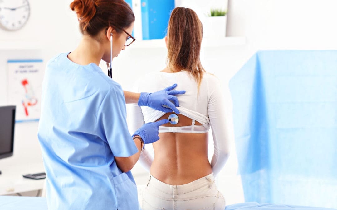
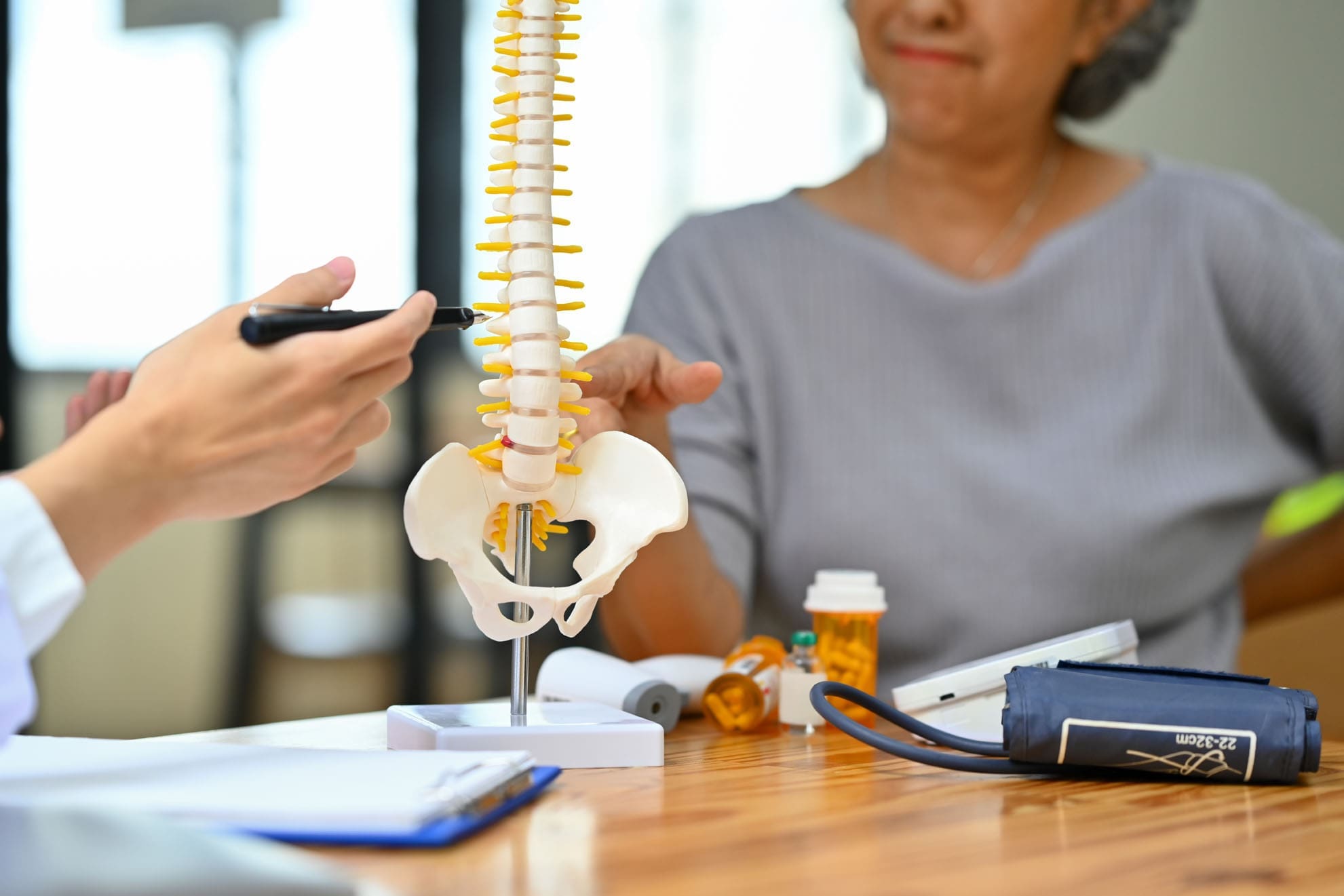
What Is Sciatica?
Sciatica is pain that travels along the sciatic nerve. This nerve starts in the lower back and travels through the buttock, hip, leg, and foot. When a spinal nerve root becomes irritated or compressed, pain can travel down the leg.
Common sciatica symptoms may include:
Low back pain
Buttock or hip pain
Burning pain down the leg
Numbness or tingling
Weakness in the leg or foot
Pain that worsens with sitting
Pain that improves when lying down or changing position
Sciatica is not always caused by the same problem. It may come from a herniated disc, disc degeneration, spinal stenosis, facet arthritis, muscle tension, pelvic imbalance, scar tissue, or inflammation. This is why a complete exam matters.
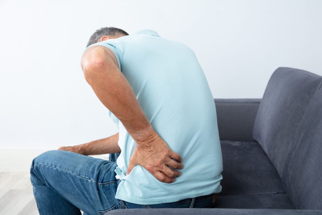
Why Chronic Back Pain Needs More Than Temporary Relief
Chronic back pain is pain that lasts longer than expected. It often continues for more than 12 weeks. By that time, the body may start to change how it moves. Muscles tighten. Joints stiffen. Nerves become more sensitive. The patient may avoid activity, which can lead to weakness and more pain.
Traditional care often focuses on short-term pain relief. That can help during a flare-up, but it may not be enough when the deeper problem is structural or inflammatory.
A more complete plan may look at:
Spinal alignment and joint motion
Disc health
Nerve irritation
Ligament and tendon stress
Muscle weakness
Core control
Inflammation
Nutrition
Sleep
Blood sugar and metabolic health
Prior auto, work, or sports injuries
This whole-person view is important because healing is not only about one painful spot. The spine is part of a larger system.
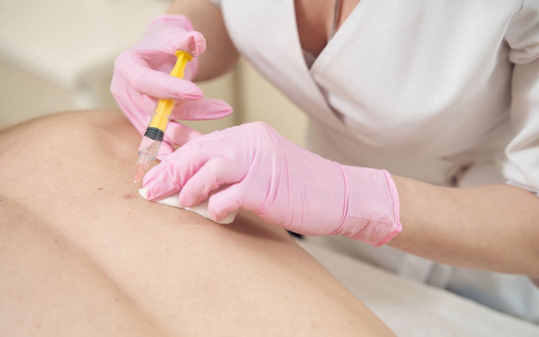
How Epidural Spinal Injections May Help Sciatica
An epidural spinal injection places medication or biologic material near an irritated spinal nerve. The goal is to reduce inflammation around the nerve root and help calm leg pain.
For a patient with strong nerve pain, this can be helpful. When pain is severe, the patient may not be able to move, stretch, exercise, or sleep well. If an epidural injection reduces the pain enough, the patient may be able to begin rehabilitation and chiropractic care more safely.
Epidural steroid injections are commonly used for spinal stenosis and nerve-related back and leg pain. However, long-term outcomes may vary. In one PCORI-supported report on lumbar spinal stenosis, epidural injections with corticosteroid plus lidocaine did not show long-term benefits over lidocaine alone for pain, function, opioid use, or surgery rates in the studied group (Friedly et al., 2019).
This does not mean epidural injections are useless. It means they should be used carefully and as part of a larger care plan.
Why Some Patients Look Beyond Repeated Steroid Injections
Steroids can reduce inflammation. That is why they are often used during painful flare-ups. But repeated steroid use may carry risks. Cortisone injections can have side effects, including cartilage damage, tendon weakening, blood sugar changes, infection risk, and bone thinning, especially when used too often or in high amounts (Mayo Clinic, 2026).
For some patients, this raises an important question:
Can we reduce pain while also supporting tissue repair?
This is where regenerative therapies may enter the conversation. Regenerative care does not simply try to hide symptoms. It aims to support the body’s natural healing response.
What Are Regenerative Spine Therapies?
Regenerative spine therapies use biologic materials, often from the patient’s own body, to support healing. These treatments may be considered for chronic spine pain, disc-related pain, ligament injury, facet joint pain, and nerve irritation when the patient is a proper candidate.
Common regenerative options include:
PRP: platelet-rich plasma
PFP: platelet-fibrin plasma or platelet-fibrin products
Platelet lysate: a platelet-derived fluid rich in growth factors
mFAT: microfragmented adipose tissue
These therapies are often called orthobiologics. “Ortho” refers to bones, joints, muscles, ligaments, and spine structures. “Biologics” refers to healing materials that come from living tissue.
The University of Iowa Health Care describes regenerative medicine as care that may use a person’s own cells, tissues, or biologic materials to support healing and repair (University of Iowa Health Care, n.d.).
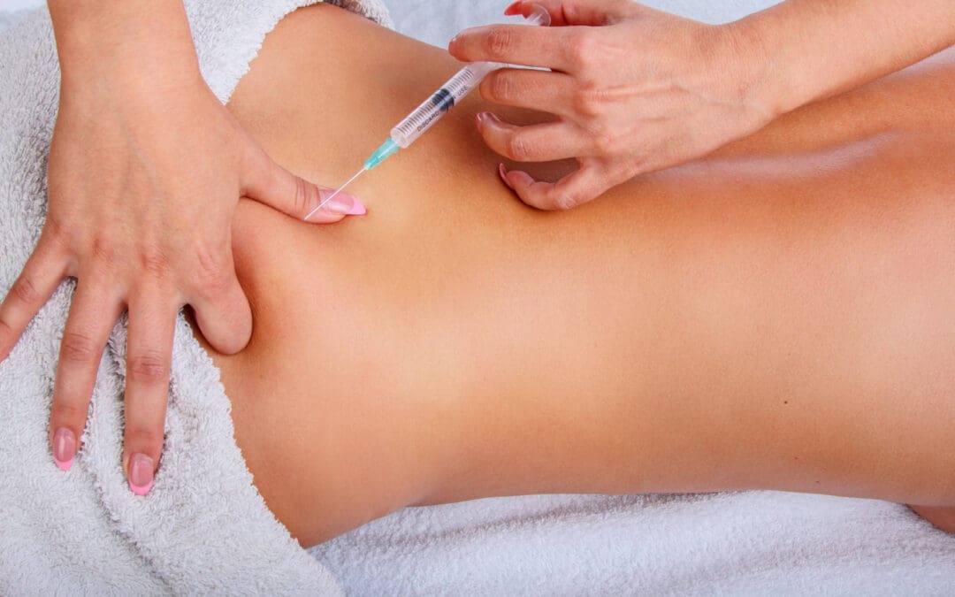
PRP: Platelet-Rich Plasma for Spine and Nerve-Related Pain
PRP is made from a small sample of the patient’s blood. The blood is processed to concentrate platelets. Platelets are best known for helping blood clot, but they also carry growth factors and healing signals.
In spine care, PRP may be used to support damaged or irritated tissues, such as:
Disc-related pain areas
Facet joints
Ligaments
Tendons
Soft tissues around the spine
Research on PRP for low back pain is still growing. A narrative review on regenerative medicine for chronic low back pain described PRP and other biologic therapies as promising options, while also noting that more high-quality research is needed (Wang et al., 2023). A systematic review of PRP for low back pain found PRP was generally effective and safe for degenerative low back pain but also called for stronger studies and better treatment standards (Machado et al., 2023).
In simple terms, PRP is not a magic cure. But for selected patients, it may help support a better healing environment.
Platelet Lysate and Epidural Biologic Injections
Platelet lysate is made from platelets, but it is processed differently than PRP. The platelets are broken open, releasing growth factors into a thinner fluid. Because it is less thick than PRP, platelet lysate may be considered for nerve-related areas, including epidural use in some regenerative medicine settings.
A study of lumbar epidural platelet lysate for radicular pain reported improvements in pain and function through 24 months, with mild adverse events reported in a small percentage of patients (Centeno et al., 2017). More research is still needed, but this area is important because it examines biological support for nerve-related back and leg pain.
A 2025 meta-analysis also compared epidural PRP with steroid injections for lumbar disc disease with radiculopathy. The authors reviewed randomized controlled trials and examined pain and function outcomes over several time points (Muthu et al., 2025). This growing research shows why biologic epidural options are becoming a major topic in modern spine care.
PFP: A Natural Scaffold for Healing
PFP, or platelet-fibrin plasma, is similar to PRP but includes more fibrin activity. Fibrin is a natural protein involved in clotting and wound repair.
You can think of fibrin as a healing web. It may help hold platelets and growth factors in one area longer. This may be useful when the care plan is focused on damaged ligaments, tendons, or joint tissues.
PFP may support:
Local repair signaling
Tissue stability
Collagen remodeling
Longer contact time for healing factors
A more organized repair response
Like other regenerative options, PFP should be used after a detailed exam and proper diagnosis.
mFAT: Microfragmented Adipose Tissue
mFAT stands for microfragmented adipose tissue. Adipose tissue is fat tissue. In this treatment, a small amount of a patient’s own fat is collected, processed, and prepared for injection into a target area.
Fat tissue contains signaling cells and support structures that may help with tissue repair. mFAT is often discussed in regenerative medicine for joint, soft tissue, and orthopedic problems. It does not “regrow” a spine overnight. Instead, it may help support the local repair environment in selected cases.
For chronic spine problems, mFAT may be considered when there is deeper tissue degeneration, joint wear, or long-standing injury patterns. The key is proper patient selection, medical screening, imaging review, and follow-up care.
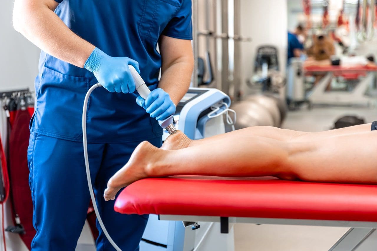
Shockwave Therapy: The Biological Catalyst
Shockwave therapy, also called extracorporeal shockwave therapy (ESWT), uses sound waves to stimulate tissue. It is non-surgical and does not involve medication.
Shockwave therapy may help painful tissues by creating a controlled healing signal. This process is called mechanotransduction. That means the body turns mechanical energy into a biological response.
ESWT may support healing by helping:
Increase local blood flow
Stimulate new small blood vessel formation
Improve cell activity
Reduce pain signaling
Break down scar-like tissue
Improve collagen remodeling
Support tissue repair pathways
A systematic review and meta-analysis found that ESWT improved pain and lumbar function in patients with chronic low back pain, with no serious adverse effects reported in the included studies (Liu et al., 2023). Another review described shockwave as a tool that may support tissue repair through blood vessel growth, anti-inflammatory effects, and cell signaling (Cheng & Wang, 2015).
Why Shockwave and Regenerative Injections May Work Well Together
Regenerative injections bring healing signals to damaged tissue. Shockwave therapy may help prepare the tissue to respond better.
This is why ESWT can be described as a biological catalyst. A catalyst helps a process move forward. Shockwave does not replace PRP, PFP, platelet lysate, or mFAT. It may help create a better local environment for healing.
A simple way to picture it is this:
PRP, PFP, platelet lysate, and mFAT bring healing signals.
Shockwave therapy helps wake up slow-healing tissue.
Chiropractic care improves joint motion and biomechanics.
Rehabilitation rebuilds strength, balance, and control.
Functional medicine looks for healing barriers inside the body.
When combined correctly, these tools may help the body repair itself more effectively than a single treatment alone.
The Role of Chiropractic Care at El Paso Back Clinic
Chiropractic care is often central to sciatica and back pain recovery because movement matters. If spinal joints, hips, pelvis, and soft tissues are not moving well, stress can build up around the nerves and discs.
At El Paso Back Clinic, chiropractic care may support:
Better spinal motion
Less joint stiffness
Improved posture
Better pelvic and hip mechanics
Reduced muscle guarding
Safer return to activity
Better rehab progress
Dr. Alexander Jimenez, DC, APRN, FNP-BC, CCST, CFMP, IFMCP, ATN, uses a dual-scope clinical view that connects chiropractic evaluation, injury care, functional medicine, and rehabilitation. His clinical observations often focus on how spinal structure, inflammation, metabolic health, and movement patterns work together.
This matters because many patients do not only have “a bad disc.” They may have a body system that is under stress.
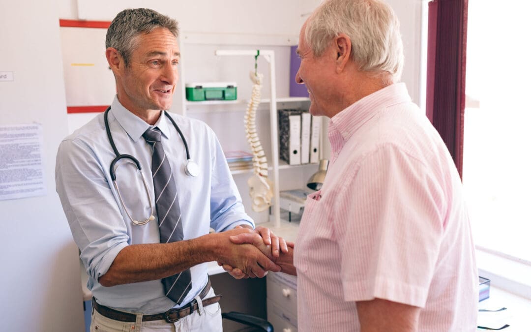
Medical Oversight With Dr. Maria Guadalupe Cardenas, MD
At Injury Medical Clinic PA and within the larger integrative care model connected with El Paso Back Clinic, Dr. Maria Guadalupe Cardenas, MD, serves as Medical Director and Collaborative Physician. She is Board Certified in Internal Medicine, has over 40 years of experience as an internist, and is listed with NPI #1164426749 and Texas MD License #J2933.
This medical oversight is valuable because many spine patients have other health issues that can affect treatment safety and healing.
These may include:
Diabetes or blood sugar problems
High blood pressure
Autoimmune conditions
Medication use
Blood thinner use
Hormone changes
Infection risk
Poor sleep
Chronic inflammation
Older injuries or surgeries
A multidisciplinary clinic can help connect the dots between medical history, spine pain, nerve symptoms, and recovery goals.
Functional Medicine: Looking for Healing Barriers
Functional medicine asks a deeper question:
Why is this patient not healing well?
For chronic back pain and sciatica, the answer may lie beyond the spine. The body heals best when it has the right nutrients, blood flow, hormones, oxygen, sleep, and control of inflammation.
Functional medicine support may look at:
Vitamin D status
Blood sugar and insulin
Inflammation markers
Thyroid function
Hormone balance
Gut health
Nutrition
Weight management
Sleep quality
Stress load
This does not replace spine care. It supports spine care. A patient with poor blood sugar control, low protein intake, poor sleep, and high inflammation may heal more slowly. Improving these areas may help the patient respond better to chiropractic care, rehab, injections, and shockwave therapy.
Why Personal Injury Patients May Benefit
After a car crash, work injury, or sports injury, pain may not show up right away. Some symptoms appear hours or days later. Neck pain, back pain, headaches, sciatica, numbness, and stiffness can develop after the body’s stress response calms down.
Personal injury care needs careful documentation and a clear clinical plan. At El Paso Back Clinic, the care model may include:
Injury history
Orthopedic testing
Neurological testing
Range-of-motion findings
Imaging review when needed
Functional limits
Treatment response
Rehab progress
Referrals when needed
This matters because injury recovery is not only about pain relief. It is also about restoring function and documenting how the injury changed it.
A Step-by-Step Spine Recovery Plan
A patient-centered spine plan may include several phases.
Phase 1: Calm the Nerve
When sciatica is active, the first goal is to reduce irritation. This may include careful activity changes, decompression, gentle chiropractic care, targeted injection options, and pain-control strategies.
Phase 2: Improve the Healing Environment
Once pain is more controlled, regenerative therapies and shockwave therapy may be considered. The goal is to support tissue repair, improve circulation, and help chronic tissue move out of a stalled healing state.
Phase 3: Restore Motion
Chiropractic care, soft-tissue therapy, mobility work, and decompression may help the spine and pelvis move more freely.
Phase 4: Rebuild Strength
Rehabilitation helps the patient rebuild core strength, hip control, balance, posture, and endurance. This step helps protect the spine from future flare-ups.
Phase 5: Maintain Long-Term Function
The final goal is not just to feel better for a few days. The goal is to help the patient return to life with improved movement, strength, and awareness of how to prevent future problems.
Who May Be a Candidate?
A patient may be a candidate for this type of care if they have:
Sciatica
Chronic low back pain
Disc herniation
Disc degeneration
Annular tear
Facet arthritis
Ligament injury
Post-accident back pain
Pain that returns after basic care
Difficulty walking, sitting, or sleeping due to nerve pain
Not every patient is a candidate for every treatment. Severe weakness, loss of bowel or bladder control, fever, infection signs, cancer history, major trauma, or rapidly worsening nerve symptoms need urgent medical attention.
Final Thoughts
Sciatica and chronic back pain can be frustrating, but patients now have more options than short-term pain masking. Epidural spinal injections may help calm acute nerve irritation. Regenerative therapies such as PRP, PFP, platelet lysate, and mFAT may support repair in damaged or irritated tissues. Shockwave therapy may act as a biological catalyst by improving blood flow, stimulating cell activity, and helping chronic tissue respond.
At El Paso Back Clinic, this kind of care fits into a larger model that includes chiropractic care, medical oversight, functional medicine, personal injury care, and rehabilitation. With Dr. Alex Jimenez, DC, APRN, FNP-BC, working alongside Dr. Maria Guadalupe Cardenas, MD, Medical Director and Collaborative Physician, patients receive a team-based approach focused on structure, function, safety, and long-term healing.
The goal is simple: reduce pain, restore movement, support healing, and help patients return to the life they want.
Many adults notice extra weight creeping on, especially around the middle, even when they try to eat better and stay active. Hormone changes over time often play a quiet but powerful role in how the body stores fat, burns energy, and controls hunger. Bioidentical hormone replacement therapy (BHRT) offers a way to bring those internal messengers back into better balance. It is not a quick weight-loss fix or a magic pill. Instead, it helps remove some of the metabolic roadblocks that make diet and lifestyle efforts harder to sustain.
When hormone levels are optimized, many people find it easier to manage cravings, keep steady energy, and support lean muscle. This article explains how BHRT, and specifically the EvexiPEL method from Evexias Health Solutions, can work alongside smart eating and daily habits for longer-lasting results.
What Bioidentical Hormones Actually Do in the Body
Hormones act like chemical messengers. They tell the body when to store fat, when to burn it, how hungry to feel, and how well muscles can grow. Key players include estrogen, testosterone, insulin, cortisol, and thyroid hormones. When these get out of balance—often from aging, stress, or other life changes—metabolism can slow, fat can gather more easily around the belly, and cravings for sweets can grow stronger.
Bioidentical hormones are made to match the exact structure of the ones the human body produces naturally. They usually come from plant sources and are customized for each person after lab testing. The goal is to restore balance rather than force rapid change. Because they more closely match the body’s own chemistry, many patients experience smoother effects than with synthetic options.
How Balanced Hormones Help with Weight and Fat Control
Balanced hormones support weight management in several practical ways:
Fewer intense sugar cravings: When estrogen, progesterone, and cortisol signals stabilize, the brain’s hunger cues become easier to manage. People often report a less urgent desire for processed sweets or snacks.
Better insulin sensitivity: Improved insulin function helps the body use blood sugar for energy rather than store it as fat. This makes it easier to maintain a steady weight over time.
More consistent daily energy: Steady hormone levels reduce afternoon slumps. With more energy, it becomes easier to go for a walk, prepare a healthy meal, or stick to an exercise plan.
Support for lean muscle: Testosterone and other hormones help maintain or build muscle. Muscle tissue burns more calories even at rest, which supports a higher everyday metabolism.
Less stubborn abdominal fat: Hormone balance can influence where the body prefers to store fat. Many notice gradual improvement in midsection fat when levels are optimized alongside healthy habits.
These changes do not happen overnight. They create an internal environment where diet and movement efforts can finally show clearer results.
EvexiPEL Pellet Therapy: Steady Delivery Without the Roller Coaster
Evexias Health Solutions developed the EvexiPEL method as a form of BHRT that uses tiny, custom-made pellets. A trained provider places the pellets just under the skin during a short office visit. The pellets then release a steady, consistent dose of bioidentical hormones—such as testosterone or estradiol—over several months, usually three to six.
This steady release mimics the body’s natural rhythm far better than daily creams, gels, pills, or weekly shots. Many patients describe avoiding the ups and downs, or “roller coaster,” that can come with other delivery methods. Consistent levels often translate into more reliable energy, steadier moods, and fewer hormone-driven cravings throughout the day.
Because the delivery stays even, people can focus on building healthy routines instead of managing daily symptom swings. EvexiPEL is always paired with lab testing and a full wellness plan; it is never used alone.
Why Nutrition Matters Even More with BHRT
BHRT works best when paired with a diet built around fresh, whole foods. Think plenty of vegetables, quality proteins, healthy fats from avocados and nuts, and fiber-rich choices. These foods provide the body with the raw materials it needs for hormone production, detoxification, and stable blood sugar.
Cutting back on processed carbohydrates and added sugars helps too. These foods can spike blood sugar and work against the improvements in insulin sensitivity that BHRT supports. Many people find that once hormones stabilize, choosing whole foods feels more natural because energy stays higher and cravings quiet down.
Evexia’s providers often combine pellet therapy with targeted nutraceuticals—high-quality supplements designed to support metabolism, gut health, and mitochondrial energy. This root-cause approach to care addresses multiple systems at once rather than focusing on calories alone.
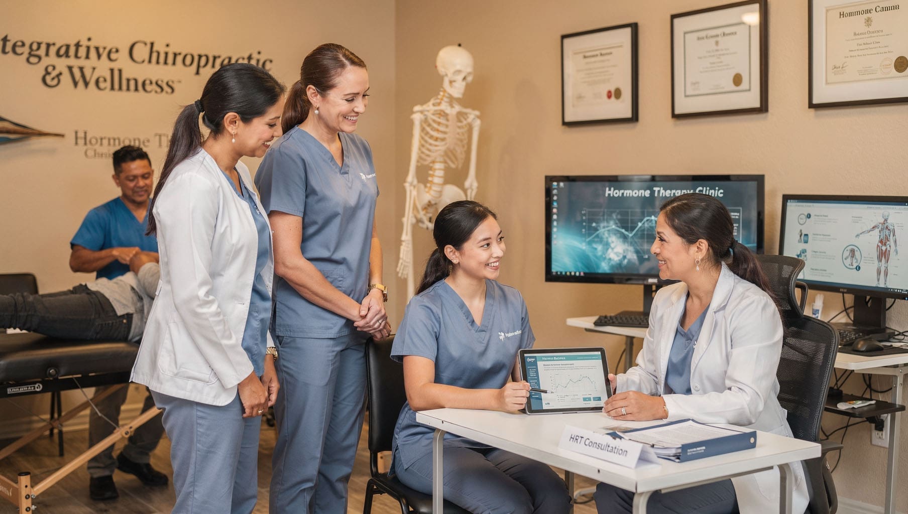
The Advantage of Multidisciplinary Integrative Care
Hormone balance does not exist in a vacuum. The nervous system, gut health, sleep, stress, and physical structure all influence how well hormones work. That is why care from a coordinated team often produces stronger, longer-lasting outcomes.
A clear example is the collaborative model at Injury Medical Clinic PA in El Paso, Texas. Dr. Alexander Jimenez, DC, APRN, FNP-BC, CFMP, IFMCP, ATN, CCST, brings chiropractic expertise, functional medicine insights, and advanced wellness protocols. He works directly with Medical Director Dr. Maria Guadalupe Cardenas, MD, a board-certified internal medicine physician with more than 40 years of experience (NPI #1164426749, Texas MD License #J2933).
In this setup:
Chiropractic care from Dr. Jimenez helps optimize nervous system function, posture, and mobility, so patients can move more comfortably and handle daily stress more effectively.
Dr. Cardenas provides medical oversight, reviews lab results, manages internal medicine needs, and ensures safe, appropriate hormone monitoring.
Functional medicine and nutrition support address gut health, inflammation, and lifestyle factors that affect metabolism.
Rehabilitation and personal injury services remove physical barriers that might otherwise limit activity and exercise.
Dr. Jimenez’s clinical observations in integrative settings show that patients achieve better metabolic and energy improvements when hormone optimization is combined with whole-person care. The spine and nervous system directly influence hormone signaling and stress responses. When both are supported, the body becomes more efficient at using the benefits of balanced hormones for weight and overall wellness.
This team approach makes BHRT one component of a larger, personalized strategy rather than an isolated treatment.
What Results Typically Look Like
People who combine EvexiPEL BHRT with whole-food nutrition and team-based support often describe:
More stable energy that lasts through the afternoon without relying on caffeine or sugar.
Reduced cravings that once derailed healthy eating plans.
Gradual improvements in body composition—less fat, better muscle tone—as insulin sensitivity and metabolism improve.
Easier adherence to daily movement because joints and energy feel better supported.
These changes build over weeks and months. The steady hormone delivery helps patients stay consistent long enough for new habits to stick. BHRT does not replace the need for healthy food choices and regular activity; it makes those efforts more effective by clearing hormonal interference.
Sample Report
Taking the Next Step Toward Balanced Health
If stubborn weight, low energy, or strong cravings have been ongoing challenges despite sincere efforts, checking hormone levels can be a useful step. A provider trained in EvexiPEL or similar BHRT methods will review full lab results, health history, and lifestyle before recommending a plan. Results vary, and therapy must always occur under proper medical supervision.
Clinics that blend chiropractic care, internal medicine oversight, functional nutrition, and regenerative approaches—like the model with Dr. Jimenez and Dr. Cardenas—can offer the coordinated support many people need. By addressing hormones, nervous system health, nutrition, and daily habits together, patients often move from frustration to steady, inside-out progress.
Balanced hormones alone will not create lasting change. But when they work in harmony with smart daily choices and a supportive care team, weight management becomes less of a constant struggle and more of a natural outcome of a body that is finally working with you instead of against you.
Extreme Temperatures and Car Accident Risks in El Paso
In El Paso, Texas, summer heat often climbs above 100 degrees. This extreme heat does more than make you uncomfortable. It increases the risk of motor vehicle crashes and can worsen injuries. Scientific studies and safety data confirm that hot days and heat waves lead to more accidents and higher severity. On the road, heat creates a dangerous mix of tired drivers, stressed vehicles, and tough conditions.
This article walks you through why heat increases crash risks, how to prepare your vehicle and spot warning signs while driving, and what to do if you are in an accident. You will also learn about helpful integrative care options available right here in El Paso for faster, whole-person recovery.
Why Extreme Heat Leads to More Motor Vehicle Crashes
Research shows a clear link between high temperatures and more crashes. One review of studies found that hotter days are connected to rising numbers of fatal car crashes across the United States and other countries (Valentine, 2023). Another analysis noted a 3.4 percent rise in fatal crashes during heat waves (Adler, n.d.). In places like Texas, summer months often see the highest numbers of deadly wrecks.
Heat affects people, cars, and roads in several ways:
Your body struggles to stay cool. You sweat to cool down, but in extreme heat, you quickly lose water and important salts. This leads to dehydration. Dehydrated drivers often feel tired, have trouble focusing, and react more slowly to traffic lights or sudden stops.
Heat changes your mood and thinking. Many people become irritable or impatient when hot. This can lead to aggressive driving, tailgating, or risky decisions. Studies link heat to slower brain function and poorer judgment (Valentine, 2023; Adler, n.d.).
Your car turns into an oven. Sunlight passes through windows and traps heat inside. On a 100-degree day, the inside of a parked car can reach 130 to 150 degrees in a short time. Even with air conditioning, it takes time to cool down. Drivers in hot cabins feel distracted, sweaty, and less alert.
Vehicles face extra stress. Hot pavement and high temperatures can cause tire blowouts, especially on worn tires. Engines work harder and may overheat if coolant is low. Air conditioning systems strain to keep the cabin comfortable.
More traffic in summer. People drive more for vacations and outdoor plans. Higher traffic volume on hot, sunny days increases the likelihood of collisions (Adler, n.d.).
In El Paso’s desert climate, these factors combine often. Research cited by legal and safety sources shows that crash risks can rise by about 2.9 percent on heat-wave days, with even higher increases for crashes involving driver fatigue or distraction (Callahan Law, n.d.; Martinez, n.d.).
Preparing Your Vehicle for El Paso’s Hot Summers
A well-prepared car helps you avoid breakdowns and stay safer in extreme heat. Take these steps before and during summer:
Check tires carefully. Heat makes air inside tires expand, but worn tread or damage increases the risk of a blowout on hot roads. Check tire pressure when the tires are cool, usually in the morning. Look for cracks, bulges, or low tread. Replace tires that show wear.
Test and service the air conditioning. A strong AC keeps you cool and focused. If the air feels weak or takes too long to cool, have a mechanic check the system. Good cooling fights the greenhouse effect inside your car.
Inspect the cooling system. Make sure the radiator, hoses, and coolant levels are in good shape. Low coolant can cause engine overheating, leaving you stranded in dangerous heat.
Have the battery checked. Extreme heat shortens battery life and can cause sudden failure. Clean any corrosion from terminals and replace old batteries before problems start.
Use simple heat blockers. Keep a windshield sunshade handy. Park in shade or a garage whenever possible. These steps stop the inside of your car from reaching dangerous temperatures.
Carry basic supplies. Keep water bottles, a first-aid kit, a flashlight, and a phone charger in the car. If you break down, you can stay hydrated and call for help safely.
These simple actions reduce mechanical failures that, when combined with driver fatigue, cause crashes.
Spotting Heat-Related Fatigue While Driving
Even with a well-prepared car, long drives or heavy traffic in El Paso’s heat can tire you out quickly. Knowing the early signs lets you act before trouble starts. Common signs include:
Yawning often or feeling your eyelids grow heavy
Trouble staying focused on the road or missing exits and signs
Your vehicle drifting between lanes without you meaning to
Feeling more grumpy or frustrated with other drivers than usual
Headache, dry mouth, thirst, or general sluggishness
Slower reactions, such as braking late or not noticing hazards quickly
If you notice any of these, pull over to a safe spot right away. Drink water, sit in shade or cool air if possible, and rest. Some drivers find that calm music helps them stay relaxed (Martinez, n.d.). Do not try to push through severe tiredness. If you feel unsafe, let someone else drive or stop for the day. Your quick action can prevent a serious crash.
Regular Vehicle Maintenance to Lower Heat Dangers
Ongoing care keeps your car reliable when temperatures soar. Schedule a full inspection before summer begins. Ask a mechanic to check belts, hoses, fluids, and the air conditioning system. Change oil and filters on time so the engine runs cooler under heavy load. Monitor brake, transmission, and power steering fluid because heat makes these systems work harder. Replace wiper blades and ensure all lights work properly for better visibility in bright sunlight or dusty conditions.
Staying ahead on maintenance means fewer surprises and safer drives.
What to Do If You Are in a Motor Vehicle Accident
Even careful drivers can face crashes. In extreme heat, the stress on your body may make symptoms like headaches, back pain, or neck pain feel stronger or last longer. Getting the right care early supports better healing.
Integrative clinics offer a multifaceted approach. These clinics often bring together chiropractors, nurse practitioners, physical therapists, and medical doctors who work as a team. They address pain, movement, inflammation, and overall health instead of treating just one symptom.
How an Integrative Team Supports Recovery in El Paso
Many people involved in motor vehicle accidents deal with whiplash, spinal misalignments, soft tissue strains, headaches, back pain, or neck pain. These injuries happen from the sudden force of impact. An integrative and holistic approach can speed healing by combining treatments that support the whole body.
At Injury Medical Clinic PA (also known as Mission Plaza Injury Medical Clinic) in El Paso, Texas, the team uses this collaborative model. Dr. Alexander Jimenez, DC, APRN, FNP-BC, provides chiropractic care focused on spinal alignment and function. Through his extensive clinical experience treating patients in El Paso, shared on platforms such as dralexjimenez.com and his professional profiles, he has observed that recovery improves when care addresses both spinal issues and the body’s broader healing needs, often using advanced imaging and combined therapies.
Working with him is Dr. Maria Guadalupe Cardenas, MD, Board Certified in Internal Medicine. She brings over 40 years of experience as an internist, holds NPI #1164426749, and maintains Texas MD License #J2933. Dr. Cardenas serves as Medical Director and Collaborative Physician at the practice. This multidisciplinary setup is common in strong integrative and injury care clinics. The MD provides medical direction and oversight for complex cases, while the chiropractor delivers hands-on spinal care. Together, they create safe, coordinated plans.
The team integrates several services:
Chiropractic adjustments to gently realign the spine, relieve nerve pressure, reduce pain and inflammation, and restore mobility. This helps with common post-accident problems such as whiplash-related headaches and neck pain or lower back injuries.
Medical oversight and evaluation by Dr. Cardenas to assess overall health, manage inflammation or other factors, and guide the treatment path.
Functional medicine support, including nutrition and lifestyle guidance, to help the body repair tissues and regain energy.
Rehabilitation and physical therapy to build strength, improve flexibility, and prevent future issues.
Personal injury care that includes proper documentation and coordination for insurance or legal needs.
This combined approach often leads to faster relief, better mobility, and a lower risk of pain becoming chronic. It focuses on root causes rather than only covering symptoms. For anyone in the El Paso area experiencing headaches, back pain, or neck pain after a recent motor vehicle accident, the team can create a personalized recovery plan based on your specific injuries and health background. They may recommend imaging or referrals to other specialists when needed.
Patients frequently report improved comfort and function when care starts soon after an accident and includes this full-team support.
Moving Forward with Safety and Stronger Recovery
Extreme heat clearly raises the risks of motor vehicle crashes in El Paso, but preparation makes a difference. Checking your vehicle, watching for signs of fatigue, and keeping up with maintenance help protect you on the road. If an accident does occur, integrative care that blends chiropractic expertise, medical oversight, and functional support can help you heal more completely and quickly.
In El Paso, teams like the one at Injury Medical Clinic PA, with Dr. Alexander Jimenez and Dr. Maria Guadalupe Cardenas, offer this kind of coordinated, patient-centered care. They focus on restoring function and addressing the whole person so you can return to daily life with less pain and more confidence.
Drive safely, stay cool, and seek professional support when needed. Effective help is available close to home.
Platelet-Rich Plasma (PRP) Therapy for Spinal Care: A Natural Path to Pain Relief and Healing
Platelet-rich plasma (PRP) therapy helps people with back pain find relief without surgery. Doctors take a small sample of the patient’s own blood and turn it into a powerful healing mixture. This mixture uses the body’s natural platelets to reduce swelling and repair damaged areas of the spine. Many patients with mild to moderate spine problems choose PRP after other treatments like physical therapy do not fully work.
What Is Platelet-Rich Plasma Therapy?
PRP therapy is a simple treatment that comes from the patient’s blood. A nurse or doctor draws a small amount of blood from the arm. Then the blood spins in a machine called a centrifuge. This step pulls out the platelets and makes them extra strong. The result is platelet-rich plasma, rich in growth factors. These growth factors act like signals that tell the body to start healing. PRP does not use drugs or chemicals from outside the body. It works with what the patient already has inside. This makes it a safe and natural choice for many people who want to avoid surgery.
How PRP Therapy Supports Spinal Healing
The spine has discs, facet joints, ligaments, and nerves that can wear down over time. PRP goes right to these spots and gets to work. The growth factors reduce inflammation and kick-start tissue repair. For example, they help degenerated discs hold more water and stay flexible. They also calm painful facet joints and strengthen loose ligaments. Because PRP comes from the patient’s own blood, the body accepts it and begins repairing the damage quickly. Studies show PRP can even help nerves heal and reduce chronic pain signals.
Releases growth factors that tell cells to grow and repair
Lowers swelling around discs and joints
Builds new blood vessels so nutrients can reach damaged areas
Helps ligaments and tendons get stronger
Supports natural disc repair without cutting into the body
Key Benefits of PRP for Back and Spine Issues
Patients often notice real changes after PRP. The treatment gives long-lasting pain relief instead of short-term fixes like steroid shots. Many people move better and feel more active in daily life. PRP also cuts the need for strong pain pills. Because it is minimally invasive, patients avoid hospital stays and big scars. Recovery is quick, and the risk of side effects stays low since the body uses its own material. Over time, PRP may slow down further spine wear.
Natural healing that lasts months or even years
Less pain without heavy medication
Better mobility and daily function
Quick return to normal activities
Lower chance of allergic reactions
Works well with other non-surgical care
Common Spinal Conditions PRP Can Help
Doctors use PRP for several spine problems that cause daily discomfort. It works best when the damage is mild to moderate. Conditions include degenerative disc disease, where discs lose height and cause stiffness. Spinal stenosis, which narrows the space around nerves, also responds well. Facet joint arthritis causes sharp pain that PRP can help ease. Herniated discs and ligament strains improve, too. Even chronic low back pain and sciatica often get better. Patients who tried rest, therapy, or meds without complete success often turn to PRP next.
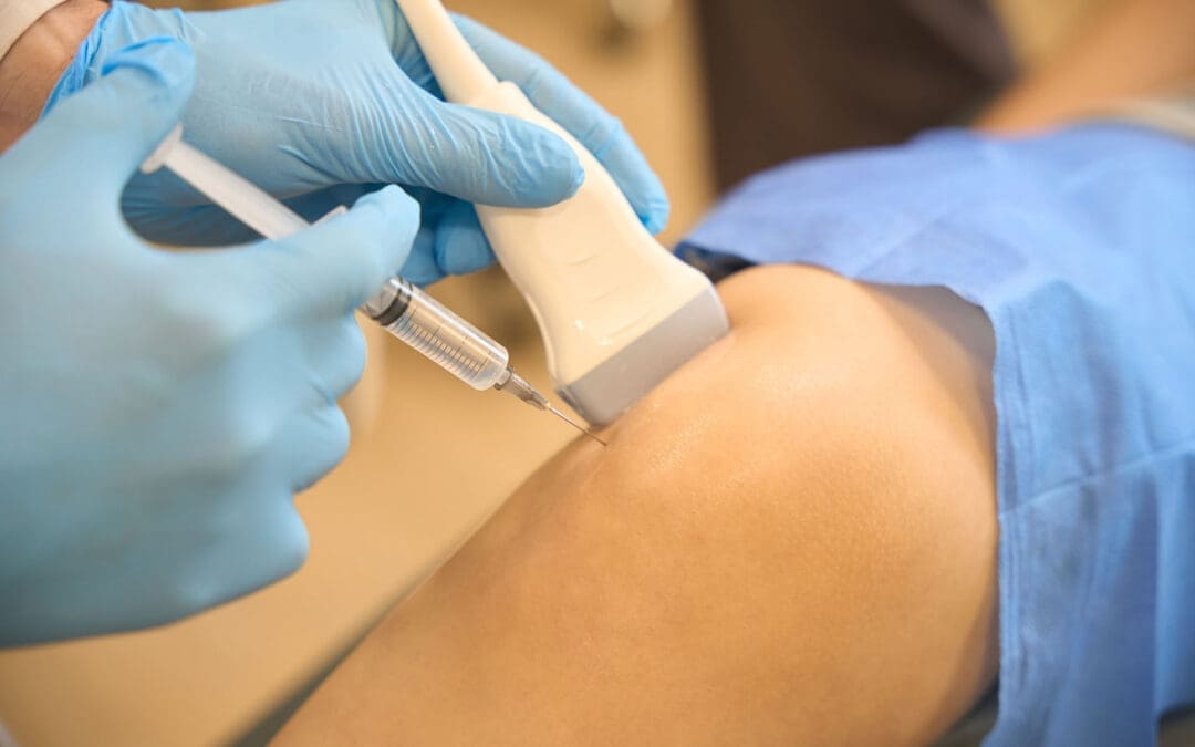
The Step-by-Step PRP Procedure
The whole process feels straightforward and takes about an hour. First, the nurse draws blood from the arm. Next, the blood spins in the centrifuge to create the PRP. Then the doctor uses ultrasound or X-ray guidance to place the PRP exactly where it is needed. Patients stay awake and feel only mild pressure. No stitches or long cuts are involved. The clinic sends the patient home the same day with simple care instructions.
Blood draw (small amount from the arm)
Centrifuge step to concentrate platelets
Ultrasound-guided injection into the spine
Short rest period before going home
Follow-up visits to check progress
Who’s a Good Candidate for PRP Therapy?
PRP is suitable for people with mild to moderate spinal wear who have not found sufficient relief from physical therapy or medication. It is not usually the first choice for very severe damage. A doctor checks imaging and health history to decide. Patients who want to stay active and avoid surgery often like this option. Good health and realistic goals help the treatment work best.
Integrative Spinal Care: Combining PRP with Chiropractic and Functional Medicine
In clinics that blend different care styles, PRP becomes even more effective. An Advanced Practice Registered Nurse (APRN/FNP-BC) with functional medicine training (CFMP, IFMCP, ATN, CCST) can administer precise, ultrasound-guided PRP injections. At the same time, chiropractic adjustments keep the spine aligned. Nutritional support from functional medicine fixes any missing vitamins or inflammation triggers in the body. This team approach creates the perfect setting for repair. The body gets structural help, cellular healing, and inside support all at once.
Insights from Dr. Alexander Jimenez on PRP and Spine Health
Dr. Alexander Jimenez, DC, APRN, FNP-BC, sees PRP as part of whole-body healing in El Paso, Texas. As both a chiropractor and nurse practitioner, he combines spinal adjustments with regenerative shots and metabolic checks. His clinical work shows that patients with sciatica or disc problems heal faster when PRP teams up with chiropractic care and proper nutrition. Dr. Jimenez notes that this mix helps clear waste from injured tissues, builds stronger blood flow, and stops pain cycles. Many of his patients return to work and sports with less discomfort and more confidence.
What to Expect During Recovery
Most people feel mild soreness for a few days after the shot, like a deep bruise. Ice packs and gentle movement help. Light activities can start right away, but heavy lifting waits one to two weeks. Full benefits build over four to six weeks as the growth factors continue to work. Some patients need a second shot after a month or two for the best results. Follow-up visits track progress and adjust the plan.
Evidence and Safety of PRP Therapy
Research backs PRP for spine care. Clinical reviews show pain drops and better movement in patients with degenerative discs and facet problems. Nerve repair studies also point to positive results. Side effects are rare because the treatment uses the patient’s own blood. No major complications appear in most studies. Doctors continue to track long-term outcomes, but current data look promising for people who want natural options.
Conclusion
Platelet-rich plasma therapy offers a fresh way to handle spinal pain and damage. It uses the body’s own tools to reduce swelling, repair tissues, and restore movement. When paired with expert chiropractic and functional medicine, the results can feel even better. Patients who have struggled with ongoing back issues often discover new hope through PRP. Talking with a trained provider helps decide if this path fits personal needs. With steady advances in regenerative care, many more people may soon enjoy life with less spine pain and more freedom.
PRP Therapy for Sports Injuries: How It May Speed Healing Without Surgery
Sports injuries can slow life down fast. A sore tendon, a strained ligament, or a muscle tear can make it difficult to train, work, sleep, or even walk comfortably. That is one reason Platelet-Rich Plasma, or PRP, has gained attention in sports medicine. PRP is made from a patient’s own blood and then injected into an injured area to support healing. Medical centers such as Yale Medicine, Penn Medicine, Johns Hopkins Medicine, and Temple Health describe PRP as a biologic or regenerative treatment that may help repair tissue, lower pain, and improve function in certain musculoskeletal injuries. It is often used for tendon, ligament, muscle, cartilage, and joint problems, including some cases of osteoarthritis. (Johns Hopkins Medicine, n.d.; Penn Medicine, 2025; Yale Medicine, n.d.).
PRP is appealing because it is non-surgical and uses the body’s own healing tools. Still, it is not a miracle fix for every athlete or every injury. Research shows promising results in many cases, but outcomes can vary depending on the tissue involved, how long the injury has been present, how the PRP is prepared, and whether the person also follows a successful rehab plan. In other words, PRP works best as part of a comprehensive care strategy rather than a stand-alone shot. (Saini et al., 2021; Jimenez, n.d.).
What PRP Therapy Is
PRP stands for Platelet-Rich Plasma. Plasma is the liquid part of blood, and platelets are blood components best known for their role in clotting. However, platelets also carry growth factors and signaling molecules that help tissue repair. To make PRP, a clinician draws a small amount of blood, spins it in a centrifuge, and separates out a platelet-rich portion. That concentrated solution is then placed into the injured area. The goal is to increase healing signals directly at the site of tissue damage. (Johns Hopkins Medicine, n.d.; Yale Medicine, n.d.; HSS, n.d.; Penn Medicine, 2025).
A simple way to think about PRP is this: it does not just try to numb pain. It tries to support the body’s repair response. Hospital for Special Surgery describes PRP as a form of regenerative medicine that amplifies natural growth factors in blood cells to help damaged tissue heal. Johns Hopkins Medicine similarly explains that the concentrated growth factors in PRP may stimulate tissue regeneration and speed healing in the treated area. (HSS, n.d.; Johns Hopkins Medicine, n.d.).
What the procedure usually includes
A small blood draw from the patient
Processing the sample in a centrifuge
Preparing the platelet-rich portion
Injecting the PRP into the injured tissue
In some cases, using ultrasound to guide the injection
A visit that often takes less than an hour
This basic process is described by major medical centers, including Penn Medicine, Yale Medicine, and Johns Hopkins Medicine. (Johns Hopkins Medicine, n.d.; Penn Medicine, 2025; Yale Medicine, n.d.).
How PRP May Help Sports Injuries Heal
When tissue is injured, the body sends platelets to the area early in the healing process. Temple Health explains that platelets contain growth factors that help promote cell growth, repair tissue, and reduce inflammation. Yale Medicine notes that PRP contains concentrated platelets, cytokines, and growth factors with anti-inflammatory properties. This is why PRP is often used for injuries that have been slow to heal on their own. (Temple Health, 2021; Yale Medicine, n.d.).
PRP may be especially useful in tissues that do not receive a strong blood supply. The 2021 review in the Indian Journal of Orthopaedics notes that tendons heal more slowly than many other tissues because of their poor vascularity. That same review also explains that PRP has been studied in tendon disorders such as Achilles tendinopathy, rotator cuff tendinitis, and epicondylitis, as well as in muscle strains and osteoarthritis. (Saini et al., 2021).
For athletes, this matters because many sports injuries are overuse or repetitive-stress injuries. If a tendon stays irritated for months, or a ligament strain never fully calms down, the body may need extra support to restart a healthier repair process. Some research suggests earlier PRP use in select injuries may help guide inflammation toward recovery and restore tissue balance. Even so, researchers also note there is no universal PRP formula or perfect protocol yet, so treatment must be individualized. (Saini et al., 2021).
Common Sports Injuries PRP Is Used For
Medical centers and sports medicine sources commonly describe PRP for the following problems:
Chronic tendinitis or tendinopathy
Tennis elbow
Patellar tendinopathy or “jumper’s knee”
Achilles tendon problems
Ligament strains
Muscle strains and some muscle tears
Cartilage irritation
Osteoarthritis in active adults
These uses are repeatedly listed by Penn Medicine, Yale Medicine, Temple Health, and HSS. (Penn Medicine, 2025; Temple Health, 2021; Yale Medicine, n.d.; HSS, n.d.).
Temple Health highlights tennis elbow and jumper’s knee as common orthopedic conditions that may benefit from PRP. In its overview, Penn Medicine also lists structures such as the Achilles tendon, ACL, hamstring, patellar tendon, and cartilage as areas in sports medicine where PRP is used. Yale Medicine adds tendon, ligament, and muscle conditions, as well as degenerative joint conditions, to that list. (Penn Medicine, 2025; Temple Health, 2021; Yale Medicine, n.d.).
There is also supportive evidence for muscle injury care when injections are placed carefully. A 2014 study in Blood Transfusion reported that athletes with grade II muscle lesions who received ultrasound-guided PRP showed full healing on ultrasound, pain resolution, and return to sport, with only one relapse reported a year later. That does not prove PRP is right for every muscle injury, but it does show why sports clinicians remain interested in it. (Borrione et al., 2014).
What Recovery Feels Like After PRP
One important point for patients is that PRP can cause short-term soreness. Yale Medicine says the most common side effects are discomfort, pain, and stiffness at the injection site. Penn Medicine also notes that mild soreness, swelling, or stiffness is common for the first few days. Johns Hopkins Medicine adds that some people notice soreness and bruising after the procedure. In most cases, these effects are temporary. (Johns Hopkins Medicine, n.d.; Penn Medicine, 2025; Yale Medicine, n.d.).
Patients also need realistic expectations. PRP is not usually an instant pain reliever. Penn Medicine says improvement may take a few weeks to become noticeable, with fuller benefits developing over months. Yale Medicine reports that some people notice pain improvement in four to six weeks, with continued progress for up to a year. (Penn Medicine, 2025; Yale Medicine, n.d.).
Aftercare often includes
Resting the area for a short time
Avoiding hard exercise right away
Using a guided rehab plan
Following instructions about pain control
Avoiding some anti-inflammatory medicines when advised
Penn Medicine and HSS both note that anti-inflammatory medicines may interfere with the early healing response that PRP is meant to support, so patients should follow their treating clinician’s advice. (HSS, n.d.; Penn Medicine, 2025).
Why Ultrasound-Guided PRP Matters
Not every injection needs the same level of precision, but many sports injuries benefit from careful image guidance. Both Johns Hopkins Medicine and Yale Medicine acknowledge the use of ultrasound during PRP procedures. Research in athletes also supports this approach. The 2014 study on muscle injuries emphasized that ultrasound was important for both locating the lesion and guiding the needle accurately into it. The 2021 sports injury review similarly reported that ultrasound-guided injections improve accuracy, particularly for musculoskeletal conditions. (Johns Hopkins Medicine, n.d.; Yale Medicine, n.d.; Borrione et al., 2014; Saini et al., 2021).
On Dr. Alexander Jimenez’s public clinical website, one recent educational article describes ultrasound-guided intra-articular hip PRP as a precision-focused procedure in which ultrasound helps the clinician visualize anatomy, confirm correct placement, and improve safety. That same article stresses that biologic injections work best when they are combined with rehabilitation and movement-based recovery rather than used alone. (Jimenez, n.d.).
Dr. Alexander Jimenez’s Clinical Observations and the Value of Integrated Care
Dr. Alexander Jimenez, DC, APRN, FNP-BC, describes his El Paso practice as a multidisciplinary and integrative model that combines chiropractic care, functional medicine thinking, sports medicine principles, rehabilitation, and regenerative strategies. His website presents regenerative medicine as a natural, non-surgical option designed not only to reduce pain but also to improve structure, movement, and function. (Jimenez, n.d.).
That point matters in sports injury care. A tendon or muscle may not stay healthy if the athlete still has poor joint mechanics, weak stabilizers, incorrect loading patterns, or nutrition and recovery habits that slow healing. Dr. Jimenez’s site repeatedly frames recovery as a full process that includes a detailed history, physical evaluation, attention to biomechanics, regenerative options when appropriate, chiropractic care to improve motion, rehab planning, and follow-up focused on function. (Jimenez, n.d.).
In a comprehensive clinic model, that means PRP can be paired with structural care, progressive rehabilitation, and functional medicine support. The injection may help the tissue biologically, while rehab helps the athlete move better and reduce repeated stress on the injured area. This combined approach aligns with the broader message from both sports medicine research and Dr. Jimenez’s clinical content: better recovery usually comes from treating the tissue and the movement pattern together. (Borrione et al., 2014; Jimenez, n.d.; Saini et al., 2021).
Benefits and Limits of PRP
Possible benefits
Uses the patient’s own blood
Minimally invasive
May reduce pain and improve function
May help some chronic tendon, ligament, muscle, and joint problems
Can be part of a non-surgical recovery plan
Can be combined with rehab and other supportive care
These benefits are commonly described by Yale Medicine, Penn Medicine, Johns Hopkins Medicine, and HSS. (HSS, n.d.; Johns Hopkins Medicine, n.d.; Penn Medicine, 2025; Yale Medicine, n.d.).
Important limits
Results vary from person to person
Some injuries still need surgery or other procedures
Relief may take weeks or months, not days
PRP preparation methods are not fully standardized
Some tissues have stronger evidence than others
Those limits are important because proper medicine depends on the right treatment for the right injury at the right time. PRP may be a strong option, but it should be chosen carefully after a full exam and diagnosis. (Saini et al., 2021; Penn Medicine, 2025).
Final Thoughts
PRP therapy offers a promising non-surgical option for sports injuries because it delivers a concentrated dose of the patient’s own platelets to damaged tissue, where growth factors may support repair, reduce inflammation, and improve recovery. It is commonly used for chronic tendinopathy, ligament strain, muscle injury, and some joint conditions. Short-term soreness at the injection site can happen, but serious side effects are uncommon. The best results usually come when PRP is matched to the right injury and combined with smart rehabilitation, movement correction, and careful follow-up. (Johns Hopkins Medicine, n.d.; Penn Medicine, 2025; Yale Medicine, n.d.; Jimenez, n.d.).
Sciatica Relief in El Paso: How Integrative Chiropractic Care Supports Healing and Mobility
Sciatica can make daily life challenging. It often causes pain that starts in the lower back or buttocks and travels down the leg. Some people also feel tingling, numbness, burning, or weakness. In many cases, the problem begins when a lumbar disc, tight soft tissue, joint irritation, or spinal narrowing compresses a nerve root. Because sciatica can have multiple causes, treatment works best when it focuses on the whole person, not just the pain. That is why a chiropractic rehabilitation model aligns well with this topic for El Paso Back Clinic. The clinic publicly describes itself as a chiropractic rehabilitation and integrated medicine center focused on injury recovery, movement, function, and whole-person care. (Berry et al., 2019; El Paso Back Clinic, n.d.-a; El Paso Back Clinic, n.d.-b).
At El Paso Back Clinic, the public-facing message centers on chiropractic care, rehabilitation, mobility, flexibility, nutrition, and integrated support. The site describes Dr. Alexander Jimenez as both a chiropractor and a family nurse practitioner, leading a multidisciplinary team that blends evidence-based care with natural and functional approaches. That positioning is relevant for sciatica because many people improve with conservative care built around assessment, education, movement, and structured rehabilitation before more invasive options are considered. (El Paso Back Clinic, n.d.-a; El Paso Back Clinic, n.d.-c; Jimenez, n.d.).
What Sciatica Really Means
Sciatica is a symptom pattern, not a stand-alone diagnosis. It usually describes pain that follows the path of the sciatic nerve, often from the lower back into the buttocks, thigh, calf, or foot. A careful exam usually includes a history, strength testing, reflexes, sensation testing, and nerve tension testing. This matters because sciatica-like pain can arise from lumbar disc herniation, degenerative disc changes, facet joint irritation, spinal stenosis, piriformis-related irritation, or combined movement-related problems. When the source is correctly identified, treatment can be more specific and effective. (Berry et al., 2019).
Why a Chiropractic and Physical Rehabilitation Approach Fits So Well
Current guidance for lumbosacral radicular pain supports a stepped, conservative approach as first-line treatment. That usually means education, staying active, exercise therapy, and treatment matched to the patient’s symptoms and function. Recent guideline work also emphasizes clear communication, a gradual return to activity, and exercise therapy tailored to the person’s needs and tolerance. In other words, successful care is not just about lying down and waiting. It is about restoring motion, building support around the spine, and helping the nervous system calm down while the tissues recover. (Apeldoorn et al., 2024; Schmid & Tampin, 2023).
This conservative framework matches the public model of El Paso Back Clinic. The clinic’s website describes a whole-person plan that addresses posture, movement, daily habits, flexibility, strength, and nutrition. It also highlights chiropractic adjustments, rehabilitation-based care, and functional support rather than making injections the center of the message. That is a strong fit for a sciatica article aimed at a chiropractic and physical therapy audience. (El Paso Back Clinic, n.d.-d; El Paso Back Clinic, n.d.-e; El Paso Back Clinic, n.d.-f).
How Integrative Chiropractic Care May Help Sciatica
Chiropractic care for sciatica is not just one quick adjustment. In a more integrative setting, it can include a mix of spinal manipulation or mobilization, soft-tissue work, guided stretching, core-stability work, gait and posture correction, mobility drills, and progressive strengthening. The goal is to reduce mechanical stress, improve joint motion, improve movement patterns, and support the body’s own recovery. El Paso Back Clinic’s public materials describe a broader plan, including adjustments, exercises, and wellness strategies designed to restore mobility and reduce pressure on irritated structures. (El Paso Back Clinic, n.d.-b; El Paso Back Clinic, n.d.-d; El Paso Back Clinic, n.d.-e).
A 2024 narrative review on lumbar disc herniation with radiculopathy reported that spinal mobilization with leg movement, lumbar stabilization exercises, and manipulation can reduce symptoms and improve stability and mobility in selected patients. The same review emphasized that weak core muscles and poor spinal stability can delay healing, which is why structured rehabilitation matters so much. This supports a chiropractic rehabilitation strategy that focuses on both pain relief and rebuilding support around the lumbar spine. (El Melhat et al., 2024).
The Role of Exercise, Rehab, and Movement Training
For many people with sciatica, movement is medicine when it is used the right way. Recent physical therapy guidance recommends exercise therapy for patients who need help with daily activities, participation, or movement-related limits. The program should match irritability, tolerance, and function. In early stages, that may mean gentle pain-relieving movements, walking progressions, and avoiding positions that sharply increase symptoms. Later, it often expands into core work, hip strength, endurance, balance, and return-to-activity training. (Apeldoorn et al., 2024).
This is one of the biggest advantages of an integrative chiropractic clinic with a rehabilitation mindset. A patient is not just told where the pain is. They are shown how to move better, sit and lift with less strain, rebuild spinal support, and reduce the repeated stresses that may have contributed to the problem. El Paso Back Clinic’s site repeatedly highlights mobility, flexibility, sports medicine concepts, rehabilitation, and personalized exercise support as part of care. (El Paso Back Clinic, n.d.-d; El Paso Back Clinic, n.d.-f).
Common parts of a chiropractic rehabilitation plan for sciatica
Spinal adjustments or mobilization to improve motion
Soft tissue work for tight lumbar, hip, and gluteal tissues
Nerve-friendly movement progressions
Core stabilization exercises
Hip and pelvic strength work
Posture and ergonomic coaching
Walking programs and activity modification
Nutrition and inflammation support when needed
These tools do not all apply to every patient, but together they show why conservative care can be more than temporary pain relief. It can help correct the patterns that keep irritating the sciatic nerve. (Apeldoorn et al., 2024; El Melhat et al., 2024; El Paso Back Clinic, n.d.-e).
Clinical Observations from Dr. Alexander Jimenez
Dr. Alexander Jimenez’s public pages describe a dual-scope model that blends chiropractic care with nurse practitioner-level medical evaluation, functional medicine, and individualized rehabilitation planning. His clinic materials emphasize non-surgical recovery, movement restoration, advanced assessment, and whole-person healing. At El Paso Back Clinic, sciatica care is presented as a process of locating the source of the problem, improving alignment and mechanics, and guiding the patient back toward better function. That practical, layered approach is especially useful for chronic or recurring sciatica, where structural, inflammatory, stress-related, and lifestyle factors may overlap. (Jimenez, n.d.; El Paso Back Clinic, n.d.-a; El Paso Back Clinic, n.d.-b).
Where PRP Fits In
Platelet-Rich Plasma is made from a patient’s own blood and is used in regenerative medicine to deliver concentrated platelets and growth factors to a target area. In lumbar radiculopathy research, PRP injections have shown promising results in pain and function, and some studies suggest longer-lasting improvement than steroid injections in selected patients. Still, PRP is best presented as an adjunct option for carefully chosen cases, not as the foundation of care for every person with sciatica. (Gupta et al., 2024; Saraf et al., 2023).
That is also the most natural fit for a chiropractic and rehab-focused clinic. The main message should remain focused on conservative care, mechanical correction, mobility, strength, and function. PRP can be discussed as a secondary option for patients with persistent disc-related irritation who have not improved sufficiently with conservative care and who want a non-surgical option that goes beyond short-term symptom control. (Schmid & Tampin, 2023; Gupta et al., 2024; Saraf et al., 2023).
Why Whole-Person Care Matters
Sciatica is often worse when movement quality, stress load, inflammation, sleep, conditioning, and work demands are ignored. That is why integrative care can be valuable. A patient may need chiropractic treatment for joint motion, rehabilitation for core support and hip control, coaching on posture and lifting, and broader wellness strategies to reduce ongoing irritation. El Paso Back Clinic publicly describes this kind of combined approach, which includes chiropractic, rehabilitation, functional medicine, nutrition, and injury recovery planning. (El Paso Back Clinic, n.d.-c; El Paso Back Clinic, n.d.-f; Jimenez, n.d.).
Final Thoughts
For El Paso Back Clinic, the strongest sciatica message is clear: chiropractic rehabilitation should lead the conversation. People searching for help with sciatic pain often want answers that feel practical, natural, and functional. They want to know whether they can move again, work again, sleep better, and get back to life without jumping straight to drugs or procedures. A chiropractic and physical therapy-based strategy speaks directly to those goals. PRP can stay in the background as an advanced regenerative option for selected cases, but the heart of the article should stay on spinal mechanics, rehabilitation, movement, and whole-person recovery. That approach is consistent with both modern stepped-care guidance and the public identity of El Paso Back Clinic. (Apeldoorn et al., 2024; Schmid & Tampin, 2023; El Paso Back Clinic, n.d.-a).
Restore Flexibility and Mobility with Integrative Chiropractic Care and Shockwave Therapy at El Paso Back Clinic
Many El Paso residents wake up with stiff joints or tight muscles, making simple daily tasks feel hard. Reaching overhead, bending down, or walking for long stretches can become painful or limited. At El Paso Back Clinic, integrative chiropractic care combined with Extracorporeal Shockwave Therapy (ESWT) offers a natural solution. This approach restores proper joint alignment, reduces muscle tension, and resolves soft-tissue restrictions, allowing patients to move freely again. Led by Dr. Alexander Jimenez, DC, APRN, FNP-BC, the clinic’s team uses gentle adjustments, stretching, exercises, and advanced shockwave treatments to help people regain flexibility and enjoy life in El Paso.
What Integrative Chiropractic Care Does for Flexibility at El Paso Back Clinic
Integrative chiropractic care at El Paso Back Clinic treats the whole body instead of just one problem area. It corrects small misalignments, called subluxations, in the spine and joints. These misalignments put pressure on nerves and tighten muscles. Regular adjustments gently move everything back into place. This restores proper joint alignment, eases tension, and lets the nervous system send clearer signals to the muscles.
When joints line up correctly, range of motion improves right away. Stiffness fades, and daily movements become smoother and more efficient. Patients at the clinic often say they feel looser and more energetic after just a few visits. (Gentle Chiro, n.d.) The care also includes stretching and therapeutic exercises to maintain gains over time. Muscles and joints start working together as a team, building resilience that lasts.
How Chiropractic Adjustments Restore Joint Alignment and Reduce Stiffness
Adjustments form the core of care at El Paso Back Clinic. The team uses precise, gentle pressure to correct subluxations. This simple step brings clear benefits that patients notice quickly:
Better range of motion, so joints glide freely without catching
Less muscle tension around the back, neck, and limbs
Improved nervous system function for better balance and coordination
Smoother daily activities like turning your head while driving or reaching for groceries
Lower risk of future stiffness because proper alignment trains the body to stay balanced
Many people in El Paso report that these changes make physical activities feel easier and less tiring. (Rodgers Stein Chiropractic, n.d.) The adjustments help the body move more efficiently without pain, supporting an active lifestyle.
Adding Stretching and Therapeutic Exercises for Long-Term Results
Adjustments open the door to better movement, but stretching and exercises keep it open. At El Paso Back Clinic, the rehabilitation team creates simple home programs that match each patient’s needs. Dynamic stretches warm up the body before activity. Static stretches hold the new mobility after adjustments. Therapeutic exercises strengthen the muscles that support the joints.
These steps build endurance and agility. Patients find they can stay active longer without soreness. The clinic’s sports medicine approach helps people return to hiking in the Franklin Mountains, playing with family, or working without the same old limitations. (Chiropractic Fitness, n.d.) Consistent practice turns short-term gains into lasting flexibility.
Introducing Extracorporeal Shockwave Therapy (ESWT) at El Paso Back Clinic
ESWT uses focused sound waves to reach deep into muscles, tendons, and ligaments. The waves create tiny pulses that restart healing in areas stuck with scar tissue or chronic tightness. This noninvasive treatment increases blood flow, breaks down old buildup, and reduces inflammation. At El Paso Back Clinic, ESWT is available as a key component of advanced care plans for patients who need additional support for soft tissue problems.
Why Combining Chiropractic Care and ESWT Delivers Stronger Flexibility Gains
The real power at El Paso Back Clinic comes from pairing chiropractic adjustments with ESWT. Adjustments fix the mechanical side—joint position and nerve signals—while ESWT handles the soft-tissue side—scar tissue, poor circulation, and stubborn tension. Together, they create faster, longer-lasting results than either method alone.
This dual approach works in several key ways:
Chiropractic restores spinal and joint mobility
ESWT breaks down scar tissue and releases tight fascia
The pair reduces inflammation and collagen cross-linking that causes stiffness
Blood flow improves, helping muscles and tendons heal
Patients regain a greater range of motion because both structure and tissue health get better at once
Clinic reports show that this combination can significantly improve outcomes compared with standard care. Many El Paso patients with ongoing tightness notice a real return of freedom of movement.
Common Conditions That Benefit from This Integrated Approach
El Paso Back Clinic uses this combined approach to treat several conditions that rob people of flexibility. Here are some of the most common:
Frozen shoulder – Adjustments free stuck joints while ESWT dissolves scar tissue and calcium deposits. Patients often regain full arm motion without pain.
Achilles tendinopathy – Chiropractic realigns the lower body to ease strain. Shockwave therapy stimulates the growth of new blood vessels and clears chronic buildup, so walking and running feel normal again.
General chronic muscle tension – Tightness in the back, neck, or legs from stress, work, or old injuries—responds well. The therapies release trigger points and restore smooth movement.
Post-injury stiffness from car accidents or sports – The clinic specializes in personal injury care. The combination speeds recovery and safely rebuilds mobility.
Other issues, such as plantar fasciitis and tennis elbow, also improve because the care addresses both alignment and tissue damage. (Bend Total Body Chiropractic, n.d.)
Clinical Insights from Dr. Alexander Jimenez at El Paso Back Clinic
Dr. Alexander Jimenez, DC, APRN, FNP-BC, leads El Paso Back Clinic with more than 30 years of experience. As both a Doctor of Chiropractic and a board-certified Family Nurse Practitioner, he brings a unique integrative perspective to every patient. In his clinical work in El Paso, Dr. Jimenez sees how chiropractic adjustments correct subluxations and improve nervous system function, thereby boosting flexibility and range of motion. When combined with ESWT, the results are even stronger for soft tissue injuries from accidents or overuse.
Dr. Jimenez often notes that this teamwork helps patients break down scar tissue, reduce inflammation, and restore proper movement patterns faster than traditional methods alone. His approach includes personalized functional medicine, nutritional support, and rehabilitation exercises to help patients build lasting resilience. At the clinic’s convenient El Paso locations, patients receive complete care that addresses the root causes of stiffness and helps them return to daily life and favorite activities with confidence.
Tips to Get the Most from Care at El Paso Back Clinic
Start with a full evaluation so the team can build a plan that fits your body and lifestyle. Attend regular adjustments and ESWT sessions as recommended. Follow the simple stretching and exercise routine at home every day. Support your progress with good posture, daily walks, proper hydration, and enough rest. The friendly staff at El Paso Back Clinic makes the process easy and supportive. Many patients see big improvements in flexibility within just a few weeks when they stay consistent.
A Natural Path to a More Flexible, Resilient Life in El Paso
Integrative chiropractic care and ESWT at El Paso Back Clinic offer a powerful, drug-free way to fight stiffness and reclaim natural movement. By correcting joint alignment, releasing muscle tension, and healing soft tissues, this approach makes daily life and physical activity feel effortless again. Muscles and joints work in harmony, the nervous system functions smoothly, and the body stays strong through the years.
Whether you deal with occasional tightness or a specific injury, the experienced team at El Paso Back Clinic can help. Contact the clinic today to schedule an evaluation and discover how these natural tools can work for you. With the right plan, better flexibility and mobility are well within reach for El Paso residents.
IFM's Find A Practitioner tool is the largest referral network in Functional Medicine, created to help patients locate Functional Medicine practitioners anywhere in the world. IFM Certified Practitioners are listed first in the search results, given their extensive education in Functional Medicine