Can individuals dealing with pain and inflammation in their bodies can incorporate a ketogenic diet and have beneficial results?
Introduction
When it comes to a person’s health and wellness journey, many people often wonder where to start making changes in their bodies. Many people begin exercising not only to reduce stress levels in the musculoskeletal system but also to help clear their minds. This is a great cause, as any form of physical activity can help reduce muscle fatigue and weakness and strengthen the extremities and quadrants in the musculoskeletal system. However, even though exercising is part of the health and wellness journey, another component plays a crucial part in the health and wellness journey: eating nutritional foods and dieting. Now, dieting can be scary for some newcomers who are just entering their health and wellness journey, but when people start making small changes to their eating habits, like incorporating more vegetables and fruits in their meals, making more meals at home, and doing portion control to eat enough till they are full can provide beneficial results. These small changes can empower individuals to take control of their health and wellness journey. Dieting and changing eating habits can give useful results for people dealing with chronic issues correlated with environmental factors. In today’s article, we will look at a particular diet known as the ketogenic diet, its beneficial properties, and how to incorporate it to reduce chronic conditions affecting the body. We talk with certified associated medical providers who provide our patients’ information to assess how incorporating the ketogenic diet can help reduce chronic conditions. We also inform patients while asking their associated medical provider intricate questions to formulate customized treatment plans to help with chronic conditions by incorporating the ketogenic diet along with physical activities. Dr. Alex Jimenez, D.C., includes this information as an academic service. Disclaimer.
What Is the Ketogenic Diet?
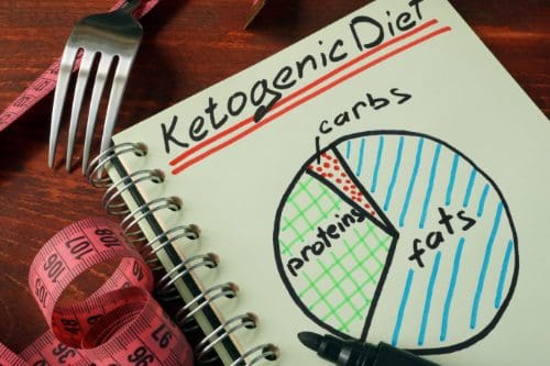
Do you often feel extremely thirsty throughout the day, and do you chug at least a couple of gallons of water daily? Do you feel heat or see redness in various locations around your body that seem tender when touched? Or do your joints become stiff in the mornings when you wake up and feel better throughout the entire day? Often, people have a love-hate relationship with food. However, it all depends on what the person is eating and what environmental factors correlate with their food. So, when a person has chronic conditions that cause pain and inflammation in their joints, muscles, and organs, the negative side effects are that the food they consume can induce the inflammatory effects. In contrast, the positive impact of food can help individuals dampen the pain and inflammation in the body. (Fifi & Holton, 2020) Many individuals dealing with inflammation and pain in their bodies can incorporate a ketogenic diet to reduce the inflammatory effects.
Now, what is the ketogenic diet? The ketogenic diet is where carbohydrates are low, and various levels of lean proteins and healthy fats induce ketosis. (McGaugh & Barthel, 2022) This, in turn, helps with alternating the metabolic pathways to induce weight loss, improve health conditions like lipid profile improvements, and reduce hyperglycemia. When environmental factors like obesity, sedentary lifestyles, or physical inactivity can cause chronic issues like lipedema, the body will cause overlapping risk profiles to induce inflammation. When the body is dealing with lipedema, it can cause the body to be in a constant inflammatory state that induces cell damage and cell death through apoptosis. Hence, when a person is going to do a ketogenic diet, it can help reduce the inflammatory effects while alleviating pain symptoms. (Verde et al., 2023)
Eating Right To Feel Better-Video
The Beneficial Properties Of A Ketogenic Diet
One of the beneficial properties that a person can take with a ketogenic diet is that it can help reduce cardiovascular diseases with its anti-inflammatory properties. Since a ketogenic diet helps place the body in a state of nutritional ketosis, incorporating omega-3 fatty acids can help the body exert systemic anti-inflammatory effects and begin healing. (Dynka et al., 2023) At the same time, the ketogenic diet can help individuals who are suffering from chronic conditions like epilepsy, diabetes, or obesity to not only induce weight loss but also help improve body composition. (Sjodin et al., 2020) This is because when individuals get a customized treatment plan incorporating the ketogenic diet, it must include physical activities that help strengthen muscles in the body’s quadrants. In contrast, the ketogenic diet helps slow down muscle glycogen depletion. Additionally, the ketogenic diet is beneficial by:
Reducing glucose levels so individuals with diabetes don’t need insulin.
When it comes to the ketogenic diet, many individuals have dealt with chronic conditions like epilepsy, diabetes, or cardiovascular conditions. Understand that incorporating a healthy diet filled with healthy fats, lean protein, plenty of fruits and vegetables, and physical activity can benefit many individuals in the long run. The ketogenic diet can help the liver by producing more ketone bodies to help with the production of ATPs and reduce ROS (reactive oxygen species); this, in turn, helps improve a person’s quality of life. (Abboud et al., 2021) Informing individuals who are dealing with chronic conditions that they must make small changes to their routines. This can be incorporated into their customized treatment plan and help reduce the pain-like effects from the person’s chronic conditions correlating to the inflammatory effects. The ketogenic diet can be a stepping stone to a person’s health and wellness and help them be motivated to see positive results.
References
Abboud, M., AlAnouti, F., Georgaki, E., & Papandreou, D. (2021). Effect of Ketogenic Diet on Quality of Life in Adults with Chronic Disease: A Systematic Review of Randomized Controlled Trials. Nutrients, 13(12). https://doi.org/10.3390/nu13124463
Dowis, K., & Banga, S. (2021). The Potential Health Benefits of the Ketogenic Diet: A Narrative Review. Nutrients, 13(5), 1654. https://doi.org/10.3390/nu13051654
Dynka, D., Kowalcze, K., Charuta, A., & Paziewska, A. (2023). The Ketogenic Diet and Cardiovascular Diseases. Nutrients, 15(15). https://doi.org/10.3390/nu15153368
Sjodin, A., Hellstrom, F., Sehlstedt, E., Svensson, M., & Buren, J. (2020). Effects of a Ketogenic Diet on Muscle Fatigue in Healthy, Young, Normal-Weight Women: A Randomized Controlled Feeding Trial. Nutrients, 12(4). https://doi.org/10.3390/nu12040955
Verde, L., Camajani, E., Annunziata, G., Sojat, A., Marina, L. V., Colao, A., Caprio, M., Muscogiuri, G., & Barrea, L. (2023). Ketogenic Diet: A Nutritional Therapeutic Tool for Lipedema? Curr Obes Rep, 12(4), 529-543. https://doi.org/10.1007/s13679-023-00536-x
For individuals with cervical arthritis, can physical therapies help manage symptoms and bring pain relief?
Cervical Arthritis
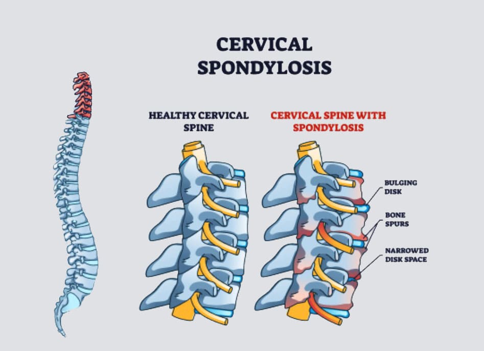
Cervical spondylosis, more commonly known as cervical arthritis or arthritis of the neck, refers to the wearing down of neck bones, discs, tendons, ligaments, and joints. The primary symptoms are neck pain and stiffness. However, it is also possible to have cervical spondylosis and not have any symptoms. The condition affects over 85% of individuals over age 65. (American Academy of Orthopaedic Surgeons, 2021) Treatment can consist of conservative therapies and includes physical therapies, alternative medicine therapies, at-home self care, and over-the-counter and prescription medications. Severe cases of cervical spondylosis are treated with surgery to repair damaged parts of the spine.
Symptoms
Neck pain and headaches at the back of the head are usually the first symptoms. (Kazeminasab S. et al., 2022) The neck can also feel stiff, with worse morning symptoms that improve throughout the day. (Johns Hopkins Medicine, 2024) Symptoms can range from mild discomfort to severe pain. As cervical spondylosis progresses, individuals can experience:
Inability to turn the head or bend the neck.
A clicking or grinding noise when turning the neck.
Tenderness with pressure on the neck.
Pain that radiates to the shoulders or shoulder blades.
Pain and symptoms that disrupt sleep, sometimes causing waking up throughout the night.
Symptoms that decrease with rest.
More severe symptoms include:
Cervical Bone Spurs – Osteophytes
Some with cervical spondylosis have bony growths that can place pressure on the spinal nerves (a pinched nerve) (Bon Secours, 2024). Compression of spinal nerve roots produces cervical radiculopathy, which leads to pain, tingling, and weakness that radiates into the shoulders, arms, and hands.
Cervical Myelopathy
This refers to spinal impingement that leads to spinal cord dysfunction. (Spinal cord dysfunction is a nervous system disorder with interruptions in the spinal cord’s motor, sensory, and autonomic functions.) Symptoms include pain, tingling, numbness, muscle spasms, and weakness in areas below the neck. Spinal cord dysfunction can affect mobility, hand use, and bladder or bowel function control.
Causes
Where degenerative changes are commonly associated with cervical spondylosis, other conditions, and factors can lead to it and include:
Autoimmune Diseases
Rheumatoid arthritis and psoriatic arthritis can cause chronic inflammation in the cervical spine.
Trauma
Neck trauma, including injury and repetitive stress on the neck.
Cervical spondylosis is commonly seen in occupations that involve neck-stressing activities, such as sports.
Age
Wearing down of the spinal discs cartilage between the vertebrae.
Genetic components have been identified in connection with cervical spondylosis, meaning that some types of arthritis that lead to spinal damage are hereditary. (Kazeminasab S. et al., 2022)
Treatment
Treatment begins conservatively, using protocols to preserve function and avoid surgery. Nonsurgical treatments include medications, physical therapy, at-home exercises, and alternative medicine. The treatment method a healthcare provider chooses will depend on how severe the spondylosis is and other factors like age, how much pain is being experienced, the cause, and overall health. The main objectives are to relieve pain, prevent long-term damage to the spinal cord and nerves, and help maintain performing daily activities. (Bon Secours, 2024)
Medications
Medicines used to treat cervical spondylosis include:
Nonsteroidal Anti-inflammatory Drugs NSAIDs
NSAIDs, including ibuprofen and naproxen sodium, are available without a prescription to relieve pain and inflammation.
A healthcare provider can prescribe a more powerful NSAID to help manage severe symptoms.
Corticosteroids
A corticosteroid injection or a short course of an oral corticosteroid, like prednisone, can ease pain and reduce inflammation.
Muscle Relaxants
If cervical spondylosis causes muscle spasms, a healthcare provider can prescribe cyclobenzaprine, a muscle relaxant, to manage symptoms.
Antidepressants
Some types of antidepressants can ease neck pain from cervical spondylosis.
Anti-seizure Meds
Some anti-seizure drugs can cause nerve pain resulting from damaged nerves.
Physical Therapies
Physical therapy will help manage pain and stiffness and keep muscles loose and relaxed.
A physical therapist will teach the patient exercises to stretch and strengthen neck and shoulder muscles.
At-Home Self Care
At-home exercises can help relieve pain, stiffness, and swelling. Some can include:
Reducing inflammation and stress on the neck through posture training.
At-home targeted stretches and exercises will help bring pain relief.
Ice treatment reduces swelling.
Heat will increase circulation.
A neck brace may be recommended briefly to avoid muscle weakness and stiffness.
Alternative Medicine
Chiropractic adjustments and massage therapy are alternative treatments that will help manage cervical spondylosis.
Acupuncture can also be beneficial in reducing neck pain and increasing energy circulation. (Gu C. L. et al., 2019)
Various therapeutic massage therapies will help relieve neck pain and stiffness. Talk to a healthcare provider before starting treatment so they can advise on whether neck massages are safe.
Surgery
A healthcare provider may recommend surgical treatment when all other treatments have failed, if neurological symptoms are severe, or if neck arthritis causes extreme pain or disability. Surgery to treat cervical spondylosis can involve removing bone spurs, part of the cervical vertebra, or a herniated disc. The removed portions of the cervical spine are fused with hardware and bone grafts.
Injury Medical Chiropractic and Functional Medicine Clinic
Chiropractic therapy is among the more conservative treatment options and may be tried first before proceeding with surgery. Injury Medical Chiropractic and Functional Medicine Clinic works with primary healthcare providers and specialists to develop an optimal health and wellness solution.
Arthritis Explained
References
American Academy of Orthopaedic Surgeons. (2021). Cervical spondylosis (arthritis of the neck). https://orthoinfo.aaos.org/en/diseases–conditions/cervical-spondylosis-arthritis-of-the-neck/
Kazeminasab, S., Nejadghaderi, S. A., Amiri, P., Pourfathi, H., Araj-Khodaei, M., Sullman, M. J. M., Kolahi, A. A., & Safiri, S. (2022). Neck pain: global epidemiology, trends and risk factors. BMC musculoskeletal disorders, 23(1), 26. https://doi.org/10.1186/s12891-021-04957-4
Johns Hopkins Medicine. (2024). Spinal arthritis (arthritis in the back or neck). https://www.hopkinsmedicine.org/health/conditions-and-diseases/spinal-arthritis
Bon Secours. (2024). Cervical osteoarthritis (arthritis in the neck). https://www.bonsecours.com/health-care-services/spine-care/conditions/cervical-osteoarthritis
American Chiropractic Association. (2024). Neck pain. https://www.acatoday.org/patients/neck-pain-and-chiropractic/
Jenkins, H. J., Downie, A. S., Moore, C. S., & French, S. D. (2018). Current evidence for spinal X-ray use in the chiropractic profession: a narrative review. Chiropractic & manual therapies, 26, 48. https://doi.org/10.1186/s12998-018-0217-8
Gu, C. L., Yan, Y., Zhang, D., & Li, P. (2019). An evaluation of the effectiveness of acupuncture with seven acupoint-penetrating needles on cervical spondylosis. Journal of pain research, 12, 1441–1445. https://doi.org/10.2147/JPR.S199798
Can individuals support their parasympathetic nervous system to maintain a relaxed state and avoid excessive agitation, stress, anxiety, and dysregulation?
Parasympathetic Nervous System
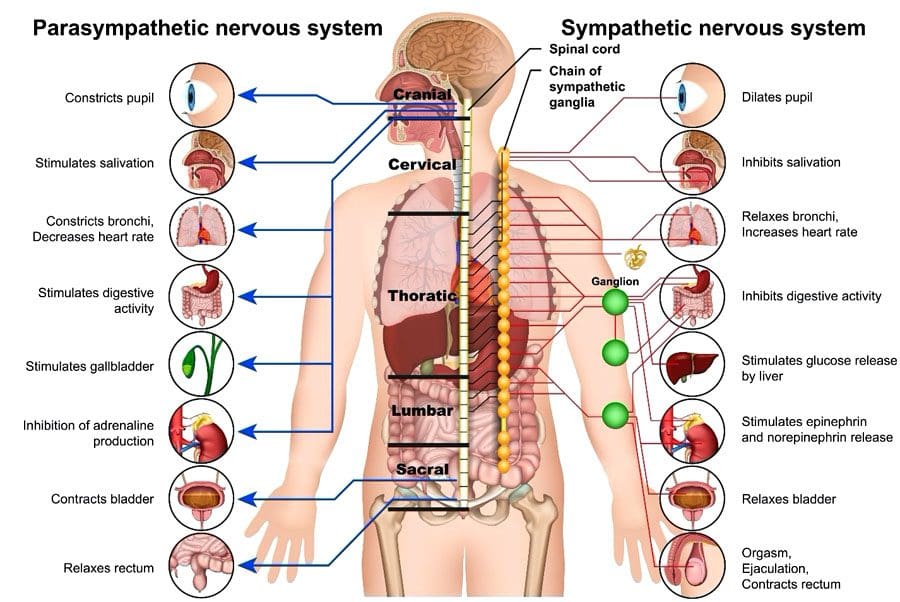
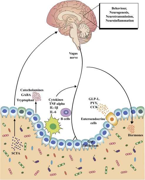
The parasympathetic nervous system (PSNS) involves organs and cells that release neurotransmitters that help the body maintain essential functions like heart rate, breathing, digestion, relaxation, thinking, and sleep. It is part of the autonomic nervous system, which includes the nerves and neurotransmitters that control the body’s internal organs to maintain reliable function. The autonomic nervous system regulates a continual balance between the parasympathetic nervous system’s rest-and-digest functions and the sympathetic nervous system’s fight-or-flight responses.
Functions and Responses
The parasympathetic nervous system includes nerves and neurotransmitters (chemical messengers) distributed throughout the body and is often described as promoting the body’s rest-and-digest state. The body’s needs regulate the control and effects of the system. Certain medications and health issues can modify or alter the function of the autonomic nervous system, including sympathetic and parasympathetic areas. The parasympathetic and sympathetic nervous systems collaborate to control cells, tissues, and organs. The body requires constant functioning and adjustment of both systems. (McQuade J. D. et al., 2017) Functions include: (Valenti V. E. et al., 2024)
Saliva secretion to break down food.
Maintaining blood pressure, resting heart, and breathing rate.
Producing enzymes in the stomach and intestines to break down food further and absorb nutrients.
Kidney balancing of fluid and mineral levels by producing urine.
Constricting pupils (making the dark circle in the eye smaller) to see details up close.
Maintaining focus and concentration to think, remember, and make decisions.
Releasing hormones to fall asleep and stay asleep.
Providing consistent muscle stimulation to maintain endurance for muscle activity.
All functions are adjusted based on the body’s needs.
Activation
The parasympathetic response constantly works to keep the body active, engaged, and healthy and can be stimulated simply by relaxing and resting. Individuals can sometimes focus on promoting their body’s parasympathetic activity through stretching, slow movements, relaxing sounds or music, meditation, and similar activities. Individuals who frequently feel anxious or stressed and have difficulty relaxing could benefit from guidance from a therapist or coach on learning to engage the parasympathetic nervous system. (Goren O. et al., 2024) This can involve activities such as guided meditation or talk therapy, which can help individuals work through fears and anxieties.
Dysregulation
Some medical conditions are associated with dysregulation. Heart disease, respiratory disease, sleep disorders, mental health conditions, and behavioral conditions can be associated with overactive or underactive parasympathetic or sympathetic nervous system regulation. (Veerakumar A. et al., 2022) Various mental health and physical health disorders can involve symptom fluctuations that affect the autonomic nervous system. For example, anxiety disorders can cause periods of overeating as well as loss of appetite and episodic indigestion, nausea, and vomiting. Symptoms of dysregulation can include: (McQuade J. D. et al., 2017)
Excessive sleepiness
Insomnia
Difficulty concentrating
Confusion
Anxiety – nervousness or agitation
A feeling of a rapid heart rate
Shortness of breath
Dry mouth
Throat tightness
Indigestion
Stomachaches
Nausea
Vomiting
Cold hands and feet
Sweating or clammy hands
Sadness
Depression
Dizziness or feeling physically off-balance
Any of these symptoms can develop and resolve quickly. Most individuals experience symptoms or parasympathetic nervous system dysfunction when an unexpected stressful event occurs, and the response is considered normal in traumatic, shocking, sad, or high-stress circumstances. (Veerakumar A. et al., 2022)
Medications may induce parasympathetic overactivity or underactivity until the medicine is broken down and removed from the body. (Valenti V. E. et al., 2024)
Nerves
The system comprises nerves that release hormones and neurotransmitters and nerves that respond to the hormones and neurotransmitters. Parasympathetic activation and response include: (Valenti V. E. et al., 2024)
The nerves that activate the parasympathetic nervous system run throughout the body, primarily in the brain, heart, lungs, stomach, and intestines.
The brain, heart, lungs, stomach, intestines, muscles, liver, kidneys, reproductive organs, eyes, and mouth are among the areas of the body that respond to stimulation.
The vagus nerve, one of the cranial nerves, is closely associated with parasympathetic nervous system activity. It runs from the brainstem down into the throat, heart, lungs, and digestive system. (Valenti V. E. et al., 2024) Surgical, medical, and alternative interventions are used to regulate the activity of the parasympathetic nervous system and are targeted toward regulating and controlling the activity of the vagus nerve. (Hernández-Domínguez R. A. et al., 2024)
PSNS Support
Living with any parasympathetic nervous system dysfunction can be challenging. For individuals with heart or lung disease, healthcare providers will evaluate the medical condition and recommend surgery or medication to control symptoms and avoid complications. (Hernández-Domínguez R. A. et al., 2024) Individuals who are living with a mental health disorder or behavioral disorder that is associated with any dysregulation of the parasympathetic nervous system activity may benefit from a combination of behavioral interventions and medication to help control symptoms long term. (Goren O. et al., 2024)
Injury Medical Chiropractic and Functional Medicine Clinic
The spinal cord has multiple functions in restoring, rejuvenating, and strengthening the nervous system. Chiropractic care has a highly responsive therapeutic effect on the nervous system because of its focus on the spine. Spinal decompression, traction, soft tissue manipulation, and other treatments help regulate and restore the function of the nervous system. Chiropractic benefits:
Reduce and/or eliminate pain.
Improves the quality of sleep.
Increases energy.
Improves cognition and clarity.
Reduces or eliminates headaches and migraines.
Improves digestive function.
Improves balance and coordination.
Increases flexibility and mobility.
Regulates respiration.
Regulates lower heart rate.
At Injury Medical Chiropractic and Functional Medicine Clinic, our areas of practice include Chronic Pain, Personal Injury, Auto Accident Care, Work Injuries, Back Injury, Low Back Pain, Neck Pain, Migraine Headaches, Sports Injuries, Sciatica, Scoliosis, Complex Herniated Discs, Fibromyalgia, Chronic Pain, Complex Injuries, Stress Management, Wellness & Nutrition, Functional Medicine Treatments, and in-scope care protocols. We focus on what works for every patient to restore function. If other treatment is needed, individuals will be referred to a clinic or physician best suited to their injury, condition, or ailment.
Chiropractic Secrets Exposed
References
McQuade, J. D., Penzel, T. E., Silk, J. S., & Lee, K. H. (2017). Parasympathetic Nervous System Reactivity Moderates Associations Between Children’s Executive Functioning and Social and Academic Competence. Journal of abnormal child psychology, 45(7), 1355–1367. https://doi.org/10.1007/s10802-016-0246-5
Valenti, V. E., Vanderlei, L. C. M., & Godoy, M. F. (2024). Editorial: Understanding the role of the autonomic nervous system in health and disease. Frontiers in neuroscience, 18, 1446832. https://doi.org/10.3389/fnins.2024.1446832
Goren, O., Paz, A., Bar-Kalifa, E., Gilboa-Schectman, E., Wolff, M., & Atzil-Slonim, D. (2024). Clients’ and therapists’ parasympathetic interpersonal and intrapersonal regulation dynamics during psychotherapy for depression. Psychotherapy research : journal of the Society for Psychotherapy Research, 1–15. Advance online publication. https://doi.org/10.1080/10503307.2024.2378038
Veerakumar, A., Yung, A. R., Liu, Y., & Krasnow, M. A. (2022). Molecularly defined circuits for cardiovascular and cardiopulmonary control. Nature, 606(7915), 739–746. https://doi.org/10.1038/s41586-022-04760-8
Hernández-Domínguez, R. A., Herrera-Orozco, J. F., Salazar-Calderón, G. E., Chávez-Canales, M., Márquez, M. F., González-Álvarez, F., Totomoch-Serra, A., Reyes-Cruz, T., Lip, F., & Aceves-Buendía, J. J. (2024). Optogenetic modulation of cardiac autonomic nervous system. Autonomic neuroscience : basic & clinical, 255, 103199. https://doi.org/10.1016/j.autneu.2024.103199
Can incorporating these 7 exercises help individuals dealing with back pain help promote a healthy spine and functionality?
Introduction
Many individuals have dealt with back pain in their body’s upper, middle, and lower portions, which can correlate with other issues in the upper and lower body extremities. This is due to how many environmental factors affect a person’s daily routine. From stressful days that impact a person’s day to physical inactivity or even spinal issues that have developed over time can cause back pain. When individuals decide to make changes in their health and wellness journey to not only reduce back pain but also improve how they present themselves. Many individuals can start with exercises to reduce back pain and help their spinal health by making sure that they are doing it correctly to prevent injuries. Today’s article looks at how spinal issues correlate with back pain and how these seven simple exercises and stretches can help reduce lower back pain and help you have a healthy spine. We talk with certified associated medical providers who provide our patients’ information to assess back pain correlated with their spine. We also inform patients while asking their associated medical provider intricate questions to formulate customized treatment plans to reduce back pain by integrating exercises to help reduce the pain and promote wellness. Dr. Alex Jimenez, D.C., includes this information as an academic service. Disclaimer.
Spinal Issues Correlating To Back Pain
Do you feel stiffness or muscle aches in your back’s upper, middle, or lower areas? Have you noticed that your posture is hunched more than normal when looking at the phone or being on the computer for an extended period? Or does your back ache from lifting a heavy object or sleeping incorrectly? More often than not, these pain-related scenarios are associated with back pain combined with spinal issues. As one of the leading causes of disability, loss of productivity, and more visits to a health clinic, back pain can impact the body and cause individuals to be miserable. (Bang et al., 2021) Back pain can be specific or non-specific and can cause a person’s spine to degenerate through the spinal disc. The spinal disc provides stability, flexibility, and mobility to the spine, which then helps keep the host upright. However, as the body ages, so does the spine, as lower back pain is multifactorial. When the spinal disc degenerates, the spine has a reduced capacity for intrinsic self-repair within the tissues. (Mohd Isa et al., 2022)
At the same time, when many individuals are dealing with low back pain, depending on the severity of the issue, theywill often change their gait mechanics by adapting different strategies to mitigate the loading on the primary muscles associated with the locomotion that protects the pain-producing tissues. (Smith et al., 2022) When that happens, the pain from the lower back muscles can aggravate the spine further and lead to more chronic issues; however, there are ways to reduce the effects of lower back pain and to help keep the spine healthy.
Can Core Exercises Help with Back Pain?-Video
The 7 Exercises To Incorporate For Back Pain
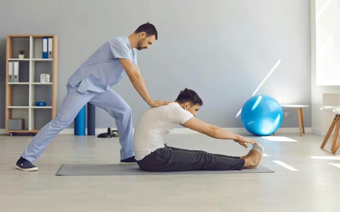
When it comes to making sure that lower back pain can be reduced and to help with keeping a healthy spine, many people often seek out physical therapy to reduce the pain. Since low back pain is costly in a clinical approach, physical therapy is cost-effective, non-invasive, and can help individuals get a kick start in their health journey. Physical therapy involves whole-body movement that emphasizes breathing coordination, reducing pain from the lower back, and helping stabilize the lumbar spine to improve physical function. (Li et al., 2023) By going through a treatment plan that incorporates physical therapy, many individuals will begin to notice their pain is improving and their quality of life is getting better. (Fischer et al., 2021) Additionally, stretching and core stability exercises can activate the deep and superficial spinal muscles by strengthening them and help stretch out sore muscles affected by low back pain to help many individuals recover. (Calatayud et al., 2019) Below are seven exercises that can help reduce back pain and, when done correctly and consecutively, can help many individuals have a healthier spine while being more mindful of their bodies.
Knee-To-Chest Exercise
This knee-to-chest exercise can help stretch the lower back muscles and can be done in the morning or evening.
Lying on your back with knees bent and feet flat for stability.
Pull one knee up with both hands and press it towards your chest.
Keep the stomach muscles tight while pressing your spine to the floor, holding for at least 30 seconds before returning to position.
Repeat with the other knee and do each stretch 2-3 times.
Lower Back Rotational Stretch (On the Floor)
This lower back rotational stretch can help stretch tight muscles in the lower back.
Laying on the mat, ensure you are on your back with knees bent and feet flat.
Make sure the shoulders are firmly on the floor, and slowly roll the knees to one side until 45 degrees.
Hold that position for 30 seconds before slowly rotating the knees back to the starting position.
Repeat on the other side and do each stretch 2-3 times.
Lower Back Flexibility Exercise
This lower back flexibility will help stretch and strengthen the lower back and core muscles.
Lay flat on the mat. For stability, make sure that the knees are bent with feet are flat on the floor.
Tighten the stomach muscles so the lower back can be pulled away from the floor.
Hold the position for 5 seconds and relax, slowly lowering to the floor.
Flatten the back as your belly button starts to go towards the floor, and hold the position for 5 seconds before relaxing.
Do five repetitions a day to slowly work up to 30 reps.
Bridge Exercise
The bridge exercise can help with core stability and help strengthen core muscles.
Laying flat on your back on the floor, with knees bent and feet flat. Make sure that your shoulders and head are relaxed.
Tighten the core and glute muscles while slowly raising from the hips to form a straight line from the knees to the shoulders.
Stay in that position for 30 seconds while taking deep breaths.
Slowly go down to the floor and relax.
Do five repetitions a day to slowly work up to 30 reps.
Cat-To-Cow Stretch
The cat-to-cow stretch helps with shoulders, upper back, and lower back muscles.
On your hands and knees, hip-width apart on the mat, be in a neutral spine position.
Slowly arch your back by pulling your belly towards the ceiling and your head down for 30 seconds.
Then, slowly let the back and belly sag towards the floor as the head rises for 30 seconds.
Return to the neutral spine position and repeat about 3-5 times twice daily.
Lower Back Rotational Stretch (Seated)
This lower back rotational stretch is seated if the floor is uncomfortable for individuals with severe back pain.
Sitting in an armless chair or stool in a seated upright position, cross one leg over the other.
Then, place the left elbow against the outside of the right knee and twist and stretch the side.
Hold the postion for 10 seconds before slowly returning to a seated position.
Repeat the stretch on the opposite side.
Do this stretch 3-5 times on each side to stretch tight back muscles about twice daily.
Shoulder Blade Squeeze
This shoulder blade squeeze helps individuals properly posture while stretching and strengthening tight upper back and shoulder muscles.
Start in a seated upright position on an armless chair or stool.
Slowly pull the shoulder blades together in the upright position and hold for 5-30 seconds.
Relax, return to the upright position, and repeat 3-5 times twice daily.
References
Bang, A. A., Bhojraj, S. Y., & Bang, A. T. (2021). Back pain and musculoskeletal pain as public health problems: Rural communities await solution. J Glob Health, 11, 01007. https://doi.org/10.7189/jogh.11.01007
Calatayud, J., Escriche-Escuder, A., Cruz-Montecinos, C., Andersen, L. L., Perez-Alenda, S., Aiguade, R., & Casana, J. (2019). Tolerability and Muscle Activity of Core Muscle Exercises in Chronic Low-back Pain. Int J Environ Res Public Health, 16(19). https://doi.org/10.3390/ijerph16193509
Fischer, S. C., Calley, D. Q., & Hollman, J. H. (2021). Effect of an Exercise Program That Includes Deadlifts on Low Back Pain. J Sport Rehabil, 30(4), 672-675. https://doi.org/10.1123/jsr.2020-0324
Li, Y., Yan, L., Hou, L., Zhang, X., Zhao, H., Yan, C., Li, X., Li, Y., Chen, X., & Ding, X. (2023). Exercise intervention for patients with chronic low back pain: a systematic review and network meta-analysis. Front Public Health, 11, 1155225. https://doi.org/10.3389/fpubh.2023.1155225
Mohd Isa, I. L., Teoh, S. L., Mohd Nor, N. H., & Mokhtar, S. A. (2022). Discogenic Low Back Pain: Anatomy, Pathophysiology and Treatments of Intervertebral Disc Degeneration. Int J Mol Sci, 24(1). https://doi.org/10.3390/ijms24010208
Smith, J. A., Stabbert, H., Bagwell, J. J., Teng, H. L., Wade, V., & Lee, S. P. (2022). Do people with low back pain walk differently? A systematic review and meta-analysis. J Sport Health Sci, 11(4), 450-465. https://doi.org/10.1016/j.jshs.2022.02.001
Individuals with hip bursitis often experience discomfort during physical activity, walking, and pain when lying on the affected side. What treatment options are available to control and manage the condition?
Hip Bursitis
Hip bursitis, also known as trochanteric bursitis, is a common condition that causes pain and discomfort in the hip and upper thigh along the outside of the hip joint. It occurs when one of the hip’s bursae, or fluid-filled sacs cushion joints, becomes inflamed. Treatment for hip bursitis is to control the inflammation caused by this condition.
Causes
Hip bursitis can be caused by injury or overuse of the hip, such as repetitive activities, twisting, or rapid joint movement. It can also be caused by a direct blow or fall to the side of the hip.
Symptoms
Pain from hip bursitis can be sharp at first and may feel dull and achy later.
It may be worse when standing up after sitting, moving, or using the hip.
Individuals may also notice pain when lying on the affected side or sitting for a long time.
Rest
This means a period of not participating in physical, exercise, and sports activities that aggravate symptoms. Any activity that causes hip pain should be avoided as this only contributes to inflammation of the bursa. (American Academy of Orthopaedic Surgeons, 2022) Modifying how particular activities are performed can help alleviate pressure on the inflamed bursa. Working with a physical therapist can also be recommended. They are experts in movement and alignment, and if certain muscles are overused compared to others, this can lead to unhealthy movement patterns, causing bursa irritation.
Anti-Inflammatory Medications
Nonsteroidal anti-inflammatory drugs, such as Motrin, Aleve, Naprosyn, etc., will help control inflammation (American Academy of Orthopaedic Surgeons, 2022). Anti-inflammatory medications can be extremely effective but should be taken cautiously. The instructions on the label need to be followed unless directed otherwise by a healthcare provider. Be aware of side effects and inform the healthcare provider if side effects present.
Cold Therapy
Applying ice to the hip area often helps alleviate the symptoms (National Library of Medicine, 2022). Ice can control inflammation by decreasing blood circulation to the area, especially after physical activity and exercise.
Aspiration
A needle is placed into the bursa to drain the fluid for those with a significant amount of fluid collected within the bursa. (National Library of Medicine, 2022) This is rarely needed in cases of hip bursitis, but when it is done, it can be combined with a cortisone injection.
Cortisone Injections
A cortisone injection may also be given into the bursa to alleviate pain. (American Academy of Orthopaedic Surgeons, 2022) The cortisone injection is helpful because it can be a diagnostic and therapeutic tool. In cases where hip bursitis may be one of several diagnoses being considered, cortisone can be given to see if it helps alleviate symptoms. Cortisone is a powerful anti-inflammatory medication that can be administered directly to the problem area. These injections are well-tolerated, but there can be possible side effects. Once the initial symptoms are under control, physical therapy strengthening and stretching exercises may be recommended.
Stretching
Most find relief by stretching the muscles and tendons over the outside of the hip, specifically the iliotibial band. The goal is for a better-conditioned muscle and tendon to glide more easily and not cause inflammation. Proper stretching techniques and posture are important in re-injury prevention.
Physical Therapy
Working with a physical therapist is an effective adjunct treatment for bursitis (American Academy of Orthopaedic Surgeons, 2022). Physical therapists correct muscle imbalances through stretching and exercise and improve alignment to prevent bursa irritation from reoccurring.
Surgery
Most patients get better with conservative treatment within about six weeks. Surgical treatment for hip bursitis is rarely needed (UCSF Health, 2024). Those who do not rest from their activities until the inflammation subsides often have a return of bursitis symptoms, and those who return too aggressively to activities and do not gradually build up also find that their symptoms return. In cases where surgery is needed, the healthcare provider may recommend an arthroscopic bursectomy. (American Academy of Orthopaedic Surgeons, 2022) The surgery is an outpatient minimally invasive procedure in which the bursa is removed through a small incision. After a short healing period, the individual can return to normal activity. Crutches may be used for a few days. Common complications are anesthetic-related complications and infection.
Injury Medical Chiropractic and Functional Medicine Clinic
As with any treatment program, always talk with your healthcare provider before initiating specific treatments. Fortunately, treatment of hip bursitis is generally accomplished with conservative therapies. Efforts to limit pressure directly on the bursa, alleviate inflammation, and restore normal movement to the hip joint will typically resolve symptoms. At Injury Medical Chiropractic and Functional Medicine Clinic, we focus on what works for you to relieve pain and restore function. Regarding musculoskeletal pain, specialists like chiropractors, acupuncturists, and massage therapists can help mitigate the pain through spinal adjustments that help the body realign itself. They can also work with other associated medical professionals to integrate into a treatment plan to improve the body’s flexibility and mobility, resolve musculoskeletal issues, and prevent future pain symptoms from reoccurring.
The Chiropractic Approach for Pain Relief
References
American Academy of Orthopaedic Surgeons. (2022). Hip bursitis. https://orthoinfo.aaos.org/en/diseases–conditions/hip-bursitis
National Library of Medicine. (2022). Bursitis: Learn More – How can bursitis be treated? InformedHealth.org [Internet]. Cologne, Germany: Institute for Quality and Efficiency in Health Care (IQWiG). https://www.ncbi.nlm.nih.gov/books/NBK525763/
Can individuals reduce stress affecting their daily routine through treatments to restore their gut health?
Introduction
Everybody in the entire world has dealt with stress at some point in their lives and has experienced general aches and pains in their bodies. This is due to the hormone cortisol, which helps regulate the entire body’s response. Stress has two forms, acute and chronic, and depending on the scenario a person is under, itcorrelates with stress and can even impact the major body systems. When dealing with acute stress, the muscles start to tense up at the stressor and go into a fight-or-flight mode until the stressor is gone and the individual can begin to relax. However, if the individual is dealing with chronic stress, it can cause numerous overlapping issues that can lead to digestive issues affecting the gut system. When the gut system is acting up from the cortisol hormone in its chronic form, many people will begin to notice small changes that are affecting their health and well-being. Today’s article examines why gut issues induce stress, how individuals can reduce stress naturally, and how people can restore their gut health. We talk with certified associated medical providers who provide our patients’ information to assess gut issues associated with stress that is affecting their health and wellness. We also inform patients while asking their associated medical provider intricate questions to formulate customized treatment plans to reduce the effects of stress correlating to gut health. Dr. Alex Jimenez, D.C., includes this information as an academic service. Disclaimer.
Why Gut Issues Induced Oxidative Stress?
How often does your body tense up and slowly relax after a stressful situation? Do you experience general aches or pains in your muscles and joints? Or do you feel tenderness and discomfort in your gut? When people are experiencing chronic oxidative stress in their bodies, they sometimes don’t realize that their gut also plays a part. This is because the gut is known as “the second brain” and helps regulate the immune response while protecting the body from unwanted pathogens. However, dealing with a large amount of stress from environmental factors can cause alterations in the immune system’s response and disrupt the delicate balance between the individual and the gut microbiota. (Sharifa et al., 2023) Since gut issues can vary from person to person, it is important to note that when environmental factors cause overlapping risk profiles, it can modify or change the gut microbiota composition of the host.
The gut system, which harbors a large number of beneficial microbial cells, plays a crucial role in the human body. These cells help stimulate the immune system’s maturity and play a role in cognitive performance and stress tolerance. (Marttinen et al., 2020) However, when a person has a poor diet, not physically active, or leads a stressful lifestyle due to their jobs, these environmental factors can cause the development of gut issues over time and contribute to the increase of oxidative stress in the body. (Vasquez et al., 2019) Additionally, many people dealing with chronic stress will often notice their skin feeling a bit warmer, they feel bloated, they will often experience fatigue throughout the day, and experience weight changes. So when the gut system is experiencing high levels of stress from environmental factors, the intestinal epithelial permeability will begin to activate the T-cells that will destroy the immunosuppressive cytokines which leads to systemic inflammation to the body. (Ni et al., 2022) However, there are numerous ways to reduce stress and to help restore gut health naturally.
Fighting Inflammation Naturally- Video
How To Reduce Stress Naturally
When it comes to reducing stress naturally, the first step is to recognize the stressors. Many individuals can start off small by making small changes to their health and wellness. Incorporating meditation, journalling, chiropractic care, acupuncture, massage therapy, and exercise can help relax the mind, reduce stress from the body, and alleviate musculoskeletal issues that are correlated with the gut and stress. These treatments can help reduce the tension from the muscles being affected by stress, while stretching and relaxing them. Additionally, these therapies are non-surgical, customized to the patient, and help realign the spine to promote healing. By recognizing the stressor, many individuals can make the small changes they need to reduce their stress and prevent them from returning.
How To Restore Gut Health
Restoring gut health is crucial to prevent inflammatory issues from reoccurring in the body. The gut microbiota, which contains trillions of bacteria aiding in digestion and nutrient transport, needs to be replenished with good bacteria and reduced bad bacteria to dampen the inflammatory effects caused by stress. One effective way to restore gut health in the body is by incorporating probiotic-rich foods into your diet. These include yogurt, kefir, sauerkraut, and kimchi. These dietary modifications can influence the gut bacteria and restore the gut structure. (Madison & Kiecolt-Glaser, 2019)
At the same time, probiotics can help stabilize stress cortisol levels and boost the good bacterial microbiota to improve body function by lowering the inflammatory cytokines. (Madabushi et al., 2023) This, in turn, helps restore the gut flora to the gut system. At the same time, when individuals incorporating a healthy diet filled with anti-inflammatory foods and pro/prebiotics can help not only reduce oxidative stress correlated with gut issues but also implement management techniques for stress and exercises that can help improve a person’s quality of life, reduce inflammation from the gut and body, and help enhance the immune response. (Jawhara, 2024) When individuals start to think about their health and well-being, making small changes can lead to positive results and help make sure that no gut issues associated with stress can reoccur. Figuring out what stressors are impacting the gut and making these changes can help prevent them from returning in the future and help that individual live a healthier lifestyle.
Madabushi, J. S., Khurana, P., Gupta, N., & Gupta, M. (2023). Gut Biome and Mental Health: Do Probiotics Work? Cureus, 15(6), e40293. https://doi.org/10.7759/cureus.40293
Madison, A., & Kiecolt-Glaser, J. K. (2019). Stress, depression, diet, and the gut microbiota: human-bacteria interactions at the core of psychoneuroimmunology and nutrition. Curr Opin Behav Sci, 28, 105-110. https://doi.org/10.1016/j.cobeha.2019.01.011
Marttinen, M., Ala-Jaakkola, R., Laitila, A., & Lehtinen, M. J. (2020). Gut Microbiota, Probiotics and Physical Performance in Athletes and Physically Active Individuals. Nutrients, 12(10). https://doi.org/10.3390/nu12102936
Ni, Q., Zhang, P., Li, Q., & Han, Z. (2022). Oxidative Stress and Gut Microbiome in Inflammatory Skin Diseases. Front Cell Dev Biol, 10, 849985. https://doi.org/10.3389/fcell.2022.849985
Sharifa, M., Ghosh, T., Daher, O. A., Bhusal, P., Alaameri, Y. A., Naz, J., Ekhator, C., Bellegarde, S. B., Bisharat, P., Vaghani, V., & Hussain, A. (2023). Unraveling the Gut-Brain Axis in Multiple Sclerosis: Exploring Dysbiosis, Oxidative Stress, and Therapeutic Insights. Cureus, 15(10), e47058. https://doi.org/10.7759/cureus.47058
Vasquez, E. C., Pereira, T. M. C., Campos-Toimil, M., Baldo, M. P., & Peotta, V. A. (2019). Gut Microbiota, Diet, and Chronic Diseases: The Role Played by Oxidative Stress. Oxid Med Cell Longev, 2019, 7092032. https://doi.org/10.1155/2019/7092032
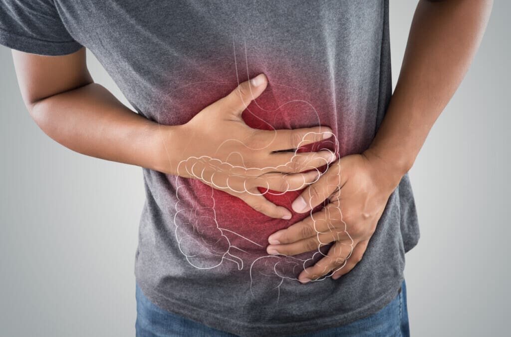
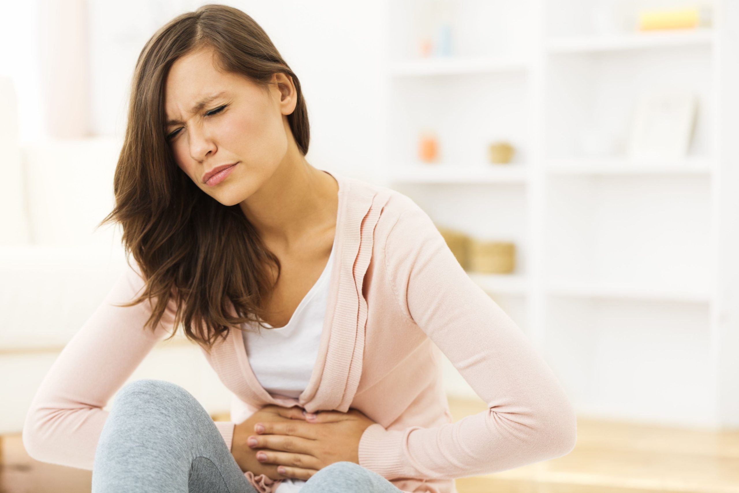
Can individuals dealing with back pain find treatment to reduce gut pain associated with SIBO to improve body health?
Introduction
Many individuals have noticed that when it comes to improving their health and well-being, many will incorporate small changes into their daily routines. From exercising for at least 30 minutes to incorporating healthy nutritional foods into their diet, many people don’t realize that the best way to have a healthy style starts with the gut. The gut system helps the body digest food and nutrients to be transported to the body, helps regulate growth and metabolism, and provides immune support to all the organ systems. However, when harmful pathogens and environmental factors start to impact the gut, it can cause the development of harmful pathogens inside the gut system and, over time, cause overlapping risk profiles in the body. This leads to gut dysfunction and musculoskeletal issues in the individual and can cause pain and discomfort if not treated right away. Luckily, numerous ways exist to improve gut health and reduce musculoskeletal issues. Today’s article focuses on a gut issue known as SIBO, how SIBO is correlated with back pain, and what treatments can help reduce SIBO. We talk with certified associated medical providers who provide our patients’ information to assess and identify how SIBO is correlated with back pain. We also inform patients while asking their associated medical provider intricate questions to formulate customized treatment plans to reduce the effects of SIBO and help restore gut health. Dr. Alex Jimenez, D.C., includes this information as an academic service. Disclaimer.
What is SIBO?
How often do you feel general aches or pain in your gut or around your lower back? Do you constantly feel tired throughout the day, even after a full night’s rest? Or have you been constantly feeling constipated or bloated after eating a meal? Many of these scenarios are associated with a gut issue known as SIBO or small intestinal bacterial overgrowth. Before diving into what SIBO is, it is important to see the gut’s main function to the body. Known as the second brain of the body, the gut system is home to trillions of good bacteria that help with food digestion and protect the body from bad bacteria. When environmental factors like poor dieting, physical inactivity, or inflammatory effects affect the body, the gut’s delicate ecosystem is also affected. This can cause gut dysfunction to the body and, over time, when it is not being treated, lead to SIBO.
SIBO is the presence of excess bad bacteria in the small intestines, which causes protective barriers that help the small intestines weaken. (Sorathia et al., 2024) Additionally, SIBO can correlate with conditions as it can accompany other gut issues by stimulating the immune system. (Banaszak et al., 2023) When the immune system becomes hyperactively stimulated by SIBO, it can cause the inflammatory cytokines to mass produce and cause a ripple effect on the entire body. Since inflammation is the body’s natural response to remove harmful pathogens that cause issues, mass production of inflammatory cytokines in the gut can cause toxins and bad bacteria to enter the bloodstream and travel to different body areas to cause pain. At the same time, SIBO can disrupt the gut-brain axis, which leads to intestinal motility changes and secretion, thus causing overlapping risk profiles like back pain to affect the body. (Carter et al., 2023)
Eating Right to Feel Better- Video
How Does Back Pain Correlate With SIBO?
Now, many people are wondering how back pain is correlated with SIBO. Since SIBO causes the immune system to be hyperactive and mass-produce inflammatory cytokines to reduce the integrity and function of the gastrointestinal barrier, it can cause chronic inflammation and induce pain, which includes musculoskeletal conditions like back pain. (Hui et al., 2023) Additionally, the gut-brain axis being over-runed by SIBO and chronic inflammation being an overlapping risk factor can cause negative influences on the gut microbiome composition, and how the individual reacts to the changes can lead to abnormal bone growth and reabsorption due to the excess bacteria. (Geng et al., 2023) The excess bacteria produced by SIBO can affect intervertebral disc homeostasis and, when combined with environmental factors, can further enhance the inflammatory damage to the back muscles. (Yao et al., 2023) However, there are ways to not only reduce the back pain but also treat SIBO from causing more issues in the gut.
Treatments To Reduce SIBO
When it comes to treating SIBO, it depends on what treatment a person will be combined. The main goals for creating a treatment plan for SIBO are:
Reducing the bad bacteria
Bio-transform the gut
Preventing a relapse
Additionally, many individuals can make small changes in their routine by making dietary changes that can modify the intestinal microbiota. (Souza et al., 2022) This, in turn, helps promote gut health and replenish the nutrients while restoring the good bacteria to the gut. Regarding back pain associated with SIBO, chiropractic care can help individuals decrease or alleviate musculoskeletal symptoms by realigning the spine while massaging the muscles. This can help increase circulation while soothing the inflammatory effects caused by SIBO. Chiropractic care can be implemented as part of a person’s customizable treatment plan through a whole body approach by incorporating lifestyle changes and restoring gut health. By making these small changes with the right treatments to manage the overlapping symptoms caused by SIBO, many individuals can make these small changes to their routine to prevent SIBO from reappearing and causing issues in the body.
References
Banaszak, M., Gorna, I., Wozniak, D., Przyslawski, J., & Drzymala-Czyz, S. (2023). Association between Gut Dysbiosis and the Occurrence of SIBO, LIBO, SIFO and IMO. Microorganisms, 11(3). https://doi.org/10.3390/microorganisms11030573
Carter, J., Bettag, J., Morfin, S., Manithody, C., Nagarapu, A., Jain, A., Nazzal, H., Prem, S., Unes, M., McHale, M., Lin, C. J., Hutchinson, C., Trello, G., Jain, A., Portz, E., Verma, A., Swiderska-Syn, M., Goldenberg, D., & Kurashima, K. (2023). Gut Microbiota Modulation of Short Bowel Syndrome and the Gut-Brain Axis. Nutrients, 15(11). https://doi.org/10.3390/nu15112581
Geng, Z., Wang, J., Chen, G., Liu, J., Lan, J., Zhang, Z., & Miao, J. (2023). Gut microbiota and intervertebral disc degeneration: a bidirectional two-sample Mendelian randomization study. J Orthop Surg Res, 18(1), 601. https://doi.org/10.1186/s13018-023-04081-0
Hui, J., Chen, Y., Li, C., Gou, Y., Liu, Y., Zhou, R., Kang, M., Liu, C., Wang, B., Shi, P., Cheng, S., Yang, X., Pan, C., Jia, Y., Cheng, B., Liu, H., Wen, Y., & Zhang, F. (2023). Insight into the Causal Relationship between Gut Microbiota and Back Pain: A Two Sample Bidirectional Mendelian Randomization Study. Adv Genet (Hoboken), 4(4), 2300192. https://doi.org/10.1002/ggn2.202300192
Souza, C., Rocha, R., & Cotrim, H. P. (2022). Diet and intestinal bacterial overgrowth: Is there evidence? World J Clin Cases, 10(15), 4713-4716. https://doi.org/10.12998/wjcc.v10.i15.4713
Yao, B., Cai, Y., Wang, W., Deng, J., Zhao, L., Han, Z., & Wan, L. (2023). The Effect of Gut Microbiota on the Progression of Intervertebral Disc Degeneration. Orthopaedic Surgery, 15(3), 858-867. https://doi.org/10.1111/os.13626
IFM's Find A Practitioner tool is the largest referral network in Functional Medicine, created to help patients locate Functional Medicine practitioners anywhere in the world. IFM Certified Practitioners are listed first in the search results, given their extensive education in Functional Medicine