Chiropractic Care for Motor Vehicle Accident Recovery: A Comprehensive Guide to Healing
Motor vehicle accidents (MVAs) can be life-changing events, often leaving individuals with injuries that range from mild discomfort to severe, debilitating conditions. Among the most common injuries sustained in MVAs are musculoskeletal injuries, which affect muscles, bones, ligaments, tendons, and nerves. These injuries can significantly impact daily life, work, and recreational activities, raising critical questions about recovery: Will I fully recover, and how long will it take? Chiropractic care offers a powerful, non-invasive solution for addressing these injuries, relieving pain, and restoring mobility. By focusing on spinal alignment, reducing inflammation, and improving nerve function, chiropractors help prevent long-term complications and expedite healing. In El Paso, Texas, Dr. Alexander Jimenez, DC, APRN, FNP-BC, stands out as a leader in this field, using a dual-scope approach that combines chiropractic expertise with nurse practitioner skills to deliver comprehensive care. This blog post explores the nature of musculoskeletal injuries from MVAs, the benefits of chiropractic care, and Dr. Jimenez’s unique approach to treatment and legal documentation, all while providing actionable insights for recovery.
Understanding Musculoskeletal Injuries from Motor Vehicle Accidents
Musculoskeletal injuries are among the most common outcomes of MVAs, even in low-speed collisions. The sudden force of a crash can cause significant trauma to the body’s musculoskeletal system, which includes muscles, bones, ligaments, tendons, and nerves. These injuries vary widely in severity, from minor sprains to severe fractures or nerve damage, and their effects can disrupt daily life.
Types and Severity of Musculoskeletal Injuries
Whiplash: One of the most frequent MVA injuries, whiplash occurs when the head and neck are abruptly jerked, often in rear-end collisions. It can damage cervical muscles, ligaments, discs, and nerves, leading to neck pain, stiffness, and headaches. If untreated, whiplash can cause long-term issues like chronic pain or spinal degeneration (Torts Law, 2023).
Soft Tissue Injuries: These involve damage to muscles, ligaments, or tendons, such as sprains or strains. Soft tissue injuries may not be immediately visible but can cause significant pain and mobility issues if ignored, potentially leading to chronic disability (Aguiar Injury Lawyers, 2023).
Fractures: Broken bones, such as those in the arms, legs, or spine, are common in high-impact crashes. Fractures require immediate medical attention and can lead to long-term complications like reduced mobility or arthritis (Ortho Sport & Spine, 2023).
Herniated Discs: The force of an MVA can compress or displace spinal discs, causing them to bulge or rupture. This can pinch nerves, leading to pain, numbness, or weakness in the arms or legs (Florida Physical Medicine, 2023).
Nerve Damage: Trauma from an MVA can compress or irritate nerves, causing symptoms like tingling, numbness, or muscle weakness. Nerve injuries can significantly affect daily activities and may require specialized care (Lonseth Pain, 2023).
Impact on Daily Life
Musculoskeletal injuries can disrupt every aspect of life. For example, whiplash may make it difficult to turn your head while driving or working at a desk. Soft tissue injuries can limit your ability to lift objects or participate in sports. Fractures or herniated discs may require time off work, affecting income and mental well-being. Chronic pain from untreated injuries can also lead to emotional challenges, such as anxiety or depression, further complicating recovery (JS Berry Law, 2023). The severity of these injuries often depends on factors like the speed of the collision, seatbelt use, airbag deployment, and the occupant’s position in the vehicle (Advantage Healthcare Systems, 2023).
The Recovery Question
A common concern after an MVA is whether full recovery is possible and how long it will take. Recovery depends on the injury’s severity, the individual’s overall health, and the timeliness of treatment. Minor injuries like sprains may heal in weeks with proper care, while severe fractures or nerve damage could take months or require ongoing management. Early intervention is critical to prevent long-term complications, such as chronic pain or reduced mobility (Wirth Chiropractic, 2023).
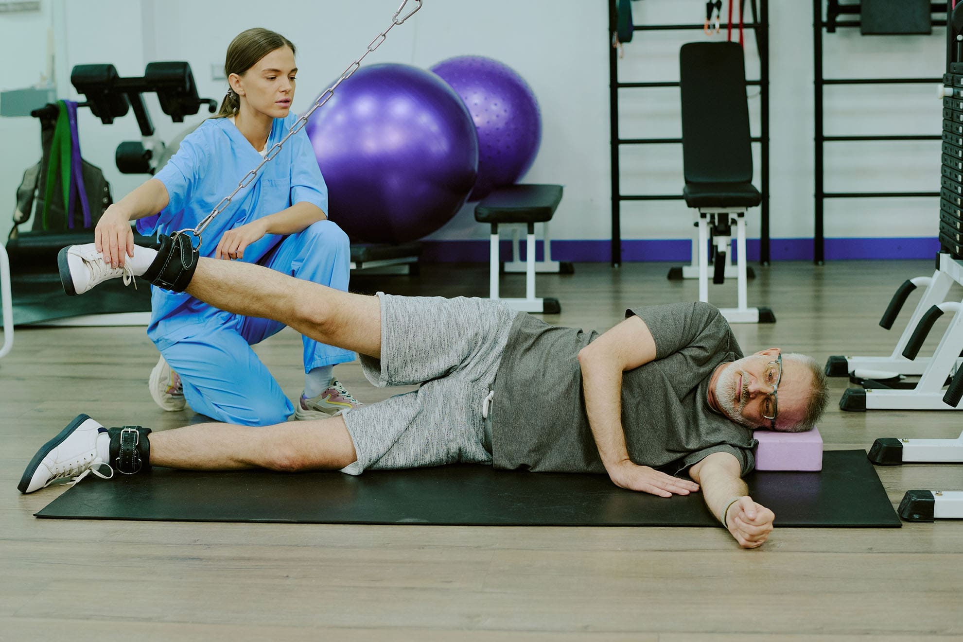
Chiropractic care is a non-invasive, drug-free approach that focuses on diagnosing and treating musculoskeletal disorders, particularly those affecting the spine and nervous system. After an MVA, chiropractors play a vital role in addressing injuries, relieving pain, and restoring function, helping patients return to their normal lives.
How Chiropractic Care Works
Chiropractors use hands-on techniques, such as spinal adjustments, to realign the spine and joints, reduce inflammation, and restore nerve function. These adjustments involve precise, controlled movements to correct misalignments (subluxations) caused by the force of an MVA. By restoring proper alignment, chiropractic care alleviates pressure on nerves, improves blood flow, and promotes the body’s natural healing processes (Hogan Chiropractic, 2024).
Benefits of Chiropractic Care
Pain Relief: Spinal adjustments release anti-inflammatory hormones and reduce nerve pressure, providing relief from neck, back, and joint pain without relying on medications, which can have side effects or risks of dependency (Brookdale Health, 2023).
Improved Mobility: MVAs often cause stiffness and reduced range of motion. Chiropractic techniques, including soft tissue therapy and corrective exercises, restore flexibility and allow patients to perform daily tasks with ease (Recovery Chiromed, 2023).
Reduced Inflammation: Chiropractic care improves circulation and reduces swelling in injured tissues, speeding up recovery and preventing scar tissue buildup (Uptown Denver Chiropractor, 2023).
Prevention of Long-Term Complications: Early chiropractic intervention can prevent chronic pain, spinal degeneration, or mobility issues by addressing injuries at their source (The Neck and Back Clinics, 2024).
Holistic Healing: Chiropractors often incorporate nutrition counseling, stress management, and lifestyle advice to support overall health, addressing both physical and emotional aspects of recovery (Artisan Chiropractic Clinic, 2023).
Recovery Timeline with Chiropractic Care
The recovery timeline varies based on the injury’s severity and the patient’s commitment to treatment. Minor injuries like whiplash may improve within 4 to 12 weeks of regular chiropractic sessions, while more severe injuries, such as herniated discs, may require several months of care. Most patients notice significant pain relief and improved mobility within a few sessions, but consistent treatment is essential for long-term healing (Tradition Health, 2024). Chiropractors create personalized treatment plans, adjusting techniques as the patient progresses to ensure optimal outcomes.
Dr. Alexander Jimenez, DC, APRN, FNP-BC, is a board-certified chiropractor and family nurse practitioner based in El Paso, Texas, with over 25 years of experience treating MVA injuries. His unique dual licensure allows him to combine chiropractic expertise with medical diagnostics, offering a comprehensive approach to personal injury cases. Dr. Jimenez’s practice, Injury Medical & Chiropractic Clinic, is renowned for its integrative treatment plans that address both the biomechanical and systemic effects of MVAs.
Dual-Scope Diagnosis and Treatment
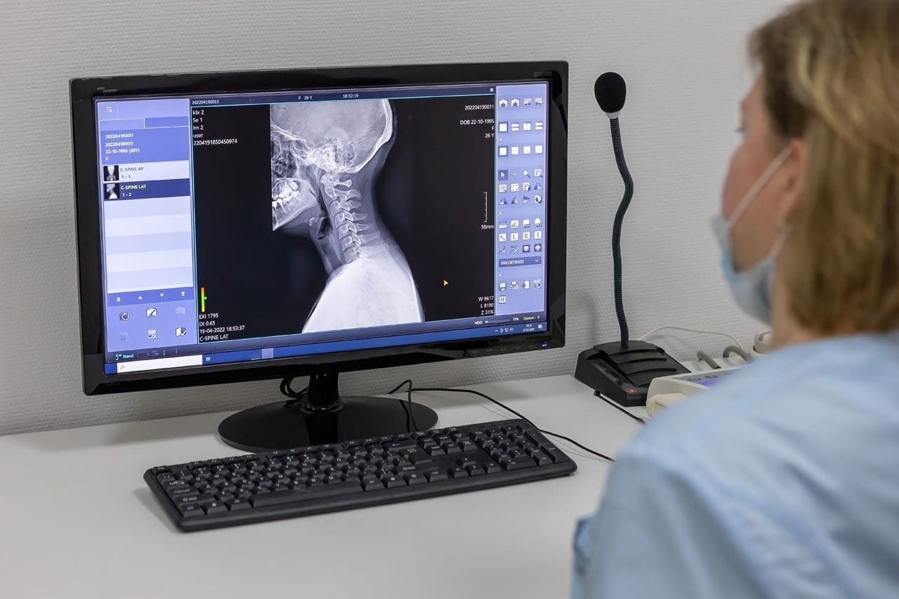
Dr. Jimenez employs a dual-scope approach, integrating chiropractic assessments (e.g., spinal alignment checks) with medical evaluations (e.g., metabolic and hormonal tests). This method ensures that both physical and physiological issues are addressed, providing a more accurate diagnosis and tailored treatment plan. For example, he uses advanced imaging techniques, such as X-rays, MRIs, and digital motion X-rays, to detect subtle injuries like ligament tears, disc herniations, or nerve compression that may not be apparent in initial exams (Jimenez, 2023a). His clinical observations emphasize the correlation between patient injuries and objective diagnostic findings, ensuring precise interventions.
Integrative Medicine Approach
Dr. Jimenez incorporates integrative medicine principles, combining chiropractic adjustments with functional medicine, nutrition counseling, and rehabilitation exercises. This holistic approach addresses the root causes of injuries rather than just masking symptoms. For instance:
Nutritional Counseling: He recommends anti-inflammatory diets rich in omega-3s and antioxidants to reduce inflammation and support tissue repair (El Paso Back Clinic, 2025).
Functional Medicine: He monitors systemic issues like inflammation or stress responses that can hinder recovery, ensuring comprehensive healing (Jimenez, 2023b).
Rehabilitation Exercises: Targeted exercises restore strength and flexibility, preventing re-injury and promoting long-term mobility (Chiropractic Scientist, 2025).
Handling Legal Paperwork in Personal Injury Cases
Dr. Jimenez’s dual licensure equips him to bridge medical treatment with legal documentation, a critical aspect of personal injury cases. He provides detailed injury reports, treatment plans, and progress updates supported by imaging results, ensuring patients receive appropriate compensation. His ability to document injuries accurately strengthens legal claims while prioritizing clinical care (Jimenez, 2024). This dual role sets him apart, as he can navigate both the healthcare and legal systems effectively, reducing stress for patients.
Clinical Observations and Expertise
Dr. Jimenez’s work, as seen on dralexjimenez.com, elpasobackclinic.com, and his LinkedIn profile, highlights his commitment to evidence-based, patient-centered care. He emphasizes early intervention to prevent chronic issues, noting that “undetected microtraumas from even minor accidents can compound over time, affecting spinal alignment and nervous system function” (Jimenez, 2025). His integrative approach has helped countless patients regain their quality of life, as evidenced by testimonials praising his ability to restore mobility and reduce pain (Chiropractic Scientist, 2025).
Seeking chiropractic care soon after an MVA is crucial for optimal recovery. The body’s stress response can mask symptoms, delaying the recognition of injuries like whiplash or soft tissue damage. These “hidden” injuries can worsen without treatment, leading to chronic pain or long-term complications.
The Critical 72-Hour Window
Clinical evidence suggests that symptoms of MVA injuries often appear within 24 to 72 hours post-accident. During this period, the body may still be in shock, and adrenaline can suppress pain signals. Chiropractic evaluation within this window allows for early detection and intervention, preventing conditions from becoming chronic (The Neck and Back Clinics, 2024). For example, early spinal adjustments can correct misalignments before they cause nerve compression or inflammation.
Preventing Chronic Pain
Untreated musculoskeletal injuries can lead to persistent pain, reduced mobility, and even psychological effects like depression. Chiropractic care addresses these issues proactively by restoring alignment, reducing inflammation, and promoting tissue healing. Studies show that patients who seek chiropractic care soon after an MVA experience greater improvements compared to those who delay treatment (Clearway Pain, 2023).
Supporting Legal Claims
Early chiropractic care also strengthens personal injury claims by providing documented evidence of injuries and treatment. Chiropractors like Dr. Jimenez ensure that medical records, imaging results, and progress reports are thorough and legally sound, helping patients secure fair compensation (Jimenez, 2024).
Chiropractic care is often most effective when combined with other therapies, creating a holistic approach to MVA recovery. Dr. Jimenez and other chiropractors integrate complementary treatments to address both the physical and systemic effects of injuries.
Complementary Therapies
Acupuncture: Thin needles stimulate acupoints to release endorphins, improve blood flow, and calm the nervous system, reducing pain and inflammation (Jimenez, 2023a).
Massage Therapy: This relaxes tense muscles, breaks down scar tissue, and improves circulation, enhancing the effects of chiropractic adjustments (Uptown Denver Chiropractor, 2023).
Physical Therapy: Targeted exercises strengthen muscles, improve flexibility, and support spinal alignment, preventing re-injury (Tradition Health, 2024).
Nutrition Counseling: Anti-inflammatory diets and supplements support tissue repair and reduce systemic inflammation, promoting faster recovery (El Paso Back Clinic, 2025).
Benefits of an Integrative Approach
Integrative care treats the whole person, not just isolated symptoms. By addressing biomechanical, physiological, and emotional factors, this approach ensures faster, more complete healing. For example, combining chiropractic adjustments with acupuncture can alleviate pain while improving mental clarity, helping patients cope with the stress of recovery (Artisan Chiropractic Clinic, 2023). Dr. Jimenez’s clinic emphasizes “therapeutic layering,” using these therapies in structured phases to avoid setbacks and empower patients with long-term self-care strategies (Jimenez, 2025).
Recovering from an MVA requires a proactive approach, combining professional care with self-management. Here are practical steps to support healing and prevent complications.
Seek Immediate Care
Visit a chiropractor or medical professional within 72 hours of the accident, even if you feel fine. Early evaluation can detect hidden injuries and establish a baseline for treatment and legal documentation (Clearway Pain, 2023).
Follow Your Treatment Plan
Adhere to your chiropractor’s recommendations, attending all scheduled sessions and performing prescribed exercises. Consistency is key to achieving full recovery and preventing setbacks (Tradition Health, 2024).
Adopt Healthy Habits
Nutrition: Eat anti-inflammatory foods like fish, leafy greens, and berries to support tissue repair (El Paso Back Clinic, 2025).
Rest: Prioritize sleep to allow your body to heal and reduce stress (Jimenez, 2023b).
Posture: Maintain proper posture during daily activities to avoid straining injured areas (Recovery Chiromed, 2023).
Communicate with Your Chiropractor
Report any changes in symptoms or challenges with your treatment plan. Your chiropractor can adjust therapies to ensure progress (Hogan Chiropractic, 2024).
Manage Legal Aspects
Work with your chiropractor to document injuries and treatments for insurance or legal claims. Accurate records can streamline the process and reduce stress (Jimenez, 2024).
Motor vehicle accidents can cause a wide range of musculoskeletal injuries, from whiplash and soft tissue damage to fractures and nerve injuries, significantly impacting daily life. Chiropractic care offers a non-invasive, holistic solution, relieving pain, restoring mobility, and preventing long-term complications. By addressing the root causes of injuries through spinal adjustments, integrative therapies, and personalized treatment plans, chiropractors help patients recover faster and more completely. Dr. Alexander Jimenez exemplifies this approach, using his dual licensure to provide comprehensive care and legal support in El Paso. Early intervention, consistent treatment, and healthy lifestyle choices are key to achieving full recovery. If you’ve been in an MVA, don’t wait—seek chiropractic care to start your healing journey today.
Understand the implications of an auto accident and how to navigate the aftermath effectively for recovering from WAD.
Understanding Whiplash-Associated Disorders from Motor Vehicle Accidents: Causes, Treatments, and Recovery
Whiplash-associated disorders (WAD) sound like something you’d get from a wild roller coaster ride, but unfortunately, they’re a lot less fun. These injuries often occur after a motor vehicle accident (MVA), leaving victims with neck pain, stiffness, and sometimes a whole laundry list of other symptoms. If you’ve ever been rear-ended at a stoplight and felt your head snap back like a bobblehead, you’ve probably experienced the forces that can lead to WAD. In this blog post, we’ll dive deep into why WAD and the cervical spine are so closely tied to MVAs, how these injuries develop, and what you can do to recover without feeling like you’re stuck in a neck brace forever. We’ll also spotlight Dr. Alexander Jimenez, a rockstar chiropractor in El Paso, Texas, who’s helping accident victims navigate the road to recovery with advanced diagnostics and a knack for bridging the medical and legal worlds. Buckle up (safely, of course) for a comprehensive, SEO-optimized guide that’s over 5,000 words, written for a high school reading level, with a sprinkle of humor to keep things light—because nobody likes reading about neck pain without a chuckle or two.
What Are Whiplash-Associated Disorders (WAD)?
Let’s start with the basics. Whiplash-associated disorders are a collection of symptoms that pop up after your neck goes through a rapid back-and-forth motion, like a whip cracking (hence the name). This usually happens in MVAs, especially rear-end collisions, where your car gets hit from behind, and your head decides to do its best impression of a ping-pong ball. The Quebec Task Force, a group of experts who’ve studied this stuff, classifies WAD into five grades, from “no pain at all” (Grade 0) to “ouch, there’s a fracture or dislocation” (Grade 4). Most people fall into Grades 1 or 2, which involve neck pain, stiffness, or tenderness, sometimes with reduced range of motion or point tenderness (Spitzer et al., 1995).
The cervical spine—those seven vertebrae in your neck (labeled C1 to C7)—is the star of this unfortunate show. It’s a flexible, hardworking structure that supports your head, protects your spinal cord, and lets you turn your head to check your blind spots (or avoid awkward eye contact). But when an MVA sends your neck into overdrive, the cervical spine can take a beating, leading to WAD symptoms like:
Neck pain and stiffness
Headaches, often starting at the base of the skull
Shoulder or upper back pain
Dizziness or vertigo
Tingling or numbness in the arms
Fatigue, irritability, or even trouble concentrating (because apparently, whiplash doesn’t think neck pain is enough)
These symptoms can show up right after the accident or sneak in days later, like an uninvited guest who overstays their welcome. So, why does the cervical spine get so cranky after an MVA? Let’s break it down.
References
Spitzer, W. O., Skovron, M. L., Salmi, L. R., Cassidy, J. D., Duranceau, J., Suissa, S., & Zeiss, E. (1995). Scientific monograph of the Quebec Task Force on Whiplash-Associated Disorders: Redefining “whiplash” and its management. Spine, 20(8S), 1S–73S.
The Clinical Rationale: Why MVAs Cause WAD and Cervical Spine Injuries
Picture this: You’re stopped at a red light, singing along to your favorite tune, when BAM! A distracted driver rear-ends you. Your car lurches forward, but your seatbelt keeps your body in place—except for your head, which snaps backward and then forward faster than you can say “whiplash.” This is what experts call a cervical acceleration-deceleration (CAD) injury, and it’s the key to understanding why MVAs and WAD are such close cousins.
The Biomechanics of Whiplash
During a rear-end collision, your cervical spine goes through a wild ride. High-speed cameras and crash test dummies (who have a worse day than you) show that the lower cervical vertebrae (C5 and C6) hyperextend—meaning they bend backward too far—while the upper vertebrae (C1 and C2) hyperflex, bending forward. This creates an S-shaped curve in your neck, which is not how your spine likes to hang out (Kaneoka et al., 1999). This abnormal motion stretches or tears soft tissues like:
Ligaments: The anterior longitudinal ligament (ALL) and facet joint capsules can get overstretched or sprained.
Muscles: The sternocleidomastoid and trapezius muscles might strain or develop trigger points.
Joints: The zygapophyseal (facet) joints can get compressed or irritated.
Discs: Intervertebral discs might bulge or herniate under pressure.
These injuries cause inflammation, edema (swelling), and sometimes tiny hemorrhages, which is why your neck feels like it’s auditioning for a role as a rusty hinge. In severe cases, the rapid motion can even affect nerves, leading to symptoms like arm tingling or weakness (Grade 3 WAD). And if you’re unlucky, you might end up with a fracture or dislocation (Grade 4), but that’s rare.
Why the Cervical Spine Is Vulnerable
The cervical spine is like the acrobatic gymnast of your body—flexible but prone to injury if pushed too far. Here’s why it’s so susceptible in MVAs:
Flexibility: The cervical spine’s range of motion makes it great for turning your head but terrible at handling sudden, forceful movements.
Weight of the Head: Your head weighs about 10–12 pounds (like a bowling ball). When it’s whipped around, it puts massive stress on the neck’s delicate structures.
Lack of Support: Unlike your torso, which is strapped in by a seatbelt, your head is free to flop around, amplifying the forces on the cervical spine.
S-Curve Formation: That S-shaped curve during whiplash creates high shearing forces, especially at the C5–C6 level, where injuries are most common (Bogduk & Yoganandan, 2001).
Dr. Alexander Jimenez, a chiropractor and family nurse practitioner in El Paso, explains that these biomechanical forces are why even low-speed collisions (think 10–15 mph) can cause significant damage. “The cervical spine isn’t designed for rapid, unnatural movements,” he says. “When you add the element of surprise in an MVA, the muscles don’t have time to brace, leaving ligaments and joints to bear the brunt” (Jimenez, n.d.).
The Role of MVAs in WAD Development
MVAs, especially rear-end collisions, are the poster child for WAD because they perfectly set up the CAD mechanism. Studies show that 62% of people in MVAs report neck pain, with 93% feeling it within 24 hours (Schofferman et al., 2015). But it’s not just rear-end crashes—side impacts, frontal collisions, or even amusement park rides can trigger WAD. The key is the sudden acceleration-deceleration that throws your neck out of whack.
Chronic WAD, where symptoms last more than six months, affects up to 50% of victims with Grades 1 or 2 injuries. This is partly because the initial injury can lead to:
Sensory Hypersensitivity: Your nervous system gets cranky, making you more sensitive to pain or pressure (Sterling et al., 2006).
Motor Deficits: Reduced neck mobility and muscle weakness can persist, making it hard to turn your head without wincing.
Psychological Factors: Stress, anxiety, or post-traumatic stress disorder (PTSD) from the accident can amplify pain perception (Gaab et al., 2004).
Dr. Jimenez emphasizes that untreated or poorly managed WAD can spiral into chronic pain, which is why early intervention is critical. “The sooner we address the injury, the better we can prevent long-term complications,” he notes (Jimenez, n.d.).
References
Bogduk, N., & Yoganandan, N. (2001). Biomechanical basis of whiplash injury. Journal of Whiplash & Related Disorders, 1(1), 85–97.
Gaab, J., Baumann, S., Budnoik, A., Gmünder, H., Hottinger, N., & Ehlert, U. (2004). Reduced reactivity of the hypothalamic–pituitary–adrenal axis in chronic whiplash-associated disorder. Pain, 111(3), 289–297.
Jimenez, A. (n.d.). About Dr. Alex Jimenez. El Paso Back Clinic. https://elpasobackclinic.com/
Kaneoka, K., Ono, K., Inami, S., & Hayashi, K. (1999). Motion analysis of cervical vertebrae during whiplash loading. Spine, 24(8), 763–770.
Schofferman, J., Bogduk, N., & Slosar, P. (2015). Chronic whiplash and whiplash-associated disorders: An evidence-based approach. Journal of the American Academy of Orthopaedic Surgeons, 23(10), 596–606.
Sterling, M., Jull, G., Vicenzino, B., & Kenardy, J. (2006). Physical and psychological factors maintain long-term predictive capacity post-whiplash injury. Pain, 122(1–2), 102–108.
How WAD Affects Individuals from Auto Injuries
If you’ve ever had whiplash, you know it’s not just a pain in the neck (pun intended). WAD can turn everyday tasks—like driving, working, or even sleeping—into a challenge. Let’s explore how these injuries impact your life and why they’re such a big deal in personal injury cases.
Physical Impacts
The most obvious effect of WAD is physical discomfort. Neck pain and stiffness can make it hard to turn your head, which is a problem when you’re trying to check your mirrors or dodge a rogue shopping cart in the grocery store. Headaches, often caused by irritation of the upper cervical spine or occipital nerves, can feel like a marching band in your skull. For some, symptoms like dizziness or arm tingling add to the misery, making it tough to focus or stay active.
Chronic WAD is particularly sneaky. Studies show that 20–88% of chronic WAD patients report headaches, often linked to upper cervical spine injuries (Panjabi et al., 2004). Muscle strains in the neck and upper back, like the splenius capitis or trapezius, can lead to trigger points—those tender spots that feel like someone’s poking you with a hot needle (Elliott et al., 2016). In rare cases, WAD can even cause visual problems, like blurred vision or accommodative spasm, where your eyes refuse to focus properly (Endo et al., 2006).
Psychological and Social Impacts
WAD doesn’t just mess with your body—it can mess with your mind, too. The stress of an MVA, combined with ongoing pain, can lead to anxiety, depression, or PTSD. One study found that a significant number of WAD patients develop PTSD symptoms, which can make pain feel worse and recovery harder (Gaab et al., 2004). Socially, WAD can isolate you—nobody wants to go bowling when their neck feels like it’s made of concrete. Work can also take a hit, with some patients needing time off or struggling with tasks that require neck movement.
Economic and Legal Impacts
In places like El Paso, where MVAs are all too common, WAD is a major player in personal injury cases. In the U.S., whiplash-related claims account for over 65% of bodily injury claims, costing around $8 billion annually (Freeman, 1999). These cases often involve proving that the injury was caused by the accident, which is where medical expertise and legal documentation come in. Victims may face medical bills, lost wages, and the hassle of dealing with insurance companies, all while trying to heal.
Dr. Jimenez sees this firsthand in his El Paso practice. “Many of my patients are dealing with not just physical pain but the stress of navigating insurance claims or legal battles,” he says. “That’s why comprehensive care and clear documentation are so important” (Jimenez, n.d.).
References
Elliott, J. M., Hoggarth, M. A., Sparks, C. L., & Weber, K. A. (2016). Advancements in imaging technology: Do they (or will they) equate to advancements in our knowledge of recovery in whiplash? Journal of Orthopaedic & Sports Physical Therapy, 46(10), 862–872.
Endo, K., Ichimaru, K., Komagata, M., & Yamamoto, K. (2006). Cervical vertigo and dizziness after whiplash injury. European Spine Journal, 15(6), 886–890.
Freeman, M. D. (1999). A review and methodologic critique of the literature refuting whiplash syndrome. Spine, 24(1), 86–98.
Gaab, J., Baumann, S., Budnoik, A., Gmünder, H., Hottinger, N., & Ehlert, U. (2004). Reduced reactivity of the hypothalamic–pituitary–adrenal axis in chronic whiplash-associated disorder. Pain, 111(3), 289–297.
Jimenez, A. (n.d.). About Dr. Alex Jimenez. El Paso Back Clinic. https://elpasobackclinic.com/
Panjabi, M. M., Ito, S., Pearson, A. M., & Ivancic, P. C. (2004). Injury mechanisms of the cervical intervertebral disc during simulated whiplash. Spine, 29(11), 1217–1225.
The Science of Motion- Video
Treatments for WAD and Cervical Spine Injuries
Good news: You don’t have to live with WAD forever. With the right treatments, most people recover within weeks or months, though chronic cases may need extra TLC. Let’s explore the evidence-based options, with insights from Dr. Jimenez, who’s been helping El Pasoans get back on their feet (or at least turn their heads) for years.
Conservative Treatments
Conservative treatments—those that don’t involve surgery—are the first line of defense for WAD Grades 1–3. These include:
Active Mobilization: Forget the soft collar (unless your doctor insists). Studies show that early movement, like gentle neck exercises, speeds up recovery compared to immobilization (Schnabel et al., 2004). Dr. Jimenez often prescribes range-of-motion exercises to restore flexibility without aggravating the injury.
Physical Therapy: A physical therapist can guide you through exercises to strengthen deep neck flexors (like the longus colli) and scapula stabilizers (like the rhomboids). This helps take pressure off the cervical spine and improves posture (Kay et al., 2005).
Manual Therapy: Chiropractic adjustments or mobilization can reduce pain and improve joint mobility. Dr. Jimenez uses spinal manipulation to realign the cervical spine, which can relieve pressure on irritated nerves and joints (Bryans et al., 2014).
Pain Management: Over-the-counter meds like ibuprofen or acetaminophen can help with pain and inflammation. For severe cases, doctors might prescribe muscle relaxants or short-term steroids (Peloso et al., 2007).
Dr. Jimenez emphasizes a multimodal approach, combining these treatments for the best results. “No single treatment works for everyone,” he says. “We tailor the plan to the patient’s symptoms, whether it’s muscle spasms, joint dysfunction, or nerve irritation” (Jimenez, n.d.).
Advanced Interventions
For chronic WAD or cases that don’t respond to conservative care, more advanced options may be considered:
Trigger Point Injections: Injecting a local anesthetic or botulinum toxin (Botox) into trigger points can reduce pain and improve range of motion. One study found that Botox injections into muscles like the trapezius helped chronic WAD patients after just four weeks (Freund & Schwartz, 2000).
Radiofrequency Neurotomy: For persistent facet joint pain, this procedure uses heat to disrupt pain signals from the nerves. It’s effective for some chronic WAD cases but requires careful diagnosis (Lord et al., 1996).
Psychological Support: If PTSD or anxiety is making pain worse, cognitive-behavioral therapy (CBT) can help. Dr. Jimenez often refers patients to counselors to address the emotional side of recovery (Teasell et al., 2010).
Dr. Jimenez’s Approach to Treatment
At El Paso Back Clinic, Dr. Jimenez combines chiropractic care, physical therapy, and advanced diagnostics to create personalized treatment plans. His dual training as a chiropractor and family nurse practitioner gives him a unique perspective, allowing him to address both the physical and systemic aspects of WAD. He often uses:
Chiropractic Adjustments: To restore proper alignment and reduce joint irritation.
Therapeutic Exercises: To strengthen neck muscles and improve stability.
Lifestyle Advice: To help patients avoid activities that strain the cervical spine, like hunching over a phone (we’re all guilty of that).
Dr. Jimenez also stresses the importance of patient education. “When patients understand their injury and what they can do about it, they’re more likely to stick with the plan and recover faster,” he says (Jimenez, n.d.).
References
Bryans, R., Decina, P., Descarreaux, M., Duranleau, M., Marcoux, H., Potter, B., … & White, E. (2014). Evidence-based guidelines for the chiropractic treatment of adults with neck pain. Journal of Manipulative and Physiological Therapeutics, 37(1), 42–63.
Freund, B. J., & Schwartz, M. (2000). Treatment of whiplash-associated disorders with botulinum toxin-A: A pilot study. Journal of Rheumatology, 27(9), 2222–2226.
Jimenez, A. (n.d.). About Dr. Alex Jimenez. El Paso Back Clinic. https://elpasobackclinic.com/
Kay, T. M., Gross, A., Goldsmith, C., Santaguida, P. L., Hoving, J., & Bronfort, G. (2005). Exercises for mechanical neck disorders. Cochrane Database of Systematic Reviews, (3), CD004250.
Lord, S. M., Barnsley, L., Wallis, B. J., McDonald, G. J., & Bogduk, N. (1996). Percutaneous radio-frequency neurotomy for chronic cervical zygapophyseal-joint pain. New England Journal of Medicine, 335(23), 1721–1726.
Peloso, P., Gross, A., Haines, T., Trinh, K., Goldsmith, C. H., & Burnie, S. (2007). Medicinal and injection therapies for mechanical neck disorders. Cochrane Database of Systematic Reviews, (3), CD000319.
Schnabel, M., Ferrari, R., Vassiliou, T., & Kaluza, G. (2004). Randomised, controlled outcome study of active mobilisation compared with collar therapy for whiplash injury. Emergency Medicine Journal, 21(3), 306–310.
Teasell, R. W., McClure, J. A., Walton, D., Pretty, J., Salter, K., Meyer, M., … & Death, B. (2010). A research synthesis of therapeutic interventions for whiplash-associated disorder (WAD): Part 3 – interventions for subacute WAD. Pain Research & Management, 15(5), 305–312.
Recovery from WAD: Preventing Further Damage
Recovering from WAD is like trying to get your car back in shape after a fender-bender—it takes time, effort, and a good mechanic (or, in this case, a chiropractor). The goal is to heal the cervical spine, reduce symptoms, and prevent chronic issues. Here’s how to do it, with tips from Dr. Jimenez and the latest research.
Early Intervention
The first 12 weeks after an MVA are critical. Research shows that active mobilization—starting gentle exercises within days of the injury—leads to faster recovery and less pain than wearing a soft collar (Rosenfeld et al., 2005). Dr. Jimenez recommends starting with simple range-of-motion exercises, like slowly turning your head side to side, to keep the cervical spine mobile without overdoing it.
Physical Therapy and Exercise
Physical therapy is your best friend during recovery. A therapist can teach you exercises to strengthen neck muscles and improve stability, which helps protect the cervical spine from further strain. For example:
Isometric Exercises: Pressing your hand against your forehead without moving your head strengthens neck flexors.
Scapular Retractions: Pulling your shoulder blades back improves posture and reduces stress on the neck.
Stretching: Gentle stretches for the trapezius and levator scapulae muscles can relieve tightness.
Dr. Jimenez often incorporates thoracic spine exercises, too, since poor upper back mobility can put extra pressure on the cervical spine (Pho & Godges, 2004).
Avoiding Reinjury
To prevent further damage, you’ll need to make some lifestyle tweaks:
Posture Check: Sit up straight and avoid slouching, especially when using your phone or computer. Dr. Jimenez calls this “text neck prevention 101.”
Ergonomics: Adjust your car seat so your headrest supports the back of your head, reducing whiplash risk in future accidents.
Activity Modification: Avoid heavy lifting or high-impact activities (sorry, no wrestling matches) until your neck is stronger.
Monitoring Progress
Recovery isn’t a straight line—some days you’ll feel great, others like you got hit by a truck (again). Regular check-ins with your healthcare provider are key to track progress and adjust your treatment plan. Dr. Jimenez uses tools like the Neck Disability Index (NDI) to measure how WAD affects your daily life and ensure you’re on the right path (Vernon & Mior, 1991).
Long-Term Recovery
For most people, WAD symptoms improve within 3–6 months, but chronic cases can linger. To avoid this, stick with your treatment plan, even when you start feeling better. Dr. Jimenez warns, “Stopping therapy too soon is like pulling a cake out of the oven before it’s done—it might look okay, but it’s not fully set” (Jimenez, n.d.). Long-term strategies include:
Maintenance Exercises: Keep up with neck and upper back strengthening to maintain stability.
Stress Management: Techniques like meditation or yoga can reduce tension that exacerbates neck pain.
Regular Chiropractic Care: Periodic adjustments can prevent minor issues from becoming major problems.
References
Jimenez, A. (n.d.). About Dr. Alex Jimenez. El Paso Back Clinic. https://elpasobackclinic.com/
Pho, C., & Godges, J. (2004). Management of whiplash-associated disorder addressing thoracic and cervical spine impairments: A case report. Journal of Orthopaedic & Sports Physical Therapy, 34(9), 511–523.
Rosenfeld, M., Seferiadis, A., & Gunnarsson, R. (2005). Active involvement and intervention in patients exposed to whiplash trauma in automobile crashes reduces costs: A randomized, controlled clinical trial and health economic evaluation. Spine, 30(16), 1799–1804.
Vernon, H., & Mior, S. (1991). The Neck Disability Index: A study of reliability and validity. Journal of Manipulative and Physiological Therapeutics, 14(7), 409–415.
Dr. Alexander Jimenez: El Paso’s Personal Injury Expert
In El Paso, where I-10 traffic can feel like a demolition derby, personal injury cases from MVAs are a fact of life. Dr. Alexander Jimenez, DC, APRN, FNP-BC, is a standout practitioner helping victims of WAD and other auto injuries get back on track. With over 30 years of experience, he’s not just a chiropractor—he’s a medical-legal liaison who bridges the gap between healing and justice.
Advanced Diagnostics and Imaging
Dr. Jimenez doesn’t mess around when it comes to diagnosing WAD. He uses advanced imaging like:
X-Rays: To rule out fractures or dislocations (Grade 4 WAD). Dynamic X-rays, where you move your head during the scan, can spot instability not visible in standard views (Ronnen et al., 1996).
MRI: To check for soft tissue damage, like ligament tears or disc herniations, which are common in WAD but often missed on X-rays (Krakenes & Kaale, 2006).
CT Scans: For detailed views of bones or suspected fractures, especially in complex cases.
These tools help him pinpoint the exact cause of your symptoms, whether it’s a sprained ligament, an irritated facet joint, or a cranky nerve. “Accurate diagnosis is the foundation of effective treatment,” Dr. Jimenez says. “Without it, you’re just guessing” (Jimenez, n.d.).
Dual-Scope Procedures
Dr. Jimenez’s dual training as a chiropractor and nurse practitioner gives him a “dual-scope” approach, blending hands-on therapies with medical expertise. For example, he might combine chiropractic adjustments with trigger point injections or refer patients for advanced procedures like radiofrequency neurotomy if needed. This holistic approach ensures that both the structural and systemic aspects of WAD are addressed.
Medical-Legal Liaison Role
Personal injury cases require airtight documentation to prove that the MVA caused your injuries, and Dr. Jimenez excels at this. He provides detailed reports that link your symptoms to the accident, using data from imaging, physical exams, and diagnostic evaluations. These reports are gold for attorneys, helping them build strong cases for compensation. He also works closely with legal teams to ensure patients get the medical care they need while their claims are processed.
In El Paso, where personal injury claims are common, Dr. Jimenez’s reputation is unmatched. His LinkedIn profile highlights his commitment to “functional medicine and injury care,” with a focus on helping patients recover physically and financially (Jimenez, n.d.). Whether you’re dealing with insurance adjusters or preparing for court, Dr. Jimenez is the guy you want in your corner.
References
Jimenez, A. (n.d.). About Dr. Alex Jimenez. El Paso Back Clinic. https://elpasobackclinic.com/
Jimenez, A. (n.d.). Dr. Alex Jimenez’s LinkedIn profile. LinkedIn. https://www.linkedin.com/in/dralexjimenez/
Krakenes, J., & Kaale, B. R. (2006). Magnetic resonance imaging assessment of craniovertebral ligaments and membranes after whiplash trauma. Spine, 31(25), 2820–2826.
Ronnen, H. R., de Korte, P. J., Brink, P. R., van der Bijl, H. J., Tonino, A. J., & Franke, C. L. (1996). Acute whiplash injury: Is there a role for MR imaging? A prospective study of 100 patients. Radiology, 201(1), 93–96.
The Importance of Personal Injury Cases in El Paso
El Paso’s busy roads, from I-10 to Loop 375, see their fair share of MVAs, making personal injury cases a big deal. WAD is a leading injury in these cases because it’s so common and can have long-lasting effects. Victims often face medical bills, lost income, and pain that disrupts their lives, which is why fair compensation is crucial.
Personal injury cases hinge on proving causation—that the MVA caused your injuries—and documenting the extent of the damage. This is where medical experts like Dr. Jimenez shine. His detailed evaluations and imaging reports provide the evidence needed to show that your neck pain or headaches are directly tied to the accident. Without this, insurance companies might try to lowball you, claiming your symptoms are “preexisting” or “not that bad.”
Dr. Jimenez’s work goes beyond the clinic. By acting as a medical-legal liaison, he ensures that patients get the care they need while their legal cases move forward. His reports can make or break a claim, helping victims secure settlements for medical costs, lost wages, and pain and suffering. In a city like El Paso, where community matters, having a trusted practitioner like Dr. Jimenez is a game-changer.
Conclusion
Whiplash-associated disorders are a serious consequence of motor vehicle accidents, driven by the cervical spine’s vulnerability to rapid acceleration-deceleration forces. These injuries, ranging from mild neck pain to chronic disability, can disrupt lives physically, emotionally, and financially. Understanding the biomechanical causes—like the S-shaped curve that stresses ligaments, muscles, and joints—helps explain why WAD is so common in MVAs. Early intervention, through conservative treatments like physical therapy and chiropractic care, is critical to prevent chronic pain and restore function. In El Paso, Dr. Alexander Jimenez stands out as a leader in WAD care, using advanced diagnostics, dual-scope procedures, and medical-legal expertise to help victims recover and seek justice.
Disclaimer: This blog post is for informational purposes only and should not be taken as medical or legal advice. Always consult a qualified healthcare provider or attorney for personalized guidance. The information provided is based on current research and clinical insights, but individual cases vary, and professional evaluation is essential.
References
Spitzer, W. O., Skovron, M. L., Salmi, L. R., Cassidy, J. D., Duranceau, J., Suissa, S., & Zeiss, E. (1995). Scientific monograph of the Quebec Task Force on Whiplash-Associated Disorders: Redefining “whiplash” and its management. Spine, 20(8S), 1S–73S.
Bogduk, N., & Yoganandan, N. (2001). Biomechanical basis of whiplash injury. Journal of Whiplash & Related Disorders, 1(1), 85–97.
Kaneoka, K., Ono, K., Inami, S., & Hayashi, K. (1999). Motion analysis of cervical vertebrae during whiplash loading. Spine, 24(8), 763–770.
Schofferman, J., Bogduk, N., & Slosar, P. (2015). Chronic whiplash and whiplash-associated disorders: An evidence-based approach. Journal of the American Academy of Orthopaedic Surgeons, 23(10), 596–606.
Sterling, M., Jull, G., Vicenzino, B., & Kenardy, J. (2006). Physical and psychological factors maintain long-term predictive capacity post-whiplash injury. Pain, 122(1–2), 102–108.
Gaab, J., Baumann, S., Budnoik, A., Gmünder, H., Hottinger, N., & Ehlert, U. (2004). Reduced reactivity of the hypothalamic–pituitary–adrenal axis in chronic whiplash-associated disorder. Pain, 111(3), 289–297.
Panjabi, M. M., Ito, S., Pearson, A. M., & Ivancic, P. C. (2004). Injury mechanisms of the cervical intervertebral disc during simulated whiplash. Spine, 29(11), 1217–1225.
Elliott, J. M., Hoggarth, M. A., Sparks, C. L., & Weber, K. A. (2016). Advancements in imaging technology: Do they (or will they) equate to advancements in our knowledge of recovery in whiplash? Journal of Orthopaedic & Sports Physical Therapy, 46(10), 862–872.
Endo, K., Ichimaru, K., Komagata, M., & Yamamoto, K. (2006). Cervical vertigo and dizziness after whiplash injury. European Spine Journal, 15(6), 886–890.
Freeman, M. D. (1999). A review and methodologic critique of the literature refuting whiplash syndrome. Spine, 24(1), 86–98.
Bryans, R., Decina, P., Descarreaux, M., Duranleau, M., Marcoux, H., Potter, B., … & White, E. (2014). Evidence-based guidelines for the chiropractic treatment of adults with neck pain. Journal of Manipulative and Physiological Therapeutics, 37(1), 42–63.
Freund, B. J., & Schwartz, M. (2000). Treatment of whiplash-associated disorders with botulinum toxin-A: A pilot study. Journal of Rheumatology, 27(9), 2222–2226.
Kay, T. M., Gross, A., Goldsmith, C., Santaguida, P. L., Hoving, J., & Bronfort, G. (2005). Exercises for mechanical neck disorders. Cochrane Database of Systematic Reviews, (3), CD004250.
Peloso, P., Gross, A., Haines, T., Trinh, K., Goldsmith, C. H., & Burnie, S. (2007). Medicinal and injection therapies for mechanical neck disorders. Cochrane Database of Systematic Reviews, (3), CD000319.
Schnabel, M., Ferrari, R., Vassiliou, T., & Kaluza, G. (2004). Randomised, controlled outcome study of active mobilisation compared with collar therapy for whiplash injury. Emergency Medicine Journal, 21(3), 306–310.
Teasell, R. W., McClure, J. A., Walton, D., Pretty, J., Salter, K., Meyer, M., … & Death, B. (2010). A research synthesis of therapeutic interventions for whiplash-associated disorder (WAD): Part 3 – interventions for subacute WAD. Pain Research & Management, 15(5), 305–312.
Pho, C., & Godges, J. (2004). Management of whiplash-associated disorder addressing thoracic and cervical spine impairments: A case report. Journal of Orthopaedic & Sports Physical Therapy, 34(9), 511–523.
Rosenfeld, M., Seferiadis, A., & Gunnarsson, R. (2005). Active involvement and intervention in patients exposed to whiplash trauma in automobile crashes reduces costs: A randomized, controlled clinical trial and health economic evaluation. Spine, 30(16), 1799–1804.
Vernon, H., & Mior, S. (1991). The Neck Disability Index: A study of reliability and validity. Journal of Manipulative and Physiological Therapeutics, 14(7), 409–415.
Krakenes, J., & Kaale, B. R. (2006). Magnetic resonance imaging assessment of craniovertebral ligaments and membranes after whiplash trauma. Spine, 31(25), 2820–2826.
Ronnen, H. R., de Korte, P. J., Brink, P. R., van der Bijl, H. J., Tonino, A. J., & Franke, C. L. (1996). Acute whiplash injury: Is there a role for MR imaging? A prospective study of 100 patients. Radiology, 201(1), 93–96.
Jimenez, A. (n.d.). Dr. Alex Jimenez’s LinkedIn profile. LinkedIn. https://www.linkedin.com/in/dralexjimenez/
Find out how cervical collars are used for MVAs, which provide crucial support during recovery from cervical injuries like whiplash.
Whiplash Injuries and the Cervical Spine: A Comprehensive Guide to Motor Vehicle Accident Recovery
Introduction: The Whiplash Wake-Up Call
Picture this: you’re cruising down the road, maybe humming along to your favorite tune, when—bam!—a car slams into your rear bumper. Your heart races, your car’s a mess, and soon, your neck starts to complain. Welcome to the world of whiplash, a neck injury that’s as common as it is misunderstood. Whiplash happens when your head is whipped back and forth, like a slinky toy caught in a windstorm, straining the delicate structures of your neck. It’s estimated that around one million whiplash injuries occur each year in the United States, making it a leading cause of neck pain from motor vehicle accidents (MVAs) (ScienceDirect, n.d.). While many people bounce back quickly, others face lingering pain, making proper understanding and treatment critical.
In this guide, we’ll dive into why whiplash is so closely tied to MVAs, how it affects the cervical spine, and what treatments work best. We’ll also spotlight Dr. Alexander Jimenez, a dual-licensed chiropractor and nurse practitioner in El Paso, who’s helping MVA victims recover and navigate personal injury cases with expertise. So, buckle up (safely, of course) as we explore this neck-twisting topic with a sprinkle of humor to keep things light!
Understanding the Cervical Spine: Your Neck’s Delicate Design
Your neck, or cervical spine, is like the unsung hero of your body—it holds up your head (all 10-12 pounds of it!) while letting you nod, shake, and tilt like a bobblehead. It’s composed of seven vertebrae, labeled C1 to C7, which form a gentle curve known as lordosis. These bones protect the spinal cord, support the skull, and allow a wide range of motion, from checking your blind spots to head-banging at a concert (Johns Hopkins Medicine, 2024).
But this flexibility is a double-edged sword. The cervical spine’s design makes it vulnerable during MVAs, especially rear-end collisions. When a car is hit, the sudden force can push the body forward while the head lags behind, stretching muscles, ligaments, and even nerves beyond their normal limits. Think of it like pulling a rubber band too far—it might not snap, but it’s not happy (Cleveland Clinic, 2022).
Table 1: Key Structures of the Cervical Spine Affected by Whiplash
The Mechanism of Whiplash: A Neck-Snapping Rollercoaster
So, how does a simple fender-bender turn your neck into a grumpy, achy mess? The biomechanics of whiplash, also known as cervical acceleration-deceleration (CAD) injury, are akin to a high-speed dance gone wrong. In a rear-end collision, the car is suddenly pushed forward, and your body follows, pressed against the seat. Your head, however, plays hard to get, staying put due to inertia before snapping backward and then forward. This rapid motion creates an S-shaped curve in the cervical spine, stressing the lower vertebrae into extension and the upper ones into flexion (Physiopedia, n.d.).
Research suggests that this movement can strain muscles, sprain ligaments such as the nuchal and anterior longitudinal ligaments, and even compress facet joints. In severe cases, it may lead to disc herniation or nerve irritation. One study found that the forces in low-speed collisions (8.7-14.2 km/h) are enough to cause these injuries, debunking the myth that only high-speed crashes cause whiplash (PubMed, 1998). It’s like your neck is trying to do a gymnastics routine it never practiced for!
Symptoms and Diagnosis: When Your Neck Says “Ouch”
Whiplash symptoms can be as sneaky as a cat burglar, sometimes not showing up until hours or days after the accident. Common complaints include:
Neck pain and stiffness
Headaches, often starting at the base of the skull
Dizziness or vertigo
Shoulder or upper arm pain
Tingling or numbness in the arms (Healthline, 2023).
In some cases, patients report cognitive issues like trouble concentrating or irritability, which may signal a concurrent concussion (Cognitive FX, 2023). Diagnosing whiplash can be challenging because soft tissue damage often doesn’t show up on X-rays or MRIs. Doctors rely on a patient’s history and physical exam, checking for tenderness, range of motion, and neurological signs. It’s a bit like being a detective, piecing together clues without a clear picture (Mayo Clinic, 2024).
Treatment Options: Getting Your Neck Back in the Game
Treating whiplash is all about relieving pain and restoring function without turning your neck into a permanent grumpy cat. In the past, cervical collars were the go-to, like a neck brace straight out of a sci-fi movie. But research now suggests they may do more harm than good for most whiplash cases. A 2008 study found that collars are primarily beneficial for spinal fractures, rather than soft tissue injuries, and prolonged use can lead to muscle degeneration and reduced function (El Paso Chiropractor Blog, 2016). 68% of whiplash patients in one study were prescribed collars, but those who wore them were more likely to miss work compared to those who didn’t.
Instead, active treatments shine. Chiropractic care, including neck adjustments and manual manipulation, can improve symptoms in up to 93% of patients (El Paso Chiropractor Blog, 2016). Physical therapy, gentle stretches, and exercises also help strengthen the neck and speed recovery. Pain relievers like ibuprofen may be used short-term, but the goal is to keep moving, not stay still like a statue.
Table 2: Comparison of Whiplash Treatment Approaches
In El Paso, Dr. Alexander Jimenez stands out as a beacon of hope for whiplash victims. As a dual-licensed Doctor of Chiropractic (DC) and Family Nurse Practitioner (FNP-BC), he brings a unique blend of expertise to the table. At his El Paso Chiropractic Rehabilitation Clinic and Integrated Medicine Center, Dr. Jimenez utilizes evidence-based protocols to treat complex injuries, such as whiplash. His approach includes chiropractic adjustments, functional medicine assessments, and sports medicine techniques, tailored to each patient’s needs (El Paso Back Clinic, n.d.).
Dr. Jimenez doesn’t just stop at treatment. He employs advanced imaging and diagnostic evaluations to pinpoint the extent of injuries, ensuring a comprehensive recovery plan. His dual-scope procedures—combining chiropractic and medical perspectives—make him a trusted partner for patients navigating the aftermath of MVAs. Plus, he’s got a knack for making patients feel at ease, like a friendly guide helping you through a neck-pain maze.
Getting rear-ended is bad enough, but dealing with insurance claims and legal battles can feel like adding insult to injury. Personal injury cases arise when someone’s negligence, like a distracted driver, causes harm. In these cases, victims may seek compensation for medical bills, lost wages, and pain and suffering. Accurate medical documentation is the backbone of these claims, and this is where Dr. Jimenez shines in El Paso.
With his expertise, Dr. Jimenez provides detailed medical reports that link injuries to the MVA, using advanced imaging and diagnostics. He acts as a liaison between medical care and legal proceedings, ensuring patients’ injuries are properly documented for court or insurance purposes. It’s like having a translator who speaks both “doctor” and “lawyer,” making sure nothing gets lost in translation (El Paso Back Clinic, n.d.).
Whiplash from MVAs can turn your life upside down, but with the right care, recovery is within reach. The cervical spine’s vulnerability makes it prone to injury, but active treatments like chiropractic care and physical therapy offer the best chance of returning to normal. In El Paso, Dr. Alexander Jimenez provides expert care, combining medical and chiropractic expertise to help patients heal and navigate personal injury cases. Disclaimer: This article is for informational purposes only and is not a substitute for professional medical advice. Always consult a qualified healthcare provider for diagnosis and treatment.
Texting and motor vehicle accidents are preventable. Explore the dangers and advocate for responsible driving practices.
The Perils of Distraction: Texting While Driving and Its Impact on Auto Injuries
Introduction
Picture this: you’re cruising down the highway, your favorite song blasting, when your phone pings with a new text. It’s tempting to glance at it—just for a second, right? But that quick peek could change everything. Texting while driving is a leading cause of motor vehicle accidents (MVAs), often resulting in serious injuries like whiplash, which can lead to chronic neck pain and long-term health challenges. In El Paso, Texas, experts like Dr. Alexander Jimenez, DC, APRN, FNP-BC, are helping victims recover from these injuries while supporting their personal injury cases with advanced diagnostics and medical expertise.
This comprehensive blog post explores the dangers of texting while driving, the clinical reasons it leads to MVAs and injuries like whiplash, and how these injuries contribute to chronic neck pain. We’ll also delve into Dr. Jimenez’s role as a distinguished practitioner in El Paso, highlighting his utilization of advanced imaging and diagnostic tools to bridge the gap between medical and legal needs. To keep things engaging, we’ll sprinkle in a bit of humor—because who doesn’t need a chuckle when discussing serious topics? However, don’t worry, we’ll conclude with a serious note and a disclaimer to ensure the gravity of this issue is clear.
The Dangers of Distracted Driving
Distracted driving is any activity that takes your focus away from driving, and texting is the most alarming culprit. According to the National Highway Traffic Safety Administration (NHTSA), distracted driving resulted in 3,275 fatalities in the U.S. in 2023 (NHTSA, 2023). Texting while driving is particularly dangerous because it involves three types of distractions:
Visual: Taking your eyes off the road to read or send a text.
Manual: Removing your hands from the steering wheel to type.
Cognitive: Diverting your mental focus from driving to the message.
A study by the Virginia Tech Transportation Institute found that texting increases the risk of a crash or near-crash by 23 times compared to driving without distractions (Virginia Tech Transportation Institute, 2009). When you text, your eyes are off the road for an average of 5 seconds. At 55 mph, that’s like driving the length of a football field blindfolded (NHTSA, 2023).
The statistics are sobering:
In 2019, over 3,100 people were killed and about 424,000 were injured in crashes involving distracted drivers (CDC, 2024).
About one in five people killed in distraction-related crashes were not in vehicles—they were pedestrians or cyclists (Gruel Mills Nims & Pylman PLLC, 2022).
Distracted driving accounts for over 58% of teen crashes, with texting being a major factor (Gruel Mills Nims & Pylman PLLC, 2022).
A Touch of Humor: If cars could talk, they might honk and say, “Put the phone down, pal—I’m not a texting booth!” Or maybe, “I’m built for driving, not for typing love notes!” These playful reminders serve as a reminder to stay focused behind the wheel.
Table 1: Distracted Driving Statistics (2023)
Metric
Data
Deaths in distraction-affected crashes
3,275 (NHTSA, 2023)
Injuries in distraction-affected crashes
~325,000 (NHTSA, 2023)
Teen crashes due to distraction
>58% (Gruel Mills Nims & Pylman PLLC, 2022)
Crash risk increase from texting
23 times higher (Virginia Tech Transportation Institute, 2009)
Virginia Tech Transportation Institute. (2009). Impact of Text Messaging on Driver Behavior in Naturalistic Driving.
Whiplash Injuries: The Clinical Connection to MVAs
Whiplash is a neck injury caused by a sudden, forceful back-and-forth movement of the head, most commonly in rear-end collisions. These accidents are often linked to distracted driving, as texting drivers may fail to stop in time when traffic slows. The clinical impact of whiplash is significant, with research indicating that it affects approximately one million people annually in the U.S., resulting in medical expenses of up to $29 billion and lost productivity (Pearson et al., 2006).
Clinical Rationale for Whiplash
A study in BMC Musculoskeletal Disorders provides key insights into why whiplash is so debilitating:
Whiplash significantly weakens neck ligaments, with a failure force of 149.4 N compared to 186.0 N in controls (P = 0.036) (Pearson et al., 2006).
Ligaments, such as the ligamentum flavum and interspinous ligaments, exhibit increased laxity, which can lead to chronic pain due to altered muscle responses and inflammation (Pearson et al., 2006).
Pain relief techniques, such as nerve blocks and radiofrequency ablation, confirm that damage to capsular ligament nerves contributes to symptoms (Pearson et al., 2006).
These findings support the ligament-injury hypothesis, suggesting that even minor collisions can cause microscopic tears in neck ligaments, which can lead to long-term issues.
Chronic Neck Pain: A Lasting Impact
Whiplash often leads to chronic neck pain, which can persist for months or years. Symptoms include stiffness, headaches, and reduced range of motion. In severe cases, it may cause cervical radiculopathy (nerve root compression) or myelopathy (spinal cord compression). A study in Pain found that patients with chronic whiplash-associated disorder (WAD) have altered sensorimotor control, which may explain persistent symptoms (Sterling et al., 2003).
A Touch of Humor: If your neck could complain after a whiplash injury, it might say, “Ouch! I wasn’t built for this rollercoaster ride!” Or, “Next time, tell that distracted driver to keep their eyes on the road, not their phone!” Humor aside, whiplash is no laughing matter, and proper treatment is essential.
Table 2: Whiplash Injury Statistics
Metric
Data
Annual U.S. incidence
~1 million (Pearson et al., 2006)
Societal cost
Up to $29 billion (Pearson et al., 2006)
Ligament failure force (whiplash vs. control)
149.4 N vs. 186.0 N (P = 0.036) (Pearson et al., 2006)
Chronic symptom prevalence
5-8% of patients (Pearson et al., 2006)
References:
Pearson, A. M., et al. (2006). Whiplash causes increased laxity of cervical capsular ligament. BMC Musculoskeletal Disorders, 7, 103. Retrieved from http://www.biomedcentral.com/1471-2474/7/103
Sterling, M., et al. (2003). Sensory hypersensitivity occurs soon after whiplash injury and is associated with poor recovery. Pain, 104(3), 509-517.
Pain Relief Through Chiropractic- Video
Dr. Alexander Jimenez: A Leader in Auto Injury Treatment
Dr. Alexander Jimenez, DC, APRN, FNP-BC, is a highly respected chiropractor and functional medicine practitioner in El Paso, Texas, with over 30 years of experience (Dr. Alexander Jimenez, n.d.). His clinic, El Paso Back Clinic, specializes in treating complex injuries from auto accidents, including whiplash and soft tissue damage (El Paso Back Clinic, n.d.).
Clinical Approach to Auto Injuries
Dr. Jimenez employs a patient-centered approach, utilizing advanced imaging techniques (e.g., MRI, CT scans) and comprehensive diagnostic evaluations to assess injuries accurately. He reportedly uses dual-scope procedures—though specific details are limited—to develop personalized treatment plans (Personal Injury Doctor Group, n.d.). His methods focus on non-invasive treatments, avoiding drugs or surgery when possible.
Dr. Jimenez emphasizes the biomechanics of whiplash, noting that rapid acceleration-deceleration forces can cause microscopic tears in neck ligaments and muscles, leading to inflammation and pain (Jimenez, n.d.). He states, “Whiplash is often underestimated because there are no broken bones or visible injuries. However, the damage to soft tissues can be significant and lead to chronic pain if not properly treated” (Jimenez, n.d.).
Role in Personal Injury Cases
In El Paso, personal injury cases from MVAs are common, and Dr. Jimenez is a key figure in supporting victims. His ability to provide detailed, evidence-based medical reports is crucial for insurance claims and legal proceedings. These reports link patient injuries to the accident’s circumstances, helping victims secure fair compensation (Personal Injury Doctor Group, n.d.). Dr. Jimenez acts as a liaison between medical and legal services, ensuring accurate documentation.
A Touch of Humor: If Dr. Jimenez’s clinic had a slogan, it might be, “We’ll fix your neck and help you get that check!” Or, “From whiplash to winning your case, we’ve got your back!” These lighthearted phrases reflect his dual role in healing and advocacy.
Table 3: Dr. Jimenez’s Expertise
Area
Details
Experience
Over 30 years in chiropractic care (Dr. Alexander Jimenez, n.d.)
Specialties
Auto injuries, whiplash, soft tissue injuries (El Paso Back Clinic, n.d.)
Diagnostic Tools
MRI, CT scans, dual-scope procedures (Personal Injury Doctor Group, n.d.)
Role in Legal Cases
Provides evidence-based reports for personal injury claims (Personal Injury Doctor Group, n.d.)
Texting while driving is a triple threat, as it distracts drivers visually, manually, and cognitively. The Centers for Disease Control and Prevention (CDC) reports that nine people are killed daily in the U.S. in crashes involving distracted drivers (CDC, 2024). In Texas, where distracted driving caused nearly one in five crashes in 2024, 373 people died and 2,587 were seriously injured (TxDOT, n.d.).
Dr. Jimenez notes that distracted drivers, particularly those texting, have delayed reaction times, increasing the likelihood of high-impact collisions (Jimenez, n.d.). These crashes often result in severe injuries like whiplash, which can have lasting effects if not treated promptly.
A Touch of Humor: Imagine a road sign that reads, “Texting Zone: Next Crash Ahead!” Or a car’s GPS chiming in, “Recalculating… because you’re too busy texting!” These playful nudges remind us to keep our phones down and our eyes on the road.
El Paso sees a high volume of personal injury cases due to MVAs, many linked to distracted driving. Dr. Jimenez’s clinic is a trusted resource for victims, offering acute injury treatment and rehabilitation for conditions like whiplash (El Paso Back Clinic, n.d.). His detailed medical reports, supported by advanced imaging and diagnostics, provide critical evidence for legal claims, helping victims navigate the complex process of seeking compensation (Personal Injury Doctor Group, n.d.).
Texting while driving is a dangerous habit that significantly increases the risk of motor vehicle accidents, leading to injuries like whiplash and chronic neck pain. The clinical evidence is clear: distraction delays reaction times, resulting in high-impact collisions that damage neck ligaments and cause lasting pain. Experts like Dr. Alexander Jimenez in El Paso play a vital role in treating these injuries and supporting personal injury cases with advanced diagnostics and detailed medical reports.
Drivers must prioritize safety by avoiding distractions, such as texting. For those injured in MVAs, seeking expert medical care and legal advice is essential to recovery and justice. Let’s all commit to keeping our eyes on the road and our hands on the wheel.
Disclaimer: This blog post is for informational purposes only and does not constitute medical or legal advice. If you’ve been in a motor vehicle accident, seek immediate medical attention from a qualified healthcare professional. For legal matters, consult an attorney specializing in personal injury law.
Discover how auto injuries can lead to whiplash and weakened ligaments, affecting your daily life and mobility.
Chronic Neck Pain and Whiplash Injuries from Motor Vehicle Accidents
Key Points
Prevalence and Impact: Research suggests that 30% to 50% of people experience neck pain, with whiplash from motor vehicle accidents (MVAs) being a leading cause of chronic neck pain, affecting millions annually.
Whiplash Mechanism: Whiplash-associated disorders (WAD) occur due to sudden neck movement in MVAs, often causing ligament damage and cervical instability, which may lead to long-term pain.
Ligament Damage: Evidence indicates that capsular ligaments, crucial for neck stability, can stretch or tear during whiplash, contributing to chronic pain and related symptoms.
Treatment Options: While conventional treatments like medications provide temporary relief, chiropractic care and prolotherapy may offer more lasting solutions by addressing underlying issues.
Dr. Alexander Jimenez’s Role: Dr. Jimenez, a chiropractor and nurse practitioner in El Paso, TX, uses advanced diagnostics to treat MVA-related injuries and supports personal injury cases with detailed medical documentation.
Controversy: Some debate exists around the long-term effects of whiplash and the efficacy of certain treatments, highlighting the need for personalized care plans.
What Are Whiplash and Chronic Neck Pain?
Whiplash is an injury caused by a sudden, forceful back-and-forth movement of the neck, often from rear-end car accidents. This motion can strain or tear muscles, ligaments, and other soft tissues in the neck, leading to symptoms like pain, stiffness, headaches, and dizziness. When these symptoms persist beyond six months, they are classified as chronic neck pain, which can significantly impact daily life. Research suggests that up to 50% of whiplash victims may develop chronic pain, making it a serious concern for those involved in MVAs.
Why Are MVAs Linked to These Injuries?
The link between MVAs and chronic neck pain lies in the biomechanics of whiplash. During a collision, the rapid movement of the vehicle causes the body to lurch forward while the head lags behind, then snaps forward. This places immense stress on the neck’s ligaments, particularly the capsular ligaments that stabilize the spine’s facet joints. Studies indicate these ligaments can absorb up to 10 times more force than other spinal structures, leading to stretching or tearing that destabilizes the neck and causes ongoing pain.
How Does Dr. Jimenez Help?
Dr. Alexander Jimenez, a chiropractor and nurse practitioner in El Paso, TX, specializes in treating whiplash and chronic neck pain from MVAs. With over 30 years of experience, he uses advanced imaging, like X-rays and MRIs, and diagnostic evaluations to pinpoint injuries. His dual expertise allows him to create tailored treatment plans, combining chiropractic adjustments with medical insights. Dr. Jimenez also plays a key role in personal injury cases, providing detailed medical reports that bridge healthcare and legal needs, helping victims secure fair compensation.
Treatment Options
Treatments for whiplash and chronic neck pain vary. Common approaches include pain medications, physical therapy, and cervical collars, but these often provide only temporary relief. Chiropractic care, which focuses on realigning the spine, has shown promising results, with studies reporting improvement in up to 93% of chronic whiplash patients. Prolotherapy, a regenerative therapy, may also help by strengthening damaged ligaments. Early intervention is crucial to prevent chronic pain, and consulting a specialist like Dr. Jimenez can ensure a comprehensive approach.
Comprehensive Guide to Chronic Neck Pain and Whiplash Injuries from Motor Vehicle Accidents
Introduction
Picture this: you’re cruising down the road, maybe singing along to your favorite tune, when—wham!—someone rear-ends you at a stoplight. Your head snaps back and forth like a bobblehead on a bumpy ride, and soon, you’re dealing with neck pain that just won’t quit. Welcome to the world of whiplash, where even a minor fender-bender can feel like you’ve gone a few rounds in a boxing ring.
Chronic neck pain affects 30% to 50% of the general population, with women over 50 particularly prone. About one-third of these individuals experience pain lasting over six months, and 5% face significant disability (Steilen et al., 2014). A leading cause of this pain is whiplash injuries from motor vehicle accidents (MVAs), which can lead to whiplash-associated disorders (WAD). These injuries, caused by sudden neck movements, can result in long-term discomfort and affect quality of life.
This comprehensive guide explores why MVAs cause chronic neck pain and whiplash, the role of ligament injuries, and effective treatment options, with a focus on chiropractic care. We’ll also spotlight Dr. Alexander Jimenez, a distinguished chiropractor in El Paso, TX, whose expertise in treating MVA injuries and supporting personal injury cases makes him a trusted resource for victims.
Citations:
Steilen, D., Hauser, R., Woldin, B., & Sawyer, S. (2014). Chronic neck pain: Making the connection between capsular ligament laxity and cervical instability. The Open Orthopaedics Journal, 8, 326-345. https://doi.org/10.2174/1874325001408010326
Understanding Whiplash-Associated Disorders (WAD)
Whiplash-associated disorders (WAD) refer to a range of neck injuries caused by sudden, forceful neck movements, most commonly from MVAs like rear-end collisions. The term “whiplash” describes the whip-like motion of the head, which strains muscles, ligaments, and tendons beyond their normal range.
In a typical MVA, the vehicle’s sudden acceleration pushes the occupant’s body forward while the head lags, causing hyperextension (backward motion) followed by hyperflexion (forward motion). This can damage soft tissues, leading to symptoms like:
Neck pain and stiffness
Headaches
Shoulder or arm pain
Dizziness
Cognitive issues, such as difficulty concentrating
Symptoms may not appear immediately, often emerging days or weeks later, making medical evaluation critical. The Quebec Task Force classifies WAD into grades:
Grade 0: No neck complaints or physical signs.
Grade I: Neck pain, stiffness, or tenderness without physical signs.
Grade II: Neck pain with musculoskeletal signs (e.g., reduced range of motion).
Grade IV: Neck pain with fractures or dislocations.
Most cases fall into Grades I and II, but without proper treatment, these can progress to chronic pain. Over two million Americans experience whiplash annually, primarily from car accidents, with up to 50% developing chronic symptoms (Rush University Medical Center, n.d.).
The Role of Ligament Injuries in Chronic Neck Pain
The cervical spine relies on ligaments to maintain stability and support movement. Capsular ligaments, which surround the facet joints, are particularly vital. During whiplash, these ligaments face intense stress, absorbing up to 10 times more force than intervertebral discs (Steilen et al., 2014). This can cause stretching or tearing, leading to cervical instability—a key driver of chronic neck pain.
A Yale University study found that ligaments in cadaver spines exposed to simulated rear-end collisions were significantly weaker than controls, resulting in altered joint motion, tissue compression, inflammation, and pain (El Paso Chiropractor Blog, 2016). Whiplash can increase ligament elongation by 85% to 275%, compromising their ability to stabilize the spine.
This instability can cause:
Chronic neck pain
Muscle spasms
Reduced range of motion
Nerve irritation, leading to symptoms like tingling or numbness
Weakened ligaments also accelerate spinal degeneration, potentially causing osteoarthritis over time.
Citations:
Steilen, D., Hauser, R., Woldin, B., & Sawyer, S. (2014). Chronic neck pain: Making the connection between capsular ligament laxity and cervical instability. The Open Orthopaedics Journal, 8, 326-345. https://doi.org/10.2174/1874325001408010326
Clinical Rationale Linking MVAs to Chronic Neck Pain
The biomechanics of whiplash explain why MVAs are a major cause of chronic neck pain. The cervical spine, comprising seven vertebrae (C1-C7), intervertebral discs, facet joints, ligaments, and muscles, is designed for flexibility but vulnerable to sudden forces.
During a rear-end collision, the torso moves forward while the head lags, causing hyperextension. As the vehicle decelerates, the head snaps forward into hyperflexion. This rapid motion places excessive stress on the capsular ligaments, which can stretch beyond their elastic limit, leading to permanent laxity (Steilen et al., 2014).
Cinephotographic studies show that facet joints experience high impact forces during whiplash, with head rotation at impact increasing ligament strain by 34%—and up to 196% at 60° rotation (Steilen et al., 2014). This damage causes cervical instability, where vertebrae move abnormally, irritating nerves and blood vessels.
This instability can lead to:
Upper Cervical Spine (C0-C2): Symptoms like vertigo, tinnitus, facial pain, and migraines due to nerve irritation or vertebrobasilar insufficiency.
Lower Cervical Spine (C3-C7): Muscle spasms, crepitation, and chronic pain.
Conditions like post-concussion syndrome and Barré-Liéou syndrome, which share symptoms with WAD, may also arise due to cervical instability, with 87% of patients reporting symptoms post-injury (Steilen et al., 2014).
Citations:
Steilen, D., Hauser, R., Woldin, B., & Sawyer, S. (2014). Chronic neck pain: Making the connection between capsular ligament laxity and cervical instability. The Open Orthopaedics Journal, 8, 326-345. https://doi.org/10.2174/1874325001408010326
Personal Injury Rehabilitation- Video
Treatment Options for WAD and Chronic Neck Pain
Treating WAD and chronic neck pain requires addressing both symptoms and underlying causes. Here’s a look at common approaches:
Conventional Treatments
Medications: NSAIDs, muscle relaxants, and pain relievers reduce inflammation and pain but offer temporary relief.
Physical Therapy: Exercises improve range of motion and strengthen neck muscles.
Cervical Collars: Once common, their prolonged use is now discouraged as it may weaken muscles and slow recovery (Mayo Clinic Health System, 2023).
Nerve Blocks: Injections provide short-term pain relief.
These methods often fail to address ligament damage, leading to persistent symptoms (Steilen et al., 2014).
Chiropractic Care
Chiropractic care focuses on spinal alignment through adjustments and manipulations. It’s like calling a plumber to fix a leaky pipe instead of just mopping the floor. Benefits include:
Reduced pain and inflammation
Improved range of motion
Strengthened muscles
Enhanced natural healing
A study found 93% of chronic whiplash patients improved with chiropractic care (Woodward et al., 1996). A systematic review also supports its effectiveness for WAD (Shaw et al., 2010).
Prolotherapy
Prolotherapy involves injecting a solution (e.g., dextrose) to stimulate tissue repair. Studies show over 85% of patients with ligament-related neck pain had minimal residual pain post-treatment, with significant Neck Disability Index improvements (Steilen et al., 2014).
Other Therapies
Acupuncture: Reduces pain and promotes relaxation.
Massage Therapy: Relieves muscle tension.
Exercise Programs: Strengthen neck muscles and improve posture.
Importance of Early Intervention
Seeking treatment soon after an MVA is critical to prevent chronic pain. Early interventions like ice therapy, gentle exercises, and chiropractic care can reduce inflammation and promote healing (Mayo Clinic Health System, 2023).
Treatment
Benefits
Limitations
Medications
Quick pain relief
Temporary, doesn’t address root cause
Physical Therapy
Improves mobility, strengthens muscles
May not fix ligament damage
Chiropractic Care
Aligns spine, reduces pain
Requires skilled practitioner
Prolotherapy
Repairs ligaments, long-term relief
Limited availability, ongoing research
Acupuncture
Reduces pain, promotes relaxation
Variable efficacy, complementary role
Citations:
Steilen, D., Hauser, R., Woldin, B., & Sawyer, S. (2014). Chronic neck pain: Making the connection between capsular ligament laxity and cervical instability. The Open Orthopaedics Journal, 8, 326-345. https://doi.org/10.2174/1874325001408010326
Woodward, M. N., Cook, J. C., Gargan, M. F., & Bannister, G. C. (1996). Chiropractic treatment of chronic ‘whiplash’ injuries. Injury, 27(9), 643-645. https://doi.org/10.1016/s0020-1383(96)00096-4
Shaw, L., Descarreaux, M., Bryans, R., Duranleau, M., Marcoux, H., Potter, B., Ruegg, R., Watkin, R., & White, E. (2010). A systematic review of chiropractic management of adults with Whiplash-Associated Disorders: Recommendations for advancing evidence-based practice and research. Work, 35(3), 369-394. https://doi.org/10.3233/WOR-2010-0996
Dr. Alexander Jimenez: Expertise in Treating MVA Injuries
Dr. Alexander Jimenez, a chiropractor and board-certified nurse practitioner in El Paso, TX, brings over 30 years of experience to his Injury Medical & Chiropractic Clinic. His dual licensure allows him to combine chiropractic techniques with medical diagnostics, offering comprehensive care for MVA-related injuries like whiplash.
Using advanced imaging (e.g., X-rays, MRIs) and diagnostic evaluations, Dr. Jimenez accurately identifies injuries such as ligament damage and cervical instability. His treatment plans often include spinal adjustments, manual therapies, and functional medicine to address root causes and promote healing.
Dr. Jimenez’s role extends beyond treatment. In personal injury cases, he provides detailed medical reports that serve as critical evidence for legal claims, acting as a liaison between patients, attorneys, and insurance companies. His clinic’s multidisciplinary team, including nutritionists and physical therapists, ensures holistic care.
The Importance of Personal Injury Cases in El Paso
El Paso sees thousands of MVAs annually, with 19,150 traffic accidents reported in 2021, 25% to 27% of which resulted in injuries or fatalities (Safe Roads USA, 2022). These incidents drive a significant number of personal injury cases, as victims seek compensation for medical costs, lost wages, and pain.
Accurate medical documentation is vital in these cases. Dr. Jimenez’s expertise in diagnosing and treating MVA injuries, coupled with his ability to provide detailed reports, makes him a key figure in El Paso’s personal injury landscape. His work ensures victims receive proper care and fair legal outcomes.
Chronic neck pain and whiplash injuries from MVAs stem from the intense forces that damage the cervical spine’s ligaments, leading to instability and persistent symptoms. While conventional treatments offer temporary relief, chiropractic care and prolotherapy address underlying issues for lasting recovery. In El Paso, Dr. Alexander Jimenez’s expertise and advanced diagnostics make him a trusted resource for MVA victims, supporting both their health and legal needs.
Disclaimer: This information is for educational purposes only and is not a substitute for professional medical advice. Consult a qualified healthcare provider for personalized guidance.
Auto injuries in rotator cuffs can lead to significant discomfort. Discover how to manage and recover from these injuries effectively.
Shoulder Pain and Injuries from Motor Vehicle Accidents: A Comprehensive Guide
Welcome to the grim reality of motor vehicle accidents (MVAs), where the road to recovery can be as bumpy as a poorly paved highway. If you’ve ever found yourself clutching your shoulder after a car crash, you’re not alone. Shoulder injuries are a common souvenir of MVAs, and they come with a side of pain that can make you feel like you’re auditioning for a role in a tragic play. But fear not, dear reader, for we’re about to dissect the clinical reasons behind this misery with a touch of Wednesday Addams’ dark humor—because nothing says “fun” like exploring the mechanics of bodily harm.
In this extensive guide, we’ll unravel why shoulder pain and injuries are so prevalent after MVAs, focusing on the rotator cuff’s starring role in this unfortunate drama. We’ll also spotlight Dr. Alexander Jimenez, a distinguished chiropractor in El Paso, Texas, whose expertise in treating MVA victims and navigating personal injury cases is nothing short of legendary. So, let’s buckle up (safely, of course) and dive into the twisted world of shoulder injuries.
Anatomy of the Shoulder: A Delicate Dance of Bones and Tendons
To understand why your shoulder throws a tantrum after an MVA, let’s first take a peek under the hood. The shoulder is like a gothic ballroom, where bones, muscles, and tendons perform a delicate dance—until a car crash turns it into a mosh pit.
The shoulder is a ball-and-socket joint, where the head of the humerus (upper arm bone) nestles into the glenoid cavity of the scapula (shoulder blade). This design allows for a wide range of motion, letting you wave dramatically or throw a punch in slow motion. However, this flexibility comes at a cost: stability. The shoulder relies heavily on soft tissues for support, particularly the rotator cuff.
The rotator cuff is a quartet of muscles—supraspinatus, infraspinatus, teres minor, and subscapularis—and their tendons, which wrap around the humeral head to keep it snug in the socket. These tendons pass through a tight space called the subacromial space, making them vulnerable to pinching or tearing when subjected to sudden forces. It’s like trying to thread a needle during an earthquake—not exactly a recipe for success.
When an MVA strikes, the rotator cuff can take a beating, leading to pain that feels like a personal vendetta from your own body. Understanding this anatomy sets the stage for why shoulder injuries are so common in car accidents.
References
Frank, C. B., Shrive, N. G., Lo, I. K. Y., & Hart, D. A. (2004). Form and function of tendon and ligament. In J. A. Buckwalter, T. A. Einhorn, & S. R. Simon (Eds.), Orthopaedic Basic Science: Biology and Biomechanics of the Musculoskeletal System (pp. 229-253). American Academy of Orthopaedic Surgeons.
Lo, I. K. Y., Thornton, G., Miniaci, A., Frank, C. B., Rattner, J. B., & Bray, R. C. (2003). Structure and function of diarthrodial joints. In J. B. McGinty (Ed.), Operative Arthroscopy (3rd ed., pp. 41-126). Lippincott Williams & Wilkins.
Mechanisms of Shoulder Injury in MVAs: A Crash Course in Chaos
Picture this: you’re cruising along, minding your own business, when another car decides to play bumper cars with your vehicle. The result? Your shoulder becomes the unwilling star of a physics experiment gone wrong. Let’s break down how MVAs wreak havoc on your shoulder, with a nod to the grim absurdity of it all.
Mechanism
Description
Impact on Shoulder
Impact and Force Transmission
Collisions transfer massive forces through the vehicle to your body, even with seatbelts and airbags.
Shoulders absorb shock, straining muscles and tendons.
Bracing for Impact
Instinctively gripping the steering wheel or dashboard positions shoulders awkwardly.
Rotator cuff tendons are stretched or torn under sudden force.
Whiplash Effect
Rapid back-and-forth head and neck movement is often associated with rear-end collisions.
Strains shoulder muscles and tendons, mimicking neck injury patterns.
Direct Trauma
Shoulder hitting the steering wheel, door, or dashboard during a crash.
Causes fractures, dislocations, or soft tissue damage.
Indirect Forces
Seatbelt pulling on the shoulder or body twisting during impact.
Leads to sprains, tears, or nerve injuries.
Imagine you’re in a rear-end collision. Your hands grip the steering wheel, shoulders tensed, as your body lurches forward and snaps back. The seatbelt digs into your shoulder, and the rotator cuff tendons scream in protest. It’s like your shoulder is caught in a medieval torture device, only with worse music playing in the background.
These mechanisms explain why shoulder injuries are so prevalent in MVAs. The sudden, forceful movements exploit the shoulder’s delicate balance of mobility and stability, often targeting the rotator cuff (Alexander Orthopaedics, 2023).
MVAs can leave your shoulder with a variety of injuries, each with its brand of discomfort. Here’s a rundown of the most common culprits, served with a side of dark humor to keep things grimly entertaining.
Injury Type
Description
Symptoms
Rotator Cuff Tears
Tears in the tendons stabilizing the shoulder, ranging from partial to full-thickness.
Severe pain, weakness, difficulty lifting arm, popping sounds.
Sprains and Strains
Damage to ligaments (sprains) or muscles/tendons (strains).
Damage to nerves supplying the arm, often from side-impact or rollover crashes.
Numbness, weakness, tingling, or paralysis in the arm.
Acromioclavicular (AC) Joint Injuries
Damage to the joint connecting the collarbone and shoulder blade.
Pain at the top of the shoulder, instability, swelling.
Rotator cuff tears are the grim celebrities of MVA injuries, often stealing the spotlight due to their frequency and impact. A 2008 study noted nearly 2 million Americans sought medical attention for rotator cuff issues, many linked to trauma like car accidents (El Paso Chiropractor Blog, 2016). Brachial plexus injuries, meanwhile, are the shadowy villains, causing nerve-related chaos that can leave your arm feeling like it’s auditioning for a zombie flick.
If your shoulder is staging a protest after an MVA, it’s likely sending you some clear signals. Recognizing these symptoms and getting a proper diagnosis is crucial unless you enjoy living in a world of perpetual discomfort.
Common symptoms include:
Pain: Sharp or aching, especially when moving the arm.
Weakness: Difficulty lifting objects or raising the arm.
Limited Range of Motion: Feeling like your shoulder is stuck in a straitjacket.
Swelling or Bruising: Visual cues that your shoulder is not happy.
Popping or Clicking: Sounds that make you wince with every movement.
Numbness or Tingling: Suggesting nerve involvement, like a brachial plexus injury.
Diagnosing these injuries requires a mix of clinical evaluation and advanced imaging. Healthcare providers may use:
X-rays: To detect fractures or dislocations.
MRI: For detailed views of soft tissues like rotator cuff tendons.
CT Scans: To assess complex bone injuries.
Ultrasound: Occasionally used for soft tissue evaluation.
Electromyography (EMG): To check nerve function in suspected brachial plexus injuries.
Dr. Alexander Jimenez, with his expertise in chiropractic care, utilizes advanced imaging and dual-scope procedures to assess the extent of shoulder injuries precisely. His approach ensures accurate diagnoses, which are critical for both treatment and legal documentation in personal injury cases (El Paso Back Clinic, n.d.).
Chiropractic Care After Accidents and Injuries- Video
Treatment Options: Mending the Broken Dance
Treating shoulder injuries from MVAs is like trying to fix a broken music box—delicate work that requires patience and precision. Treatment depends on the injury’s severity, but here are the main approaches:
Treatment
Description
Best For
Conservative Treatment
Rest, ice, compression, elevation (RICE), and over-the-counter pain relievers.
Mild sprains, strains, or minor tears.
Chiropractic Care
Adjustments and manipulations are made to realign the spine and shoulders, thereby reducing pain.
Exercises to restore strength, flexibility, and range of motion.
Most shoulder injuries, post-surgical recovery.
Medications
Prescription pain relievers or anti-inflammatories.
Moderate to severe pain and inflammation.
Injections
Corticosteroids to reduce inflammation.
Persistent pain not responding to conservative measures.
Surgery
Repairing large tears, stabilizing joints, or fixing fractures.
Severe rotator cuff tears, dislocations, fractures.
Dr. Jimenez’s multidisciplinary approach combines chiropractic care with functional medicine, addressing not just the injury but the whole person. His clinic offers tailored rehabilitation programs, including gentle therapies for post-surgical recovery, rooted in evidence-based practices (El Paso Back Clinic, n.d.). It’s like giving your shoulder a new script for a less tragic performance.
Importance of Early Treatment: Avoiding a Lifetime of Gloom
Ignoring shoulder pain after an MVA is like ignoring a creaking floorboard in a haunted house—it’s only going to get worse. Early treatment is critical for several reasons:
Preventing Chronic Pain: Untreated injuries can lead to persistent pain syndromes, turning your shoulder into a lifelong nemesis.
Avoiding Muscle Atrophy: Inactivity due to pain can weaken shoulder muscles, leading to increased instability.
Reducing Re-injury Risk: Proper rehabilitation strengthens the shoulder, making it less prone to future damage.
Supporting Legal Claims: Early documentation of injuries is vital for personal injury cases, ensuring fair compensation.
Dr. Jimenez emphasizes the importance of prompt evaluation, as delays can complicate recovery and impact legal outcomes. His clinic’s focus on early intervention helps patients avoid the grim fate of chronic pain (AICA Orthopedics College Park, 2022).
Personal Injury Cases in El Paso: Dr. Jimenez’s Expertise
El Paso, with its bustling border traffic, sees its fair share of MVAs, making personal injury cases a significant concern. Navigating these cases is like walking through a legal labyrinth, but Dr. Alexander Jimenez serves as a guiding light for victims.
With over 30 years of experience, Dr. Jimenez is a top-rated chiropractor in El Paso, recognized by Three Best Rated® from 2015 to 2024. His clinic specializes in treating complex injuries from MVAs, including shoulder pain, using a blend of chiropractic care, physical therapy, and functional medicine. What sets him apart is his ability to bridge the worlds of medicine and law.
Dr. Jimenez uses advanced imaging (like MRI and CT scans) and dual-scope procedures to provide detailed diagnoses, which are crucial for personal injury claims. His comprehensive medical reports serve as vital evidence, helping victims secure compensation for medical costs, lost wages, and pain and suffering. In El Paso’s unique legal landscape, his expertise is invaluable (El Paso Back Clinic, n.d.; LinkedIn, n.d.).
Shoulder pain and injuries from motor vehicle accidents are a serious matter, driven by the complex interplay of forces on the shoulder’s delicate structure. Rotator cuff tears, sprains, fractures, and nerve injuries can significantly impact your quality of life, but early diagnosis and treatment can make a world of difference. In El Paso, Dr. Alexander Jimenez stands out as a beacon of hope, offering expert care and legal support to MVA victims. His holistic approach and meticulous documentation ensure that patients not only recover but also receive the justice they deserve.
Disclaimer: This blog post is for informational purposes only and should not be taken as medical advice. Always consult a qualified healthcare professional for diagnosis and treatment of shoulder injuries.
Chiropractic Care for Motor Vehicle Accident Injuries: A Holistic Approach
Introduction
Motor vehicle accidents (MVAs) are a significant public health concern, with the National Highway Traffic Safety Administration reporting over three million injuries annually in the U.S. (Novus Spine Center, 2025). These accidents often result in injuries like whiplash, back pain, and nerve damage, which can have delayed symptoms and long-term consequences if untreated. While traditional medical approaches may focus on symptom relief, an integrated medicine approach—combining chiropractic care with nurse practitioner services, naturopathy, rehabilitation, nutrition counseling, and acupuncture—offers a holistic solution. Clinics like those in El Paso, Texas, provide comprehensive care that addresses the root causes of injuries, promoting pain relief, mobility restoration, and overall well-being. This article explores how such integrated chiropractic care can help individuals recover from MVA injuries, emphasizing the importance of early intervention and ongoing care.
Types of Injuries from Motor Vehicle Accidents
MVAs can cause a variety of injuries due to the sudden and forceful impact. Understanding these injuries is essential for effective treatment and preventing long-term complications. Below are the primary types of injuries associated with MVAs:
Musculoskeletal Injuries: These include sprains and strains of muscles and tendons, with whiplash being a prevalent example. Whiplash occurs when the head is rapidly jerked forward and backward, causing neck pain, stiffness, headaches, and restricted movement (Cedar Park Chiropractic & Acupuncture, n.d.).
Ligamentous Injuries: Ligaments, which connect bones, can be torn or stretched, leading to joint instability and chronic pain. These injuries often require advanced imaging, such as MRIs, for accurate diagnosis (Meridian Healthcare, n.d.).
Back and Neck Pain: MVAs are a significant contributor to back and neck pain, which can affect work, sleep, and overall quality of life. The World Health Organization notes that musculoskeletal conditions are a leading cause of disability globally (World Health Organization, 2020).
Nerve Injuries: Sudden movements can compress or irritate nerves, causing numbness, tingling, or weakness. For example, sciatica, characterized by pain radiating along the sciatic nerve, can be triggered by spinal pressure from an MVA (Mayo Clinic, 2021).
Many MVA injuries have delayed symptoms, appearing days or weeks later due to adrenaline-masking pain (Tradition Family Chiropractic, 2025). Early evaluation at clinics offering integrated chiropractic care is crucial for preventing chronic conditions.
Injury Type
Description
Common Symptoms
Musculoskeletal
Sprains and strains, e.g., whiplash from rapid head movement
Neck pain, stiffness, headaches
Ligamentous
Torn or stretched ligaments causing joint instability
Role of Chiropractic Care in Treating MVA Injuries
Chiropractic care is a cornerstone of MVA recovery, focusing on the musculoskeletal system, particularly the spine, to restore function and reduce pain without invasive procedures or medications. In an integrated medicine setting, chiropractic care works synergistically with therapies like acupuncture and nutritional counseling to enhance healing and address all aspects of health.
At clinics like El Paso Back Clinic, chiropractors employ techniques tailored to each patient’s injuries, including:
Spinal Adjustments: Gentle forces to correct spinal misalignments, relieving nerve pressure and reducing pain (American Chiropractic Association, 2021).
Soft Tissue Therapies: Massage, myofascial release, and trigger point therapy to reduce inflammation and improve circulation in damaged muscles and ligaments (International Association of Myotherapy, 2020).
Rehabilitation Exercises: Customized programs to strengthen muscles, enhance flexibility, and prevent re-injury (Physical Therapy & Rehabilitation Journal, 2019).
Research supports these benefits. A study in the Journal of Manipulative and Physiological Therapeutics found that chiropractic care significantly reduced pain and improved function in patients with whiplash-associated disorders (Blanchfield et al., 2019). Another study reported that 85% of MVA patients saw significant improvement within three months of chiropractic care (Tradition Family Chiropractic, 2025). By integrating chiropractic care with other therapies, healthcare providers can offer a holistic approach that maximizes recovery outcomes.
Blanchfield, B. P., et al. (2019). Chiropractic management of whiplash-associated disorders: A systematic review. Journal of Chiropractic Medicine, 18(2), 105-115. https://doi.org/10.1016/j.jcm.2019.01.003
Integrative medicine combines conventional and alternative therapies to provide holistic care, addressing both physical and emotional aspects of MVA recovery. Nurse practitioners (NPs) play a pivotal role in this model, offering advanced diagnostic and coordination skills to create comprehensive treatment plans.
At clinics like El Paso Back Clinic, NPs contribute through:
Diagnostic Assessments: Conducting physical exams and ordering tests like X-rays or MRIs to confirm injuries such as herniated discs or nerve damage (American Association of Nurse Practitioners, 2021).
Pain Management: Providing non-opioid options like transcutaneous electrical nerve stimulation (TENS) therapy and educating patients on lifestyle changes (Journal of Pain Research, 2020).
Care Coordination: Referring patients to specialists, such as neurologists or orthopedists, for complex cases, ensuring seamless treatment (Nurse Practitioner Journal, 2018).
Complementary therapies enhance this approach:
Acupuncture: Reduces pain and promotes relaxation, aiding recovery from soft tissue injuries (Metro Health NYC, 2023).
Nutritional Counseling: Supports tissue repair and reduces inflammation through evidence-based dietary plans (El Paso Back Clinic, n.d.).
Naturopathy: Addresses underlying health issues to optimize healing (Johnson et al., 2020).
A study published in the Journal of Alternative and Complementary Medicine found that integrative care improved pain levels and quality of life for patients with MVA compared to standard care alone (Johnson et al., 2020). Clinics offering these services under one roof provide seamless, personalized care, enhancing recovery outcomes.
Integrative Approach
Description
Benefits
Acupuncture
Uses needles to stimulate points, reducing pain and stress
Pain relief, improved relaxation
Nutritional Counseling
Personalized dietary plans to reduce inflammation and support healing
Enhanced tissue repair, reduced inflammation
Naturopathy
Addresses underlying health issues to optimize recovery
Johnson, A., et al. (2020). Integrative medicine for chronic pain: A systematic review. Journal of Alternative and Complementary Medicine, 26(5), 381-390. https://doi.org/10.1089/acm.2019.0456
Ongoing care is critical for MVA patients to ensure complete recovery and prevent long-term complications. In an integrated medicine setting, regular chiropractic adjustments, acupuncture, and nutritional support work together to maintain progress and support overall health.
Key benefits include:
Detecting Hidden Injuries: Regular evaluations can identify micro-tears or other subtle injuries, preventing chronic pain (Chiropractor in Oviedo, n.d.).
Preventing Long-Term Complications: Maintaining spinal alignment and muscle balance reduces the risk of conditions like degenerative disc disease (Metrowest Spine Clinic, n.d.).
Supporting Overall Wellness: Integrated care enhances nervous system function, reduces stress, and boosts immunity, contributing to holistic health (Tradition Family Chiropractic, 2025).
Research indicates that 85% of MVA patients see significant improvement within six weeks of integrated care, with recovery times varying by injury:
Whiplash: 2-3 months (92% improvement)
Back strain: 6-8 weeks (88% improvement)
Neck pain: 4-6 weeks (85% improvement)
Joint issues: 3-4 months (79% improvement)
(Tradition Family Chiropractic, 2025)
Prompt care within 72 hours is critical for optimal outcomes (Tradition Family Chiropractic, 2025).
Chiropractors and Nurse Practitioners in Personal Injury Cases
Chiropractors and NPs play a dual role in MVA recovery, providing medical treatment and legal support. Their expertise is crucial for navigating insurance claims and personal injury cases.
At clinics like El Paso Back Clinic, professionals provide:
Detailed Documentation: Reports linking injuries to the accident, essential for insurance claims and legal proceedings (Lorfing Law, n.d.).
Accurate Court Paperwork: Ensuring compliance with insurance requirements (El Paso Back Clinic, n.d.).
Collaboration with Attorneys: Offering expert testimony and medical evidence to strengthen cases, maximizing compensation for medical expenses and pain (Dominguez Injury Centers, n.d.).
In Texas, Personal Injury Protection (PIP) insurance covers up to $2,500 in medical expenses, including chiropractic care, but requires treatment to be initiated within 14 days (Lorfing Law, n.d.).
Legal Aspect
Details
Documentation
Detailed reports linking injuries to the accident
Court Paperwork
Accurate records for insurance claims and legal proceedings
Attorney Collaboration
Expert testimony and medical evidence to support personal injury cases
Texas PIP Requirements
Treatment within 14 days to access up to $2,500 in medical coverage
Research supports the effectiveness of integrated medicine for MVA injuries:
A Journal of Chiropractic Medicine study found significant improvements in pain and disability for patients with whiplash who received chiropractic care (Blanchfield et al., 2019).
The Journal of Manipulative and Physiological Therapeutics reported that chiropractic treatment reduced the need for surgery and medications in MVA patients (Haldeman et al., 2015).
A study cited by Complete Spine Solutions reported 100% treatment satisfaction among 190 whiplash patients who received chiropractic care (Complete Spine Solutions, n.d.).
A study published in The Journal of Alternative and Complementary Medicine found that integrative care improved pain and quality of life for patients with MVA (Johnson et al., 2020).
These findings highlight the efficacy of integrated approaches, combining chiropractic care with complementary therapies, for MVA recovery.
References
Blanchfield, B. P., et al. (2019). Chiropractic management of whiplash-associated disorders: A systematic review. Journal of Chiropractic Medicine, 18(2), 105-115. https://doi.org/10.1016/j.jcm.2019.01.003
Haldeman, S., et al. (2015). The role of spinal manipulative therapy in the treatment of low back pain. Journal of Manipulative and Physiological Therapeutics, 38(9), 644-651. https://doi.org/10.1016/j.jmpt.2015.10.002
Johnson, A., et al. (2020). Integrative medicine for chronic pain: A systematic review. Journal of Alternative and Complementary Medicine, 26(5), 381-390. https://doi.org/10.1089/acm.2019.0456
Conclusion
An integrated medicine approach, combining chiropractic care with nurse practitioner services, naturopathy, rehabilitation, nutrition counseling, and acupuncture, offers a comprehensive solution for recovering from motor vehicle accident injuries. This approach addresses musculoskeletal, ligamentous, and nerve injuries, providing pain relief, restoring mobility, and preventing long-term complications. For MVA patients, seeking care from clinics like El Paso Back Clinic within the first 72 hours can maximize recovery and support insurance claims, ensuring both medical and financial recovery.
Blanchfield, B. P., et al. (2019). Chiropractic management of whiplash-associated disorders: A systematic review. Journal of Chiropractic Medicine, 18(2), 105-115. https://doi.org/10.1016/j.jcm.2019.01.003
Haldeman, S., et al. (2015). The role of spinal manipulative therapy in the treatment of low back pain. Journal of Manipulative and Physiological Therapeutics, 38(9), 644-651. https://doi.org/10.1016/j.jmpt.2015.10.002
Johnson, A., et al. (2020). Integrative medicine for chronic pain: A systematic review. Journal of Alternative and Complementary Medicine, 26(5), 381-390. https://doi.org/10.1089/acm.2019.0456
IFM's Find A Practitioner tool is the largest referral network in Functional Medicine, created to help patients locate Functional Medicine practitioners anywhere in the world. IFM Certified Practitioners are listed first in the search results, given their extensive education in Functional Medicine