Chiropractic
Back Clinic Chiropractic. This is a form of alternative treatment that focuses on the diagnosis and treatment of various musculoskeletal injuries and conditions, especially those associated with the spine. Dr. Alex Jimenez discusses how spinal adjustments and manual manipulations regularly can greatly help both improve and eliminate many symptoms that could be causing discomfort to the individual. Chiropractors believe among the main reasons for pain and disease are the vertebrae’s misalignment in the spinal column (this is known as a chiropractic subluxation).
Through the usage of manual detection (or palpation), carefully applied pressure, massage, and manual manipulation of the vertebrae and joints (called adjustments), chiropractors can alleviate pressure and irritation on the nerves, restore joint mobility, and help return the body’s homeostasis. From subluxations, or spinal misalignments, to sciatica, a set of symptoms along the sciatic nerve caused by nerve impingement, chiropractic care can gradually restore the individual’s natural state of being. Dr. Jimenez compiles a group of concepts on chiropractic to best educate individuals on the variety of injuries and conditions affecting the human body.
by Dr Alex Jimenez DC, APRN, FNP-BC, CFMP, IFMCP | Back Pain, Chiropractic, Imaging & Diagnostics, Injury Care, Lower Back Pain, Nerve Injury
A New Injury or an Old Problem? Understanding Disc Changes After a Fall
Imagine stepping off a curb in El Paso and feeling a sharp jolt travel up your spine. Within hours, the low back that felt ordinary yesterday now radiates pain down one leg, with numbness in the foot and difficulty standing straight. An MRI later shows disc desiccation, a bulge, and an annular fissure at L4-L5. The insurance adjuster or defense reviewer labels everything “pre-existing degeneration.” The question that decides both clinical care and legal causation is whether the fall created a new injury, activated a silent process, or permanently aggravated an older one.
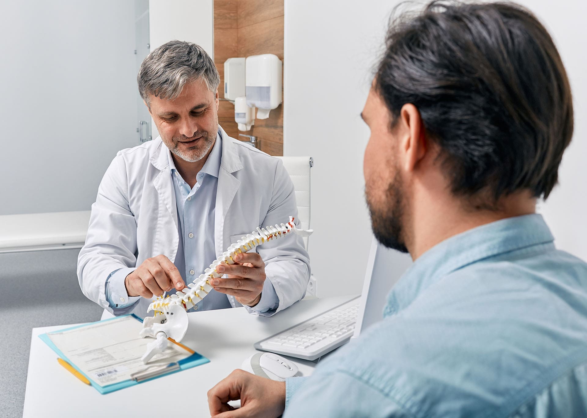
Clinicians at El Paso Back Clinic, led by Dr. Alex Jimenez, DC, APRN, FNP-BC, CFMP, IFMCP (Texas License #1191402, Prescriptive Authority #59628, NPI: 1205907805), and collaborating with Medical Director Dr. Maria Guadalupe Cardenas, MD (Texas License #J2933, NPI: 1164426748), treat this distinction as the foundation of both recovery and documentation. Dual licensure allows structural alignment work to sit alongside medical diagnostics and internal-medicine coordination. The goal is root-cause healing that reduces reliance on heavy medication or early surgery while giving patients clear information so they remain the informed decision-makers.
How Pre-Existing Degeneration Is Separated from Traumatic Aggravation
Disc degeneration is nearly universal with age. Desiccation (loss of water content), height loss, and annular fissures appear on MRI in many people who have never had significant pain. The North American Spine Society, American Society of Spine Radiology, and American Society of Neuroradiology nomenclature deliberately prefers “annular fissure” over “tear” because the finding itself does not prove a traumatic event. Less-than-violent trauma superimposed on degeneration is a clinical judgment that cannot be made from images alone; in the absence of fracture or clear violent disruption, such discs are classified morphologically as degeneration (NASS, 2014).
Texas law follows the eggshell-plaintiff principle. A defendant takes the injured person as found. Pattern Jury Charges instruct that damages cover the aggravation caused by the occurrence, not the underlying condition itself. Even when a disc already showed wear, if the fall produced new or substantially worse symptoms, functional limits, and objective findings, recovery is available for that change (PJC 28.8B and 28.8C guidance). The Fifth Circuit has confirmed that the defendant remains responsible for the full consequences that flowed from the event, even if a healthier person would have been less affected.
Clinicians therefore build a timeline rather than relying on a single film. Key questions include:
- Was the patient asymptomatic or only mildly symptomatic in the same region before the fall?
- Did new or sharply increased pain, radiating symptoms, or functional loss begin immediately or within days?
- Do prior records show a gap in treatment or only distant, resolved complaints?
A patient who worked full days, walked without limitation, and sought no care for the lumbar spine for years, then developed leg pain and positive nerve-tension signs after a fall, presents a different picture from someone already under active treatment for the identical level. Comparison imaging, when available, can show new neural contact or increased herniation size, but absence of prior films does not defeat the case when history, exam, and response to care align.
Acute Symptoms and the Clinical Timeline
Acute disc-related symptoms after a fall typically include sudden or rapidly progressive axial pain that worsens with sitting, bending, coughing, or Valsalva maneuvers, plus radicular pain in a dermatomal pattern. Delayed onset is possible but must be carefully documented; the strongest records note the fall, the immediate or early symptoms, and the progression. Inflammatory mediators released from disrupted annular fibers and nuclear material can sensitize nearby nerve roots, turning a previously compensated segment into a pain generator.
In clinic, history is paired with functional testing and, when indicated, timed inflammatory or metabolic panels ordered under Dr. Cardenas’s medical direction. Elevated markers dated to the post-fall period help isolate traumatic aggravation from baseline age-related change. Objective electronic notes that meet Colossus-style documentation standards record the mechanism (rapid deceleration or axial load of the fall), the anatomical level, and the temporal link. This creates a continuous record that supports both clinical decisions and Letters of Protection.
Neurological Findings That Anchor the Diagnosis
A careful neurological examination localizes the problem and distinguishes it from purely degenerative stiffness. For a lumbar disc herniation, clinicians look for:
- Positive straight-leg-raise or femoral-nerve-stretch tests that reproduce the patient’s radiating pain.
- Dermatomal sensory changes (for example, lateral foot numbness with L5 involvement).
- Myotomal weakness (ankle dorsiflexion or great-toe extension weakness).
- Reflex asymmetry (diminished ankle jerk with S1).
When three of the four classic findings (dermatomal pain, sensory deficit, reflex change, motor weakness) are present, the clinical diagnosis of radiculopathy is supported (StatPearls, 2023). Cervical findings follow the same logic: Spurling’s test, reduced biceps or triceps reflexes, and arm sensory or motor deficits that match the imaged level.
MRI is the preferred study for soft-tissue detail. Signs supporting an acute component include high T2 signal within the annulus (high-intensity zone), focal edema, or asymmetric disc-space signal change, especially when accompanied by ligamentous or soft-tissue injury at the same level. Muscle edema on T2 or STIR sequences can indicate acute denervation, though this is less common. Chronic findings such as advanced disc-space narrowing, large bridging osteophytes, and fatty muscle atrophy suggest longer-standing change. Importantly, an annular fissure or high-intensity zone alone does not prove the date of injury; clinical correlation remains essential.
Electrodiagnostic studies (EMG/NCS) can confirm active denervation when performed at the appropriate interval, but a normal study does not rule out symptomatic nerve-root irritation. The exam and symptom pattern still carry substantial weight.
Integrating Care for Root-Cause Healing
Once the picture of traumatic aggravation is clear, treatment focuses on reducing inflammation, restoring motion, and protecting the nervous system without unnecessary risk. Chiropractic adjustments improve segmental alignment and mobility, decreasing mechanical stress on the aggravated disc and facet joints. Patients often report better sleep, easier walking, and reduced reliance on pain medication. Neurofunctional acupuncture and electroacupuncture modulate pain pathways and support nervous-system recovery. The dual-licensed model offers advanced options—including MLS Class IV laser, extracorporeal shockwave, and carefully selected regenerative or orthobiologic approaches—to target tissue healing when conservative care alone is insufficient. Dr. Cardenas provides medical direction for complex metabolic factors that can slow recovery.
These approaches embody beneficence by addressing the mechanical and inflammatory drivers of symptoms. They embody non-maleficence by favoring non-invasive, drug-sparing methods that lower the chance of surgery or long-term opioid exposure. Autonomy is preserved through clear education: patients understand the difference between baseline degeneration and post-fall change, review their imaging and exam findings, and participate in decisions about care intensity and duration. Coordination with any existing medical team remains seamless.
Documentation produced in this setting—digital imaging reports, functional outcome measures, timed laboratory results, and detailed progress notes—gives treating providers and attorneys objective evidence of proximate causation. The fall’s biomechanical forces (axial load, flexion-compression, or shear) are linked to the specific disc level that now shows neural irritation and matching clinical deficits. Pre-existing wear may have lowered the injury threshold, but the eggshell rule and the clinical timeline show the fall caused the current disability.
Patients who complete a structured course of care frequently regain the ability to sit longer, walk farther, and sleep through the night. The same records that guide that recovery also withstand scrutiny when the question is whether the problem is new, old, or an old problem made newly disabling by a fall.
Chiropractic Care for Healing After Trauma | El Paso, TX
References
NASS, ASSR, & ASNR. (2014). Lumbar disc nomenclature: Version 2.0. The Spine Journal. https://doi.org/10.1016/j.spinee.2014.04.022
StatPearls. (2023). Lumbar disc herniation. https://www.ncbi.nlm.nih.gov/books/NBK560878/

by Dr Alex Jimenez DC, APRN, FNP-BC, CFMP, IFMCP | Chiropractic, Chronic Pain, Health, Injury Care, Treatments, Viscerosomatic Reflex
Uncover the importance of causation with chiropractic care in proving the connection between trauma and injuries in personal injury cases.
Abstract
Causation is the essential legal and medical bridge connecting an incident—such as a motor vehicle collision, slip-and-fall, or workplace trauma—to the injuries that justify recovery in personal injury claims. Without clear proof that the event caused or materially contributed to the harm, even severe pain and disability may not support full compensation. In my practice at Injury Medical Clinic PA in El Paso, Texas, I have observed for more than three decades that musculoskeletal injuries from trauma rarely exist in isolation. They disrupt joint mechanics, muscle balance, nerve signaling, and overall function, often leading to secondary problems such as chronic pain, reduced mobility, sleep disruption, and compensatory strain in distant body regions. Chiropractic care addresses these issues directly by restoring joint motion, reducing abnormal afferent input to the nervous system, decreasing local inflammation, and facilitating the body’s natural healing processes. When integrated with medical oversight from Dr. Maria Guadalupe Cardenas, MD, board-certified in Internal Medicine, functional medicine principles, targeted rehabilitation, and non-surgical regenerative options, this approach provides both clinical recovery and the objective documentation needed to establish medical causation. This educational post explains what causation means in personal injury, what chiropractic care is, how the two work together, why chiropractic care reduces musculoskeletal injuries and their comorbidities, and how an integrative team model supports durable, non-surgical recovery. Drawing on current evidence from leading researchers and my clinical observations, the goal is to help readers understand the journey from injury event to functional restoration.
Understanding Causation in Personal Injury Cases
Causation is the legal and medical requirement that links a defendant’s negligent or wrongful act to the plaintiff’s injury. Courts and insurance evaluators require evidence that the incident was both the actual cause and the proximate cause of the harm. Actual cause, often called the “but-for” test, asks whether the injury would have occurred but for the incident. Proximate cause, or legal cause, asks whether the harm was a reasonably foreseeable result of the conduct and whether the connection is close enough to justify liability. In practice, medical causation is established to a reasonable degree of medical probability—more likely than not—that the trauma caused or substantially contributed to the diagnosed condition.
The Bradford Hill criteria, originally proposed in 1965 by epidemiologist Sir Austin Bradford Hill, provide a structured way to evaluate whether an observed association is likely causal. These include strength of association, consistency across studies, specificity, temporality (the exposure must precede the outcome), biological gradient or dose-response relationship, biological plausibility, coherence with existing knowledge, experimental evidence, and analogy. In musculoskeletal personal injury, temporality is especially important: symptoms that begin or clearly worsen after a documented collision, with a mechanism consistent with known tissue tolerances, support causation. Medical records that capture the mechanism of injury, the timing of symptom onset, objective findings such as restricted range of motion, positive orthopedic tests, and imaging that correlates with clinical presentation become central evidence. Expert testimony from treating providers who have examined the patient, reviewed the history, and followed the response to treatment carries significant weight because causation opinions are inherent in clinical care.
In personal injury, delayed symptom onset does not automatically defeat causation. Soft-tissue injuries such as strains, sprains, and disc injuries can produce progressive inflammation and pain that peaks hours or days later as chemical mediators accumulate and protective muscle guarding develops. Thorough documentation of the incident details, the patient’s position and restraint use, the direction of impact, and the progressive clinical findings helps distinguish trauma-related injury from pre-existing conditions. When alternative explanations are less likely, and treatment produces measurable improvement consistent with the injury’s expected natural history, the causal link is strengthened.
What Is Chiropractic Care
Chiropractic care is a licensed healthcare profession focused on diagnosing, treating, and preventing disorders of the neuromusculoskeletal system, particularly those involving the spine and extremities. Doctors of Chiropractic are trained to evaluate joint dysfunction, soft-tissue injury, and related functional impairments, then deliver manual therapies—most commonly spinal and extremity adjustments or mobilizations—along with rehabilitative exercise, soft-tissue techniques, and patient education. The core clinical goal is to restore normal joint motion, reduce abnormal mechanical stress, modulate pain signaling, and support the body’s capacity for recovery without reliance on surgery or long-term pharmaceuticals when those interventions can be avoided or delayed.
Modern, evidence-based chiropractic practice is grounded in biomechanics and neurophysiology rather than older theories of “vertebral subluxation” as a cause of systemic disease. High-velocity, low-amplitude thrusts and graded mobilizations stimulate mechanoreceptors in joint capsules, ligaments, and muscle spindles. This afferent barrage can produce short-term changes in segmental spinal excitability, alter descending pain-modulatory pathways, and reduce local muscle hypertonicity. Clinical practice guidelines from multiple organizations support spinal manipulative therapy as part of multimodal care for acute, subacute, and chronic low back pain, neck pain, and certain headache presentations. Systematic reviews and randomized trials have shown reductions in pain intensity and improved function, particularly when chiropractic care is combined with exercise and education.
In the personal injury setting, chiropractic care is often the patient’s first and most continuous contact after trauma. Early evaluation identifies red flags that require imaging or medical referral, establishes objective baseline measures, and begins conservative management while the inflammatory phase of healing is still active. Because many musculoskeletal injuries from collisions or falls are graded sprains and strains rather than fractures or complete ruptures, non-surgical restoration of motion and load-sharing is both clinically appropriate and consistent with the principle of beginning with the least invasive effective option.
How Chiropractic Care Works Hand in Hand with Causation in Personal Injury
Chiropractic care and the proof of causation reinforce each other in several practical ways. First, the initial examination captures the patient’s mechanism of injury in their own words and correlates it with clinical findings. A restrained driver who reports neck pain after a rear-end impact, who demonstrates restricted cervical rotation, and who exhibits positive orthopedic tests for soft-tissue strain supplies the temporality and biological plausibility elements of causation analysis. Subsequent visits document the recovery trajectory: decreasing pain scores, increasing measurable range of motion, improved strength, and reduced functional limitations. These serial observations create a clear timeline that is difficult to dismiss as unrelated to the incident.
Second, chiropractic providers routinely record objective data—goniometric measurements, muscle testing, orthopedic special tests, and postural observations—that can be compared over time. When these findings improve in parallel with the expected phases of tissue healing (inflammatory, proliferative, and remodeling), the records support the conclusion that the trauma produced a treatable injury whose resolution is consistent with the care provided. Third, chiropractic documentation often includes the absence of prior similar symptoms or the clear aggravation of pre-existing conditions. In my clinical experience, patients who had stable, low-level back or neck complaints before a collision frequently report a distinct increase in intensity, new radiation of pain, or new functional loss after the event. Documenting that distinction is essential when defense argues the current symptoms are merely the natural progression of degenerative changes.
Finally, the treating chiropractor’s ongoing relationship with the patient supplies continuity that a one-time independent medical examination cannot match. When the same provider observes both the acute presentation and the response to care, the opinion that the incident caused or materially contributed to the injury rests on direct clinical observation rather than retrospective chart review alone. Courts and juries give substantial weight to this contemporaneous evidence, especially when it is consistent across chiropractic and medical notes within the same clinic.
Physiological Underpinnings of Chiropractic Care for Musculoskeletal Injuries
Spinal and extremity joints that lose normal motion after trauma generate continuous abnormal input from mechanoreceptors and nociceptors. This input can facilitate segmental spinal cord neurons, expand pain receptive fields, and maintain protective muscle co-contraction that further restricts motion. High-velocity, low-amplitude manipulation produces a brief, high-frequency burst of discharge from muscle spindle primary afferents and other dynamically sensitive receptors. Animal and human studies show that this afferent volley can reduce the excitability of dorsal horn neurons involved in pain transmission and modulate descending inhibitory pathways originating in the brainstem and higher centers.
Beyond neural effects, restoring joint motion improves local circulation and reduces concentrations of inflammatory mediators in peri-articular tissues. When synovial fluid redistributes and capsular tension normalizes, the chemical environment that sustains peripheral sensitization improves. Soft-tissue techniques used with joint mobilization address myofascial trigger points and adhesions that develop from muscle guarding. Together, these changes reduce the mechanical and chemical drivers of pain, allowing patients to resume more normal movement patterns that further promote healing.
For disc-related injuries, controlled distraction or flexion-distraction techniques can reduce intradiscal pressure and may improve nutrient diffusion into the avascular disc. Combined with core stabilization exercise, this strategy aims to restore load-sharing among the spine’s passive (ligaments and discs), active (muscles), and neural control subsystems. Clinical observations in my practice confirm that patients who regain segmental motion early and then progress through graded loading recover function more reliably than those who remain immobilized or who rely solely on passive modalities.
How Chiropractic Care Reduces Musculoskeletal Injuries and Their Comorbidities
Musculoskeletal injuries from trauma commonly produce a cascade of secondary problems. Restricted motion leads to muscle atrophy and altered recruitment patterns. Persistent nociceptive input can contribute to central sensitization, sleep fragmentation, and reduced physical activity. Reduced activity then worsens cardiovascular fitness, glycemic control, and mood. Chiropractic care interrupts this cascade at several points.
Randomized trials in active-duty military personnel have shown that adding chiropractic care to usual medical management produces moderate improvements in low-back-pain intensity and disability at six weeks, along with higher patient satisfaction and lower self-reported pain-medication use. Observational studies of older Medicare beneficiaries with chronic low back pain and multiple comorbidities found lower overall costs and lower rates of subsequent surgery among those who received chiropractic manipulative treatment. Systematic reviews support spinal manipulative therapy as part of multimodal care for neck pain and cervicogenic headache, with reductions in both pain and associated disability.
In personal injury populations, the comorbidities I most frequently observe are compensatory shoulder or hip strain from altered gait or posture, tension-type or cervicogenic headache secondary to upper cervical dysfunction, and sleep disruption that prolongs recovery. By restoring cervical and lumbar motion, reducing muscle hypertonicity, and enabling earlier return to graded activity, chiropractic care decreases the likelihood that these secondary problems become entrenched. Patients who recover segmental mobility and core endurance report better tolerance for work and daily activities, which in turn supports mental health and reduces the risk of prolonged opioid exposure.
Integrative Chiropractic Care in Non-Surgical Treatments
Non-surgical management of traumatic musculoskeletal injury is most effective when mechanical, neurological, and systemic factors are addressed together. Integrative chiropractic care combines joint mobilization or manipulation with rehabilitative exercise, soft-tissue therapy, nutritional support for tissue repair, and, when indicated, regenerative options such as platelet-rich plasma under appropriate medical direction. The reasoning is straightforward: restoring motion reduces abnormal loading and afferent drive, while progressive loading stimulates collagen remodeling and motor-pattern retraining. Functional medicine evaluation can identify co-factors—such as vitamin D insufficiency, sleep apnea, or inflammatory dietary patterns—that slow healing even when local mechanics are improved.
Spinal decompression is used when imaging and clinical findings indicate disc-related nerve compression; controlled traction reduces intradiscal pressure and can create a window for nutrient exchange and symptom relief, allowing patients to participate more fully in active rehabilitation. Soft-tissue techniques and instrument-assisted mobilization address adhesions that limit excursion. Acupuncture or electroacupuncture is sometimes added for additional pain modulation via endogenous opioid and non-opioid pathways. The plan adjusts throughout based on the healing phase and the patient’s response, with medical oversight ensuring red-flag conditions are identified and any necessary imaging, lab evaluation, or referral occurs promptly.
The Multidisciplinary Team at Injury Medical Clinic PA and the Role of Dr. Maria Guadalupe Cardenas
At Injury Medical Clinic PA (Mission Plaza Injury Medical Clinic) in El Paso, Texas, a coordinated team delivers care under dual clinical leadership. I provide chiropractic evaluation and treatment together with nurse-practitioner-level medical assessment. Dr. Maria Guadalupe Cardenas, MD, Board Certified in Internal Medicine, NPI #1164426749, Texas MD License #J2933, serves as Medical Director and Collaborative Physician. With more than 40 years of experience as an internist, Dr. Cardenas provides physician-level oversight for complex cases, reviews systemic medical issues that may affect recovery, and collaborates on treatment plans that require medical decision-making beyond the scope of chiropractic or advanced-practice nursing alone.
This structure is common in integrative injury clinics. The chiropractor focuses on joint mechanics, soft-tissue function, and progressive rehabilitation. The internist evaluates comorbidities such as hypertension, diabetes, or inflammatory conditions that can influence healing, coordinates any necessary imaging or specialty referral, and ensures that documentation meets both clinical and medicolegal standards. Physical therapists, massage therapists, and additional nurse practitioners contribute specific modalities and exercise progression. The result is a single, coherent record that captures the mechanism of injury, objective findings, response to care, and medical correlation—precisely the elements needed to support causation analysis.
Patients benefit because decisions consider both mechanical and systemic perspectives. When a patient with pre-existing diabetes sustains a disc injury, for example, glycemic control is optimized alongside spinal decompression and core training so that microvascular factors do not impede soft-tissue healing. When sleep disruption or mood changes appear, we address them rather than ignore them. The collaborative model therefore supports both clinical recovery and the clear, consistent documentation that personal injury claims require.
Clinical Observations from Dr. Alexander Jimenez
In more than three decades of practice in El Paso, I have repeatedly observed that the patients who recover most completely are those whose care begins with a precise description of the mechanism of injury and whose treatment plans are adjusted according to measured progress rather than a fixed schedule. A mid-20s heavy-labor worker who sustained neck and low-back injuries in a collision recovered approximately 80 percent of function over nineteen weeks, and then experienced a second collision, requiring careful re-evaluation of both the new trauma and the residual deficits from the first event. The integrated record—chiropractic measurements of motion and strength together with medical assessment of systemic factors—allowed the team to distinguish the effects of each incident and to modify the plan accordingly.
I have also noted that delayed symptom onset is common after low-speed collisions when the head is rotated or when the patient is looking over the shoulder. Soft-tissue strain and facet irritation can cause progressive inflammation that peaks 1 to 3 days later. Early documentation of the exact body position and the progressive clinical findings has proven essential when causation is later questioned. Patients who receive early restoration of joint motion, soft-tissue care, and graded activity consistently report better sleep, fewer compensatory pains, and faster return to work than those whose care is limited to passive modalities or prolonged rest.
These observations align with the broader literature: multimodal care that includes spinal manipulative therapy produces meaningful reductions in pain and disability, and contemporaneous records from the treating team provide the strongest evidence of both the injury and its causal relationship to the incident.
Why Specific Techniques and Protocols Are Used
Spinal and extremity adjustments are used because restricted joint motion after trauma generates ongoing abnormal afferent input that facilitates pain and maintains muscle guarding. By restoring motion, the technique reduces that input and allows more normal movement patterns to re-emerge. The reasoning is both biomechanical—normalization of load distribution—and neurophysiological—modulation of segmental and descending inhibitory pathways.
Spinal decompression is selected when clinical and imaging findings indicate disc-related compression because controlled distraction can lower intradiscal pressure and may improve fluid exchange. It is paired with stabilization exercise so the temporary pressure reduction is followed by improved muscular support.
Soft-tissue techniques and therapeutic exercise are included because muscle guarding and trigger points develop secondary to joint dysfunction and nociceptive drive. Addressing them directly reduces local ischemia and referred pain, while progressive loading stimulates collagen alignment during remodeling. The protocol includes medical oversight and functional-medicine evaluation because systemic factors—inflammation, nutrient status, sleep, and comorbidities—modulate tissue repair. Optimizing these factors increases the probability that local mechanical improvements translate into durable functional gains.
The protocol is individualized throughout according to injury grade, healing phase, and measured response. Conservative care continues when progress is documented; imaging or specialty referral occurs when red flags appear, or expected improvement does not occur. This reasoned, stepwise approach supports both clinical recovery and the clear causal narrative required in personal injury documentation.
Conclusion
Causation in personal injury is established by demonstrating that an incident more likely than not caused or substantially contributed to the diagnosed musculoskeletal injuries and their functional consequences. Chiropractic care supports this process by providing early, continuous, objective documentation of the mechanism, findings, and treatment response while restoring joint motion, modulating pain signaling, and facilitating tissue healing. When delivered within an integrative model that includes medical direction from an experienced internist such as Dr. Maria Guadalupe Cardenas, functional-medicine principles, and progressive rehabilitation, the approach reduces both the primary injury and the secondary comorbidities that commonly prolong disability. The physiological rationale is grounded in current neurophysiological and biomechanical research, and clinical results in practice align with published trials showing improved pain, function, and reduced reliance on more invasive interventions. Readers seeking recovery after trauma are encouraged to pursue care that addresses both the mechanical and systemic dimensions of injury and that produces the clear, contemporaneous records needed to support both healing and the proof of causation.
References
Hill, A. B. (1965). The environment and disease: Association or causation? Proceedings of the Royal Society of Medicine, 58(5), 295–300. https://www.ncbi.nlm.nih.gov/pmc/articles/PMC1898525/
Goertz, C. M., Long, C. R., Vining, R. D., Pohlman, K. A., Walter, J., & Colter, I. (2018). Effect of usual medical care plus chiropractic care vs usual medical care alone on pain and disability among US service members with low back pain: A comparative effectiveness clinical trial. JAMA Network Open, 1(1), e180105. https://jamanetwork.com/journals/jamanetworkopen/fullarticle/2680417
Weeks, W. B., Leininger, B., Whedon, J. M., Lurie, J. D., Tosteson, T. D., Swenson, R., O’Malley, A. J., & Goertz, C. M. (2016). The association between use of chiropractic care and costs of care among older Medicare patients with chronic low back pain and multiple comorbidities. Journal of Manipulative and Physiological Therapeutics, 39(2), 63–75. https://www.jmptonline.org/article/S0161-4754(16)00007-5/abstract
Pickar, J. G. (2002). Neurophysiological effects of spinal manipulation. The Spine Journal, 2(5), 357–371. https://www.sciencedirect.com/science/article/abs/pii/S152994300200400X
Gevers-Montoro, C., Provencher, B., Descarreaux, M., Ortega de Mues, A., & Piché, M. (2021). Neurophysiological mechanisms of chiropractic spinal manipulation for spine pain. European Journal of Pain, 25(7), 1429–1448. https://doi.org/10.1002/ejp.1773
Côté, P., Hartvigsen, J., Axén, I., Leboeuf-Yde, C., Corso, M., Shearer, H., Wong, J., Marchand, A. A., Cassidy, J. D., French, S., Kawchuk, G. N., Mior, S., Poulsen, E., Srbely, J., Ammendolia, C., Blanchette, M. A., Busse, J. W., Bussières, A., Cancelliere, C., … Yu, H. (2021). The global summit on the efficacy and effectiveness of spinal manipulative therapy for the prevention and treatment of non-musculoskeletal disorders: A systematic review of the literature. Chiropractic & Manual Therapies, 29, 8. https://doi.org/10.1186/s12998-021-00362-9
SEO Tags
causation personal injury, chiropractic care personal injury, medical causation Bradford Hill, integrative chiropractic El Paso, non-surgical musculoskeletal injury treatment, Dr Alex Jimenez DC APRN FNP-BC, Dr Maria Guadalupe Cardenas MD, Injury Medical Clinic PA El Paso, spinal manipulation neurophysiology, whiplash recovery chiropractic, low back pain evidence-based care, personal injury documentation, multidisciplinary injury clinic Texas
by Dr Alex Jimenez DC, APRN, FNP-BC, CFMP, IFMCP | Chiropractic Examination, Hip Pain & Disorders, Lower Back Pain, Spine Care
Is It Your Low Back—or Your SI Joint? Why Pain Near the Belt Line Can Be Hard to Pinpoint
Abstract
Pain on one side near the belt line often feels like a classic low-back problem, but it can come from the lumbar spine, the sacroiliac region, the hip joint, or an irritated muscle. This article explains how a careful history, movement testing, and targeted examination separate those possibilities without labeling every one-sided complaint as sacroiliac dysfunction. It also explains when imaging helps and how rehabilitation restores everyday function for programmers, Amazon associates, data center technicians, runners, drivers, and other adults.
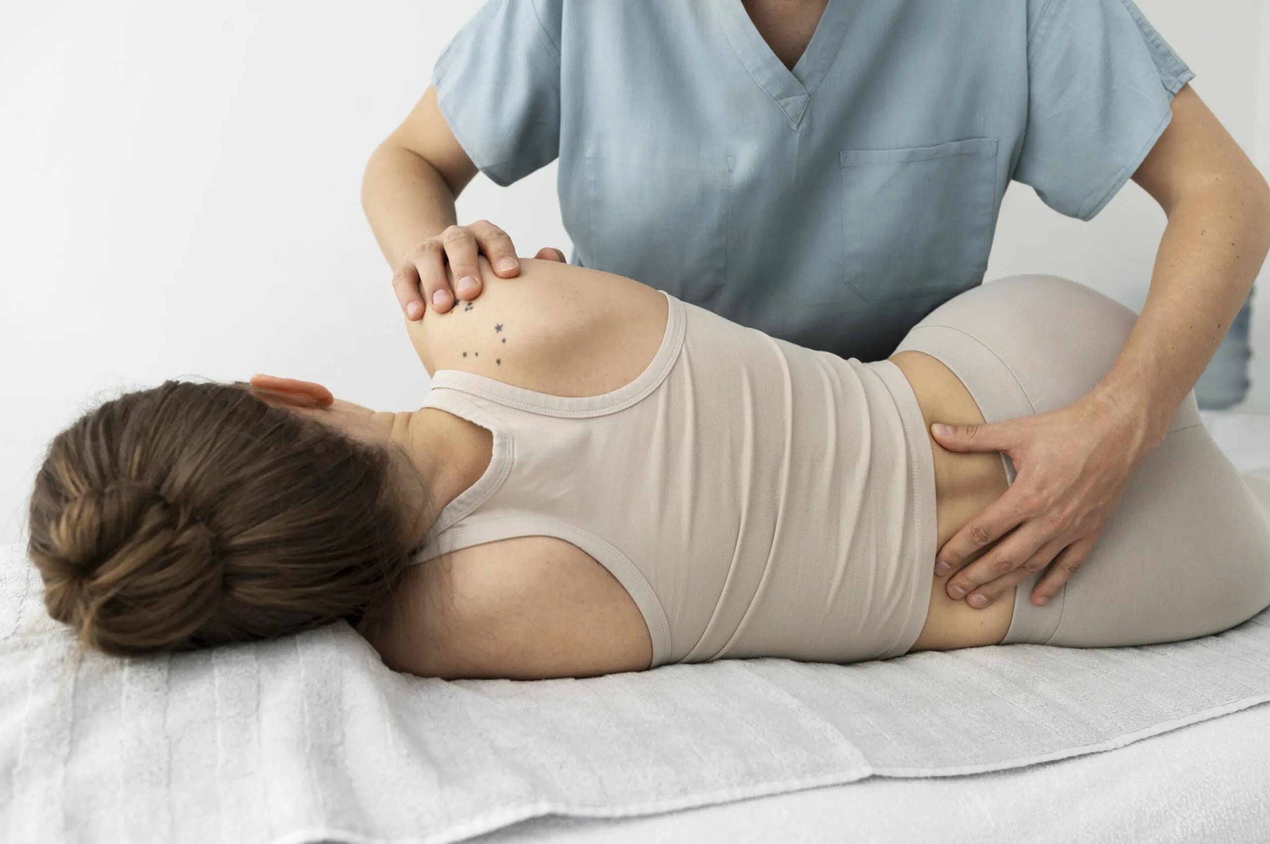
A programmer finishes a long coding block, stands, and feels a sharp catch just below the belt on one side. An Amazon associate steps off a ladder after a high reach and notices the same spot lighting up. A data center technician crouches beside a server rack, twists to connect a cable, and the pain returns on the drive home. A runner finishes a longer weekend loop and feels it the next morning near the belt line. A driver climbs out after a multi-hour haul, and the area stiffens before the first step. None of these people has classic sciatica shooting down the leg. The pain stays local or drifts into the buttock. The question is the same: is it the low back, the sacroiliac joint, the hip, or something else?
Why the Belt-Line Area Is Hard to Pinpoint
The area just above and below the belt line is a busy intersection. The lower lumbar joints, sacroiliac joints, hip joints, and surrounding muscles sit close together. Their referral patterns overlap. A problem in one place can feel like a problem in another area. That overlap is why one-sided pain near the belt line is difficult to pinpoint and why a single label—especially “SI dysfunction”—does not fit every case.
The sacroiliac joint connects the sacrum and the ilium. It has very little motion compared with the lumbar spine or the hip. Strong ligaments hold it. When the joint or its supporting tissues become irritated, pain is often felt in the buttock, just below the belt, or along the posterior pelvis. It may also refer to the upper thigh or groin. The same areas can be caused by a lumbar disc or facet joint, hip joint irritation, or muscle overload in the gluteals or deep hip rotators. Distinguishing them requires more than location alone.
Comparing the Common Sources
Lumbar spine pain often changes when you bend forward or arch backward. It may feel more central or switch sides. When a nerve root is involved, symptoms follow a clearer path down the leg, may include numbness or tingling in a specific skin area, and can show weakness on testing. A positive straight-leg raise that reproduces the leg pain raises the possibility of nerve irritation. Many people with pure lumbar pain do not have those nerve findings.
Sacroiliac-region pain is frequently one-sided and sits below the belt line. Patients often point with one finger to the area just beside the dimple of the lower back—the posterior superior iliac spine region. Activities that load one side tend to aggravate it:
- standing on one leg
- climbing stairs
- getting out of a car
- sitting for long periods and then standing
Prolonged sitting can also bother it, which is common for programmers and drivers. Not every person with these features has a primary sacroiliac problem. The pattern is a clue, not a diagnosis.
Hip-related pain can refer to the buttock or the front of the thigh. It is more likely when hip motion is limited, especially internal rotation or flexion, or when specific hip tests provoke the familiar pain in the groin. Runners and people who spend hours standing on concrete sometimes develop hip stiffness that can radiate to the low back or sacroiliac region.
Muscle irritation—particularly in the gluteus medius, gluteus maximus, or piriformis—can create tender spots and pain that mimic either a joint issue or a mild nerve pattern. These muscles work hard during single-leg stance, scanning motions, and directional changes on a warehouse floor.
What a Careful Examination Looks For
A careful examination starts with the story. When did it begin? Did a specific twist, lift, run, or long sit start it? What makes it better or worse? Does it change with position or time of day? Observation follows: how the person stands, walks, and moves from sitting to standing. Lumbar range of motion is checked. Hip mobility and strength are tested. Watch gait for a limp or a drop of the opposite hip when standing on one leg. These findings narrow the possibilities before any special test is performed.
Understanding Academic Low Back Pain: Impact and Chiropractic Solutions | El Paso, TX
Provocation Testing: Useful but Not Perfect
Provocation testing adds another layer. One commonly used set is the cluster described by Laslett and colleagues. It includes distraction, compression, thigh thrust, sacral thrust, and Gaenslen’s tests. When three or more of these tests reproduce the person’s familiar pain, the likelihood that the sacroiliac region is contributing rises (Laslett et al., 2005). Earlier work suggested high sensitivity and specificity for this combination. Later analyses have tempered expectations: the tests help, but they do not deliver certainty by themselves. Negative tests are often more useful for lowering the chance that the sacroiliac joint is the main source. When needed, the most precise confirmation remains an image-guided injection that temporarily relieves the pain. In everyday practice, the goal is not to force a label. The goal is to identify what is limiting function and address it (Cohen et al., 2013).
Hip Mobility, Gait, and Rehabilitation
Hip mobility and gait matter for almost every person in this group. Programmers and analysts who sit for long blocks often develop tight hip flexors and under-used gluteal muscles. Amazon associates and data-center technicians repeatedly load one leg while reaching or scanning. Runners accumulate high step counts. Drivers sit, then suddenly stand and twist to exit the vehicle. Rehabilitation therefore focuses on restoring usable motion and control rather than chasing a single joint name.
Practical steps include:
- brief position changes during long sits or stands
- hip and lumbar mobility work where testing shows restriction
- progressive strengthening of the gluteals and trunk muscles that stabilize the pelvis
- single-leg balance and controlled step-downs for stairs, ladders, and uneven floors
For many people, these changes reduce the catch near the belt line within a few weeks. Chiropractic care can address related spinal and pelvic mechanics when joint motion is restricted. The emphasis stays on function: being able to sit through a coding session, finish a shift, complete a run, or get out of the car without the sharp reminder.
When Imaging Is Useful
Imaging is useful when red flags appear—recent significant trauma, unexplained weight loss, night pain that wakes a person, progressive weakness, or changes in bowel or bladder control. It also helps rule out other conditions, such as a lumbar disc herniation with nerve compression, a hip joint problem, a sacral fracture, or an inflammatory arthritis affecting the sacroiliac joints. Plain radiographs of the lumbar spine and pelvis are often the first step. Consider magnetic resonance imaging when neurological signs persist or when the history suggests something beyond ordinary mechanical pain. Imaging does not reliably prove that the sacroiliac joint itself is the pain generator in the absence of clear inflammatory or structural findings. A normal study does not exclude mechanical irritation, and an abnormal study does not automatically explain the symptoms (American Academy of Family Physicians, 2022).
Working Together for Your Best Outcome
Collaborative care at Injury Medical Clinic PA brings these pieces together. Dr. Alex Jimenez, DC, APRN, FNP-BC, CFMP, IFMCP, combines chiropractic structural work with the ability to order and interpret advanced testing under collaborative oversight with Dr. Maria Guadalupe Cardenas, MD. The focus remains on what serves the patient: reducing pain, restoring mobility, and supporting recovery without unnecessary procedures. The team prioritizes non-invasive options such as targeted exercise, manual care, and activity modification to avoid the risks of surgery or heavy medication when they are not required. Patients remain the decision-makers. Education about the findings, the options, and the expected timeline lets each person choose a plan that fits work, family, and activity demands. Many group insurance plans cover this coordinated approach, providing you with access to both the mechanical and medical sides of the evaluation.
If one-sided pain near the belt line is limiting your shift, run, commute, or time at the desk, a structured examination can identify the contributors and point you to practical next steps. The same team that evaluates the lumbar spine, the sacroiliac region, the hip, and the surrounding muscles can also coordinate any needed imaging or further medical review. Call 915-850-0900 or visit the clinic at Mission Plaza in El Paso to schedule.
References
Laslett, M., Aprill, C. N., McDonald, B., & Young, S. B. (2005). Diagnosis of sacroiliac joint pain: Validity of individual provocation tests and composites of tests. Manual Therapy, 10(3), 207–218.
Cohen, S. P., Chen, Y., & Neufeld, N. J. (2013). Sacroiliac joint pain: A comprehensive review of epidemiology, diagnosis, and treatment. Expert Review of Neurotherapeutics, 13(1), 99–116.
American Academy of Family Physicians. (2022). Sacroiliac joint dysfunction: Diagnosis and treatment. American Family Physician, 105(3), 239–245.

by Dr Alex Jimenez DC, APRN, FNP-BC, CFMP, IFMCP | Lower Back Pain, Posture, Spine Care, Work Related Injuries
Why Your Low Back Burns After Standing at a Counter or Workbench
Abstract: Standing still at a counter or workbench loads the low back differently from walking. This post explains static lumbar loading, trunk endurance, hip position, and forward reach, then outlines chiropractic assessment and ways to vary position and build standing tolerance for data-center technicians, Amazon workers, lab and network technicians, and hybrid employees.
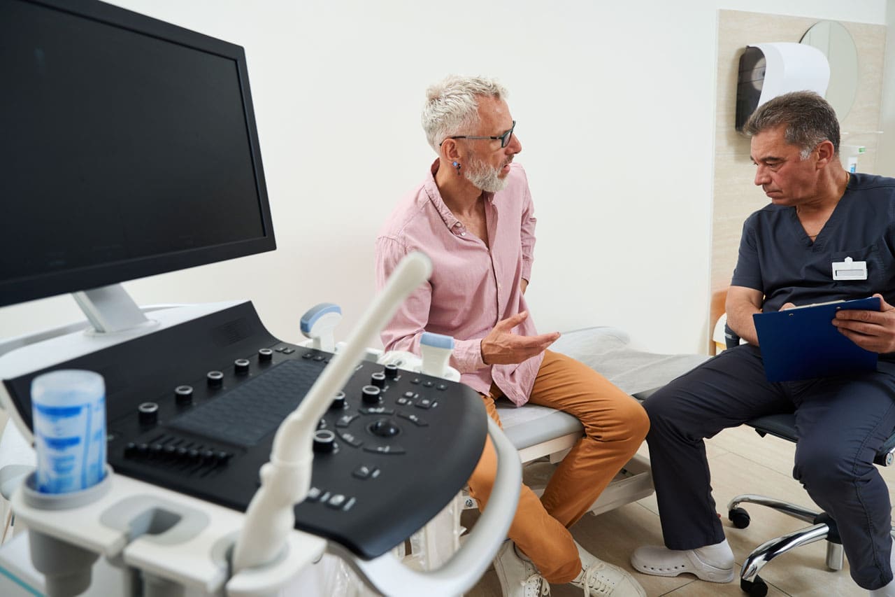
The burn starts quietly. You finish a server-rack check, a packing-station count, or a lab-bench setup, and your low back feels tight and hot. Walking the aisle felt fine. Standing still at the counter, however, did not feel fine. Static standing holds the lumbar spine under continuous load without the regular movement that walking provides.
Data-center technicians, Amazon fulfillment associates, lab personnel, network technicians, and hybrid employees often move between desk time and standing tasks. The low back may tolerate walking across the floor, then complain once the body locks in place at the workbench. Building better standing tolerance starts with understanding that difference, not simply being told to “stand straight.”
How Static Standing Loads the Lumbar Spine
When you walk, the pelvis tilts slightly with each step, and the back extensors alternate between brief work and brief rest. Standing still removes that rhythm. The lumbar extensors must hold an isometric contraction to keep the trunk upright. That continuous demand is called static lumbar loading.
Research shows that people who develop low back pain during prolonged standing often start with lower trunk endurance and different muscle activation patterns. One study found that participants who reported pain after two hours of standing had shorter side-bridge endurance times and higher co-activation of the gluteus medius muscles (Marshall et al., 2011). Another analysis linked higher estimated intervertebral disc compressive force during quiet standing with low back pain (Hasegawa et al., 2018). The load isn’t huge compared with heavy lifting, yet it stays on the tissues without the brief unloading movement provides.
Reaching forward at a counter or workbench increases the moment arm, so the back extensors work harder to keep the trunk from falling forward. Leaning onto the counter can feel like a rest, yet it often shifts the pelvis and may increase uneven loading if the hips are already tight from earlier desk time. Hybrid workers who sit for long blocks and then stand at a bench often carry that sitting posture into standing.
Why Standing Still Feels Different from Walking
Walking activates the calf muscle pump and repeatedly changes spinal posture. Standing still reduces that activity and keeps joints in the same positions for minutes at a time. A laboratory comparison found that standing produced less lower-leg swelling and muscle fatigue than walking (Wall et al., 2020). The same principle applies higher up: the low back receives less recovery when the body remains fixed.
People who develop standing-related low back pain also tend to move less early in a standing period and show higher gluteus medius co-activation (Khoshroo et al., 2023). Less movement means the tissues stay under the same stress longer. For an Amazon associate at a packing station or data center technician at a raised bench, those minutes add up across a shift.
Trunk Endurance and Hip Position Matter More Than “Stand Straight”
Telling someone to stand straight often increases effort if trunk endurance is low or if the hips cannot easily reach a neutral position. After hours at a desk, many hybrid employees and network technicians have shortened hip flexors and reduced glute endurance. When they stand, the pelvis may tip forward, increasing lumbar lordosis and the demand on the back extensors. Side-bridge endurance is a practical marker of the capacity that helps people tolerate standing without early pain (Marshall et al., 2011).
Isometric trunk training that builds endurance while keeping the spine relatively neutral has been shown to reduce pain and disability in chronic low back pain. The goal is the ability to hold useful positions for the minutes a work task requires, not maximum strength in one lift.
Chiropractic Assessment for Standing Tolerance
A focused chiropractic evaluation looks at how you stand, how your hips move, how long you can hold a side-bridge or similar endurance position, and whether forward reach or counter-lean changes your symptoms. We check lumbar joint motion, pelvic alignment, and your ability to shift weight because limited joint motion can force muscles to work harder.
Dr. Alex Jimenez, DC, APRN, FNP-BC, CFMP, IFMCP, brings dual licensure as a chiropractor and board-certified family nurse practitioner. At Injury Medical Clinic PA in El Paso, he works with Dr. Maria Guadalupe Cardenas, MD, the medical director with more than forty years of internal medicine experience. Together, they match structural findings with any medical factors that could affect recovery. Beneficence means the plan exists to improve function and reduce unnecessary risk. Non-maleficence is served by starting with non-invasive options—joint motion restoration, endurance work, and position changes. Autonomy is protected when you understand the findings and help decide which habits fit your shifts.
Building Standing Tolerance with Position Variation and Conditioning
Position variation supports the research on movement and recovery. Useful options include:
- Shifting weight from one foot to the other every minute or two.
- Placing one foot on a low step or the bottom shelf of a workbench for short periods.
- Taking two or three short steps in place or away from the counter before returning.
- Alternating a slight lean with an upright hold rather than locking into one angle.
- Avoiding locked knees and continuous forward reach when the task allows a closer stance.
These are not substitutes for conditioning. Progressive hip and trunk work builds the endurance linked to better standing tolerance. Side bridges, bird-dog holds, glute bridges, and controlled hip-hinge patterns can be practiced in short sessions. The target is the ability to stand for a typical task block with less burn, not a perfect posture held for an entire shift.
For data-center technicians moving between racks and benches, Amazon workers at packing stations, lab personnel at fixed counters, and hybrid employees alternating desk blocks with standing work, the same principles apply. The surface and reach distance change; the need for endurance and variation does not.
When to Seek Coordinated Care
Most standing-related low back pain improves with better load management and capacity. Red flags such as progressive leg weakness, bowel or bladder changes, or pain that wakes you from sleep need prompt medical review. Dr. Cardenas provides medical oversight for complex cases, while Dr. Jimenez coordinates the mechanical plan. Many group insurance plans cover the initial evaluation; the clinic can help clarify benefits.
The aim is to raise the threshold at which the low back begins to burn so you can finish the shift with less residual tightness. That outcome comes from matching assessment to your work demands, then practicing the variation and conditioning that support them.
Call 915-850-0900 or visit the clinic scheduler to arrange an evaluation focused on your standing tolerance and work tasks. You remain the decision-maker; the team supplies the information you need to choose the next step.
Spinal Decompression in Depth | El Paso, TX
References
Hasegawa, T., Katsuhira, J., Oka, H., Fujii, T., & Matsudaira, K. (2018). Association of low back load with low back pain during static standing. PLOS ONE, 13(12), e0208877. https://doi.org/10.1371/journal.pone.0208877
Khoshroo, F., Seidi, F., Bayattork, M., & Minoonejad, H. (2023). Distinctive characteristics of prolonged standing low back pain developers’ and the associated risk factors: Systematic review and meta-analysis. Scientific Reports, 13, 6392. https://doi.org/10.1038/s41598-023-33590-5
Marshall, P. W. M., Patel, H., & Callaghan, J. P. (2011). Gluteus medius strength, endurance, and co-activation in the development of low back pain during prolonged standing. Human Movement Science, 30(1), 63–73. https://doi.org/10.1016/j.humov.2010.08.017
Wall, R., Garcia, G., Läubli, T., Seibt, R., Rieger, M. A., Martin, B., & Steinhilber, B. (2020). Physiological changes during prolonged standing and walking considering age, gender and standing work experience. Ergonomics, 63(5), 579–592. https://doi.org/10.1080/00140139.2020.1725145

by Dr Alex Jimenez DC, APRN, FNP-BC, CFMP, IFMCP | Epidural Spinal Injections, Herniated Disc, Nerve Injury, Sciatica, Sciatica Nerve Pain
When Leg Pain Is Too Irritated for Rehab: What an Epidural Injection Is Actually Trying to Accomplish
Abstract
When radiating leg pain from lumbar nerve-root inflammation becomes so intense that walking, sitting, sleeping, or starting rehabilitation feels impossible, an image-guided epidural injection may be considered. This post explains radicular pain, why the procedure aims to reduce inflammation rather than repair a disc, how fluoroscopic guidance places medication in the epidural space, what patients typically experience, and how temporary symptom relief can open a practical window for decompression, movement, and active recovery. It also explains when an injection isn’t the right first step.
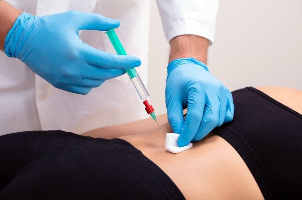
You finish a long Amazon fulfillment shift, a night of data-center rack work, a full day of programming, or a long drive, and the walk to the car lights the leg up so badly that even the idea of starting rehabilitation the next day feels out of reach. Sitting for more than a few minutes becomes a negotiation. Sleep is broken by position changes. Walking the length of a parking lot or a warehouse aisle turns into a calculated risk. For many adults with significant radiating leg pain, the barrier is not motivation. The barrier is that the nerve is too irritated to tolerate the very movements that would otherwise help.
What Radicular Pain Actually Is
Radicular pain travels along the path of a spinal nerve root. In the lumbar spine, it often follows the sciatic nerve distribution or one of its roots, causing burning, shooting, or electric sensations that can reach the buttock, thigh, calf, or foot. Numbness, tingling, or a feeling of weakness in the leg can accompany it.
A herniated disc, foraminal narrowing, or other structural change can mechanically crowd the nerve root. At the same time, biochemical factors amplify the problem. Degenerated or herniated disc material releases inflammatory mediators such as phospholipase A2, cytokines, and prostaglandins. These substances increase local edema, sensitize the nerve, and can cause ectopic firing even with only moderate mechanical pressure (McLain et al., 2005). The result is a nerve root that is both compressed and chemically inflamed. Pain and limited function often exceed what imaging alone would predict.
Why an Epidural Is Not Trying to “Repair the Disc”
An epidural steroid injection places medication into the epidural space—the area outside the dural sac that surrounds the spinal cord and nerve roots.
- It does not enter the disc.
- It does not shrink, reseal, or surgically remove a herniation.
- It does not enlarge a stenotic canal or reverse degenerative change.
The intended effect is to locally reduce inflammation around the irritated nerve root. Corticosteroids decrease inflammatory mediator production, reduce capillary permeability and edema, and can dampen ectopic nerve firing (Patel et al., 2024). When included, local anesthetic may provide more immediate, temporary relief and help confirm that the targeted root is relevant. The goal is symptom reduction, not structural repair.
Guidelines that synthesize higher-quality fluoroscopically guided trials describe meaningful short- to intermediate-term benefit for lumbar radicular pain associated with disc herniation when the injection is properly placed (Manchikanti et al., 2021). Duration varies widely; some patients get weeks of useful relief, while others experience several months. The injection is best understood as a bridge, not a destination.
Fluoroscopic Guidance, the Epidural Space, and Medication Placement
Modern lumbar epidural procedures are performed under fluoroscopic (live X-ray) guidance. The patient is positioned on a table, the skin is cleaned, and local anesthetic is used at the insertion site. A thin needle is advanced toward the epidural space. Approaches include interlaminar (through the ligamentum flavum between the laminae) and transforaminal (more selective placement near a specific exiting nerve root). The physician injects contrast dye under continuous imaging to confirm the medication spreads in the epidural space and does not enter a blood vessel or the subarachnoid space.
Once the physician verifies correct placement, they inject the corticosteroid (often with local anesthetic). The entire procedure typically lasts 10–20 minutes. Patients remain awake or lightly sedated so they can report unusual sensations. After a short observation period, most people go home the same day.
What Patients Commonly Experience
During the injection, some people feel pressure or a brief reproduction of their usual leg symptoms as the medication spreads. Immediate relief from the local anesthetic can last a few hours. The steroid effect usually begins over the following 2–7 days. A temporary increase in soreness at the injection site or a short flare of familiar leg pain can occur in the first day or two as the medication disperses; this is usually mild and self-limited.
Relief is not guaranteed and varies from person to person. When it occurs, patients often notice that they can sit longer, walk farther, sleep with fewer position changes, or tolerate the early stages of guided movement. The practical value is that these improvements create a usable window.
The Window for Movement, Decompression, and Rehabilitation
When chosen, the central purpose of the injection is to lower nerve-root irritation enough that active care becomes possible. Once leg pain no longer dominates every transition, spinal decompression therapy can be introduced or progressed more comfortably. Chiropractic adjustments and mobilization that respect the irritated root can be applied with clearer feedback. Targeted exercises for hip mobility, core control, and walking tolerance can start without an immediate severe flare.
- For Amazon associates, this may mean returning to a modified floor shift with better standing and walking tolerance.
- For data center technicians, it may mean safer ladder and rack work.
- For programmers and drivers, it may mean longer sitting blocks without the same electric build-up.
- For any adult, it may mean restoring sleep and basic daily mobility.
At Injury Medical Clinic PA, we never view the injection as a stand-alone fix. Dr. Alex Jimenez, DC, APRN, FNP-BC, CFMP, IFMCP, works under the collaborative medical direction of Dr. Maria Guadalupe Cardenas, MD, so the procedure, when indicated, is coordinated with structural care, rehabilitation, and any needed functional medicine support. The aim is beneficence: using the least invasive, effective option that serves the patient’s overall recovery and safety.
When an Injection Is Not the Appropriate First Step
Several situations call for a different sequence or urgent evaluation rather than an elective epidural:
- Progressive motor weakness, saddle anesthesia, or new bowel or bladder changes (possible cauda equina) require immediate medical assessment, not a scheduled injection.
- Active infection, uncontrolled coagulopathy, or allergy to the planned medication are contraindications.
- Pure axial low-back pain without a clear radicular pattern is not a classic indication; other sources (facet, discogenic, myofascial) should be considered first.
- Mild or improving symptoms that still allow basic walking, sleep, and a trial of conservative care often respond to decompression, targeted exercise, and activity modification without injection.
- If the clinical pattern doesn’t correlate with imaging, or if a properly placed prior injection fails, the clinic should re-examine the diagnosis rather than repeat the same procedure.
Non-maleficence guides these boundaries. The clinic prioritizes options that avoid unnecessary procedural risk, prolonged high-dose medication, or premature surgery. Autonomy remains central: patients receive a clear explanation of expected benefits, realistic duration, risks, and alternatives so they can decide in coordination with their existing medical team and any group insurance requirements.
A Practical Next Step
If radiating leg pain is currently blocking rehabilitation, sleep, or basic work and home function, an evaluation can determine whether an image-guided epidural is appropriate or whether a different sequence is safer. Many patients with excellent group insurance coverage can coordinate the integrated evaluation, procedure (when indicated), and subsequent rehabilitation plan efficiently.
Watch a brief discussion of sciatica patterns and recovery approaches:
To schedule a visit at Injury Medical Clinic PA in El Paso, call 915-850-0900 or use the online scheduler. The multidisciplinary team—led by Dr. Alex Jimenez, DC, APRN, FNP-BC, and Dr. Maria Guadalupe Cardenas, MD—will review your history, examination, and imaging to develop a plan that supports lasting movement and function.
References
Manchikanti, L., Knezevic, N. N., Navani, A., Christo, P. J., Limerick, G., Calodney, A. K., Grider, J., Harned, M. E., Cintron, L., Gharibo, C. G., Shah, S., Nampiaparampil, D. E., Candido, K. D., Soin, A., Kaye, A. D., Kosanovic, R., Magee, T. R., Beall, D. P., Atluri, S., … Hirsch, J. A. (2021). Epidural interventions in the management of chronic spinal pain: American Society of Interventional Pain Physicians (ASIPP) comprehensive evidence-based guidelines. Pain Physician, 24(S1), S27–S208.
McLain, R. F., Kapural, L., & Mekhail, N. A. (2005). Epidural steroid therapy for back and leg pain: Mechanisms of action and efficacy. The Spine Journal, 5(2), 191–201.
Patel, K., Chopra, P., & Upadhyayula, S. (2024). Epidural steroid injections. In StatPearls. StatPearls Publishing.

by Dr Alex Jimenez DC, APRN, FNP-BC, CFMP, IFMCP | Chiropractic Examination, Lower Back Pain, Spinal Hygiene, Spine Care
Why Rolling Over in Bed Can Trigger Back Pain Even When Lying Still Feels Fine
Abstract
Lying still in bed can feel comfortable, while rolling over triggers sudden back pain. This article explains rotational loading through the lumbar spine, muscle guarding after inactivity, facet-joint irritation, disc sensitivity, and hip contributions. It covers why transitions between positions often hurt more than any single posture, what a focused examination looks for, practical sleep-positioning adjustments, trunk and hip rehabilitation steps, and signs that warrant further evaluation. This discussion is written for tech workers, warehouse employees, data-center personnel, parents, and other adults who wake up when they change sleep positions.

You settle into bed after a long shift. The mattress supports your back. The room is quiet. Then you roll onto your side, and a sharp catch or deep ache stops you. Moments later, the pain eases if you stay still. This pattern is common among El Paso tech workers who sit for hours, warehouse associates who bend and reach repeatedly, data center technicians who crouch or stand, and parents who lift children or car seats before bedtime. The position itself may not be the problem. The rotational load from changing positions is a factor.
Why the Turn Hurts More Than the Position
The lumbar spine allows only a few degrees of rotation at each segment. Facet joints at the back of the vertebrae limit twisting, rather than absorbing large amounts of it. When you roll over, the rib cage and pelvis must move together. If the pelvis lags or the thoracic spine rotates first, extra rotation concentrates at one or two lumbar levels (Biology Insights, 2026). Overnight inactivity stiffens the surrounding muscles. They guard more readily when a sudden twist arrives. The result is a brief but intense signal that can wake you even though the same spine felt fine while you were still.
Disc pressure also changes with posture and time. Standing and sitting load the discs during the day. Lying down reduces that load and allows some rehydration. A quick rotational movement can then stress an already sensitive outer annulus or facet capsule (Biology Insights, 2026). For many people, the final trigger is simply the last small load on a structure that has been irritated by cumulative daytime demands—scanner reaches, ladder climbs, or prolonged keyboard work.
Muscle Guarding, Facet Irritation, and Disc-Related Symptoms
After several hours of limited movement, the deep stabilizers and larger erector muscles hold a protective tone. A rapid roll can stretch that guarded tissue before it has warmed. People often describe the sensation as a catch rather than a constant ache. Facet joints can become irritated when one segment rotates more than its neighbors because neighboring segments are stiffer from arthritis or a prior disc bulge. Local inflammation around the joint capsule causes pain that worsens with rotation and improves with stillness.
Disc-related symptoms follow a different pattern. A disc that has developed annular fissures over time may not tolerate the shear that accompanies uncontrolled lumbar rotation. Pain can be sharp and then settle once the spine returns to a neutral position. In some cases, the same motion that feels manageable during the day becomes provocative at night because the disc has regained fluid and internal pressure has risen slightly (Biology Insights, 2026). Neither finding automatically means a large herniation has occurred. Many people experience transient mechanical irritation that improves once they restore movement variability and manage load.
The Hip’s Contribution to Nighttime Rotation Pain
The hips and sacroiliac region often influence how the lumbar spine handles a roll. Tight hip flexors from desk work or prolonged standing can tilt the pelvis. When you then attempt to roll, the lumbar segments must accommodate more motion. Gluteal or piriformis tightness can restrict pelvic rotation so the lumbar spine absorbs the difference. Parents who carry children on one hip or warehouse workers who pivot under load may develop asymmetric hip mobility. That asymmetry becomes noticeable only when the body tries to turn as a unit in bed. Addressing hip mobility and control often reduces the rotational demand placed on the lower back.
What a Focused Examination Looks For
At Injury Medical Clinic PA, evaluation begins with a clear history of the pain pattern. Does the catch occur only during the turn, or does pain persist once you are in the new position? Do you have leg symptoms, numbness, or weakness? Dr. Alex Jimenez, DC, APRN, FNP-BC, CFMP, IFMCP, and the collaborative medical team look at lumbar range of motion, hip mobility, pelvic control, and segmental stiffness. Simple tests such as repeated end-range movements or controlled rotation in side-lying help identify whether the pain is primarily facet, disc, or muscular. Neurological screening checks strength, reflexes, and sensation. When needed, Dr. Maria Guadalupe Cardenas, MD, provides medical oversight for metabolic factors or red-flag conditions that can amplify musculoskeletal pain. Imaging is reserved for cases with progressive neurological findings or failure to improve with conservative care. The goal is to match the mechanical finding to a practical plan rather than to label every nighttime catch as a surgical problem.
Sleep Positioning That Reduces Rotational Stress
Supportive positioning keeps the pelvis and rib cage closer to neutral, so the lumbar spine doesn’t absorb large twists. Side sleepers often benefit from a pillow between the knees that prevents the top leg from pulling the pelvis into rotation. Back sleepers may place a pillow under the knees to reduce lumbar extension. Avoiding a full prone position limits both lumbar extension and the cervical rotation needed to breathe. A medium-firm mattress that maintains spinal curves without deep sagging is generally more helpful than an extremely soft surface (Saini et al., 2025). When rising, a log-roll technique—moving the shoulders and hips together onto the side, then pushing up with the arms while the legs swing down—keeps the spine from flexing and rotating at the same time. These adjustments do not cure an underlying mechanical issue, but they reduce the number of provocative turns during the night.
Trunk and Hip Rehabilitation Steps
Rehabilitation focuses on restoring controlled movement rather than forcing large rotations through the lumbar spine. Early work often includes gentle hip mobility drills, pelvic tilts, and isometric trunk bracing in positions that don’t provoke pain. Once symptoms settle, progressive exercises help the hips and thoracic spine share rotational demand. Single-leg balance, controlled step-downs, and bird-dog variations build the stability needed for both work tasks and nighttime turns. For data center technicians and warehouse associates, the program can include rehearsal of job-specific transitions—rising from a crouch or pivoting under a light load—so the same control carries into the shift. You can add shockwave or MLS laser therapy later if a specific tendon remains irritable after weeks of mechanical care, but these modalities support, rather than replace, movement training. Many El Paso tech, data center, and Amazon fulfillment workers have excellent group insurance that covers this coordinated chiropractic and rehabilitative approach when it is medically necessary.
When Nighttime Pain Deserves Further Evaluation
Most mechanical catches improve with positioning changes and graded rehabilitation. Certain features prompt earlier medical review. Progressive leg weakness, saddle anesthesia, or changes in bowel or bladder function require prompt assessment. Pain that is severe, unrelenting, and unrelated to position—especially if accompanied by fever, unexplained weight loss, or a history of cancer—needs medical evaluation to rule out non-mechanical causes. Persistent night pain that fails to improve after several weeks of appropriate conservative care may also warrant imaging or specialist input. Non-invasive options such as chiropractic adjustments, targeted rehabilitation, and, when indicated, image-guided epidural injections can often reduce symptoms enough to avoid more invasive procedures. The informed patient, working alongside the existing medical team, makes the final decision.
Collaborative Care That Supports Function and Autonomy
Beneficence guides the plan: structural alignment, hip and trunk control, and sleep positioning address the mechanical load that interrupts rest and work. Non-maleficence is supported by prioritizing drug-free, non-surgical strategies first so that medication dependence or surgical risk is reduced whenever possible. Autonomy is preserved by explaining the findings in plain language and outlining realistic options so each person can decide the next step. At Injury Medical Clinic PA in El Paso, Dr. Jimenez and Dr. Cardenas coordinate chiropractic care with medical oversight, functional laboratory review when indicated, and rehabilitative progressions. The result is a plan that fits shift schedules, family demands, and the specific demands of desk, rack, or fulfillment work.
If rolling over wakes you repeatedly, a focused examination can identify whether the issue is primarily rotational stiffness, hip contribution, or a combination that responds to positioning and progressive loading. Call 915-850-0900 or visit the clinic at Mission Plaza to discuss an evaluation that respects both your schedule and your long-term spinal function.
El Paso, TX Lower Back Pain Chiropractor Treatment
References
Biology Insights. (2026). Why do you have lower back pain when turning in bed? https://biologyinsights.com/why-you-have-lower-back-pain-when-turning-in-bed/
Saini, A., et al. (2025). Relationship between sleep posture and low back pain: A systematic review. Musculoskeletal Care. https://onlinelibrary.wiley.com/doi/10.1002/msc.70114

by Dr Alex Jimenez DC, APRN, FNP-BC, CFMP, IFMCP | Lower Back Pain, Power & Strength, Spine Care, Work Related Injuries
Why Repeated Small Bends Can Bother Your Back More Than One Heavy Lift
Abstract
Repeated small bends for packages, cables, laundry, or drawers can irritate the lower back more than one controlled heavy lift. This article explains cumulative spinal loading, how fatigue and reach distance change forces on the discs and ligaments, and why limited hip mobility and trunk endurance shift more work onto the lumbar spine. It outlines a chiropractic assessment process, progressive hip and trunk conditioning, practical task changes, and a plan to build bending tolerance safely rather than avoid spinal flexion. The goal is better function for Amazon associates, data center technicians, parents, warehouse workers, and desk professionals.
Maria finishes her Amazon shift without a single heavy box. She reaches into totes and bends for packages dozens of times an hour. By the time she gets home, her low back feels tight. At home, she loads the washer and picks up toys. One controlled lift earlier never bothered her as much. The pattern shows up in data center technicians, parents, warehouse workers, and desk professionals who repeatedly bend for cables, laundry, tools, or drawers.
The idea that only heavy objects matter misses how the spine handles repeated work. A single well-executed lift is one event. One hundred lighter bends spread the work across tired muscles, changing angles, and longer reaches. The tissues never fully recover between cycles, so the result can feel different—and sometimes worse.
The Myth of the Heavy Object
Advice to “lift with your legs” and “never bend your back” protects against sudden overload. It does not fully describe repeated bending for hours. Research shows both the size of each force and the number of cycles matter. High-frequency moderate loads still contribute substantially to total spinal stress (Coenen et al., 2012).
A controlled heavy lift usually uses a planned technique and finishes before fatigue sets in. Repeated light bends are hurried, done at the end of a reach, and performed after the trunk muscles have already worked. The spine sees a different pattern of compression, shear, and bending moment.
What Cumulative Loading Means for Your Spine
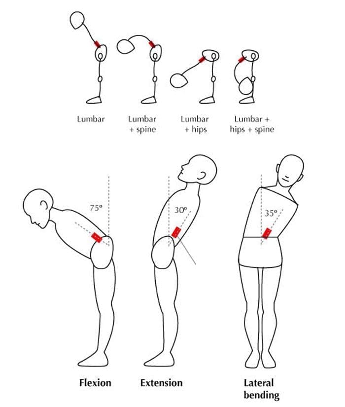
Each bend compresses and bends the lumbar discs and ligaments. When erector spinae muscles fatigue, they allow more lumbar flexion. Volunteers who lifted a 10 kg weight 100 times showed increased peak lumbar flexion and a 36 percent rise in estimated bending moment by the end of the task (Dolan & Adams, 1998).
Fatigue failure studies reinforce the point. Motion segments cyclically loaded to simulate lifting a 9 kg weight failed after far fewer cycles as torso flexion increased. Neutral posture allowed thousands of cycles; 45 degrees of flexion reduced survival to a few hundred cycles on average (Gallagher et al., 2005). Spinal posture during bending changes tissue tolerance.
Fatigue and Creep
As back muscles tire, the spine relies more on passive tissues. Repeated flexion produces creep—a slow increase in flexibility. After 20 minutes of sustained flexion, peak flexion rose by about 5.5 degrees, and full stiffness recovery took longer than the loading period (McGill & Brown, 1992). Short rests do not always fully reset the tissues. Residual creep can leave the spine more flexible for the next cycle.
Reach Distance and the Moment Arm
How far the hands are from the body matters as much as the weight. Holding a package at arm’s length multiplies the lumbar bending moment even if the package is light. Reaching into a rack, tote, or across a bed lengthens the lever arm. Bringing the object closer or raising the work surface shortens that distance and reduces demand.
How Hips and Trunk Endurance Share the Work
The lumbar spine favors stability; the hips handle large ranges of flexion. Limited hip mobility forces earlier and deeper lumbar flexion. Over dozens of bends, this shifts cumulative load onto the discs and ligaments.
Trunk endurance matters as much as peak strength. Someone who can deadlift once may still fatigue during 40 minutes of light bending if deep stabilizers and hip extensors lack endurance. Building that capacity protects the spine later in the shift.
Chiropractic Assessment: Finding the Real Pattern
A thorough evaluation at El Paso Back Clinic looks beyond the single heavy lift. Dr. Alex Jimenez, DC, APRN, FNP-BC, CFMP, IFMCP, and the team examine how the patient moves during the tasks that bother them. Assessment covers lumbar and hip range of motion, hip-hinge ability with a relatively neutral lumbar spine, trunk and hip endurance, control during repeated bending, and postural habits. The evaluation promptly identifies red-flag signs that require imaging or referral.
The evaluation identifies limited hip mobility, poor trunk endurance, excessive reach distance, residual stiffness, or a combination. Medical Director Dr. Maria Guadalupe Cardenas, MD, provides collaborative oversight, interpreting structural findings alongside medical factors when needed. Accurate diagnosis lets treatment address the real pattern. Beneficence guides the process: coordinated care improves safety, function, and recovery.
Low Back Pain Chiropractic Care | El Paso, TX
Building Bending Tolerance Instead of Avoiding Flexion
Fear of spinal flexion can create its own problems. Avoiding all bending leads to deconditioning. The tissues that are never loaded gradually lose capacity. A better approach is to restore control and then gradually reintroduce bending under conditions the spine can tolerate.
Chiropractic care restores restricted joint motion and reduces protective guarding. Adjustments and soft-tissue work free the hips and thoracic spine so the lumbar spine compensates less. Once motion improves, focus shifts to endurance work that matches real tasks:
- Hip-hinge practice with light loads held close to the body
- Controlled step-ups or partial squats that train the hips to share work
- Timed trunk endurance holds in positions that match the job
- Progressive increases in the number of bends per set, starting well below the threshold that produces next-day stiffness
The plan matches the job. An Amazon associate who bends 200 times per shift needs a different progression from a parent who bends mainly after a desk day. Progress is measured by how the back feels during and after the real task. Non-invasive options reduce risks of surgical complications or heavy medication dependence.
Practical Task Modifications for Daily Work and Home
While capacity builds, small changes reduce the total load the spine sees each day:
- Bring packages, cables, or laundry closer before lifting or lowering them.
- Use a low stool or kneel for tasks that would otherwise require deep lumbar flexion.
- Alternate sides and change stance so one pattern does not dominate.
- Take brief standing or walking breaks after longer periods of repeated bending.
- Raise frequently used items so the average bend is shallower.
- For parents, place a sturdy surface at hip height for folding or packing.
These changes reduce peak moments and high-flexion cycles while improving capacity. Patients remain the informed decision-makers, choosing modifications that fit their shift and family schedule.
The Multidisciplinary Path Forward
Integrative care at Injury Medical Clinic PA combines chiropractic structural work with medical oversight from Dr. Cardenas. Dr. Jimenez’s dual licensure supports coordinated attention to mechanical alignment and contributing factors. The emphasis remains on non-invasive strategies that restore function and reduce the need for surgery. Many patients with excellent group insurance coverage find coordinated care covered when it addresses documented functional limits.
If repeated bending has become the part of your day you dread, a focused assessment can clarify the drivers and how to rebuild tolerance safely. Call 915-850-0900 or visit the clinic in El Paso. The aim is to keep the spine working for the tasks you actually do.
References
Coenen, P., Kingma, I., Boot, C. R. L., Bongers, P. M., & van Dieën, J. H. (2012). The contribution of load magnitude and number of load cycles to cumulative low-back load estimations: A study based on in-vitro compression data. Clinical Biomechanics, 27(9), 871–877.
Dolan, P., & Adams, M. A. (1998). Repetitive lifting tasks fatigue the back muscles and increase the bending moment acting on the lumbar spine. Journal of Biomechanics, 31(8), 713–721.
Gallagher, S., Marras, W. S., Litsky, A. S., & Burr, D. (2005). Torso flexion loads and the fatigue failure of human lumbosacral motion segments. Spine, 30(20), 2265–2273.
McGill, S. M., & Brown, S. (1992). Creep response of the lumbar spine to prolonged full flexion. Clinical Biomechanics, 7(1), 43–46.