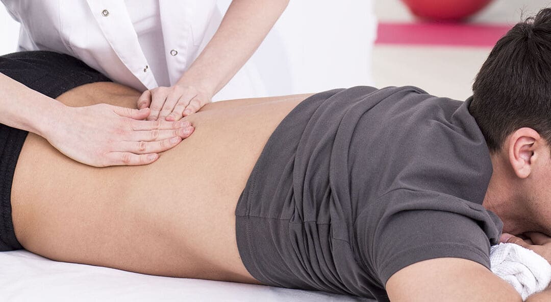
A physiotherapist is having a therapy session with a mature client with an injured knee
Introduction
Think of your body as a high-performance vehicle, needing the right fuel to glide through daily activities. Healthy foods like omega-3-packed salmon, antioxidant-rich spinach, and protein-filled chicken keep your joints limber, muscles strong, and movements smooth (Orthopedic Institute of SF, n.d.). These nutrients fight inflammation, protect tissues, and rebuild what daily wear tears down (Human Care NY, n.d.). Without them, you might feel stiff, achy, or tired just walking or bending.
Chiropractic integrative care enhances this by aligning your spine to improve nerve function, paired with nutrition to fuel healing and strengthen bones and muscles (Rangeline Chiropractic, n.d.). At El Paso Back Clinic, Dr. Alexander Jimenez, DC, APRN, FNP-BC, blends these approaches to help patients move freely, especially after injuries (Jimenez, n.d.a). This article explores how nutrition drives mobility, how chiropractic boosts it, and Dr. Jimenez’s methods for injury recovery. You’ll find simple food tips, movement tricks, and ways to stay pain-free, all grounded in science.
Mobility is for everyone—whether climbing stairs or playing sports. With the right diet and care, you can keep moving easily and avoid aches or injuries (Dr. Alex Jimenez, n.d.).
The Power of Mobility
Mobility is your ability to move without pain, from reaching for a shelf to running a mile. It relies on flexible joints, strong muscles, and a healthy spine (Alter Chiropractic, n.d.). Good mobility means tackling tasks with ease, staying energized, and lowering risks for falls or strains (Dallas Accident and Injury Rehab, n.d.a). Poor mobility can make simple movements, like bending, feel tough and lead to chronic pain.
Nutrition provides the raw materials for movement. Omega-3s in fish like mackerel reduce swelling that stiffens joints (Best Grand Rapids Chiropractor, n.d.). Antioxidants in berries protect cartilage from wear, keeping you flexible (417 Spine, n.d.). Proteins like eggs rebuild muscles after activity, preventing weakness (Better Day Chiro, n.d.). Chiropractic care ties it together by fixing spinal misalignments, ensuring nerves signal muscles for smooth motion (Grove Chiropractic, n.d.). Together, they keep you active, as patients at El Paso Back Clinic often see after a few sessions (Jimenez, n.d.b).
Nutrients That Drive Movement
Your body needs specific foods to move well. Omega-3 fatty acids in salmon or walnuts cut inflammation, easing joint pain for smoother motion (Best Grand Rapids Chiropractor, n.d.). Antioxidants in leafy greens like kale or berries like blueberries fight oxidative stress, protecting joints and keeping them flexible (417 Spine, n.d.).
Lean proteins—turkey, beans, or tofu—supply amino acids to repair muscles and tendons after exercise or injury (Human Care NY, n.d.). Calcium and vitamin D from yogurt or sunlight strengthen bones, while magnesium in nuts prevents cramps (PMC, n.d.; Foot and Ankle Experts, n.d.). Potassium-rich bananas support muscle function during activity (PMC, n.d.). Eating these foods daily builds a foundation for pain-free movement.
Top Foods to Enhance Mobility
Let’s get practical with foods that help you move. Fatty fish like sardines, eaten twice a week, can reduce joint stiffness by 20% over time (Orthopedic Institute of SF, n.d.). Walnuts or chia seeds are easy snacks that provide omega-3s and fight inflammation (Human Care NY, n.d.).
Leafy greens like arugula provide vitamin K for bones and antioxidants for flexibility (Dr. Alex Jimenez, n.d.). Berries—strawberries or raspberries—add flavor and protect cartilage (417 Spine, n.d.). Lean proteins like grilled chicken or lentils repair tissues, keeping muscles ready (Dallas Accident and Injury Rehab, n.d.a). Almonds offer magnesium to ease soreness (Better Day Chiro, n.d.).
Try simple meals: a salmon salad for lunch or a berry smoothie for breakfast. These choices fuel mobility fast.
How Poor Nutrition Slows You Down
Skipping healthy foods can hurt movement. Without omega-3s, inflammation spikes, stiffening joints, and causing pain (Best Grand Rapids Chiropractor, n.d.). Low antioxidants from a few fruits lead to cartilage wear, like a rusty hinge (Ease Well, n.d.). Protein shortages weaken muscles, making stairs or lifting tough (Alter Chiropractic, n.d.).
Low calcium or vitamin D risks brittle bones, increasing fall chances (Peak Portland, n.d.). Magnesium shortages cause cramps, limiting activity (Foot and Ankle Experts, n.d.). Sugary processed foods worsen inflammation, adding stiffness (Grove Chiropractic, n.d.). This can lead to weight gain, stress on joints, and reduced mobility (Dr. Marc Rogers, n.d.). Switching to nutrient-rich foods can reverse this in weeks.
Chiropractic Care: Unlocking Mobility
Chiropractic care boosts mobility by aligning the spine, freeing nerves to signal muscles and joints properly (New Edge Family Chiropractic, n.d.). Misalignments can cause uneven movement, leading to pain or weakness (Rangeline Chiropractic, n.d.). Adjustments address this, improving joint function and motion, often easing stiffness quickly (Texas Medical Institute, n.d.).
It also reduces inflammation by relieving nerve pressure, aiding healing (Dallas Accident and Injury Rehab, n.d.b). Paired with nutrition, chiropractic builds a strong base for mobility, helping prevent issues like arthritis (417 Spine, n.d.). Patients at El Paso Back Clinic often move more easily after adjustments (Jimenez, n.d.a).
Dr. Jimenez’s Approach at El Paso Back Clinic
At El Paso Back Clinic, Dr. Alexander Jimenez, DC, APRN, FNP-BC, connects injuries to mobility issues using his dual expertise as a chiropractor and nurse practitioner. Trauma from work, sports, personal falls, or motor vehicle accidents (MVAs) can misalign the spine, limiting movement and healing (Jimenez, n.d.b). “Injuries block nutrient delivery, slowing recovery,” he notes (Jimenez, n.d.a).
His clinic uses advanced diagnostics: X-rays spot misalignments, and blood tests check inflammation from diet gaps (Jimenez, n.d.a). A sports injury might pinch nerves, weakening leg motion. Treatments are non-surgical: adjustments restore alignment, ultrasound reduces swelling, and exercises rebuild strength. For MVAs, Dr. Jimenez provides detailed medical-legal documentation, partnering with specialists for smooth claims.
Integrative therapies enhance recovery. Nutrition plans with omega-3s cut inflammation, massage boosts blood flow for nutrient delivery, and acupuncture eases pain for better motion (Jimenez, n.d.b). A worker regained leg strength after a fall with adjustments and protein-rich meals. Dr. Jimenez targets root causes, like poor diet or posture, to prevent chronic mobility loss.
Nutrition and Chiropractic Synergy
Pairing nutrition with chiropractic maximizes mobility. Adjustments improve nerve signals for muscle control, while omega-3s reduce joint inflammation (Best Grand Rapids Chiropractor, n.d.). Greens’ vitamins strengthen bones, enhancing adjustment benefits (Dallas Accident and Injury Rehab, n.d.a). Proteins speed tissue repair post-session, reducing soreness (Human Care NY, n.d.).
This combo cuts pain faster than either alone, improving flexibility (Rangeline Chiropractic, n.d.). At the clinic, patients follow anti-inflammatory diets with care, seeing quicker movement gains (Jimenez, n.d.a).
Exercises to Amplify Nutrition
Food works better with movement. Core exercises like planks, paired with protein, build muscle stability (Sport and Spinal Physio, n.d.). Stretches with berries’ antioxidants protect joints during activity (Start PT Now, n.d.). Yoga, fueled by omega-3s, increases flexibility (Alter Chiropractic, n.d.).
Walking after green-heavy meals boosts circulation, delivering nutrients to muscles (PMC, n.d.). Start with 10-minute daily sessions, growing as strength improves. These pair with a nutrient-rich diet for mobility gains.
Preventing Long-Term Mobility Issues
Stay mobile with consistent habits. Eat omega-3s and greens daily for joint health (Orthopedic Institute of SF, n.d.). Regular chiropractic visits catch misalignments early (New Edge Family Chiropractic, n.d.). Exercise, like balance drills, prevents stiffness (Sport and Spinal Physio, n.d.).
Keep weight in check with nuts to ease joint stress (Better Day Chiro, n.d.). Sleep well, aided by magnesium foods, for tissue repair (Foot and Ankle Experts, n.d.). These steps maintain mobility for years.
Patient Stories of Success
At El Paso Back Clinic, a driver post-MVA eased knee pain with adjustments and salmon-rich meals. A runner with a sports injury moved freely again after a massage and greens. These stories show how nutrition and chiropractic restore mobility.
Conclusion
Healthy foods like omega-3 fish, leafy greens, and proteins fuel mobility by fighting inflammation and building strength. Chiropractic care at El Paso Back Clinic, led by Dr. Jimenez, aligns the spine and pairs with nutrition for optimal movement. Try fish tacos, daily stretches, and a clinic visit. Move stronger, live better.
Discover the connection between myofascial pain syndrome and chiropractic care, and find your path to relief today.
Chiropractic Care for Myofascial Pain Syndrome: Natural Relief, Root Causes, and Your Path to Wellness
Hey there, pain warriors! Ever feel like your muscles are throwing a secret party—knotty, achy, and refusing to let you join the fun? That’s myofascial pain syndrome (MPS) gatecrashing your day, turning simple moves into a comedy of errors. But fear not; chiropractic care is like the wise party pooper who gently clears the room with hands-on magic, easing those trigger points without the drama of drugs or surgery. In this epic guide (over 5,000 words of straightforward, science-backed goodness), we’ll unpack what MPS is, its sneaky causes and symptoms, how it messes with your musculoskeletal system, and why environmental factors like stress or pollution can turn up the volume on your pain. We’ll spotlight how chiropractic care, teamed with nonsurgical treatments, slashes inflammation, releases those muscle knots, and gives you a head start on a vibrant wellness journey. Plus, we’ll weave in clinical insights from Dr. Alexander Jimenez, DC, APRN, FNP-BC, a top El Paso expert who’s all about linking your injuries to cutting-edge diagnostics for real, lasting relief. Think of this as your playbook to evicting MPS—no eviction notice required, just smart, natural strategies!
We’ll keep it easy-breezy, like chatting over smoothies (or ice packs). If MPS has you feeling tied in knots, chiropractic care might just untangle things. Let’s roll!
What Is Myofascial Pain Syndrome? The Basics
Let’s kick off with the fundamentals. Myofascial pain syndrome, or MPS, is like a stubborn cramp that sets up camp in your muscles and the fascia—the tough, spiderweb-like connective tissue that wraps around them like cling wrap (Mayo Clinic, 2024a). It’s a chronic pain condition where hypersensitive spots, called trigger points, form in tight muscle bands, causing local aches or even zapping pain to distant body parts, known as referred pain (Cleveland Clinic, 2023a). Unlike a one-off muscle pull from a weekend hike, MPS lingers, making everyday tasks like stretching for a high shelf or walking the dog feel like an uphill battle.
At its heart, MPS is a musculoskeletal disorder, zeroing in on those knotty trigger points rather than widespread tenderness like in fibromyalgia (Shah et al., 2015). It’s super common—up to 85% of people might tangle with it sometimes—and it plays no favorites; desk jockeys, athletes, and couch potatoes alike can get snagged (Gerwin, 2010). The silver lining? It’s highly treatable, especially with chiropractic techniques that target those trigger points head-on, no meds or incisions needed.
Humor alert: MPS is like your muscles deciding to host a flash mob—knots dancing everywhere—but chiropractic care is the DJ who changes the tune to “relax”!
Gerwin, R. D. (2010). Myofascial pain syndrome. In S. Mense & R. D. Gerwin (Eds.), Muscle pain: Diagnosis and treatment (pp. 15–83). Springer. https://doi.org/10.1007/978-3-540-85021-2_2
Shah, J. P., Thaker, N., Heimur, J., Aredo, J. V., Sikdar, S., & Gerber, L. H. (2015). Myofascial trigger points then and now: A historical and scientific perspective. PM&R, 7(7), 746–761. https://doi.org/10.1016/j.pmrj.2015.01.024
Causes of Myofascial Pain Syndrome: The Hidden Triggers
MPS doesn’t just pop up—it’s often sparked by a mix of physical, emotional, and environmental factors that overload your muscles and fascia (StatPearls, 2023a). Physical trauma is a prime suspect: a slip on icy stairs, a fender-bender whiplash, or repetitive strain from assembly line work or marathon typing sessions can create tiny muscle tears, forming those pesky trigger points (Jimenez, 2016). Overuse is another culprit—think of a painter’s shoulder from constant overhead reaches or a runner’s calves from pounding pavement without rest (Healthline, 2024).
Poor posture sneaks in too: slouching at your desk or hunching over your phone tightens neck and shoulder muscles, setting the stage for knots (WebMD, 2024). Emotional stress amps it up—clenching your jaw during a tense meeting or tensing up in traffic can make muscles rigid, inviting trigger points (Medical News Today, 2022). Now, environmental factors? They’re the silent accomplices: cold, damp weather can stiffen muscles (like shoveling snow in winter chills), while vitamin D deficiency from indoor lifestyles weakens tissues (StatPearls, 2023b). Air pollution or toxins irritate the system, ramping up inflammation, and even ergonomic nightmares like a bad office chair contribute by promoting poor alignment (PMC, 2024).
Dr. Alexander Jimenez highlights that in his practice, MPS often stems from everyday stressors, such as prolonged sitting, which leads to postural imbalances that strain the upper back (Jimenez, 2016). Systemic factors like sleep deprivation, chronic infections, or hormonal imbalances (e.g., thyroid issues) can also play a role, making MPS a perfect storm of modern life (AAPM&R, 2024).
Humor: Causes of MPS? It’s like your muscles collecting bad habits like stamps—posture slumps, stress stamps, and cold weather postmarks—time to cancel that subscription!
Symptoms of Myofascial Pain Syndrome: The Red Flags
MPS symptoms can be sneaky, starting as a dull ache and building to a full-blown nuisance. The main event is deep, throbbing muscle pain that feels like a persistent bruise, often worsening with activity or pressure (Mayo Clinic, 2024b). Trigger points steal the show: these tender knots, when poked, cause sharp local pain or shoot discomfort elsewhere—like a back knot zinging down your leg (Cleveland Clinic, 2023b).
You’ll spot taut, stringy muscle bands, restricted movement (turning your head feels like twisting a rusty knob), and weakness that makes lifting groceries a workout (Physiopedia, n.d.). Sleep gets hijacked—pain amps up at night, leaving you tossing like a salad (WebMD, 2024). Headaches from neck triggers are frequent, and some experience fatigue or mood slumps from the endless ache (Healthline, 2024). In athletes, it might manifest as reduced speed or strength, like a swimmer with shoulder pain losing stroke power.
Dr. Jimenez notes symptoms often mimic other issues, but reproducing pain by pressing a trigger point is a telltale sign—unpleasant but revealing (Jimenez, 2016). If it’s MPS, you’ll feel that “jump sign” twinge.
Humor: Symptoms of MPS? It’s like your muscles texting “SOS”—knots that yelp when touched, aches that crash your sleep party, and a range of motion that’s on strike!
How Myofascial Pain Syndrome Affects the Musculoskeletal System
MPS is a real wrecker for your musculoskeletal system—the network of muscles, bones, tendons, ligaments, and fascia that keeps you upright and active. Trigger points mess with muscle function, creating stiff bands that hinder smooth contraction and relaxation, leading to weakness and imbalance (StatPearls, 2023a). This domino effect strains joints, accelerating wear on your spine or hips, like a misaligned wheel wobbling your car (PMC, 2019).
Fascia gets glued and restricted, limiting flexibility and causing referred pain that confuses your nerves (Shah et al., 2015). Long-term, it sparks compensatory habits—limping on one leg overuses the other—upping injury risk, like shoulder pain turning into elbow trouble (Gerwin, 2010). For athletes, it tanks performance: a calf knot alters a runner’s stride, stressing knees; a back trigger limits a golfer’s swing (AAPM&R, 2024).
Chronic MPS feeds into bigger problems, like poor sleep, ramping up inflammation, and creating a loop (Medical News Today, 2022). Dr. Jimenez explains that untreated MPS can snowball into fibromyalgia-like symptoms or nerve compression, but catching it early stops the cascade (Jimenez, 2016).
Humor: MPS on the musculoskeletal system? It’s like a bad orchestra—knots playing off-key, referred pain joining the wrong section, and your joints begging for a conductor!
Gerwin, R. D. (2010). Myofascial pain syndrome. In S. Mense & R. D. Gerwin (Eds.), Muscle pain: Diagnosis and treatment (pp. 15–83). Springer. https://doi.org/10.1007/978-3-540-85021-2_2
Shah, J. P., Thaker, N., Heimur, J., Aredo, J. V., Sikdar, S., & Gerber, L. H. (2015). Myofascial trigger points then and now: A historical and scientific perspective. PM&R, 7(7), 746–761. https://doi.org/10.1016/j.pmrj.2015.01.024
Chiropractic Care: Your Natural Ally Against Myofascial Pain Syndrome
Chiropractic care is like a skilled negotiator for MPS, stepping in to ease trigger points and restore muscle harmony without the need for meds or surgery (PubMed, 2009). Adjustments realign the spine and joints, reducing nerve pressure and improving blood flow to knotted areas, which helps flush out inflammation and relax taut bands (Integrative Physical Health, 2022). It’s non-invasive, focusing on the whole body to address imbalances that fuel MPS.
How does it work? Chiropractors use manual manipulations to release fascia restrictions, stretch muscles, and break up trigger points, often combining it with soft-tissue techniques like myofascial release (Gonstead Chiropractic Center, 2023). This boosts mobility, cuts pain, and prevents knots from returning. For environmental triggers like poor posture from desk work or stress from a hectic lifestyle, chiro restores alignment, easing the load on muscles (Radix Chiro, 2023).
Dr. Jimenez, with his dual expertise in chiropractic and nursing, uses hands-on assessments to spot trigger points, then tailors plans that include adjustments to reduce inflammation tied to factors like cold weather or repetitive strain (Jimenez, 2016). His approach not only targets pain but promotes overall wellness, helping patients dodge future flare-ups.
Humor: Chiropractic for MPS? It’s like sending a peacekeeper to your muscle’s knotty rebellion—adjust, release, and suddenly everyone’s chilling!
PubMed. (2009). Chiropractic management of myofascial trigger points and myofascial pain syndrome: A systematic review of the literature. https://pubmed.ncbi.nlm.nih.gov/19121461/
Environmental Factors and Myofascial Pain Syndrome: The Connection
Environmental factors are sneaky amplifiers for MPS, turning minor muscle stress into major pain (Best Practice & Research Clinical Rheumatology, 2024). Cold, damp weather stiffens muscles, making trigger points more likely—think shivering through a winter run without warming up (Pain Free Nottingham, 2024). Pollution and toxins irritate the system, ramping up inflammation that tightens fascia and creates knots (ScienceDirect, 2024).
Poor ergonomics, like a wonky desk setup or repetitive factory work, promote posture slumps that strain neck and back muscles (LWW, 2021). Stress from urban hustle or job pressure clenches muscles, fostering trigger points (JOSPT, 2025). Nutritional gaps, such as low vitamin D from indoor lifestyles, weaken tissues, while sleep deprivation from noisy environments exacerbates the issue (AAPM&R, 2024).
Chiropractic care shines here: adjustments correct posture imbalances from desk life, release tension from stress, and improve circulation to counter cold-weather stiffness (PubMed, 2009). Dr. Jimenez often sees MPS linked to these factors and uses tailored plans to break the cycle (Jimenez, 2016).
Humor: Environmental factors and MPS? It’s like Mother Nature pranking your muscles with cold snaps and stress bombs—chiro’s the hero who calls her bluff!
PubMed. (2009). Chiropractic management of myofascial trigger points and myofascial pain syndrome: A systematic review of the literature. https://pubmed.ncbi.nlm.nih.gov/19121461/
Chiropractic Care Combined with Nonsurgical Treatments: A Winning Team
Chiropractic care shines solo for MPS, but how does it team up with nonsurgical treatments? That’s a wellness super squad, slashing pain faster and kickstarting your health journey (ScienceDirect, 2009). Adjustments pair perfectly with myofascial release or massage to break up trigger points, while physical therapy adds stretches and exercises to build strength and flexibility (Integrative Physical Health, 2022).
Add acupuncture or dry needling to zap knots with precision, or laser therapy to boost healing without touch (LWW, 2021). TENS (transcutaneous electrical nerve stimulation) zings nerves to block pain signals, and ultrasound waves heat deep tissues for relief (PubMed, 2009). These combos tackle MPS’s multifactorial nature—chiro fixes alignment, PT builds resilience, and acupuncture eases tension—for quicker recovery and prevention (SE Pain and Spine Care, 2024).
Dr. Jimenez integrates these in his plans, using chiro as the anchor for nonsurgical synergy, helping patients ditch pain and embrace wellness (Jimenez, 2016).
Humor: Chiro and nonsurgical treatments? It’s like a band jamming—chiro on lead guitar, PT on drums, acupuncture on bass—hitting all the high notes of relief!
PubMed. (2009). Chiropractic management of myofascial trigger points and myofascial pain syndrome: A systematic review of the literature. https://pubmed.ncbi.nlm.nih.gov/19121461/
Getting a Head Start on Health and Wellness with Chiropractic and Nonsurgical Treatments
Chiropractic care with nonsurgical treatments isn’t just pain relief—it’s your fast pass to a healthier, more vibrant life (JMPT, 2009). By easing MPS, it boosts mobility, letting you hike, dance, or chase kids without wincing. Reduced inflammation means better sleep, more energy, and fewer mood dips—hello, happier you (Dynamic Care, n.d.)!
Nonsurgical add-ons like PT or acupuncture build on chiro’s foundation, strengthening muscles and preventing relapses, while nutrition tweaks (e.g., anti-inflammatory diets) fuel your body right (All Star Chiropractic, 2023). This holistic mix jumpstarts wellness: lower stress, stronger immunity, and balanced hormones for overall glow-up (Urban Chiros, n.d.).
Dr. Jimenez’s patients often report this head start—less pain opens doors to exercise, better eating, and stress-busting habits (Jimenez, 2016). It’s like upgrading from economy to first-class on your health flight!
Humor: Chiro and nonsurgical treatments for wellness? It’s like giving your body a VIP pass—skip the pain line and head straight to “feeling awesome”!
Dr. Alexander Jimenez’s Clinical Approach: Linking Injuries with Advanced Tools
Dr. Alexander Jimenez, DC, APRN, FNP-BC, is a standout in El Paso for associating patient injuries with precise diagnostics (LinkedIn, n.d.). He uses advanced imaging like MRI and CT scans to visualize soft-tissue damage, such as fascia restrictions in MPS or spinal misalignments from trauma (DrAlexJimenez.com, n.d.). These tools reveal hidden issues, like trigger points causing referred pain.
Diagnostic evaluations, including functional assessments and lab tests, pinpoint inflammation or nutritional deficiencies contributing to MPS (DrAlexJimenez.com, n.d.). Dual-scope procedures—combining endoscopy with arthroscopy—allow real-time views of joint and tissue damage, guiding minimally invasive fixes (NYS DOH, 2013; FACS, 2018).
This multifaceted method ensures accurate diagnosis, linking symptoms to causes for effective, tailored plans (Jimenez, 2016). Patients receive comprehensive reports for insurance or legal purposes, blending chiropractic care with medical precision.
Humor: Dr. Jimenez’s diagnostics? It’s like giving your injury a full body scan—trigger points can’t hide from this super sleuth!
Real-Life Stories: Overcoming MPS with Chiropractic Care
Meet Sarah, a 35-year-old office worker whose desk job sparked MPS in her neck, causing headaches that felt like a daily hammer. After chiropractic adjustments and myofascial release, she ditched the pain and now stretches like a pro (inspired by patient testimonials from Dr. Jimenez’s practice) (Jimenez, 2016).
Or take Mike, a weekend warrior with shoulder knots from golf swings. Combining chiro with PT, he swung back into action pain-free, crediting the combo for his “head start” on fitness (similar to cases in PubMed, 2009).
These stories show chiro’s real-world wins—reducing pain, boosting mobility, and sparking wellness.
Humor: Sarah’s story? From “desk zombie” to “stretch queen”—chiro turned her headaches into history!
PubMed. (2009). Chiropractic management of myofascial trigger points and myofascial pain syndrome: A systematic review of the literature. https://pubmed.ncbi.nlm.nih.gov/19121461/
The Science Behind Chiropractic’s Success for MPS
Chiropractic isn’t magic—it’s science. Adjustments restore joint function, reducing muscle tension and trigger point activity (PubMed, 2009). This lowers inflammation by improving blood flow, flushing toxins, and releasing endorphins for natural pain relief (ScienceDirect, 2009).
Studies show chiro outperforms meds for chronic pain, with lasting effects (JMPT, 2009). Combined with nonsurgical options like ultrasound or TENS, it accelerates healing by addressing fascia and nerve issues (LWW, 2021).
Dr. Jimenez’s method, using diagnostics to link injuries, ensures science-backed plans (LinkedIn, n.d.).
Humor: The science of chiro? It’s like your spine’s TED Talk—adjust, align, and applaud the relief!
PubMed. (2009). Chiropractic management of myofascial trigger points and myofascial pain syndrome: A systematic review of the literature. https://pubmed.ncbi.nlm.nih.gov/19121461/
Preventing MPS: Lifestyle Hacks for Long-Term Relief
Prevention is MPS’s kryptonite. Maintain good posture with ergonomic setups—your desk shouldn’t be a pain factory (WebMD, 2024). Stay active with regular stretches; even desk-side yoga counts. Eat anti-inflammatory foods like turmeric or omega-3s to keep muscles happy (Healthline, 2024).
Manage stress with meditation—don’t let tension turn muscles into rocks. Get enough sleep; it’s your body’s repair shop (Medical News Today, 2022). For environmental foes, bundle up in cold weather and stay hydrated to flush toxins (Pain Free Nottingham, 2024).
Chiro check-ups catch early knots, keeping you ahead (Jimenez, 2016).
Humor: Preventing MPS? It’s like muscle maintenance—stretch like a cat, eat like a rainbow, and stress less, or your knots will tie you up!
If pain persists despite rest or home remedies, it’s chiro time. Signs like constant aches, knots that don’t budge, or referred pain zapping your limbs scream “professional help!” (Mayo Clinic, 2024b). Early intervention prevents escalation (Cleveland Clinic, 2023b).
Dr. Jimenez recommends seeking care if symptoms disrupt daily life or sleep—he’ll use diagnostics to rule out mimics like arthritis (Jimenez, 2016).
Humor: When to see a chiro for MPS? When your muscles are more knotted than your earbuds after a run, it’s time to untangle!
Nutrition is your secret weapon against MPS. Anti-inflammatory diets rich in omega-3s (fish, flaxseeds) and antioxidants (berries, spinach) reduce trigger point flare-ups (LWW, 2021). Vitamin D and magnesium supplements ease muscle tension, as low levels from indoor lifestyles worsen knots (AAPM&R, 2024).
Avoid sugar and processed foods that spike inflammation (Healthline, 2024). Dr. Jimenez incorporates nutritional assessments in his plans, linking deficiencies to MPS triggers (Jimenez, 2016).
Humor: Nutrition for MPS? Eat like a rainbow warrior—berries battling knots, fish fighting inflammation—your plate’s the new battlefield!
Exercise is MPS’s frenemy—right ones soothe, wrong ones irritate. Low-impact activities like swimming or yoga stretch fascia without stress (Mayo Clinic, 2024b). Strength training with light weights builds muscle balance, preventing knots (Physiopedia, n.d.).
Start slow: trigger point self-massage before workouts, then gentle stretches. Dr. Jimenez recommends tailored routines to complement chiro, like core exercises for back MPS (Jimenez, 2016).
Humor: Exercise for MPS? It’s like whispering to your muscles—”Let’s stretch, not stress”—they’ll thank you with less complaining!
Athletes are MPS magnets—repetitive motions like pitching or running create trigger points, which can tank performance (Gerwin, 2010). A swimmer’s shoulder knots might slow strokes, or a runner’s calf trigger might cause limps (Shah et al., 2015).
Chiro helps by releasing points and restoring balance, while nonsurgical add-ons like laser therapy speed healing (All Star Chiropractic, 2023). Dr. Jimenez’s athlete-focused plans use diagnostics to link overuse to MPS, helping athletes get back in the game (Jimenez, 2016).
Humor: MPS in athletes? It’s like your muscles saying, “We trained hard, now we’re on strike”—chiro’s the mediator calling a truce!
Gerwin, R. D. (2010). Myofascial pain syndrome. In S. Mense & R. D. Gerwin (Eds.), Muscle pain: Diagnosis and treatment (pp. 15–83). Springer. https://doi.org/10.1007/978-3-540-85021-2_2
Shah, J. P., Thaker, N., Heimur, J., Aredo, J. V., Sikdar, S., & Gerber, L. H. (2015). Myofascial trigger points then and now: A historical and scientific perspective. PM&R, 7(7), 746–761. https://doi.org/10.1016/j.pmrj.2015.01.024
MPS and Mental Health: The Mind-Body Link
MPS isn’t just physical—it’s a mind-body tango. Pain disrupts sleep, spiking stress hormones that tighten muscles further (Medical News Today, 2022). Anxiety or depression can amplify symptoms, creating a loop where pain fuels mood dips, and vice versa (AAPM&R, 2024).
Chiro breaks this by reducing pain, improving sleep, and lowering stress—adjustments release endorphins for natural mood boosts (PubMed, 2009). Combined with counseling or mindfulness, it’s a holistic win (LWW, 2021).
Dr. Jimenez includes stress management in plans, recognizing the emotional side of MPS (Jimenez, 2016).
Humor: MPS and mental health? It’s like your muscles and mind in a bad rom-com—lots of tension, no happy ending—until chiro directs a rewrite!
PubMed. (2009). Chiropractic management of myofascial trigger points and myofascial pain syndrome: A systematic review of the literature. https://pubmed.ncbi.nlm.nih.gov/19121461/
The Future of MPS Treatment: Emerging Trends
MPS treatment is evolving with tech like ultrasound-guided dry needling for precise trigger point hits (SE Pain and Spine Care, 2024). Regenerative therapies, like platelet-rich plasma, show promise in healing fascia (PMC, 2024).
Chiro remains central, integrating these for personalized care (JOSPT, 2025). Dr. Jimenez stays ahead, using advanced diagnostics to blend old and new (LinkedIn, n.d.).
Humor: Future of MPS treatment? It’s like upgrading from flip phones to smartphones—chiro’s the app that ties it all together!
This deep dive into myofascial pain syndrome, its causes, symptoms, and impact on the musculoskeletal system underscores the value of chiropractic care in addressing this chronic condition. By targeting trigger points, reducing inflammation linked to environmental factors, and combining with nonsurgical treatments, chiropractic offers a natural, effective path to relief and a head start on your health journey. Dr. Jimenez’s expertise in using advanced imaging, diagnostics, and dual-scope procedures to precisely link injuries exemplifies how personalized care can transform lives.
Serious Note: While this post provides educational insights, it’s crucial to approach MPS seriously, as untreated symptoms can lead to long-term complications. Always prioritize professional medical advice.
Disclaimer: This article is for informational purposes only and is not a substitute for professional medical advice, diagnosis, or treatment. Consult a qualified healthcare provider before starting any new treatment or lifestyle change, especially with existing conditions. The content is based on research and should be taken seriously for informed health decisions. Individual results may vary, and no guarantees are made regarding outcomes.
Optimizing Movement: Chiropractic and Integrative Care for Dynamic Posture
Side view of a backpacker traveler walking against an orange wall in the city
The Importance of Dynamic Posture
Think of your body as a smoothly operating system, staying balanced and aligned whether you’re jogging, lifting, or playing a game. This ability to maintain coordination during motion is called dynamic posture, which is distinct from static posture, the way you hold yourself when still, like sitting or standing (MedlinePlus, 2023a). Good dynamic posture ensures your muscles and joints work together, distributing movement stress evenly to prevent injuries and boost performance (Cleveland Clinic, n.d.). It’s essential for safe, efficient motion in daily life or sports (Massapequa Pain Management and Rehabilitation, n.d.).
However, poor dynamic posture can lead to pain, fatigue, or injuries like strains. At El Paso Back Clinic, Dr. Alexander Jimenez, DC, APRN, FNP-BC, uses chiropractic care and integrative therapies like exercise and massage to enhance movement, especially after injuries (Jimenez, n.d.a). This article explores why dynamic posture matters, what disrupts it, and how Dr. Jimenez’s holistic approach restores balance for pain-free living.
Dynamic Posture: The Key to Fluid Movement
Dynamic posture is how your body stays aligned and stable while active, like walking to work or playing basketball. Unlike static posture—your position when not moving, like at a desk—dynamic posture involves coordinating your spine, hips, and muscles during motion (MedlinePlus, 2023a). When done right, it reduces joint stress, improves energy efficiency, and lowers injury risks, like twisting a knee (Cleveland Clinic, n.d.). It’s vital for athletes, workers, or anyone active to ensure smooth, safe movement (NYDN Rehab, n.d.).
Poor dynamic posture can cause issues, like back pain during a run or wobbling while climbing stairs. Over time, it increases the risk of chronic pain or injuries, such as sprains, and can also affect static posture (Texas Medical Institute, n.d.). Effective dynamic posture means moving with ease, recovering quickly, and staying strong, whether hiking or carrying groceries (Harrison Integrative, n.d.a).
What Leads to Poor Dynamic Posture?
Poor dynamic posture often develops from habits or injuries. Long hours of slouching, such as when using a phone, weaken core muscles, making it hard to stay aligned when active (MedlinePlus, 2023b). Repetitive tasks, like lifting heavy items incorrectly, strain the spine and disrupt movement patterns (Massapequa Pain Management and Rehabilitation, n.d.). Injuries, such as a fall or sports mishap, can lead to compensatory movements, like limping, that throw off balance (NYDN Rehab, n.d.).
Lifestyle factors contribute too. Weak core muscles from inactivity, tight hips from sitting, or stress-induced tension can disrupt natural motion (Cleveland Clinic, n.d.). These issues cause uneven stress on joints, raising risks for back pain or leg strains (Texas Medical Institute, n.d.). For instance, running with a slouched posture can overload knees, leading to pain or injury (Start PT Now, n.d.). Recognizing these patterns early helps prevent bigger problems.
Recognizing Poor Dynamic Posture
Signs of poor dynamic posture appear during activity. You might feel lower back or hip pain while walking, indicating uneven joint stress (NYDN Rehab, n.d.). Feeling unsteady on stairs or during sports can signal weak core muscles or misalignment (Cleveland Clinic, n.d.). Fatigue during tasks like carrying bags often means muscles are overworking due to poor coordination (Massapequa Pain Management and Rehabilitation, n.d.).
In the long term, poor dynamic posture increases injury risks, such as pulled muscles, and can worsen static posture, causing slouching even when still (MedlinePlus, 2023a). This leads to chronic pain in the back, neck, or knees, making movement less efficient (Harrison Integrative, n.d.a). Noticing discomfort or clumsiness during motion allows early action to avoid lasting damage.
Chiropractic Care for Improved Movement
Chiropractic care enhances dynamic posture by correcting spinal misalignments, or subluxations, that disrupt nerve signals to muscles, causing uneven movement (Harrison Integrative, n.d.b). Gentle adjustments realign the spine, improving muscle coordination and movement flow (Jimenez, n.d.a). Patients often feel steadier and less pain during activity after a few sessions (Start PT Now, n.d.).
Adjustments also relieve muscle tension, helping maintain alignment during tasks like running or lifting (Texas Medical Institute, n.d.). Regular care strengthens posture, reduces injury risks, and boosts performance for athletes or active individuals (Cleveland Clinic, n.d.). It’s like calibrating a machine for smoother operation.
Dr. Jimenez’s Expertise at El Paso Back Clinic
At El Paso Back Clinic, Dr. Alexander Jimenez, DC, APRN, FNP-BC, uses his dual expertise as a chiropractor and nurse practitioner to connect poor dynamic posture to injuries from work, sports, personal falls, or motor vehicle accidents (MVAs). “Injuries misalign the spine, disrupting movement patterns,” he explains (Jimenez, n.d.b).
His clinic employs advanced diagnostics, like X-rays for neuromusculoskeletal imaging and blood tests for inflammation, to pinpoint posture issues. A work injury, for instance, might misalign the pelvis, causing uneven strides (Jimenez, n.d.a). Treatments are non-surgical: adjustments restore alignment, ultrasound reduces swelling, and exercises rebuild muscle balance. For MVAs, Dr. Jimenez provides detailed medical-legal documentation, collaborating with specialists for seamless claims.
Integrative therapies boost recovery. Massage relaxes tight muscles, improving movement; acupuncture eases pain for natural motion; and core exercises strengthen posture-supporting muscles (Jimenez, n.d.b). A patient with back pain from a fall regained smooth walking after adjustments and yoga. Dr. Jimenez targets root causes, like poor habits, to prevent chronic posture issues.
Integrative Therapies for Movement Health
El Paso Back Clinic’s integrative approach uses natural methods to enhance dynamic posture. Core exercises, like planks, strengthen muscles for better stability during motion (Start PT Now, n.d.). The NHS recommends 150 minutes of weekly exercise, like walking or yoga, to improve coordination (MedlinePlus, 2023a).
Massage therapy loosens tight muscles, boosting blood flow for fluid movement (Texas Medical Institute, n.d.). Acupuncture reduces pain, improving joint mobility for natural motion (Jimenez, n.d.b). Spinal decompression relieves disc pressure, enhancing range of motion (Harrison Integrative, n.d.c). These therapies improve posture, prevent injuries, and aid recovery.
Everyday Habits for Better Posture
Simple habits support chiropractic care. Walk 30 minutes daily with shoulders back to practice alignment (Cleveland Clinic, n.d.). Stretch hips and hamstrings to prevent tightness that pulls the spine (Start PT Now, n.d.). Do core exercises like bridges to support movement (Massapequa Pain Management and Rehabilitation, n.d.).
Keep your back straight when lifting, bending at the knees, and avoid twisting (MedlinePlus, 2023b). Break up long sitting periods to prevent stiffness, and use ergonomic chairs to support static posture, aiding dynamic motion (NYDN Rehab, n.d.). These habits build strong, pain-free movement.
Preventing Long-Term Posture Issues
Ongoing care prevents chronic posture problems. Dr. Jimenez’s plans include regular exercises to maintain alignment, massage to keep muscles flexible, and posture checks to catch issues early (Jimenez, n.d.a). Monitoring pain during activities, like running, helps adjust care. This ensures lasting dynamic posture and fewer injuries.
Patient Success Stories
At El Paso Back Clinic, a soccer player with knee pain from poor running form improved after adjustments and core exercises. A driver with back pain from an MVA regained smooth movement with massage and acupuncture. These stories highlight the power of integrative care.
Conclusion
Dynamic posture keeps you balanced and strong during movement, reducing injury risks and boosting performance. At El Paso Back Clinic, Dr. Alexander Jimenez uses chiropractic adjustments, exercise, massage, and acupuncture to enhance alignment and recovery. Start with small steps—walk tall, stretch daily, and visit the clinic. Your body will move better and feel stronger.
Join the body-strengthening movement for a healthier you. Discover how Pilates can transform your body and fitness level.
Pilates Power: Easing Inflammation with Strength, Chiropractic Care, and Daily Wellness Tips
Hey, health nuts! Think of your body as a busy town where inflammation acts like an unexpected roadblock. It’s important for signaling problems, but it can be a pain when it stops everything. Now, imagine Pilates, the graceful exercise system that came from a dancer’s idea, swooping in like a hero to clear the way and restore smooth flow. When you combine it with exercises that build strength and chiropractic care, you have a powerful trio that can help with musculoskeletal problems and get you on the road to better health. In this in-depth guide (more than 5,000 words of clear, interesting information), we’ll explore how Pilates and strength training can help with inflammation, environmental stress, and work with chiropractic knowledge to keep you moving without pain. Dr. Alexander Jimenez, DC, APRN, FNP-BC, a top doctor in El Paso, will help us with some clinical advice. We’ll also add some humor to make it more fun. This is your guide to feeling great, whether you’re dealing with back pain or healing from an injury. You don’t need any fancy equipment, but a mat can help!
We’ll make it easy enough for a high school student to understand, with useful tips and facts backed by science. Pilates and chiropractic care could be the answer to your problems if inflammation is making you slow down. Let’s get going!
What Is Inflammation and Why Does It Matter?
Let’s go over the basics first. When something goes wrong, inflammation is like a fire alarm going off. It protects your body. When you hurt your wrist or get sick, your immune system sends white blood cells, chemicals, and fluids to the area, which can make it red, swollen, warm, or painful (Cleveland Clinic, n.d.). This is a clutch move for a healthy body: it traps germs, clears away damaged tissue, and starts healing while keeping everything in balance, which is called homeostasis (Yale Medicine, 2020).
Think about this: Carrying heavy boxes puts a lot of stress on your lower back. Inflammation comes in quickly, bringing blood full of nutrients to fix the damage. Injuries might last longer than a bad pop song stuck in your head, and infections could take over. It controls your immune system, which helps you fight off germs, and it even helps your muscles heal after a workout—your body saying, “Let’s get stronger!” (Vanderbilt Medicine, 2015). But if it stays too long, it can cause serious problems like arthritis, heart disease, or chronic pain (Yale Medicine, 2022). So, inflammation is like your body’s security guard. It can handle threats well, but it can also cause problems if it starts acting up.
Why does inflammation make you swell? Your body is having a “stop the invaders” block party, and it’s getting puffy, but someone has to clean up afterward!
Acute vs. Chronic Inflammation: What’s the Difference?
Let’s split it up into two parts: acute and chronic inflammation. Acute inflammation is the quick responder, like a superhero rushing in to save the day and then leaving after a few hours or days (Harvard Health, 2020). A pulled muscle hurts, swells, and then goes back to normal. It’s your body’s way of getting blood and immune cells to the site of injury faster.
Chronic inflammation, on the other hand, is like an unwanted guest who stays too long, simmering for months or years and possibly hurting tissues (Cleveland Clinic, n.d.). Autoimmune disorders, persistent irritants, or unresolved acute injuries can all cause chronic neck pain or sciatica (NCBI, 2023).
The main differences are that acute conditions are short, helpful, and temporary, while chronic conditions are long, harmful, and persistent. Acute helps repair by improving blood flow and cleaning up (Physiopedia, n.d.). Chronic conditions drain energy, cause constant pain, and raise the risk of getting sick (Encompass Health, 2021). Time to laugh: Acute inflammation is like a quick cameo in a big movie. It only lasts for one scene. Long-term? It’s the reboot that no one wanted, and it just keeps going on and on!
This knowledge shapes recovery: ice for sudden flare-ups and holistic methods like Pilates for long-term battles.
Environmental Factors Fueling Inflammation and Musculoskeletal Issues
Your environment isn’t just where you live; it’s also a significant factor in inflammation and musculoskeletal problems. Pollution, diet, stress, and daily habits can all make things worse (Nature Medicine, 2019). Polluted air introduces harmful chemicals into your body, which can cause oxidative stress and inflammation, making your muscles tight or putting stress on your joints (The University of Queensland, n.d.). It’s like your body is fighting a sneaky bad guy that pollutes it.
Diet is very important: Processed foods, sugars, and unhealthy fats can worsen inflammation, which can, in turn, worsen back pain or herniated discs. Antioxidant-rich foods like greens or berries can help calm it down (PMC, 2019). Pesticides and metals can get into your gut and cause systemic inflammation, which puts stress on your spine and joints (ScienceDirect, 2013). Stress raises cortisol levels, worsening inflammation and causing muscles to tense, which in turn worsens neck or shoulder pain (Northwestern University, 2017).
Other causes include smoking, which irritates tissues; being overweight, which puts pressure on joints and sends inflammatory signals; and poor posture from desk jobs or repetitive tasks, which strains your spine and causes chronic pain (PMC, 2019). Poor nutrition and other things that happen early in life can even lead to musculoskeletal problems in adults (Northwestern University, 2017). Environmental triggers make pain and inflammation worse in conditions like fibromyalgia (CGH Journal, 2024). Be careful when you laugh: Do you spend all day hunched over a desk? Your spine is begging for a break from the chair torture that makes it hurt!
Make smart choices like eating better, dealing with stress, or doing Pilates or other movement-based activities to fight back. We’ll talk more about that next.
Pilates isn’t just for people who like to work out or dance; it’s a great way for anyone who wants to reduce inflammation and build strength without pain. Joseph Pilates came up with this exercise system to work on your “powerhouse”—your core, hips, glutes, and lower back. It builds a strong, flexible base for your spine (El Paso Back Clinic, n.d.). Pilates is great for relieving stress on the muscles and bones because it uses controlled, flowing movements to stretch and strengthen them. This is different from intense workouts that can make you sore.
How does it deal with inflammation? Pilates improves circulation by sending oxygen to tissues to help reduce swelling and strengthens deep stabilizing muscles to support joints, which helps relieve stress from factors such as bad posture or repetitive movements (Siler, 2000). Because it doesn’t put stress on inflamed areas and focuses on mindful movement, it lowers cortisol levels and calms systemic inflammation (El Paso Back Clinic, n.d.). It’s like a chill pill for your body, and it’s great for everyone, from office workers to those recovering from an injury.
Humor: Pilates is like a tropical vacation for your muscles. It stretches and strengthens them, telling inflammation to take a break. No leotard needed!
Siler, B. (2000). The Pilates body: The ultimate at-home guide to strengthening, lengthening, and toning your body—without machines. Broadway Books.
How Pilates and Body-Strengthening Exercises Reduce Musculoskeletal Issues
Pilates and strength-building exercises work well together to help with musculoskeletal problems caused by inflammation. Here’s the clinical news: Pilates works on the core and stabilizing muscles, like the transversus abdominis and multifidus, which help the spine and ease joint strain (Siler, 2000). This fixes problems caused by activities like sitting for long periods or doing the same thing repeatedly, which can make muscles tight and tissues inflamed (PMC, 2019). Strength exercises, such as bodyweight movements, enhance resilience in muscles and joints, alleviating pain caused by stressors like obesity or inadequate ergonomics (Shah et al., 2015).
Pilates’ controlled movements make joints more flexible and muscles more flexible, which can help with conditions like sciatica or low back pain by putting less pressure on nerves and tissues (Cunha et al., 2018). Strength exercises add load-bearing capacity, countering wear-and-tear from environmental toxins or stress-induced tension (Northwestern University, 2017). They work together to improve circulation, eliminate inflammatory markers, and help your muscles remember how to hold themselves up, which is important for long-term relief (El Paso Back Clinic, n.d.).
Pilates is like your body’s zen master, stretching you out, and strength exercises are like a tough love coach building muscle. Together, they tell inflammation to hit the bench!
Chiropractic Care: A Head Start on Your Wellness Journey
Chiropractic care is the best way to get ready for your Pilates and strength-training workouts. It will help you live a pain-free, active life. Chiropractic adjustments realign the spine and joints, which lowers nerve pressure and improves blood flow. This helps lower inflammation and ease pain in the muscles and joints (Cleveland Clinic, n.d.). This works especially well for conditions like sciatica, neck pain, or herniated discs, where environmental stressors like bad posture or repetitive strain make symptoms worse (Western Reserve Hospital, n.d.).
Dr. Alexander Jimenez, DC, APRN, FNP-BC, a top doctor in El Paso, says that the best way to treat inflammation is to combine chiropractic adjustments with exercises like Pilates (DrAlexJimenez.com, n.d.). His method, which you can read about at https://dralexjimenez.com/, uses advanced imaging (like MRIs) and dual-scope procedures to find the source of injuries and make sure that treatment plans are accurate. Chiropractic helps with alignment, Pilates builds core strength, and body exercises make you more resilient. Together, these three things help you get a head start on health by easing pain and stopping future flare-ups.
Chiropractic care is like giving your spine a pep talk, while Pilates and strength training prepare it for the day.
Pilates and Body-Strengthening Exercises You Can Do at Home or the Gym
Ready to get moving? Here are five Pilates and body-strengthening exercises you can do at home or the gym to reduce musculoskeletal issues and inflammation. These are beginner-friendly, with modifications, and align with Dr. Jimenez’s insights on mobility and recovery (El Paso Back Clinic, n.d.).
1. Pilates Hundred
What It Does: Strengthens the core, improves circulation, and reduces lower back strain.
How to Do It: Lie on your back, legs extended or bent at 90 degrees (easier option). Lift your head and shoulders slightly, arms extended by your sides. Pump your arms up and down while inhaling for 5 counts and exhaling for 5 counts, aiming for 100 pumps. Keep your core engaged.
Why It Helps: Boosts blood flow to reduce inflammation and strengthens the powerhouse to support your spine (Siler, 2000).
Tip: Start with 50 pumps if you’re new, and keep your lower back pressed to the mat to avoid strain.
2. Bodyweight Squats
What It Does: Strengthens glutes, quads, and core, easing knee and hip stress.
How to Do It: Stand with feet hip-width apart, toes slightly out. Lower your hips as if sitting in a chair, keeping your chest up and knees over toes. Return to standing. Do 3 sets of 10-12 reps.
Why It Helps: Builds lower body strength to counter posture-related inflammation and supports joint stability (Shah et al., 2015).
Tip: Hold onto a chair for balance if needed, and don’t let knees collapse inward.
3. Pilates Roll-Up
What It Does: Stretches the spine and strengthens the core, reducing back pain.
How to Do It: Lie flat, arms extended overhead. Slowly roll up to a seated position, reaching for your toes, then roll back down with control. Do 5-8 reps.
Why It Helps: Enhances spinal flexibility and core stability, countering stress-induced tension (El Paso Back Clinic, n.d.).
Tip: Bend knees slightly for beginners, and move slowly to avoid jerking.
4. Plank
What It Does: Builds full-body strength, especially core and shoulders, to support posture.
How to Do It: Start in a push-up position, forearms on the ground, elbows under shoulders. Keep your body in a straight line, core tight, for 20-30 seconds. Repeat 3 times.
Why It Helps: Stabilizes the spine, reducing inflammation from poor posture or repetitive strain (Siler, 2000).
Tip: Drop to your knees for a modified version, and avoid sagging hips.
5. Pilates Side-Lying Leg Lift
What It Does: Strengthens hips and glutes, easing sciatica and lower back pain.
How to Do It: Lie on your side, legs stacked and straight. Lift your top leg slowly to hip height, then lower with control. Do 10-12 reps per side.
Why It Helps: Stabilizes the pelvis, reducing strain on the lower spine and nerves (Cunha et al., 2018).
Tip: Place a hand on the floor for balance, and keep movements smooth to avoid jerking.
Humor: These exercises are like giving your body a standing ovation—strengthening, stretching, and telling inflammation to take a bow and exit stage left!
Chiropractic Care: A Head Start on Your Wellness Journey
Chiropractic care is the best way to get ready for your Pilates and strength-training workouts. It will help you live a pain-free, active life. Chiropractic adjustments lower nerve pressure and improve blood flow by realigning the spine and joints. This helps reduce inflammation and relieve musculoskeletal pain (Cleveland Clinic, n.d.). This works best for conditions like sciatica, neck pain, or herniated discs, where factors such as bad posture or repetitive strain worsen the symptoms (Western Reserve Hospital, n.d.).
Dr. Alexander Jimenez, DC, APRN, FNP-BC, a top doctor in El Paso, stresses the importance of integrative care that combines chiropractic adjustments with exercises like Pilates to treat the root causes of inflammation (DrAlexJimenez.com, n.d.). His method, which you can read about at https://dralexjimenez.com/, uses advanced imaging (like MRIs) and dual-scope procedures to find the exact source of an injury, ensuring that the treatment plans are accurate. This combination of chiropractic for alignment, Pilates for core strength, and body exercises for resilience gets you started on your path to health by relieving pain and stopping future flare-ups.
Chiropractic care is like giving your spine a pep talk, and Pilates and strength training are like the training montage. Your body is ready to star in its own comeback story!
Dr. Alexander Jimenez’s Expertise in Injury Recovery
Dr. Alexander Jimenez is a well-known personal injury doctor in El Paso who combines his knowledge of chiropractic and functional medicine (LinkedIn, n.d.). He uses advanced imaging techniques, like MRIs and X-rays, and dual-scope procedures, which combine clinical exams with diagnostic tools, to find the source of injuries like whiplash or herniated discs. This accuracy makes sure that treatments are focused, which helps reduce pain and inflammation (Dr. Alex Jimenez, n.d.).
Dr. Jimenez also connects medical care and legal paperwork by writing detailed reports for injury claims. Because of his more than 30 years of experience, which you can read about at https://www.linkedin.com/in/dralexjimenez/, he is the best person to see for injuries from accidents. He uses non-invasive methods like adjustments, Pilates, and strength training to help people regain their mobility and energy.
Funny: Dr. Jimenez is like a superhero for your health. He uses high-tech imaging to figure out what’s wrong with you and Pilates to get it out of town!
Everyday Tweaks to Kickstart Your Wellness Journey
Dr. Jimenez’s clinical insights, drawn from https://dralexjimenez.com/, emphasize small, sustainable changes to reduce inflammation and musculoskeletal issues:
Nutrition: Add bromelain-rich pineapple or supplements to your diet to fight inflammation (Hikisz & Bernasinska-Slomczewska, 2021).
Movement: Incorporate 10-15 minutes of Pilates or strength exercises daily to strengthen your core and improve posture.
Posture: Set up an ergonomic workspace to counter desk-related strain.
Stress Management: Practice mindfulness or deep breathing to lower cortisol and muscle tension.
Hydration: Drink plenty of water to support tissue repair and reduce inflammation.
These tweaks, combined with regular chiropractic check-ins, build resilience against environmental stressors like pollution or repetitive tasks (El Paso Back Clinic, n.d.).
Humor: Think of these tweaks as your body’s daily tune-up—like giving your car a quick oil change to keep inflammation from revving up!
This look at Pilates, body-strengthening exercises, and chiropractic care shows a strong, evidence-based way to deal with inflammation and musculoskeletal problems. You can start a wellness journey that will help you stay healthy and mobile for a long time by dealing with environmental triggers and using Dr. Jimenez’s integrative knowledge. These strategies give you the tools you need to deal with stress, heal from injuries, and do well in an active community like El Paso.
Disclaimer: This article is only for informational purposes and is not a replacement for professional medical advice, diagnosis, or treatment. Before starting any new exercises, supplements, or treatments, especially if you already have a health problem, always talk to a qualified healthcare professional. The information comes from research and should be taken seriously when making health choices. Results are different, and there are no guarantees.
Gut Health for Faster Recovery—El Paso Back Clinic
Why your gut matters when you’re healing
After a back or neck injury—from daily strain, sports, work, or a car crash—pain and limited mobility can dominate your life. But there’s a powerful helper inside you: the gut microbiome. These trillions of microbes influence digestion, inflammation, immunity, energy, and even sleep. When they fall out of balance (called dysbiosis), bloating, irregular stools, fatigue, and higher inflammation can slow your rehab progress. The positive news is that simple daily steps can reset the balance and support your recovery. (Cleveland Clinic, 2023/2022). (Cleveland Clinic)
At El Paso Back Clinic, we often combine spine-focused care—such as chiropractic adjustments when appropriate, therapeutic exercise, soft-tissue work, and, if indicated, imaging—with practical gut-support strategies, helping patients recover more comfortably and steadily. (Dr. Alex Jimenez, El Paso clinic pages). (El Paso, TX Doctor Of Chiropractic)
Dysbiosis in plain language
Dysbiosis means your gut community is out of balance—too many “unhelpful” species, not enough beneficial ones, or less diversity overall. Diets high in refined sugars and ultra-processed foods, repeated courses of antibiotics, stress, poor sleep, and alcohol/environmental toxins are common triggers. (Cleveland Clinic, 2024; Better Health Channel, 2023; USDA ARS, 2025). (Cleveland Clinic)
Ultra-processed foods tend to be low in fiber and high in additives; over time, they’re linked with inflammation and a less favorable gut environment—exactly what you don’t want while healing. (Cleveland Clinic Newsroom, 2023). (Cleveland Clinic)
How “unhealthy” bacteria gain ground
Unwanted bacteria flourish when conditions favor them. Three everyday drivers:
Low fiber, high ultra-processed intake. Beneficial microbes feed on plant fibers and resistant starches from beans, whole grains, vegetables, and fruit. Starve them, and opportunistic species take over. (Wilson et al., 2020; Singh et al., 2017). (PMC)
Antibiotics and antimicrobials. Essential when needed, but they can also reduce helpful species; rebuilding with fiber-rich foods (and sometimes probiotics) helps restore balance. (Cleveland Clinic, 2024). (Cleveland Clinic)
Stress and poor sleep. Both alter motility and immune signaling via the brain–gut axis, nudging the microbiome toward dysbiosis. (Better Health Channel, 2023). (Better Health Channel)
SIBO: a special case to know about
Small Intestinal Bacterial Overgrowth (SIBO) happens when excess bacteria build up in the small intestine, which normally has low counts. Symptoms can include bloating, abdominal discomfort, diarrhea, early fullness, weight loss, or malnutrition. (Mayo Clinic, 2024). (Mayo Clinic)
Treatment often pairs targeted antibiotics with nutrition and root-cause fixes (e.g., motility support or addressing structural issues). Without tackling the cause, SIBO can recur. (Mayo Clinic, 2024). (Mayo Clinic)
If you notice persistent bloating, pain, or weight loss, ask your clinician about evaluation and a phased plan that treats the cause, then carefully re-expands fibers and fermented foods.
How better gut habits speed musculoskeletal recovery
Lower, steadier inflammation: A fiber-rich, plant-forward pattern boosts short-chain fatty acids (SCFAs) like butyrate that help protect the gut lining and may dampen systemic inflammation tied to pain. (Singh et al., 2017). (PMC)
Energy and participation: Balanced digestion supports energy, sleep, and mood—key drivers of successful physical therapy and home exercise. (Cleveland Clinic, 2022). (Cleveland Clinic)
Medication tolerance: If you need antibiotics or other meds, a microbiome-friendly plan can reduce GI side effects. (Cleveland Clinic, 2024). (Cleveland Clinic)
The El Paso Back Clinic approach (dual-scope care)
Our team—led by Alexander Jimenez, DC, APRN, FNP-BC—blends chiropractic care with nurse-practitioner medical evaluation. When appropriate, we use X-ray/MRI to clarify the diagnosis, and we coordinate conservative therapies with nutrition and lifestyle coaching. For injury cases, we also provide the documentation insurers and attorneys require. (El Paso, TX Doctor Of Chiropractic)
Common elements of a plan:
Dual-scope assessment: History, neuro/orthopedic testing, and imaging when indicated to pinpoint pain drivers (joint, nerve, soft tissue). (El Paso, TX Doctor Of Chiropractic)
Conservative therapies: Chiropractic adjustments (as indicated), therapeutic exercise, massage/soft-tissue work; acupuncture may be added to modulate pain and stress. (El Paso, TX Doctor Of Chiropractic)
Gut-support basics: Plant variety, fiber targets, and live-culture foods; stress and sleep tools that calm the gut–brain axis. (Cleveland Clinic Magazine; Penn State Health). (Cleveland Clinic)
Medical-legal readiness: Structured notes, imaging reports, and measurable outcomes for personal-injury and MVA cases. (El Paso, TX Doctor Of Chiropractic)
Clinical observation: Patients with back/neck pain who improve sleep and add one fermented food daily—while increasing beans/whole grains and veggies—often report less bloating and steadier energy within weeks, which helps them stay consistent with rehab.
A 4–6 week “gut-reset” that fits rehab
1) Make plants the base (daily)
Aim for colorful vegetables and fruits, beans/lentils 4–5 days/week, and whole grains (oats, barley, brown rice, quinoa). These choices feed beneficial microbes and boost SCFAs. (Wilson et al., 2020). (PMC)
2) Add one fermented food most days
Yogurt or kefir with live active cultures, kimchi, sauerkraut, or kombucha. Not all fermented foods have live microbes after processing—check the label. (Healthline; Cleveland Clinic Magazine). (Healthline)
3) Tame ultra-processed foods
Swap sugary drinks for water/unsweetened tea; favor whole-grain staples; keep packaged snacks as occasional treats. (Cleveland Clinic, 2023). (Cleveland Clinic)
4) Support sleep and stress
Target 7–9 hours with a consistent wind-down; try 5 minutes of slow breathing before bed; walk 20–30 minutes most days, and add two short strength sessions weekly. (Better Health Channel, 2023). (Better Health Channel)
5) Medications—coordinate with your clinician
Don’t stop prescribed meds on your own. If antibiotics are necessary, ask whether a food-first strategy and a short-term probiotic make sense for you. (Cleveland Clinic, 2024). (Cleveland Clinic)
6) Hygiene matters
Wash hands, rinse produce, and avoid kitchen cross-contamination to reduce exposure to harmful bacteria. (Better Health Channel, 2023). (Better Health Channel)
Two-week starter plan (easy, budget-minded)
Breakfast: Oats + kefir or yogurt + berries + nuts.
Dinner: Slow-cooker chili or lentil curry; salad with olive oil; baked potato (cool leftovers for resistant starch).
Snacks: Fruit + nut butter; carrots + hummus; plain popcorn; small kefir smoothie.
Small, steady changes add up; focus on what you can repeat during busy treatment weeks. (Penn State Health, 2018). (Penn State)
When to seek medical care now
Unintended weight loss, blood in stool, fever, severe or night-time symptoms, or a history of GI surgery.
Talk with your clinician about evaluation, including possible SIBO testing when appropriate. (Mayo Clinic, 2024). (Mayo Clinic)
Local help in El Paso
If you’re recovering from a back or neck injury and want a plan that connects spine care, gut health, and documentation for injury cases, our team can help you build a sustainable routine while we treat the root musculoskeletal drivers. (El Paso Back Clinic/Dr. Jimenez). (El Paso, TX Doctor Of Chiropractic)
Find out how bromelain can be a natural solution for reducing inflammation and promoting recovery from pain.
How Bromelain Fights Inflammation: Natural Benefits, Chiropractic Support, and Wellness Tips for Everyday Health
Hey folks! Imagine your body as a bustling city where inflammation acts like traffic cops directing chaos after a fender-bender – necessary at first, but a nightmare if it gridlocks everything. Now, picture a natural ally straight from the pineapple patch: bromelain, zapping that swelling like a tropical traffic clearer. In this in-depth guide (clocking in over 5,000 words of easy-to-digest info), we’ll unpack bromelain’s powerhouse role in taming inflammation, backed by solid science with a sprinkle of laughs to keep it fun. We’ll cover inflammation’s good and bad sides, environmental triggers, and how combining natural remedies with hands-on care like chiropractic adjustments can help dial down chronic risks. Drawing from experts in integrated health, we’ll spotlight practical tweaks for better living, especially for those dealing with pain or injuries in active communities like El Paso. No superhero cape needed – just smart, natural strategies to feel your best!
Let’s break it down simply, at a level anyone can grab onto. If inflammation feels like your body’s overeager alarm clock, bromelain might just be the snooze button you’ve been missing. Ready to dive in?
What Is Inflammation and Its Role in a Healthy Body?
Let’s kick off with the fundamentals. Inflammation is your body’s natural defense squad – like firefighters rushing to douse a blaze before it spreads. When you scrape your knee or battle a virus, your immune system deploys cells, chemicals, and fluids to isolate the threat, leading to those classic signs: redness, swelling, warmth, and ouch-factor pain (Cleveland Clinic, n.d.). In a well-tuned body, this process is a hero: it neutralizes invaders, clears out damaged bits, and paves the way for repair, all while maintaining balance or what pros call homeostasis (Yale Medicine, 2020).
Visualize this: You strain a muscle hiking. Inflammation swoops in, boosting blood flow with healing goodies to mend the tear. Without it, injuries could linger like unwanted houseguests, and infections might run wild. It’s crucial for immune regulation, helping you shrug off daily germs, and even signals muscle growth post-workout – your body’s subtle nudge to “level up!” (Vanderbilt Medicine, 2015). But when it overstays, it links to heavier hitters like cardiovascular woes or metabolic issues (Yale Medicine, 2022). Bottom line: Inflammation is your internal watchdog – vigilant and vital, but best kept in check.
Quick chuckle: Why does inflammation cause puffiness? It’s your body inflating like a balloon animal to trap the bad stuff – party trick gone wrong if it doesn’t deflate!
Acute vs. Chronic Inflammation: Spotting the Difference
Time to differentiate the duo: acute and chronic inflammation. Acute is the rapid responder – think elite rescue team handling a crisis swiftly, lasting mere hours or days before bowing out (Harvard Health, 2020). Example? A splinter: quick redness and throb, then poof, healed. It’s protective, ramping up blood supply and immune troops for speedy recovery.
Chronic inflammation? That’s the lingering lurker, simmering low-key for months or years, often silently eroding tissues (Cleveland Clinic, n.d.). Triggers include autoimmune mix-ups, persistent irritants, or unchecked acute episodes, potentially fueling ailments like joint woes or even tumors (NCBI, 2023).
Core contrasts: Acute is brief, beneficial, and resolves; chronic is prolonged, problematic, and pervasive. Acute aids repair via enhanced circulation and cell cleanup (Physiopedia, n.d.). Chronic drains energy, sparking ongoing discomfort and elevated disease odds (Encompass Health, 2021). Laugh break: Acute inflammation is a flash mob – exciting and gone fast. Chronic? More like a never-ending Zoom call, wearing everyone out!
Grasping this helps tailor fixes: Ice for acute flare-ups, holistic shifts for chronic battles.
Your surroundings aren’t passive – they’re inflammation influencers. Elements like smog, eats, tension, and habitat can spike levels (Nature Medicine, 2019). Air pollution sneaks in toxins, sparking body-wide stress and swelling (The University of Queensland, n.d.). It’s akin to inhaling mini agitators that stir your defenses.
Food choices matter big: Sugary, processed bites fan flames, while veggie-packed antioxidants douse them (PMC, 2019). Toxins from pesticides or metals mess with gut barriers, leaking inflammation systemically (ScienceDirect, 2013). Stress pumps cortisol, which chronically amps inflammation (Northwestern University, 2017).
More culprits: Smoking irks tissues, extra weight releases inflammatory signals from fat, and childhood exposures prime adult risks (PMC, 2019). In gut disorders like Crohn’s, enviro factors tweak permeability and microbes, heightening odds (CGH Journal, 2024). Giggle moment: Urban pollution? Your body’s throwing an uninvited inflammation bash – time to crash it with cleaner habits!
Counter with tweaks like fresh air filters or balanced plates – insights ahead from integrated care pros.
Meet Bromelain: Your Pineapple-Powered Ally Against Inflammation
Bromelain – not a sci-fi baddie, but an enzyme squad from pineapple cores and sap, long hailed in traditional remedies and now science-vetted for swelling showdowns (PubMed, 2022). It’s nature’s gentle warrior, slicing through proteins, easing puffiness, and fine-tuning immunity.
It shines in easing arthritis aches or surgery swells, often with fewer side hits than pills (PubMed, 2023). Fun tidbit: Nibbling pineapple helps, but caps deliver the knockout dose – though don’t count on fruit salad alone for that marathon recovery!
Clinical Reasons Bromelain Tackles Inflammation
Science breakdown: Bromelain curbs inflammation by blocking flare-up messengers like prostaglandins and cytokines, dialing down swell and hurt (Hikisz & Bernasinska-Slomczewska, 2021). It amps clot-busting, boosting flow to clear junk (PubMed, 2024).
In clinics, it shrinks fluid-trapping proteins for less edema, hastening fixes in sinus clogs or bruises (PubMed, 2023). For long-haul inflammation, it reins in immune overdrives (PubMed, 2024). Why it works? Targets origins naturally, safely – a step up from symptom-masking meds.
Snicker: Bromelain’s your chill pal bringing calm (and maybe piña coladas) to inflammation’s storm.
Mechanics deep-dive: Shuts off NF-kB inflammation triggers, ups protective enzymes. Helps disc issues by soothing responses (PubMed, 2018). Mirrors curcumin for workout woes, but with fruity flair (PubMed, 2020).
Fernández-Lázaro, D., et al. (2020). *Modulation of exercise-induced muscle damage, inflammation, and oxidative markers by curcumin supplementation*. https://pubmed.ncbi.nlm.nih.gov/32075287/
Bromelain’s Beneficial Traits and Body Boosts
Pulling from core research (Hikisz & Bernasinska-Slomczewska, 2021), bromelain’s protein-digesting, swell-reducing, and clot-preventing powers shine. It clears inflammatory clutter, smooths digestion, and ups nutrient uptake.
Perks: Eases joint pain, quickens wounds, thins mucus for clearer sinuses. Potential cancer fighter by targeting bad cells. Body-wide: Strengthens defenses, heart health via pressure drops, asthma relief through airway calm.
Aid mechanism: Balances cytokines for immune harmony, curbing chronic creeps. Gut-soother for IBS. Chuckle: Pineapple’s bromelain says, “Digest this – and ditch the bloat!”
Extras: Fights oxidative hits tied to inflammation (PubMed, 2016). Cuts post-gym aches akin to curcumin (PubMed, 2019).
Tan, Y. Q., & Zhang, J. (2019). *Effect of curcumin supplementation on exercise-induced oxidative stress, inflammation, muscle damage, and muscle soreness*. https://pubmed.ncbi.nlm.nih.gov/31025894/
Chiropractic Care Combined with Non-Surgical Approaches for Chronic Inflammation
Chiropractic isn’t mere snaps – it’s a non-invasive champ for curbing chronic inflammation overlaps. Adjustments realign spines, ease nerve pinches, and enhance flow to tame swells (Cleveland Clinic, n.d.). Paired with massage, nutrition, or acupuncture, it hits shared risks like stiffness, stress, and pain cycles.
Mechanism: Corrects misalignments to lower body-wide inflammation, aiding arthritis or spinal strains (Western Reserve Hospital, n.d.). Add-ons like diet tweaks and rehab foster drug-free healing (Driver Chiropractic, 2025).
In rheumatoid cases, exercise-integrated plans moderate inflammation (PubMed, 2022). Eases trigger points for lasting relief (PubMed, 2015). Humor: Chiropractors? Body tuners keeping inflammation from hitting sour notes!
Clinics blending these, like those in El Paso focusing on back health, offer tailored protocols for local active lifestyles, integrating functional assessments to address inflammation roots.
Sgreccia, E., et al. (2022). *The effect of exercise on patients with rheumatoid arthritis on the modulation of inflammation*. https://pubmed.ncbi.nlm.nih.gov/34874837/
Insights from Dr. Alexander Jimenez on Inflammation Management
In El Paso, practitioners like Dr. Alexander Jimenez, DC, APRN, FNP-BC, fuse chiropractic with functional medicine to combat inflammation (El Paso Back Clinic, n.d.). His take: Target origins like alignments and nutrition to shrink chronic threats.
Routine nudges: Fold in anti-inflammatory eats, steady motion, and calm techniques. Agility drills build toughness (El Paso Back Clinic, n.d.). Tools like Living Matrix craft custom paths.
Laugh: Dr. Jimenez plays inflammation sleuth – cracking cases with adjustments, no deerstalker hat required!
Clients report enhanced rest, reduced aches, and vigor via his whole-body strategy, ideal for bustling areas like El Paso.
Handling Personal Injury in El Paso: Expert Care Spotlight
El Paso sees its share of mishaps from commutes or sports, where specialized care shines for recovery (El Paso Back Clinic, n.d.). Pros like Dr. Jimenez link traumas to cutting-edge scans, diagnostics, and scoped views for precise mapping.
He bridges health services and legal needs, delivering solid records for cases. With decades in, he handles whiplash to sciatica via gentle rehab (El Paso Back Clinic, n.d.).
Giggle: Injuries sting, but expert care flips the script from “ouch” to “onward” like a pro upgrade!
Clinics here emphasize non-invasive paths, coordinating with teams for full-circle support in personal injury scenarios.
Everyday Tweaks: Clinical Wisdom for Inflammation Control
Dr. Jimenez advises: Weave bromelain sources like pineapple or supps into meals. Add walks to curb flames; mindfulness for stress (El Paso Back Clinic, n.d.). Build strength, stay hydrated.
Monitor with evals; dodge pollutants. These stack to trim risks, fostering wellness (El Paso Back Clinic, n.d.).
Humor: Tiny shifts? Like ditching junk for greens – your insides high-five without the inflammation hangover!
Wrapping this deep dive into bromelain’s inflammation-fighting prowess, natural perks, and synergy with chiropractic and lifestyle shifts, it’s evident these approaches pave ways to robust health. From science on bromelain to integrated strategies reducing chronic burdens, tools like these empower better living.
Disclaimer: This article provides general information only and isn’t medical advice, diagnosis, or treatment. Consult a healthcare professional before new regimens, especially with conditions. Content draws from research; take it seriously for informed choices. Results differ; no outcomes guaranteed.
Boosting Vitality: Chiropractic and Integrative Care for Optimal Circulation
Introduction
Imagine your body as a network of highways, with blood vessels carrying oxygen and nutrients to every cell, keeping you energized and healthy. This process, known as optimal circulation, ensures the circulatory system efficiently delivers these essentials while removing waste, supporting cell function, and overall vitality (Henry Ford Health, 2025). When circulation falters, you may feel tired, have cold hands, or experience issues such as swelling. Chiropractic care can help by aligning the spine to reduce nerve pressure and improve blood flow naturally. Paired with integrative therapies like massage and exercise, it promotes lasting wellness (Sierra, n.d.). This article examines how these methods enhance circulation, drawing on insights from Dr. Alexander Jimenez at El Paso Back Clinic in El Paso, TX, to demonstrate their role in aiding injury recovery and promoting overall health.
Why Optimal Circulation Matters
Optimal circulation is the body’s way of keeping everything running smoothly. Blood delivers oxygen and nutrients to cells, fueling energy production through ATP and clearing waste, such as carbon dioxide, to prevent fatigue and aches (Elevation Health, n.d.). Good circulation sharpens your mind, strengthens muscles, and speeds up immune responses, helping you fight off illness (Rodgers Stein Chiropractic, n.d.a).
When circulation slows, problems arise—cold feet, leg cramps, or even brain fog can signal trouble. Over time, poor flow increases the risk of conditions such as high blood pressure or slow healing (KC Chiropractic, 2025). Signs of healthy circulation include warm limbs and quick recovery after exercise, while sluggish flow might cause tingling or puffiness (British Heart Foundation, n.d.). Chiropractic care, combined with integrative approaches, keeps these pathways clear, enhancing energy and preventing long-term issues.
The Circulatory System Explained
The circulatory system is your body’s delivery service. The heart pumps approximately 2,000 gallons of blood daily through arteries, veins, and capillaries, delivering oxygen and nutrients while removing waste (Elevation Health, n.d.). Capillaries facilitate the exchange, ensuring that cells receive what they need and toxins are removed to the lungs or kidneys. The nervous system, especially its autonomic part, controls this by adjusting blood vessel size and heart rate, like speeding up for a workout (Sierra, n.d.).
Blockages, such as plaque from unhealthy eating or stress-tightened vessels, disrupt this flow, starving cells of oxygen and slowing waste removal. This can lead to tiredness, weak immunity, or muscle pain (Henry Ford Health, 2025). Maintaining this system supports clear thinking, strong movement, and overall health (Rodgers Stein Chiropractic, n.d.a).
How Chiropractic Care Improves Blood Flow
Chiropractic care focuses on the spine, the central control center of the nervous system. Misaligned vertebrae, or subluxations, can pinch nerves, disrupting signals that regulate blood vessels. Gentle adjustments correct these misalignments, freeing nerves to improve circulation (Sierra, n.d.). Patients often report feeling warmer hands or reduced tingling after sessions, indicating improved blood flow (Chiropractor Lakeworth, n.d.).
These adjustments enhance energy by speeding oxygen delivery to cells and clearing waste, reducing fatigue (LSM Chiropractic, n.d.). Regular care can lower blood pressure, ease heart strain, and improve focus by boosting brain blood flow (Ford Chiropractic, n.d.; Peak Portland, n.d.). Chiropractors may also use tools like ultrasound to warm tissues or suggest foods that support vascular health, helping prevent issues like neuropathy (Chiropractor Lakeworth, n.d.).
Dr. Alexander Jimenez and El Paso Back Clinic’s Approach
At El Paso Back Clinic, Dr. Alexander Jimenez, DC, APRN, FNP-BC, leverages his dual expertise as a chiropractor and nurse practitioner to enhance circulation, particularly after injuries. With over 30 years of experience, his team treats conditions from sports strains to motor vehicle accidents (MVAs), recognizing how trauma can inflame tissues or compress nerves, blocking blood flow (Jimenez, n.d.a). “Injuries create structural imbalances that disrupt circulation,” he notes (Jimenez, n.d.b).
The clinic utilizes advanced diagnostics, combining neuromusculoskeletal imaging, such as X-rays, with blood tests to identify inflammation. For instance, a work-related injury might show nerve pressure affecting leg circulation. Treatments include non-surgical adjustments to restore alignment, ultrasound to reduce swelling, and tailored exercises to strengthen blood vessels (Jimenez, n.d.a). For MVA patients, the clinic provides detailed medical-legal documentation for claims, ensuring seamless care coordination.
Integrative therapies enhance outcomes: massage improves blood flow, acupuncture eases inflammation, and nutrition plans rich in omega-3s promote vascular health. A patient recovering from a fall might regain circulation through adjustments and targeted stretches, avoiding chronic pain (Jimenez, n.d.b). Dr. Jimenez focuses on root causes—like poor posture or old injuries—to foster natural healing and sustained vitality.
Integrative Therapies for Circulation
Integrative care combines natural methods to support the body’s healing abilities. Massage therapy relaxes tight muscles, pushing blood toward the heart and reducing swelling, which boosts energy (Chiropractor Lakeworth, n.d.). The NHS recommends 150 minutes of weekly exercise, such as walking or swimming, to strengthen blood vessels and improve blood flow (British Heart Foundation, n.d.). Chiropractors design specific stretches or yoga poses to enhance spinal alignment and circulation.
Acupuncture targets points to open blood vessels, reducing stress-related constriction (Jimenez, n.d.a). Nutrient-rich foods, such as berries or fish oils, help keep blood fluid and vessels flexible (Kennedy Chiropractic, n.d.). These therapies work together to prevent injury recurrence and maintain energy (University of Minnesota Center for Spirituality & Healing, n.d.). At El Paso Back Clinic, a patient may combine adjustments, massage, and a diet rich in anti-inflammatory foods to restore circulation and alleviate discomfort.
Daily Habits to Support Circulation
Simple habits can enhance professional care. Walk 30 minutes daily to boost heart function and vascular health (British Heart Foundation, n.d.). Avoid smoking to prevent vessel narrowing, and incorporate foods like leafy greens and salmon into your diet for their iron and omega-3 content (Henry Ford Health, 2025; Kennedy Chiropractic, n.d.). Stress-relief practices, such as meditation, relax blood vessels, while elevating legs after long periods of sitting, prevent blood pooling (Rodgers Stein Chiropractic, n.d.a). Drinking water keeps blood flowing smoothly. Regular chiropractic visits amplify these efforts for long-term benefits.
Conclusion
Optimal circulation fuels your body, delivering oxygen and nutrients while clearing waste to sustain energy and health. Chiropractic care at El Paso Back Clinic aligns the spine to free nerves, enhancing blood flow, while integrative therapies, such as massage, exercise, and acupuncture, support natural healing. Dr. Jimenez’s expertise shows how these methods address injury-related circulation issues, promoting recovery and vitality. Begin with a chiropractic consultation, add daily movement, and embrace integrative care to keep your body’s pathways vibrant and strong.
IFM's Find A Practitioner tool is the largest referral network in Functional Medicine, created to help patients locate Functional Medicine practitioners anywhere in the world. IFM Certified Practitioners are listed first in the search results, given their extensive education in Functional Medicine