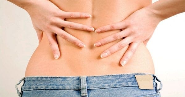
Perhaps you bent the wrong way while lifting something heavy. Or you�re dealing with a degenerative condition like arthritis. Whatever the cause, once you have low back pain, it can be hard to shake. About one in four Americans say they�ve had a recent bout of low back pain. And almost everyone can expect to experience back pain at some point in their lives.
Sometimes, it�s clearly serious: You were injured, or you feel numbness, weakness, or tingling in the legs. Call the doctor, of course. But for routine and mild low back pain, here are a few simple tips to try at home.
Chill It
Ice is best in the first 24 to 48 hours after an injury because it reduces inflammation, says E. Anne Reicherter, PhD, PT, DPT, associate professor of Physical Therapy at the University of Maryland School of Medicine. �Even though the warmth feels good because it helps cover up the pain and it does help relax the muscles, the heat actually inflames the inflammatory processes,� she says. After 48 hours, you can switch to heat if you prefer. Whether you use heat or ice � take it off after about 20 minutes to give your skin a rest. If pain persists, talk with a doctor.
Keep Moving
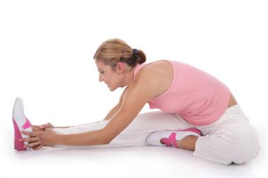
�Our spines are like the rest of our body � they�re meant to move,� says Reicherter. Keep doing your daily activities. Make the beds, go to work, walk the dog. Once you�re feeling better, regular aerobic exercises like swimming, bicycling, and walking can keep you � and your back � more mobile. Just don�t overdo it. There�s no need to run a marathon when your back is sore.
Stay Strong
Once your low back pain has receded, you can help avert future episodes of back pain by working the muscles that support your lower back, including the back extensor muscles. �They help you maintain the proper posture and alignment of your spine,� Reicherter says. Having strong hip, pelvic, and abdominal muscles also gives you more back support. Avoid abdominal crunches, because they can actually put more strain on your back.
Stretch
Don�t sit slumped in your desk chair all day. Get up every 20 minutes or so and stretch the other way. �Because most of us spend a lot of time bending forward in our jobs, it�s important to stand up and stretch backward throughout the day,� Reicherter says. Don�t forget to also stretch your legs. Some people find relief from their back pain by doing a regular stretching routine, like yoga.
Think Ergonomically
Design your workspace so you don�t have to hunch forward to see your computer monitor or reach way out for your mouse. Use a desk chair that supports your lower back and allows you to keep your feet planted firmly on the floor.
Watch Your Posture
Slumping makes it harder for your back to support your weight. Be especially careful of your posture when lifting heavy objects. Never bend over from the waist. Instead, bend and straighten from the knees.
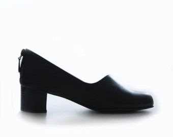
Wear Low Heels
Exchange your four-inch pumps for flats or low heels (less than 1 inch). High heels may create a more unstable posture, and increase pressure on your lower spine.
These could be signs that you have a nerve problem or another underlying medical condition that needs to be treated.
Call Today!
Are Functional Orthotics Part of Your Wellness Protocol?
Most Chiropractors advertise pain relief without drugs and care for injuries. Recently, some doctors and practices have begun labeling and promoting themselves as Wellness Centers. A wellness practice is focused on both maintaining a pre-existing level of musculoskeletal balance and postural health and preventing conditions that might alter this state of health. The challenge is, how can healthy patients be protected from problems that might arise in the future? The answer is simple: custom-made orthotics. Custom orthotics may be traditionally seen as a preventative measure, but so are most treatments of old. They are the perfect, foundational support your patients will never want to go without.
Wellness is a great concept�one of those �win-win� situations for doctor and patient. Orthotics are the perfect way to implement this concept and help establish a �preventative� approach, in addition to the traditional reactive ones, if need be. Let�s take at a look at the foundation of the body, and see just how useful they can be.
Look To The Feet
The feet are the foundation of the body. By age 40, nearly everyone has a foot condition of�some sort, many of which eventually contributing to health concerns farther up the Kinetic Chain (Figure 1). Therefore, it�s in the best interest of healthy patients to be offered a wellness program which stresses preventative care for normal, healthy feet, in order to prevent foot problems from occurring later in life.
�Pictured above, patient with severe bunions, or Hallux Valgus.
Figure 1. While 99% of all feet are normal at birth, 8% develop troubles by the first year of age, 41% at age 5, and 80% by age 20 (Fig. 1).�By age 40, nearly everyone has a foot condition of some sort.
How Can Orthotics Help?
Patients who participate in Chiropractic wellness programs can benefit from custom-made orthotics nearly as much as patients who seek Chiropractic care for musculoskeletal injuries and conditions. Foot Levelers� custom orthotics have been shown to effectively support the pedal foundation for both categories of patients, and can prevent problems well into the future with static and dynamic support.
Static support.Static support. A 1999 study using radiographic measurements found that custom-made, flexible orthotics can significantly improve the alignment of the arches when standing.2 In the wellness-practice concept of orthotic use, custom-made, flexible orthotics can be used to maintain a properly functioning arch alignment.
Dynamic support. During gait, the foot undergoes substantial changes and must permit a smooth transfer of the body�s center of mass over the leg in order to conserve energy and keep the work expenditure to a minimum.3 This requires a flexible, yet supportive orthotic that accommodates varying weights and forces and allows proper movement and function of the foot, while supporting all three arches�in order to prevent eventual arch collapse.
Postural benefits. Since the entire body structure is balanced on one foot at a time when walking and running, improving foot alignment can help maintain knee, hip, pelvic and even spinal postural alignment,4 and prevent joint degeneration (of the hip, knee, or spinal joints). A pelvic or spinal tilt or recurrent subluxations will often respond rapidly to orthotic support of the arches in the feet.
Orthotics For Everyone
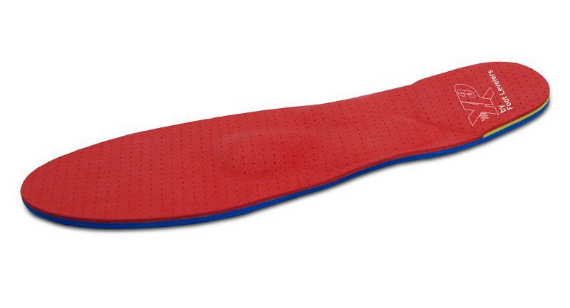
Custom-made, flexible orthotics have long been recognized as a valid adjunct to Chiropractic care for many musculoskeletal conditions. In the wellness model of Chiropractic care, Foot Levelers� custom-made, flexible orthotics (Fig. 2) can be utilized as a preventative modality for the preservation of optimal arch support and the postponement or prevention of joint imbalances in later years. Therefore, orthotics are appropriate for virtually all Chiropractic patients.
Chronic low back pain as well as radiating discomfort down one or both legs could indicate the presence of an injury or condition, such as lumbar stenosis. Spinal stenosis in the lumbar spine commonly develops with age, characterized as the narrowing of the spinal canal in the lower back. When this reduction in the vertebrae occurs, extra pressure is placed on the nerves as well as the spinal cord. Because these nerves run from the lower back to the legs, symptoms of leg pain, heaviness and/or cramping may also develop.
Anatomy of the Spinal Canal
The spinal canal located in the region of the lumbar spine is the most frequent section affected by spinal stenosis. The lumbar spine is made up of five vertebrae extending between the ribcage and pelvis, medically labelled from top to bottom as L1 through L5. Each of these vertebrae are properly separated by intervertebral discs which function as shock absorbers, cushioning and distributing the pressure being exerted onto the spine.
Each vertebrae of the spine contain what is identified as vertebral arches, protruding arch-shaped bones which create the necessary space within the spinal bones for the spinal cord. That space is referred to as the spinal canal. When the structure of the spine is healthy and it functions effectively, the spinal canal should properly be capable of protecting the spinal cord, providing the necessary and safest space required to maintain overall wellness.
Symptoms of Lumbar Spinal Stenosis
Individuals suffering from spinal stenosis in the lumbar spine commonly describe experiencing symptoms of pain and discomfort along the lower back, hips, buttocks and/or legs. Other prevalent symptoms of the condition include: lower back pain that radiated down one or both buttocks, legs, and/or feet; worsening pain in the lower extremities when walking; tingling sensations or numbness in one or both legs or feet; weakness in one or both legs or feet; restricted mobility or difficulty walking; and issues controlling bladder or bowel movements, a complication which may require immediate medical care.
Sciatica, best known as a set of symptoms rather than a single condition or disorder, can be a common diagnosis for determining the presence of an issue affecting the lower spine. Symptoms of sciatica include a collection of pain and discomfort, tingling sensations and numbness, burning sensations, and muscle weakness. Symptoms of sciatica can indicate a serious complication along the lumbar spine.
For individuals experiencing spinal stenosis in the upper back, referred to as cervical stenosis, the symptoms will be similar along the neck, shoulders, arms and/or hands.
Causes of Lumbar Spinal Stenosis
The gradual degeneration of the spine caused by the natural changes that come with age are the most common cause for the narrowing of the spinal canal, mostly due to the repetitive stress and pressure of the surrounding tissues over the course of several years. As the spinal canal becomes narrower over time, a number of conditions and disorders can develop, causing the compression or impingement of the spinal cord and leading to the irritation and inflammation of the nerve roots. This process will ultimately cause symptoms to manifest along the lower back, buttocks, and/or legs.
Lumbar spinal stenosis can also be caused by the degeneration of the intervertebral discs found between each vertebrae of the spine. Spinal disc shrinkage can impede the disc�s ability to properly separate the individual bones of the spine. This problem can generally lead to a much more severe condition referred to as a lumbar disc herniation. Also, if the spinal cord ligaments have expanded due to the natural wear and tear alteration of the structures of the body, lumbar stenosis can develop. Consequently, the degeneration of the vertebrae in the spine is the most common cause for lumbar spinal stenosis.
Diagnosing Lumbar Stenosis
When visiting a healthcare professional, such as a chiropractor, for the diagnosis of lumbar spinal stenosis, the doctor will primarily conduct a thorough physical examination of the patient to determine the source of the issue. The doctor of chiropractic, or other healthcare specialist, may also extensively review the individual�s medical history, referring them to receive other necessary or additional X-rays or MRI scans. By examining the patient�s symptoms as well as analyzing the test results, a chiropractor will be able to diagnose the individual�s injury or condition to discuss the best possible treatment options for you, including the thorough discussion of the benefits and risks of each option. Finally, the healthcare professional and patient can decide together on the preferred treatment procedure to follow to begin the rehabilitation process and restore their original health and wellness.
Treating Lumbar Spinal Stenosis
Chiropractic focuses on the diagnosis, treatment and prevention of injuries and/or conditions of the musculoskeletal and nervous system. A chiropractor may commonly utilize spinal adjustments and manual manipulations to carefully correct any misalignments in the spine that may be causing the impingement or compression of the spinal nerves. The chiropractic adjustments can help decrease the stress and pressure being placed against the structures and other tissues of the spine, reducing the symptoms of pain and discomfort associated with spinal stenosis along the different regions of the spine. Furthermore, the chiropractor may recommend a series of stretches and exercises according to the individual�s complications to speed up the rehabilitation process and help them regain their original strength, flexibility and mobility.
In the case that other forms of treatment are required to treat the individual�s injuries and/or conditions, the healthcare specialist will refer the patient to other professionals for treatment. A modification of the patient�s physical activities may be recommended as well. Other healthcare providers may provide the use of medications and other treatment methods or techniques, including physical therapy, to help improve the symptoms. While many individuals may try a number of conservative treatments to solve the issue, if the individual�s condition is severe enough to require spinal surgery, a healthcare provider may refer the patient to the appropriate doctor for treatment.
For more information, please feel free to ask Dr. Jimenez or contact us at 915-850-0900 .
By Dr. Alex Jimenez
Additional Topics: Low Back Pain After Auto Injury
After being involved in an automobile accident, the sheer force of the impact can cause damage or injury to the body, primarily to the structures surrounding the spine. An auto collision can ultimately affect the bones, muscles, tendons, ligaments and other tissues surrounding the spine, commonly the lumbar region of the spine, causing symptoms such as low back pain. Sciatica is a common set of symptoms after an automobile accident, which may require immediate medical attention to determine its source and follow through with treatment.
Athletes are specially trained to exercise and compete vigorously without experiencing injury or aggravating a previously existing condition. However, accidents and direct trauma during their specific sport or physical activity can inevitably result in damage or injury to the individual. Muscle or tissue damage are common in sports and can be dealt with accordingly but when a bone fracture occurs, these may be more delicate and may require additional diagnosis and care in order to properly help an athlete recover.
Among the general population of athletes, stress fractures can be a rare cause of pain, accounting for only 2 percent of all reported sports injuries. However, a considerably higher number of stress fractures are diagnosed in long distance runners and triathletes.
Stress fractures occurring around the pelvis are significantly uncommon although, a majority of them are often considered a differential diagnosis when athletes, specifically long distance runners and triathletes, report hip, groin or buttock pain during and after running. Because stress fractures around the pelvic/hip region, including the sacral, pubic rami and femoral neck region, are rarely diagnosed, understanding and discussing the anatomy of the injury, their clinical presentation, diagnosis and treatment for each of these types of stress fractures is important for an athlete in order to find a solution for those who do encounter it.
How Stress Fractures Occur
Stress fractures occur over a determined period of time when the bone is no longer capable of withstanding submaximal, repetitive forces. They frequently result when normal stresses cause bone fracture with decreased bone density, such as in an elderly osteoporotic individual, or as a result of an abnormal stress being placed against a normal bone and causing a fracture, such as in a long distance runner.
When the bones are exposed to loading, the introductory physiological response is a respective increase in osteoclastic activity, or bone resorption, leading to temporary structural weakening before new bone formation. If these stresses continue to occur without having the bone properly adjust to this additional, ongoing osteoclastic activity, the pressure may exceed bone regeneration, causing microfractures to occur.
The first characteristic of a stress reaction observed through the use of MRI is bone oedema as well as increased activity on bone scan. Bone scan in the acute phase has high sensitivity but an increased uptake may also be due to infection, bone infarction or neoplastic activity. Researchers from previous studies stated that 60 to 70 percent of X-rays in the acute phase of stress fractures, approximately less than 2 weeks after the injury, have a negative result. Due to its high sensitivity as well as a lack of radiation and high specificity, even despite its elevated cost, MRI is often the preferred procedure to identify stress fractures in their early phases.
Various distinct intrinsic and extrinsic elements have been determined as risk factors for stress fractures. These include but are not limited to: biomechanics, strength and flexibility, nutrition, hormonal and menstrual disturbances, and footwear. These must all be considered prior to assessing an individual with a suspected stress fracture. During an analysis of 8 female athletes with sacral stress fractures, the most significant risk factor for these types of fractures was the rapid increase in impact activity during more intense exercise programs. An increase in workload should thus be considered a significant risk factor for stress fractures.
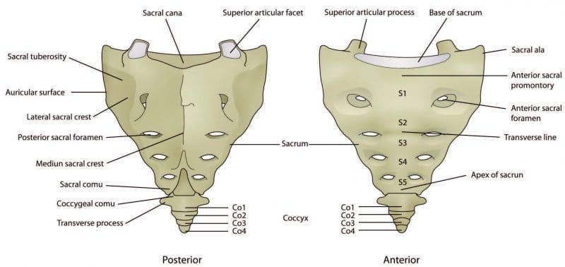
Anatomy of Sacral Stress Fractures
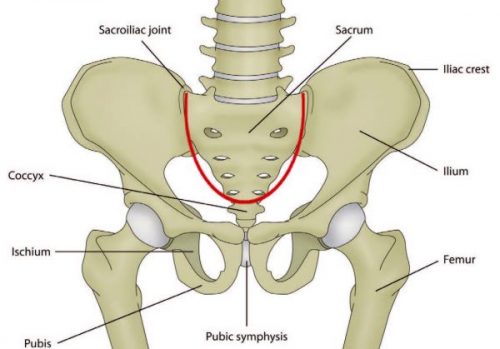
The sacrum consists of 5 fused vertebrae, S1 to S5, and is triangular in shape. It connects with the ilium at the sacroiliac joint and, due to its shape and function to distribute forces, it�s often described as the foundation to the arch of the pelvis. The sacrum, much like an inverted arch, supports the entire weight of the upper body and transfers force to the pelvis.
Sacral stress fractures most commonly occur in the lateral portion of the sacrum and are more frequently diagnosed in women. It�s been hypothesized that the shape of the female pelvis can lead create difficulty when distributing weight through the sacrum than the average male pelvis. However, it�s also been reported that several male elite Australian triathletes have experienced sacral stress fractures in recent years.
Symptoms
An athlete with a sacral stress fracture will often manifest acute onset back, buttock or hip pain which is generally described to occur suddenly during a run, making them incapable of continuing at the time. The individual may also experience limited mobility and they could or could not suffer pain on the palpation of the sacrum. Additionally, they may not experience any neurological symptoms but symptoms of sciatica may be common during this type of stress fracture. Sciatica can include pain, weakness or numbness and burning or tingling sensations along the lower back, buttock or hip, often radiating down the thigh. The individual may suffer pain or tightness when walking and they will experience symptoms when hopping on the affected side. Athletes with sacral stress fractures also frequently report pain during single leg loading tasks, for example, when putting pants on.
Diagnosis
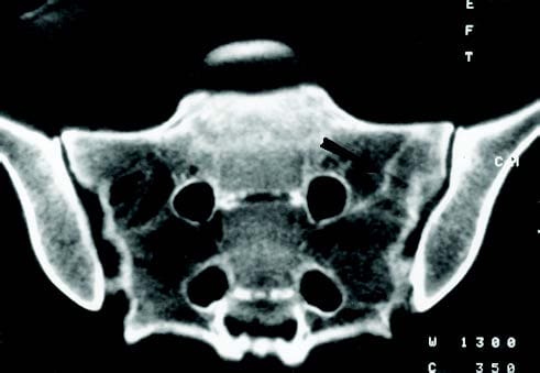
Due to the extreme overlying soft tissue and complex bone anatomy, simple radiographs can rarely conclude the presence of a sacral stress fracture. Bone scan, MRI or CT can be utilized to effectively diagnose a sacral stress fracture. CT and MRI findings suggest that sacral stress fractures occur as a result of constant compressive forces which lead to microfractures of the trabecular bone. These fractures infrequently develop a visible callus on plain radiograph, therefore, MRI or CT scans should be utilized as a follow up imaging if poor healing is detected.
Treatment
The progression of treatment for an athlete with a sacral stress fracture broadly depends on the athlete�s symptoms as these are generally stable fractures. Rehabilitation procedures will progress from non-weight bearing to weight bearing to progressive return to running activities as the symptoms decrease. In most cases, a period of 6 weeks with no running followed by a 6 to 8-week period of a return to running progression may be required. A majority of published works indicate athletes may have a full return to activity by 4 months with rare cases taking up to 14 months.
Repeated CT scans approximately 4 and 8 months after the individual�s original diagnosis can often display no signs of previous fractures which demonstrate a quicker and fuller healing of the well-vascularized trabeculae microfractures when compared to fractures involving the less well-vascularized cancellous bone. Researchers concluded that women with sacral stress fractures who had the best diets and fewer prior stress injuries or menstrual irregularities, healed the fastest.
Anatomy of Pubic Rami Stress Fractures
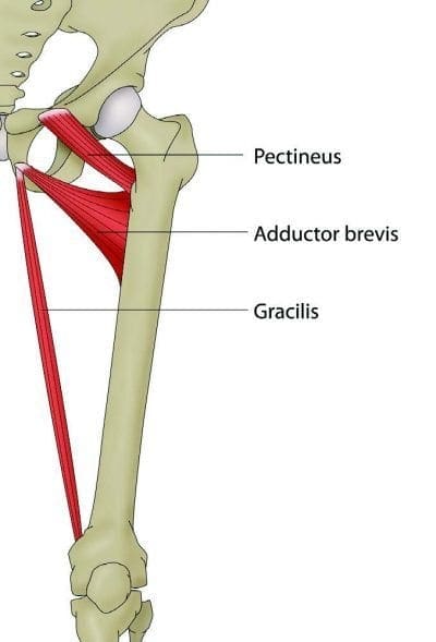
The inferior pubic ramus slopes downward and medial from the superior ramus, narrowing as it goes down and it is the region where the adductor magnus, brevis and gracilis connect, including the obturator internus and externus. Pubic Rami stress fractures have been diagnosed among runners, triathletes and military service members. These generally occur in the inferior pubic rami next to the pubic symphysis. Researchers proposed that these fractures are a result of repetitive forces being applied to and transmitted to the bone through muscle contraction or fatigue. In a study on female military service members, it was suggested that over-striding during marching procedures was a potential factor contributing to pubic rami stress fractures.
Symptoms
Pubic rami stress fractures are generally detected either in competitive races or during intensive training sessions. These frequently occur at the insertion of the adductors and/or external rotators of the hip. Athletes with pubic rami stress fractures commonly suffer from pain in the hip, buttock, inguinal or adductor region which increases with activity and decreases with rest. It�s important to remember that pain caused by irritation and swelling along these regions may also cause symptoms similar to sciatica. It�s important to receive a proper diagnosis to rule out a compression of the sciatic nerve which could be causing neurological symptoms. Athletes with this type of injury often limp and on clinical testing, they may experience symptoms with passive hip abduction, resisted hip adduction and resisted hip external rotation. Stress fractures of the pelvis can be determined even without radiographic evidence if the following criteria are met by an individual. First, running will be impossible for the athlete as a result of severe discomfort in the groin area. Then, the individual will experience discomfort in the groin with an unsupported stance on the affected leg. And last, an athlete may suffer symptoms of pain and tenderness after deep palpation procedures.
Diagnosis
Simple radiographs may demonstrate displaced fracture lines but a lack of radiographic evidence in the early phases of injury is not uncommon. Bone scan, CT or MRI may be used to determine the presence of fracture and bone oedema may be evident on MRI.
Treatment
These fractures tend to have a high rate for healing following 6 to 10 weeks of rest, however, they have a small risk of non-union and re-fracture if the appropriate amount of rest is not followed. Fractures that display delayed union will likely demonstrate full recovery when further conservative procedures are followed. Progression of treatment should be guided by pain and at first, the individual may require the utilization of crutches as walking may be painful.
Anatomy of Femoral Neck Stress Fractures
The femoral neck is the flattened, pyramid shaped piece of bone which connects the femoral head to the femoral shaft.
Athletes with femoral neck stress fractures generally report hip or groin pain when running. This pain usually has an insidious onset and the symptoms may become significantly worse depending on the intensity or duration of a run. At first, symptoms may occur at the end of a run but as the stress reaction worsens, the pain may begin showing earlier in the run where gradually more time may be required to relieve the pain and discomfort. Athletes with femoral neck stress fractures may experience hip and/or groin pain while resting and may suffer restless nights of sleep due to the symptoms. Often, individuals will also report pain while rolling in bed, single leg stance and during active straight leg raise.
Femoral neck stress fractures are described as either tension or compression stress fractures. Fracture displacement determines the outcome of an injury and tension stress fractures generally have a higher rate of displacement as a result of non-union, malunion or osteonecrosis. Due to this fact, tension stress fractures are considered more serious than compression fractures and may require surgical fixation.
Diagnosis
Conventional radiographs are often negative in the acute setting but may shown signs during instances where symptoms have been present for 2 weeks or more. MRI is the favored standard for diagnosis and should be ordered when a stress fracture of the femoral neck is suspected.
Treatment
Tension side stress fractures require diagnosis from a healthcare professional immediately after its occurred due to their risk of displacement. Compression side fractures are often managed conservatively with protected weight bearing and ongoing monitoring to keep track of the individual�s healing process. Initial phases of management should include non-weight bearing on crutches until there are no symptoms at rest, then progress to partial weight bearing to full weight bearing over a period of 4-6 weeks. A gradual return to run program can be started at 8 to 12-weeks of treatment, once the individual is able to properly walk without experiencing pain and other symptoms.
Return to Activity Plan
With all stress fractures located around the pelvis, a careful, gradual return to activity plan can be an essential element of the rehabilitation process. To make sure the athlete receives progressive loading without sudden increases in workload, the return to activity plan should be at least as long as the time off the individual�s specific activity. For instance, if the athlete had a sacral stress fracture which required 6 weeks of no running, then that athlete needs at least a 6-week gradual return to running plan before they can return to their previous running load.
Hip Strengthening Exercises
A strengthening program of the lower extremities can additionally be implemented early in the rehabilitation process, first beginning with non-weight bearing exercises, which can gradually change as the individual becomes able to weight bear without pain. Early strengthening can also help decrease muscle loss and address any biomechanical complications the athlete might be facing. As the stress fractures heal and the tolerance for load improves, these exercises can be progressed to other higher-load exercises to provide the athlete�s body for the return of their specific sports activity.
Proper stretching and exercising techniques are effective methods and techniques that can help increase an athlete’s strength, mobility and flexibility to prevent experiencing an injury or aggravating a condition. Bone fractures, in this case, pelvic stress fractures, can be challenging to heal but with proper treatment, an athlete will be able to return-to-play in no time.
For more information, please feel free to ask Dr. Jimenez or contact us at 915-850-0900 .
By Dr. Alex Jimenez
Additional Topics: Low Back Pain After Auto Injury
After being involved in an automobile accident, the sheer force of the impact can cause damage or injury to the body, primarily to the structures surrounding the spine. An auto collision can ultimately affect the bones, muscles, tendons, ligaments and other tissues surrounding the spine, commonly the lumbar region of the spine, causing symptoms such as low back pain. Sciatica is a common set of symptoms after an automobile accident, which may require immediate medical attention to determine its source and follow through with treatment.
Low back pain is one of the most prevalent symptoms that lead people to seek diagnosis and treatment with a healthcare professional. When the individual�s low back pain is accompanied with pain in one or both legs or buttocks, resulting in symptoms similar to sciatica, it may be an indicator that the patient may have a lumbar disc herniation, also referred to as a herniated disc, ruptured disc, or slipped disc.
Anatomy of the Lumbar Spine
The lumbar spine consists of five vertebrae that extend through the length of the ribcage and pelvis. From top to bottom, these vertebrae are medically labeled L1 through L5 and they�re each separated by intervertebral discs. The discs are made up of a fibrous tissue known as the annulus with a soft nucleus found at the center of each disc. These discs are fundamental towards the proper function of the spine, performing the important roles of shock absorption and distribution of pressure.
In the instance the annulus becomes ruptured or torn, the nucleus can become separated from the disc. This complication can decrease the disc�s ability to properly separate the vertebrae, an issue which often leads to increased pressure due to the compression or impingement of the spinal nerves found between each vertebrae of the spine. Individuals with a lumbar disc herniation and symptoms of sciatica commonly experience pain and discomfort related to the pinching of the nerves, which can in turn radiate down the legs.
Generally, a herniated disc is caused by the natural degeneration of the body�s structures as we age. If not diagnosed or treated in time, however, this simple wear and tear complication can develop into a more serious injury or condition. In addition, intervertebral discs can also tear due to trauma from heavy lifting or as a result of a sudden injury, such as an automobile accident or a work injury.
Diagnosing a Lumbar Disc Herniation
A chiropractor can properly diagnose a variety of injuries or conditions relating to the musculoskeletal and nervous system, including a lumbar disc herniation. During the first consultation, the chiropractor will conduct a thorough physical exam, including a comprehensive review of your medical history and test results. Using this, the healthcare professional will be able to determine the source of the symptoms. In many cases, the specialist may require additional tests to confirm the presence of a specific injury and/or condition. Most chiropractic offices will provide you with up to date information about your diagnosis, as well as the risks and benefits of each treatment option. Chiropractors will work with the individual personally to decide on the best treatment option for their complication.
Treating a Lumbar Disc Herniation
Chiropractic adjustments and manual manipulations are the most common forms of treatment provided by a doctor of chiropractic, or DC. Using this gentle techniques, the healthcare specialist will carefully realign the spine, correcting the subluxations in order to decrease and eliminate the symptoms caused by nerve compression or impingement. Chiropractors may also redirect a patient to receive other types of treatment depending on the severity of their issue. Chiropractic care can help restore an individual�s strength, mobility and flexibility, offering a wide variety of benefits. Chiropractic treatment is well-known for its natural benefits, including the enhancement of many functions of the body.
Chiropractic Can Improve Sex Life
Many people visit the chiropractor with back pain, but after several sessions of treatment, they often return reporting that their sex life has improved. Jason Helfrich, co-founder and CEO of 100% Chiropractic, stated that the body can positively respond in many aspects when the unnecessary pressure on the nervous system is decreased or removed.
Every function of the body is controlled by the nervous system, however, when the spine is misaligned, known as a subluxation, the nerves traveling between the brain and the rest of the body, these can become blocked, compromising the body�s ability to function properly. A chiropractor�s goal is to remove these subluxations, since they can both cause pain and impede feeling. But treatment can help more than just improve symptoms of back pain. The lumbar region of the spine is where the nerves that extend into your reproductive regions are found. Correcting misalignments in the lower spine can improve nerve flow to your sexual organs, increasing things like blood flow to your clitoris or the penis.
�Correcting a spinal subluxation also allows the organs to send messages to the brain more easily. This means that not only do you become physically aroused faster, but your brain also registers that ready-for-action, heightened sense of pleasure more quickly, so you move past the mental obstacles that may be keeping you from orgasming�, quoted Helfrich.
Other Adjustments for an Improved Sex Life
Libido and fertility need a proper balance of estrogen, progesterone, and other hormones, many of which are released in the upper cervical and neck area of the body. If there are any misalignments or subluxations in the upper region of the spine, the nerve transmissions exiting the brain can be interrupted due to the compression or impingement of these tissues, which will ultimately have an effect all the way down to the reproductive organs, among others.
Including fertility is affected by the nerves and hormones coming out of the spine, as they control the reproductive cycle.
Beyond all of the physiological benefits of spinal adjustments and manual manipulations, chiropractic treatment can also simply give the muscles more range of motion. This means you can try previously difficult positions under the sheets, enhancing an individual�s sex life further.
�We want to improve people�s health, and health is about living life as its intended. Having a great sex life is huge part of that�, Jason Helfrich concluded.
For more information, please feel free to ask Dr. Jimenez or contact us at 915-850-0900 .
By Dr. Alex Jimenez
Additional Topics: Low Back Pain After Auto Injury
After being involved in an automobile accident, the sheer force of the impact can cause damage or injury to the body, primarily to the structures surrounding the spine. An auto collision can ultimately affect the bones, muscles, tendons, ligaments and other tissues surrounding the spine, commonly the lumbar region of the spine, causing symptoms such as low back pain. Sciatica is a common set of symptoms after an automobile accident, which may require immediate medical attention to determine its source and follow through with treatment.
The spine is a complex structure that consists of bone, muscle, tendons, ligaments, nerves and other tissues. Spine fitness is essential towards maintaining the body�s optimal performance as it functions to support an upright posture, provides flexibility to allow the body to bend and twist freely, and protect the spinal cord. While a healthy back can easily be taken for granted, a�back injury�or other type of complication may result in long-lasting consequences that could alter an individual�s aspect of daily life.
Unfortunately,�back injuries�are among the most common types of complications suffered after an automobile accident. Each day, thousands of individuals are involved in head-on, side-impact, and rear-end auto collisions, often leading to spinal injuries even during minor car crashes. Depending on the force of the impact, a single or multiple areas of the back may be affected. Automobile injuries can range from mild sprains and bruises to fractured vertebrae and spinal cord damage.
If you�ve had a car accident, it�s essential to take note of any symptoms of�back pain, as there are a variety of spine complications which could result after experiencing an auto collision.
Central Sensitization & Auto Injuries
Recovering From Auto Injuries
Disc Herniation
One type of spine injury among individuals who have suffered from an auto accident is a�herniated disc, or a slipped/ruptured disc. Discs are small, sponge-like structures found within the spinal column which function as cushions to separate and protect the vertebrae from the others while providing the spine with smooth flexibility. The force from an auto accident impact can damage a disc, causing it to break or deform, affecting its ability to cushion the bones of the spine.
Furthermore, a damaged disc can also place unnecessary pressure directly on the nerves surrounding it, leading to symptoms of pain, numbness and weakness on the region of the body where the affected nerve travels to.
Disc injuries to the�lower back�frequently lead to a group of symptoms commonly referred to as sciatica, which is characterized by radiating pain, numbness and tingling sensations in the leg and/or buttock on either side, or occasionally, in both sides, depending on the type of injury. The symptoms associated with sciatica can be impairing and may worsen over time if left untreated.
Auto Injury Back Pain
Spinal Cord Distress
Spinal cord injuries occur from the impact of an automobile accident. It sends a direct blow to the spine, that damages the delicate bundle of nerves within it. The spinal cord is the most important structure between the body and the brain. It is a vital link between the brain and the body, functioning to carry essential information back-and-forth from the brain to the central nervous system, facilitating motor control and sensory function. Spinal cord injuries impair the brain�s ability to communicate effectively with the rest of the body, resulting in paralysis and/or lack of sensation in all or part of the body. The more severe an injury to the spinal cord is, the more of the body will be affected.
Compression Fracture aka Bone Break
Car accidents can also cause compression fractures. Compression fractures or cracks in the bones of the spine may cause the vertebrae to collapse and deform. This can permanently alter the shape and structure of the spine.
Symptoms of a compression fracture include pain and postural changes as well as breathing difficulties. Due to the fact that compression fractures are common among older adults, many individuals mistake the symptoms for�signs of aging�or arthritis. An estimated two-thirds of compression fractures go undiagnosed.
While cars are built to withstand the great force of a collision, the human body is not. The complex structures of the body, especially the spine, are vulnerable even in low speed collisions, resulting in injuries or conditions more often than not. An individual who�s been injured in an accident should seek immediate medical attention in order to diagnose any possible injuries or conditions as early as possible to begin treatment.�Chiropractic care�can effectively treat a variety of spinal complications, including auto injuries. Through the use of spinal adjustments and manual manipulations, a chiropractor can gradually help restore the individual�s natural mobility and flexibility, as well as progressively strengthen the structures surrounding the spine to relieve the symptoms and restore the individual�s lifestyle.
After being involved in an automobile accident, injuries inflicted to the spine can be a common complication for many individuals. From herniated discs to compression fractions, the force of an auto collision can place great amounts of stress on the complex structures of the spine, often leading to damage, injuries and even aggravate an existing condition.
For more information, please feel free to ask Dr. Jimenez or contact us at 915-850-0900 .
The Clinic & Crossfit:
A Clients Story
Jacqulyn Quevas is up on her feet a lot as a hair stylist and she was�searching for�and overall become healthier and once she found Push-as-Rx ��, her amazing journey began. The motivation and enthusiasm of the trainers at Push as Rx has greatly influenced Jacqulyn.
The clinic and PUSH-as-Rx �� system is leading the field with laser focus supporting our youth sport programs.� The PUSH-as-Rx �� System is a sport specific athletic�program�of reactive agility, body mechanics and extreme motion dynamics. Through detailed and continued assessments of the athletes in motion and under stress loads offer a clear scientific picture of body dynamics. This system also has helped many athletes come back from injury faster, stronger, and ready to safely return to their sport without losing a beat after recovery. Results demonstrate clear improved agility, speed, decreased reaction time and advantageous postural-torque mechanics.� PUSH-as-Rx �� offers specialized extreme performance enhancements to our athletes no matter the age.
If you�ve had a car accident, it�s essential to take note of any symptoms of�back pain, as there are a variety of spine complications which could result after experiencing an auto collision.
Low back pain is a common symptom reported among the majority of the population. Generally caused by direct trauma from an injury or as a result of an aggravated condition, low back pain can greatly restrict an individual’s daily activities, affecting their overall physical as well as mental performance. Although there’s a variety of factors which can lead to back pain along the lumbar area of the spine, an incorrect sitting posture over an extended period of time has been known to be one of the leading causes for this well-known symptom.
Sitting is one of the most difficult positions for the body to maintain. Improper sitting postures�for prolonged periods of time while hunched over in a chair can build excessive pressure on the spine and affect the blood circulation in the legs. For an individual that spends their workdays sitting down, the long term result of an improper posture can greatly alter the individual�s overall health.
Proper Posture While Sitting
Sitting for extended periods of time while hunching over a desk can cause discomfort, numbness and spine misalignment over time if the improper sitting posture is not corrected. Holding the body upright can also further increase pressure and tension in muscles, ligaments, joints, and other tissues surrounding the spine. A sedentary lifestyle where the individual doesn�t take frequent breaks throughout their workday to stand and stretch can gradually lead to cardiovascular disease due to the decreased blood flow, tightened hip flexors, shortened hamstrings, pinched nerves and many other physical injuries and/or conditions in the long run.
How Chiropractic Differs from Traditional Care
Aches and pains on the body, particularly around the spine, herniated discs, nerve complications and painful joints are direct results of working while in improper sitting postures for prolonged hours of the day. When you’re in a seated position, a lot of pressure is being placed directly on the spine and its surrounding structures. Our bodies were designed to stand upright and maintaining a seated position can be physically stressful.
“The weight is distributed while in a standing position,” explained Kelly McGonigal, Ph.D., a health psychologist at Stanford University and a leading expert in neck and back pain, �That’s not the case with sitting. McGonigal added, “When you sit, you distort the natural curve of the spine, which means your back muscles have to do something to hold your back in shape because you’re no longer using the natural curves of the spine to lift yourself up against gravity.”
Approximately 80 percent of individuals in the United States alone may experience some form of chronic pain in their lifetime. Ultimately, working on a sitting position for extended periods of time per day is putting a tremendous mental and physical stress on our bodies.
Correcting Posture
Maintaining good posture when sitting helps preserve the three natural curves of a healthy spine; the cervical curvature, the thoracic curvature, and the lumbar curvature. The normal curvatures of the spine should form a slight S-shape. An excessive curve in the spine could suggest a possible underlying condition and may present symptoms of pain and discomfort, among other uncomfortable symptoms.
The key to maintaining a proper posture is to avoid slouching by sitting up straight as well as sitting all the way back in your chair. Additionally, the chair should be tucked in close to the desk. Placing a pillow or cushion directly behind your lower back area, or the lumbar spine, can help sustain good posture by providing enough support and allowing the spine to naturally curve inward. Furthermore, it�s also recommended to stand and stretch about every 20 minutes to give your body a break from long held positions, release pressure that is being built up on the spine and restore the body�s regular circulation by pushing the blood out of your legs. Doing this consistently throughout your day can prevent the muscles, ligaments and other tissues of the body from getting strained. Simple stretches at your desk, such as twisting, turning the head from side-to-side and chin tucks upward towards the ceiling can also help. These movements will ultimately help to relieve an individual�s chronic pain as well as restore an individual�s health progressively over time and improve their overall well-being.
Sitting is one of the most difficult positions for the body to maintain. Sitting for prolonged periods of time while hunched over in a chair can build excessive pressure on the spine and affect the blood circulation in the legs. For an individual that spends their workdays sitting down, the long term result of an improper posture can greatly alter the individual�s overall health.
While for many individuals sitting behind a desk for a prolonged period of time each day is an inevitable and often necessary part of their lifestyle, practicing a proper posture as well as seeking the proper care once the symptoms of low back pain manifest can change the effect of the issue. Chiropractic care as well as physical therapy and other forms of treatment, are available to the public to help reduce their painful symptoms and restore their original functionality. The spine is one of the body’s most important structures and it’s essential to maintain its health to achieve overall wellness.
For more information, please feel free to ask Dr. Jimenez or contact us at 915-850-0900 .
Women often seek out chiropractic care during their pregnancies. One of the main reasons for this is that if they are having musculoskeletal pain, their Obstetrician or Midwife generally has very limited options for conventional medical treatment. They are less likely to prescribe medication, which presents an amazing opportunity for us as chiropractors. What initially began as a strange combination of a sports certification and a prenatal/pediatric certification has created a successful and in-demand niche for us. For the most part, I cannot imagine treating prenatal patients without having the sports background. First, most women at some point in their lives have been or currently are an athlete. And second, one of the most common complaints women have during pregnancy is myofascial pain, and who better to treat that than a sports chiropractor? Here are 5 common and easy-to-treat complaints during pregnancy:
One: Pubic Bone Pain
This pain is very common during the second and third trimesters of pregnancy. Women will often describe it as exquisitely tender, worse with rolling over in bed at night, walking or climbing stairs. Conventional medical opinion is that the ligaments during pregnancy are much more relaxed (due to the hormone, relaxin) causing separation of the pubic symphysis and thus inflammation and pain. Sports chiropractors are uniquely qualified to assess this joint, and the common causes of pain can include:
1. Adductor hypertonicity easily addressed with myofascial release or techniques such as Graston, which can be done over leggings or on skin.
2. SI Joint fixation or Pubic Symphysis fixation. While I am generally a diversified adjuster, an activator adjustment to the superior or more-tender pubic rami will go a long way in terms of providing relief. I strongly discourage any audible manipulation of the pubic symphysis.
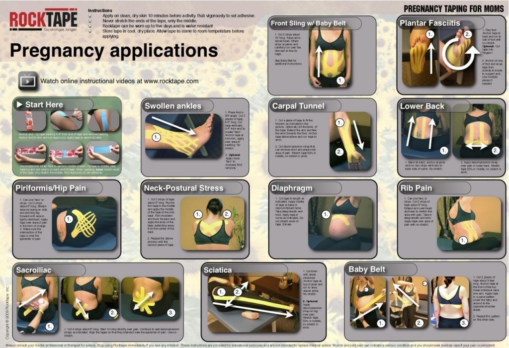
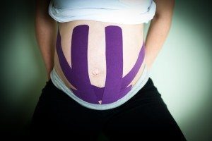
3.�Kinesiology taping�of a �RockTape Baby Belt� or modified version of this can provide significant relief and is much more comfortable than a pelvic support belt.
4. Using an ice pack for 15 minutes prior to going to bed at night will decrease pain and inflammation while sleeping.
Two: Rib Pain
Rib pain, especially in the lower and floating ribs, is common as the weight of the abdomen is pulling on the oblique abdominal muscles and their attachments at the ribs. Adjusting the ribs at the thoracic spine and incorporating myofascial release or Graston Technique will work quickly (often in just 1 treatment). Finish up with a few strips of kinesiology tape and your patient will feel significantly better.
Three: Upper Abdominal Numbness
Upper abdominal numbness is a common symptom during the later stage of pregnancy. It often presents as numbness but can also be painful and worse with sitting. One of the easiest ways to provide relief is with one simple strip of kinesiology tape over the top of the abdomen directly under the rib cage.
Four: Swelling in the arms and legs
Swelling in the arms and legs is very common and can lead to numbness, tingling or pain. Before beginning treatment, be sure to assess if the swelling in the feet is significant and test for pitting edema which can be a warning sign of preeclampsia. This can be corroborated with a high blood pressure reading and is very dangerous. Two very effective sports techniques for use with lower extremity swelling include 1.�NormaTec PULSE Recovery System�which is not contraindicated in pregnancy. Patients can do a few 20-30 minutes sessions per week to promote circulation and decrease swelling. 2. Kinesiology taping for edema on the ankles.
Five: Lower Back Pain
Lower back pain in pregnancy is very common. Evaluating a pregnant patient prone is very easy if you have pregnancy cushion that sits on top of your table. If you do not, you can evaluate the lower back in the seated or side lying position. Lower back pain can generally be addressed with diversified adjustments (without any rotation as to not stress the abdomen). In addition, the Webster Technique for pregnancy is a valuable tool for assessing and treating lower back pain during all stages of pregnancy. There are also valuable kinesiology tape applications for lower back pain,�RockTape features a pregnancy taping pdf online. In addition, there are no contraindications to using the Graston Technique to address myofascial pain in the lower back.�
Most of the taping techniques discussed above can be done by patients themselves after a one-time demonstration. Have an assistant or patient�s family member take a video of the application for reference at home. Many sports chiropractic techniques can be used very effectively on prenatal patients.
IFM's Find A Practitioner tool is the largest referral network in Functional Medicine, created to help patients locate Functional Medicine practitioners anywhere in the world. IFM Certified Practitioners are listed first in the search results, given their extensive education in Functional Medicine


 Static support.Static support. A 1999 study using radiographic measurements found that custom-made, flexible orthotics can significantly improve the alignment of the arches when standing.2 In the wellness-practice concept of orthotic use, custom-made, flexible orthotics can be used to maintain a properly functioning arch alignment.
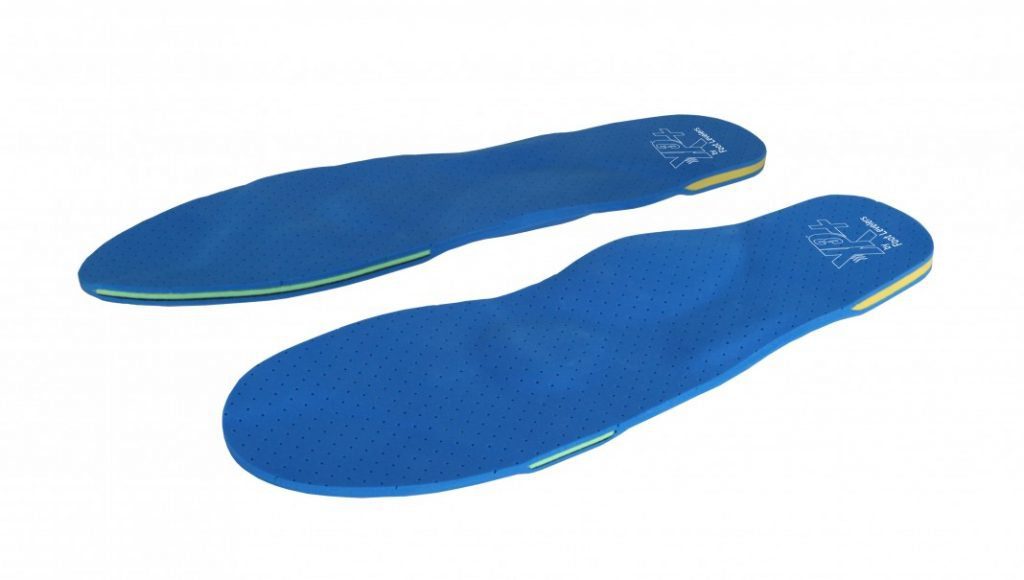

 Shoes for Kids