Restore Flexibility and Mobility with Integrative Chiropractic Care and Shockwave Therapy at El Paso Back Clinic
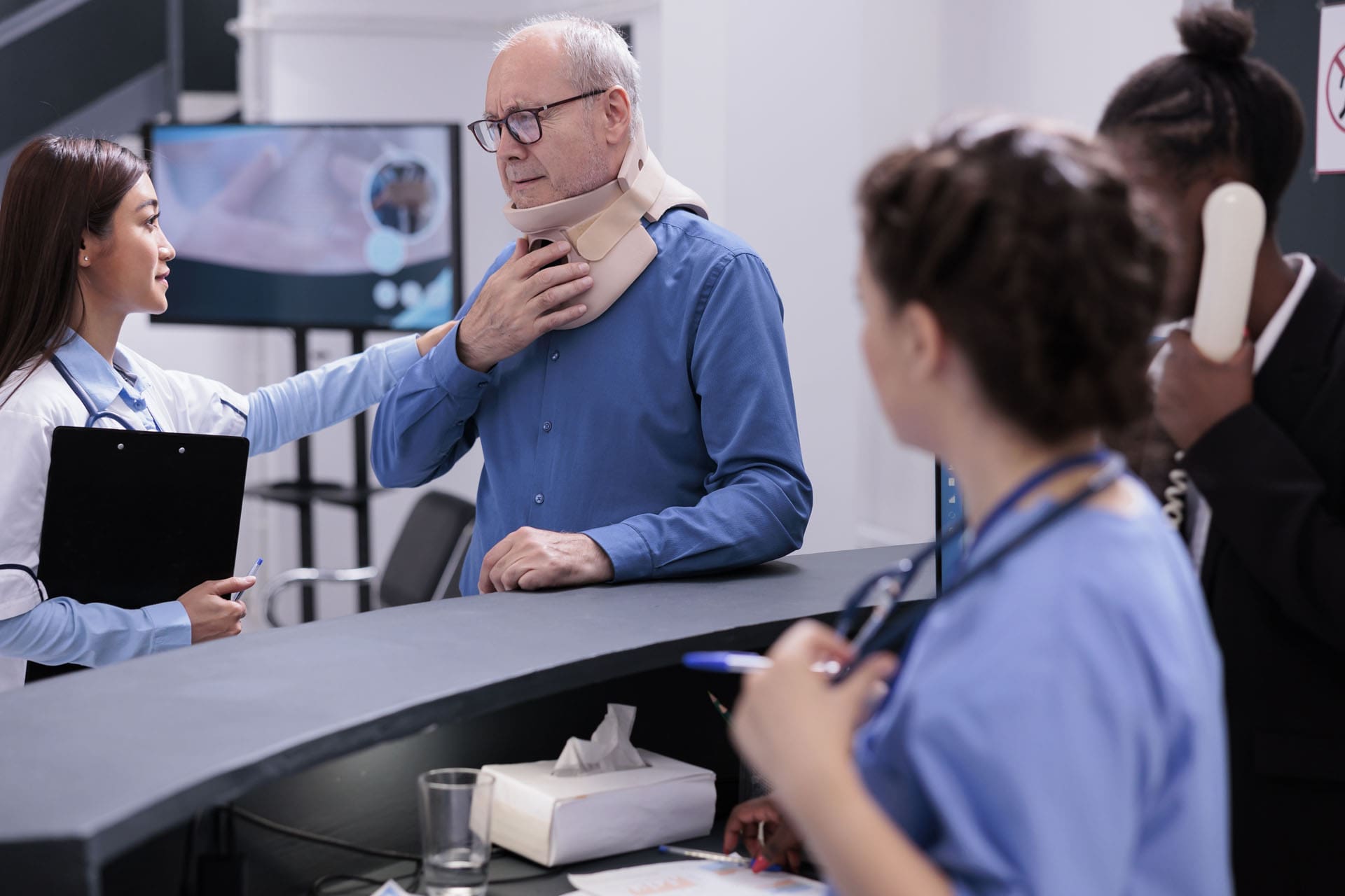
Many El Paso residents wake up with stiff joints or tight muscles, making simple daily tasks feel hard. Reaching overhead, bending down, or walking for long stretches can become painful or limited. At El Paso Back Clinic, integrative chiropractic care combined with Extracorporeal Shockwave Therapy (ESWT) offers a natural solution. This approach restores proper joint alignment, reduces muscle tension, and resolves soft-tissue restrictions, allowing patients to move freely again. Led by Dr. Alexander Jimenez, DC, APRN, FNP-BC, the clinic’s team uses gentle adjustments, stretching, exercises, and advanced shockwave treatments to help people regain flexibility and enjoy life in El Paso.
What Integrative Chiropractic Care Does for Flexibility at El Paso Back Clinic
Integrative chiropractic care at El Paso Back Clinic treats the whole body instead of just one problem area. It corrects small misalignments, called subluxations, in the spine and joints. These misalignments put pressure on nerves and tighten muscles. Regular adjustments gently move everything back into place. This restores proper joint alignment, eases tension, and lets the nervous system send clearer signals to the muscles.
When joints line up correctly, range of motion improves right away. Stiffness fades, and daily movements become smoother and more efficient. Patients at the clinic often say they feel looser and more energetic after just a few visits. (Gentle Chiro, n.d.) The care also includes stretching and therapeutic exercises to maintain gains over time. Muscles and joints start working together as a team, building resilience that lasts.
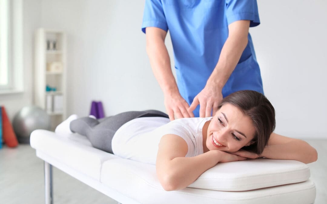
How Chiropractic Adjustments Restore Joint Alignment and Reduce Stiffness
Adjustments form the core of care at El Paso Back Clinic. The team uses precise, gentle pressure to correct subluxations. This simple step brings clear benefits that patients notice quickly:
Better range of motion, so joints glide freely without catching
Less muscle tension around the back, neck, and limbs
Improved nervous system function for better balance and coordination
Smoother daily activities like turning your head while driving or reaching for groceries
Lower risk of future stiffness because proper alignment trains the body to stay balanced
Many people in El Paso report that these changes make physical activities feel easier and less tiring. (Rodgers Stein Chiropractic, n.d.) The adjustments help the body move more efficiently without pain, supporting an active lifestyle.
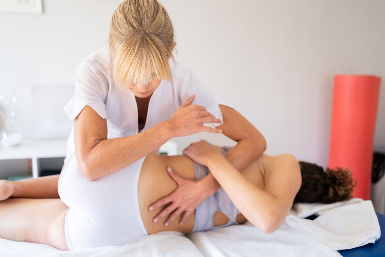
Adding Stretching and Therapeutic Exercises for Long-Term Results
Adjustments open the door to better movement, but stretching and exercises keep it open. At El Paso Back Clinic, the rehabilitation team creates simple home programs that match each patient’s needs. Dynamic stretches warm up the body before activity. Static stretches hold the new mobility after adjustments. Therapeutic exercises strengthen the muscles that support the joints.
These steps build endurance and agility. Patients find they can stay active longer without soreness. The clinic’s sports medicine approach helps people return to hiking in the Franklin Mountains, playing with family, or working without the same old limitations. (Chiropractic Fitness, n.d.) Consistent practice turns short-term gains into lasting flexibility.
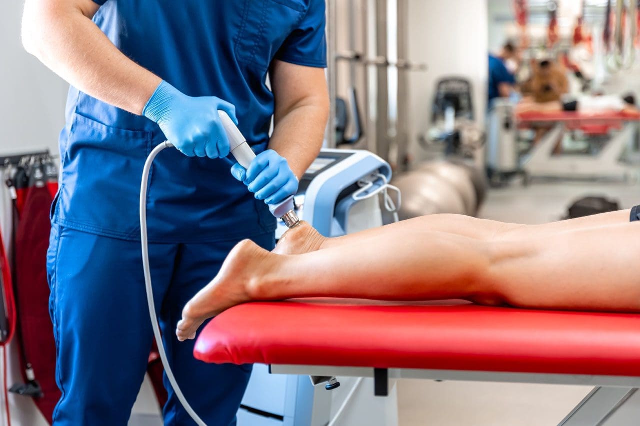
Introducing Extracorporeal Shockwave Therapy (ESWT) at El Paso Back Clinic
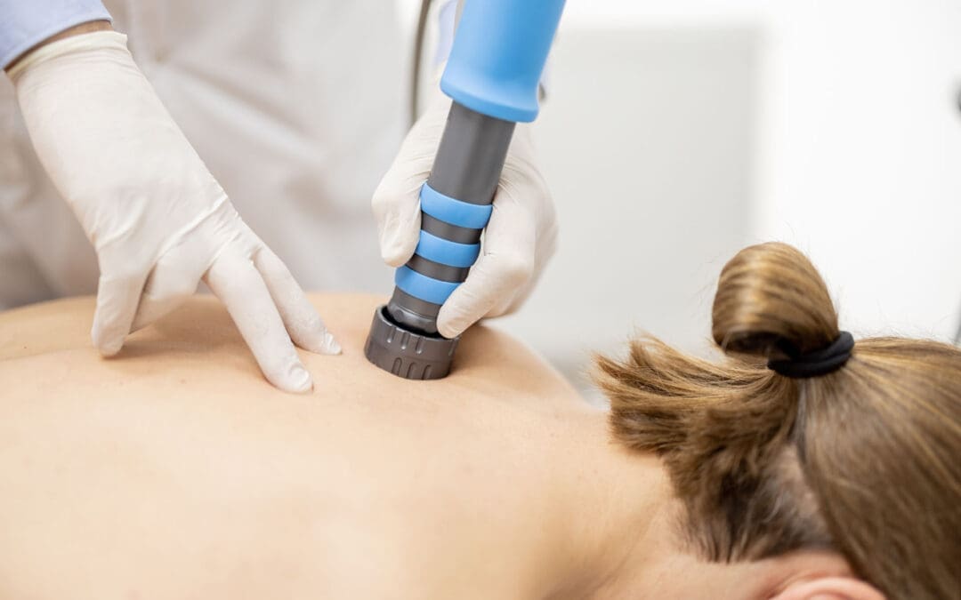
ESWT uses focused sound waves to reach deep into muscles, tendons, and ligaments. The waves create tiny pulses that restart healing in areas stuck with scar tissue or chronic tightness. This noninvasive treatment increases blood flow, breaks down old buildup, and reduces inflammation. At El Paso Back Clinic, ESWT is available as a key component of advanced care plans for patients who need additional support for soft tissue problems.
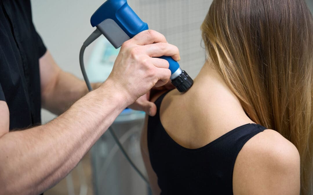
Why Combining Chiropractic Care and ESWT Delivers Stronger Flexibility Gains
The real power at El Paso Back Clinic comes from pairing chiropractic adjustments with ESWT. Adjustments fix the mechanical side—joint position and nerve signals—while ESWT handles the soft-tissue side—scar tissue, poor circulation, and stubborn tension. Together, they create faster, longer-lasting results than either method alone.
This dual approach works in several key ways:
Chiropractic restores spinal and joint mobility
ESWT breaks down scar tissue and releases tight fascia
The pair reduces inflammation and collagen cross-linking that causes stiffness
Blood flow improves, helping muscles and tendons heal
Patients regain a greater range of motion because both structure and tissue health get better at once
Clinic reports show that this combination can significantly improve outcomes compared with standard care. Many El Paso patients with ongoing tightness notice a real return of freedom of movement.
Common Conditions That Benefit from This Integrated Approach
El Paso Back Clinic uses this combined approach to treat several conditions that rob people of flexibility. Here are some of the most common:
Frozen shoulder – Adjustments free stuck joints while ESWT dissolves scar tissue and calcium deposits. Patients often regain full arm motion without pain.
Achilles tendinopathy – Chiropractic realigns the lower body to ease strain. Shockwave therapy stimulates the growth of new blood vessels and clears chronic buildup, so walking and running feel normal again.
General chronic muscle tension – Tightness in the back, neck, or legs from stress, work, or old injuries—responds well. The therapies release trigger points and restore smooth movement.
Post-injury stiffness from car accidents or sports – The clinic specializes in personal injury care. The combination speeds recovery and safely rebuilds mobility.
Other issues, such as plantar fasciitis and tennis elbow, also improve because the care addresses both alignment and tissue damage. (Bend Total Body Chiropractic, n.d.)
Clinical Insights from Dr. Alexander Jimenez at El Paso Back Clinic
Dr. Alexander Jimenez, DC, APRN, FNP-BC, leads El Paso Back Clinic with more than 30 years of experience. As both a Doctor of Chiropractic and a board-certified Family Nurse Practitioner, he brings a unique integrative perspective to every patient. In his clinical work in El Paso, Dr. Jimenez sees how chiropractic adjustments correct subluxations and improve nervous system function, thereby boosting flexibility and range of motion. When combined with ESWT, the results are even stronger for soft tissue injuries from accidents or overuse.
Dr. Jimenez often notes that this teamwork helps patients break down scar tissue, reduce inflammation, and restore proper movement patterns faster than traditional methods alone. His approach includes personalized functional medicine, nutritional support, and rehabilitation exercises to help patients build lasting resilience. At the clinic’s convenient El Paso locations, patients receive complete care that addresses the root causes of stiffness and helps them return to daily life and favorite activities with confidence.
Tips to Get the Most from Care at El Paso Back Clinic
Start with a full evaluation so the team can build a plan that fits your body and lifestyle. Attend regular adjustments and ESWT sessions as recommended. Follow the simple stretching and exercise routine at home every day. Support your progress with good posture, daily walks, proper hydration, and enough rest. The friendly staff at El Paso Back Clinic makes the process easy and supportive. Many patients see big improvements in flexibility within just a few weeks when they stay consistent.
A Natural Path to a More Flexible, Resilient Life in El Paso
Integrative chiropractic care and ESWT at El Paso Back Clinic offer a powerful, drug-free way to fight stiffness and reclaim natural movement. By correcting joint alignment, releasing muscle tension, and healing soft tissues, this approach makes daily life and physical activity feel effortless again. Muscles and joints work in harmony, the nervous system functions smoothly, and the body stays strong through the years.
Whether you deal with occasional tightness or a specific injury, the experienced team at El Paso Back Clinic can help. Contact the clinic today to schedule an evaluation and discover how these natural tools can work for you. With the right plan, better flexibility and mobility are well within reach for El Paso residents.
Sciatic Nerve Health and Sciatica Relief: An Integrative Chiropractic Approach at El Paso Back Clinic
The sciatic nerve should work like a clear, pain-free communication line between the lower spine and the lower body. When it is healthy, it carries nerve signals smoothly from the lower back through the hips, buttocks, legs, and feet. This allows comfortable walking, bending, standing, climbing, and turning. It also helps the body perceive touch, pressure, and position in the lower leg and foot. In simple terms, optimal sciatic nerve function means you can move well, feel normal sensation, and stay steady on your feet without burning, tingling, weakness, or pain traveling down the leg (Cleveland Clinic, 2026; Health.com, 2024; MedlinePlus, 2024).
The sciatic nerve is the longest and widest single nerve in the body. It is formed from spinal nerve roots L4 through S3 and travels from the lower spine through the pelvis, under the buttock area, down the back of the thigh, and toward the lower leg and foot. Because it is so long, irritation in the lower back, pelvis, or deep hip area can create symptoms that run down the leg. That is why sciatica often feels like more than just back pain. It can affect movement, balance, comfort, and daily function from the low back all the way to the foot (TeachMeAnatomy, 2025; Cleveland Clinic, 2026).
Why the Sciatic Nerve Matters So Much
The sciatic nerve has both motor and sensory jobs. On the motor side, it helps control the hamstrings and, through its branches, many muscles in the lower leg and foot. That means it plays a major role in bending the knee, moving the ankle, controlling the foot, and helping the body walk with stability. On the sensory side, it helps carry feeling from much of the lower leg and foot. Without normal sciatic nerve function, movement may feel weak or awkward, and sensation may feel dull, numb, sharp, or irritated (TeachMeAnatomy, 2025; NCBI Bookshelf, 2023).
When the sciatic nerve is functioning well, people often do not think about it at all. That is actually a positive sign. The nerve is quietly doing its job, helping the lower body move smoothly and respond to its environment.
Healthy sciatic nerve function supports:
Comfortable walking and standing
Smooth bending and lifting
Stable balance and coordination
Normal sensation in the lower leg and foot
A fuller, less painful range of motion
Better confidence in everyday movement
When any part of that nerve pathway becomes irritated, compressed, or inflamed, the result may be sciatica. Sciatica is not a separate disease by itself. It is a symptom pattern that usually happens when the sciatic nerve or the nerve roots that form it become irritated (Cleveland Clinic, 2026; Mayo Clinic, 2025).
What Can Interfere With Sciatic Nerve Function?
The sciatic nerve works best when signals can move freely without obstruction. Problems begin when pressure, inflammation, or mechanical strain affects the nerve roots or the nerve itself. One of the most common reasons is a herniated lumbar disc. Other causes include spinal stenosis, bone spurs, spondylolisthesis, muscle imbalance, piriformis syndrome, postural strain, and movement patterns that keep irritating the nerve (Mayo Clinic, 2025; MedlinePlus, 2024; Health.com, 2024).
People with sciatica may notice:
Sharp, shooting, or burning pain down one leg
Tingling or “pins and needles”
Numbness in part of the leg or foot
Weakness when walking or climbing stairs
Pain that worsens with long sitting
Tightness or pulling in the buttocks and thighs
Trouble standing up straight or moving normally
Sciatica can range from mild to severe. Some people feel a dull ache. Others feel intense nerve pain that makes simple movement difficult. Symptoms often get worse with prolonged sitting, repeated bending, lifting, twisting, or sudden spikes in activity (MedlinePlus, 2024; Hinge Health, 2025).
What Healthy Sciatic Function Feels Like
When the sciatic nerve is healthy, the lower body usually feels freer and more responsive. The hips and legs move with less guarding. Walking feels smoother. The foot responds normally. Stretching and changing position do not trigger a wave of pain down the leg. Good sciatic function also supports better posture and more efficient movement because the muscles and sensory pathways are working together the way they should (TeachMeAnatomy, 2025; Cleveland Clinic, 2026).
A healthy sciatic nerve should allow:
Nerve signals travel freely from the lower back to the foot
Stronger and more coordinated leg movement
Better lower-body flexibility
Comfortable daily activity with less compensation
Less irritation during sitting, standing, and walking
How an Integrative Chiropractic Clinic Can Help
At El Paso Back Clinic, sciatica care fits into a broader multidisciplinary model. The clinic website highlights chiropractic care, sciatica treatment, mobility and flexibility science, rehabilitation, exams and imaging diagnostics, injury care, and integrative wellness services as part of its approach to musculoskeletal recovery and function
That matters because sciatica is often more than a simple pain complaint. It can involve the spine, discs, joints, muscles, fascia, movement patterns, posture, and sometimes broader health and recovery factors. A more complete evaluation can help uncover why the nerve is irritated, rather than just covering up symptoms.
An integrative chiropractic clinic may help by focusing on:
Spinal alignment and joint motion
Disc stress and nerve root irritation
Muscle tightness and soft tissue tension
Hip and pelvic imbalance
Poor posture and repetitive strain
Weakness in the core, hips, and lower body
Mobility limits that keep the nerve irritated
When these issues are addressed together, the goal is to reduce pressure on the irritated nerve, improve motion, and help the body function better without relying only on pain medication.
Conservative, Non-Surgical Support for Sciatica
Many people with sciatica improve with conservative care. A non-surgical approach may include chiropractic adjustments, mobilization, soft tissue work, guided exercise, stretching, walking progression, posture correction, and activity modification. NICE guidance states that manual therapy, such as spinal manipulation, mobilization, or massage, may be considered as part of a treatment package that includes exercise for low back pain with or without sciatica (National Institute for Health and Care Excellence [NICE], 2016).
That kind of combined care can be helpful because the nerve usually responds best when the surrounding body is also improving. If the spine moves better, the soft tissues calm down, the hips become more balanced, and the core becomes stronger, then the lower back and nerve pathway may be under less stress.
Conservative sciatica care may include:
Chiropractic spinal adjustments or mobilization
Soft tissue therapy for the low back, gluteal area, and hips
Stretching for tight muscles that may affect nerve movement
Core and hip strengthening
Walking and mobility drills
Ergonomic and posture coaching
Recovery strategies that reduce repeated flare-ups
Cleveland Clinic also notes that stretching, light movement, and exercise can help relieve pressure, build strength, and support recovery in many cases of sciatica (Cleveland Clinic, 2026).
Clinical Observations from Dr. Alexander Jimenez
Dr. Alexander Jimenez, DC, APRN, FNP-BC, describes sciatica care as a root-cause process that should look beyond pain alone to identify why the nerve is being irritated. On his clinical and professional platforms, he emphasizes integrative, personalized treatment plans designed to improve mobility, reduce nerve irritation, and support long-term healing rather than only temporary symptom control
His published clinical perspective also supports a broader model of care. That includes chiropractic treatment, rehabilitation strategies, movement assessment, posture evaluation, and, when needed, more advanced diagnostic thinking. Because of his dual licensure as a chiropractor and nurse practitioner, Dr. Jimenez often frames sciatic pain as something that benefits from both structural and clinical evaluation, especially in more complex cases involving severe pain, weakness, chronic recurrence, or injury-related nerve irritation
That style fits the El Paso Back Clinic platform well. The site presents itself as a multidisciplinary clinic focused on severe pain, mobility, flexibility, injury recovery, rehabilitation, and advanced diagnostics, all of which are highly relevant when dealing with sciatica or nerve-related lower back pain
Restoring Mobility, Flexibility, and Daily Function
A major goal in sciatica care is not just pain relief. It is restoring function. Many people with sciatic irritation stop moving normally. They sit, stand, and walk differently, and avoid bending, lifting, or exercising. That can create a cycle where stiffness, weakness, fear of movement, and poor mechanics keep the problem going.
An integrative chiropractic approach tries to break that cycle. Early care may focus on calming pain, reducing guarding, and improving tolerance for basic movement. Later care often shifts toward strengthening, posture correction, improved movement habits, and prevention of new flare-ups.
That functional recovery may include:
Improving walking tolerance
Restoring hip and lower back mobility
Building core support
Relearning safer lifting and bending
Reducing repeated postural strain
Improving flexibility without overstretching the nerve
Helping patients return to work, exercise, and normal daily life
Ohio State Wexner Medical Center and Hinge Health both emphasize prevention strategies, such as regular movement, posture awareness, exercise, and limiting long periods of sitting, to reduce the risk of sciatic flare-ups (Hinge Health, 2025; Ohio State Wexner Medical Center, n.d.).
Why Medication Alone Is Not the Full Answer
Pain medication may sometimes help control symptoms, especially during a severe flare. But medication alone usually does not correct the mechanical or functional issue that keeps the nerve irritated. If the body still has poor spinal motion, muscle imbalance, repeated compression, or weak support systems, the symptoms may return.
That is why a more complete plan often works better for long-term progress. A patient may still need medical guidance, but the strongest long-term gains usually come from improving how the body moves, supports itself, and protects the irritated nerve pathway (NICE, 2016; Cleveland Clinic, 2026).
When Sciatica Needs Urgent Medical Attention
Even though many cases respond well to conservative care, some symptoms should be treated as urgent. Mayo Clinic advises prompt medical attention for sudden severe weakness, numbness, bowel or bladder control changes, or pain after major trauma. Those symptoms may point to a more serious problem and should not be ignored (Mayo Clinic, 2025).
Red flags include:
Sudden leg weakness
Loss of bowel or bladder control
Numbness in the groin or saddle area
Severe pain after a fall or crash
Rapidly worsening symptoms
When conservative care is appropriate, a good integrative clinic should recognize the need for referral, imaging, or urgent medical evaluation.
Conclusion
For optimal health, the sciatic nerve should function as a pain-free, unobstructed pathway for nerve signals between the lower spine and lower body. It should help the legs move with strength and coordination while providing sensory feedback that supports balance, movement, and comfort. Because it is the largest and longest nerve in the body, irritation anywhere along its pathway can significantly affect daily life, leading to symptoms such as pain, numbness, or weakness in the legs, which can hinder mobility and overall quality of life.
At El Paso Back Clinic, the sciatica model presented across the site supports a broader view of recovery that includes chiropractic care, rehabilitation, mobility work, injury support, diagnostics, and integrative wellness services. That kind of approach is useful because sciatica often involves more than pain alone. It may involve disc stress, joint restriction, muscle imbalance, posture, weakness, reduced flexibility, and repeated mechanical strain.
When care focuses on identifying and correcting underlying issues, patients may experience improved mobility, greater flexibility, reduced nerve irritation, and less dependence on medication alone. In that way, integrative chiropractic care can support not just temporary relief but also stronger long-term function and better lower-body movement.
Motivation That Lasts: Fun, Low-Impact Workouts and SMART Goal Strategies
Losing weight does not have to feel impossible, even if back pain, low energy, or busy days get in the way. Many people in El Paso start with easy exercises like short walks or gentle stretches, but staying motivated is what brings real results. The good news is that small, smart steps, plus help from a local expert team, can make all the difference. At El Paso Back Clinic, patients discover how chiropractic care and functional medicine remove roadblocks so basic weight-loss exercises feel safe, doable, and even enjoyable. This guide shares straightforward ways to set goals, track progress, choose fun movement, and get professional support right here in El Paso. You will learn practical tips that fit real life and see how the clinic’s team, led by Dr. Alexander Jimenez, helps turn “I can’t” into steady success.
Basic weight-loss exercises like walking, light yoga, or dancing burn calories without stressing your joints. When your body feels better and pain drops, motivation stays strong. El Paso Back Clinic combines chiropractic adjustments, personalized rehab, and health coaching to make these simple moves part of your everyday routine.
Setting Attainable SMART Objectives for Steady Progress
SMART goals keep your weight-loss journey clear and reachable. SMART means Specific, Measurable, Achievable, Relevant, and Time-bound. Instead of saying “I need to lose weight,” try “I will walk for 15 minutes after dinner, five days this week.” This type of goal is easy to follow and gives quick wins. (Hey Life Training, n.d.; El Paso Back Clinic, n.d.-b)
Here are SMART goal examples perfect for basic weight-loss exercises:
Walk briskly for 15 minutes, five days a week, starting this Monday.
Do gentle yoga stretches for 10 minutes each morning for the next two weeks.
Dance to favorite music for 15 minutes, three evenings a week.
Swim or walk in water for 15 minutes twice a week at a local pool.
Take the stairs instead of the elevator at least five times daily this week.
Start small, so you build confidence fast
At El Paso Back Clinic, health coaches help patients turn these goals into custom plans that match their energy and schedule.
Monitoring progress keeps motivation alive. Use a simple notebook or phone app to log your walks, steps, or how your back feels after movement. Seeing checkmarks add up or a line on a graph climb feels rewarding. Patients at the clinic often say watching their own improvements beats staring at the scale. (Zen Habits, n.d.)
To avoid burnout, pick fun, low-impact activities. Yoga, swimming, and walking ease joints and lift mood through natural feel-good chemicals. These basic exercises become something you look forward to instead of dread. (HelpGuide.org, n.d.)
Find accountability with a workout buddy or the clinic’s support network. Many patients walk with family or join gentle group sessions. Reward small wins with non-food treats like new walking shoes or a relaxing evening. Remember your “why”—more energy for family, better sleep, or less back pain. Read it daily on tough days. (Planet Fitness, n.d.-a)
Easy, Efficient Strategies to Stay Motivated Every Day
Consistency beats intensity when building habits. Here are proven strategies that work well with basic weight-loss exercises:
Start small for lasting consistency: Begin with just 10–15 minutes of movement. This avoids burnout and makes exercise a normal part of your day. (Reddit community insights, 2024)
Track your development: Write down workouts, steps, or how clothes fit. Graphs show real progress and keep you excited. (Zen Habits, n.d.)
Make it fun: Choose dancing, swimming, cycling, or active games. Fun turns movement into “me time.” (HelpGuide.org, n.d.)
Reward yourself: After five good days, celebrate with new socks, a movie, or a quiet bath. (Modern Image Aesthetics, n.d.)
Build accountability: Walk with a friend, pet, or join a beginner class. The clinic’s health coaches provide extra check-ins. (Healthline, n.d.)
Recall your “why”: Focus on deeper reasons like steady energy or pride in your posture. (Planet Fitness, n.d.-b)
Prepare for low-energy days: Have a backup like 10 minutes of gentle stretches at home. (Cleveland Clinic, n.d.)
These steps fit real El Paso life—hot days, long work hours, and family needs. Short walks during lunch or evening strolls add up fast.
Walking Your Way to Better Results: Clinic-Approved Tips
Walking is one of the easiest basic weight-loss exercises, and El Paso Back Clinic shares clear ways to burn more fat while protecting your back. Start with 15 minutes daily, five days a week, then add five minutes each week. Walk at a brisk pace faster than normal, swing your arms, and keep a healthy posture. Add short speed bursts or gentle hills for extra calorie burn without hurting knees. Wear supportive shoes and breathe steadily. (El Paso Back Clinic, n.d.-c)
Benefits include stronger bones, less joint pain, better mood, and reduced belly fat linked to heart health. Even short 15-minute walks several times a day work when time is tight. Patients at the clinic combine walking with chiropractic care for faster mobility gains and steady motivation.
Making Fitness Enjoyable and Part of Your Routine
Pick activities you actually like. If running hurts, try dancing at home, water walking, or bike rides on flat paths. Listen to music or podcasts while moving. Many patients discover they enjoy low-impact options once pain eases. (Medical Beauty and Weight Loss, n.d.)
Social support helps too. Walk with neighbors or join light classes. At El Paso Back Clinic, personalized rehab programs make movement feel safe again, so you stay consistent longer.
How El Paso Back Clinic Boosts Motivation Through Integrative Care
Back pain or low energy often stops people from exercising. El Paso Back Clinic, led by Dr. Alexander Jimenez, DC, APRN, FNP-BC, removes these barriers with chiropractic and functional medicine. Their approach helps thousands of El Paso patients move more freely and lose weight sustainably.
Chiropractic adjustments reduce chronic back, hip, and joint pain, so walking or yoga no longer hurts. Better spinal alignment improves nervous system signals that control metabolism and fat burning. When the body works more smoothly, energy rises, and motivation follows naturally. (El Paso Back Clinic, n.d.-a; Adjusted Life Chiropractic, n.d.)
Dr. Alexander Jimenez has observed over 30 years that fixing spinal misalignments breaks the pain-obesity cycle. Pain leads to less movement and comfort eating; extra weight adds more pain. His team uses gentle adjustments, advanced imaging, and lab tests to address root causes such as inflammation, hormonal imbalances, and gut issues. Patients report less pain, better sleep, steadier moods, and fewer cravings. (Jimenez, n.d.; El Paso Back Clinic, n.d.-a)
Custom low-impact exercise plans are a clinic specialty. Instead of heavy gym work, they recommend practical moves: walking programs, water exercises, light resistance bands, and core stretches that fit daily life. These plans build confidence fast because they feel safe. The clinic’s rehabilitation centers offer guided sessions with trainers who understand back issues. (Robinhood Integrative Health, n.d.; El Paso Back Clinic, n.d.-c)
Functional medicine digs deeper. The team checks for slow metabolism, insulin resistance, or stress hormones that block weight loss. Personalized nutrition advice, supplements, and lifestyle tips clear these hurdles. Health coaches then create step-by-step plans with SMART-style process goals—like “walk three to four times this week”—so patients focus on what they can control. (El Paso Back Clinic, n.d.-b, n.d.-d)
Stress management is built in
High stress raises cortisol and belly fat while lowering motivation. Chiropractic care relaxes tight muscles and calms the nervous system. Many patients report feeling more positive and ready to move on after visits. (Dr. P Chiro, n.d.)
Personalized accountability keeps progress on track. Regular check-ins, body scans, and plan updates show results beyond the scale. Improved posture from adjustments makes patients stand taller and feel stronger—boosting confidence to keep going. (Obesity Action Coalition, n.d.; Westport Chiropractic, n.d.)
Dr. Jimenez often reminds patients that big changes start with small, consistent steps. His team at El Paso Back Clinic offers multiple convenient locations across El Paso, including rehab and fitness centers with 24/7 access. Military discounts, virtual coaching options, and meal-prep support make healthy living easier. Patients with past injuries or long-term back pain often return to activities they once avoided, creating a positive cycle of more movement and faster weight-loss results.
By reducing pain, improving mobility, addressing metabolic issues, and providing expert coaching, El Paso Back Clinic turns basic weight-loss exercises into something patients actually enjoy and stick with long-term.
Putting It All Together for Real, Lasting Success
Begin today with one small change. Choose a SMART goal, schedule a 15-minute walk, and note your “why.” Add music or a friend for fun. If back pain or low energy holds you back, contact El Paso Back Clinic for a personalized evaluation. Dr. Alexander Jimenez and his multidisciplinary team combine chiropractic care, functional medicine, and health coaching to support your goals safely.
Motivation comes and goes—some days feel easier than others, and that is normal. The strategies here—SMART goals, tracking, fun movement, rewards, accountability, and professional help—help you bounce back quickly. Over weeks and months, these habits create real momentum.
Basic weight-loss exercises like daily walking or gentle yoga do more than burn calories. They improve heart health, lift mood, strengthen muscles, ease back pain, and raise self-esteem. With support from El Paso Back Clinic, you gain energy for work, family, and life. Celebrate every step, every stretch, and every healthy choice. You have local experts ready to help—one simple, consistent day at a time.
ESWT for Car Accident Injuries in El Paso: How El Paso Back Clinic Uses Shockwave Therapy With Integrative Chiropractic + NP Care
Motor vehicle accidents (MVAs) can cause injuries that do not always show up clearly on basic imaging. You might be told, “Nothing is broken,” but still feel real pain, stiffness, tightness, and limited movement. That is because many car accident injuries involve soft tissue injuries such as muscle strains, tendon irritation, ligament sprains, fascia tightness, and painful scar tissue (adhesions). These injuries can lead to chronic pain when tissues remain inflamed, circulation remains poor, and the body continues to guard the area.
At El Paso Back Clinic, an integrative approach can help people recover more completely. The clinic’s content emphasizes non-invasive care, structural assessment, chiropractic and rehab, and broader healing support as part of a multi-disciplinary recovery plan. This matters because post-MVA pain is rarely caused by just one issue. It is often a combination of tissue injury, movement dysfunction, and ongoing sensitivity.
One tool that can make a big difference in stubborn cases is genuine Extracorporeal Shockwave Therapy (ESWT). True ESWT delivers therapeutic acoustic waves into injured tissues to help break down tight scar tissue, reduce pain signaling, improve circulation, and stimulate tissue repair. Mayo Clinic describes shockwave therapy as a noninvasive option used in musculoskeletal care with generally minimal adverse effects when appropriately applied.
This article explains, in plain language, how genuine ESWT can help with MVA injuries and why it works even better when combined with integrative chiropractic care and nurse practitioner (NP) oversight, a care model frequently discussed across El Paso Back Clinic content.
What “genuine ESWT” means (and why it matters)
Not all “shockwave” or “acoustic wave” treatments are the same. Real ESWT is designed to deliver a measurable therapeutic dose of acoustic energy into tissue. In simple terms, it is meant to do more than feel like a massage tool. The goal is to create a controlled mechanical stimulus that tells your body, “Restart repair here.”
A major review in the medical literature describes ESWT as working through mechanotransduction, meaning the mechanical stimulus triggers biological healing responses in the tissue. These responses can include improved signaling for healing, pain modulation, and tissue remodeling.
At El Paso Back Clinic, ESWT is presented as a non-surgical option that can be especially useful for deeper, stubborn pain patterns and chronic soft tissue problems.
Why car accident injuries can linger for months
After an accident, your body tries to protect you. It tightens muscles, limits motion, and increases inflammation around the injured area. That is normal at first. The problem happens when this protective pattern sticks around too long.
Common reasons MVA injuries become chronic include:
Scar tissue and adhesions that limit motion and pull on pain-sensitive tissue
Poor micro-circulation around the injury, slowing repair
Trigger points and muscle guarding that keep joints stiff
Altered biomechanics (compensation patterns) that overload nearby areas
Nervous system sensitivity, where pain signals stay “turned up”
El Paso Back Clinic’s approach highlights that many chronic pain cases improve when you combine structural assessment, conservative care, and a plan that supports true recovery rather than temporary relief.
How ESWT helps MVA injuries heal
Genuine ESWT can help through several overlapping effects. Think of it as improving the tissue environment so your body can complete the healing process.
It helps break down thick, painful scar tissue
Many chiropractic and rehab clinics describe shockwave therapy as useful for breaking down scar tissue and adhesions that form after injuries, especially when those tissues stay tight and painful.
It increases circulation to injured tissue
Better blood flow helps deliver oxygen and nutrients needed for repair. This is one reason ESWT is often used for chronic injuries that feel “stuck.” UCHealth describes shockwave therapy as promoting a reparative healing process that includes changes in circulation and tissue response.
It stimulates tissue remodeling and collagen repair
Tendons, ligaments, and fascia rely heavily on collagen structure. ESWT is commonly discussed as supporting tissue regeneration and collagen-related remodeling in musculoskeletal injuries.
It can reduce pain signaling
Pain relief from ESWT is not just “numbing.” Research reviews describe pain reduction effects that may involve changes in nerve sensitivity and local biochemical signaling.
It can support recovery in stubborn muscle injuries
Some reviews describe ESWT as associated with improvements in pain and function in certain muscle injury contexts (including sports-related muscle injuries), which can be relevant when car accidents result in deep strains and protective tightness.
MVA conditions that may respond well to ESWT
ESWT is commonly used for soft tissue and chronic pain patterns. In post-accident care, it may be considered for:
Whiplash-related muscle strain patterns (neck/upper back tightness)
Shoulder strain and rotator cuff irritation
Thoracic and rib region soft tissue pain and stiffness
Low back sprains/strains and persistent tight bands
Hip and glute strain patterns (piriformis-type tightness, trigger points)
Hamstring and calf strains from bracing during impact
Tendon irritation that does not respond well to rest alone
Chronic “knots” and trigger points that restrict motion
El Paso Back Clinic’s ESWT-focused content specifically points toward accident-related soft tissue injury and stubborn pain that has not improved as situations where this approach may fit well.
How many sessions does ESWT usually take?
Many patients report improvement early, but full remodeling can take time. A common pattern described in clinic-based educational resources is:
Noticeable changes often occur within 2–3 sessions
Full treatment plans commonly range from 4 to 12 sessions, depending on severity and how long the injury has been present
What often improves first:
Reduced sharpness or intensity at the worst pain points
Better range of motion (turning the neck, lifting the shoulder, bending)
Less stiffness the next morning
Improved tolerance to rehab exercises and daily activities
Why ESWT works best when paired with integrative chiropractic + NP care
ESWT helps tissue repair, but most MVA injuries also involve movement dysfunction. If a joint is not moving well, the tissue around it can stay irritated. That is why combining tissue work and structural care often produces better results.
Clear documentation of progress and functional improvement
El Paso Back Clinic’s content highlights the value of an integrated chiropractic + nurse practitioner approach.
Why the combination accelerates healing
When ESWT improves tissue quality and pain sensitivity, it often becomes easier to:
Move better
Accept and benefit from adjustments and mobility work
Build strength and stability through rehab
Return to work, training, and daily life with fewer flare-ups
Some integrative therapy articles describe combining chiropractic care with shockwave therapy (and sometimes laser therapy or rehab) to address both tissue injury and mechanical contributors.
What an ESWT session is like at a practical level
ESWT is typically done with a handheld applicator placed on the skin over the injured area. You may feel a tapping or pulsing sensation that can be intense in tight spots.
Many people experience:
Mild soreness afterward (similar to deep tissue work)
Temporary redness or sensitivity
A sense of looseness or improved motion over the next day or two
Mayo Clinic notes that shockwave therapy is generally associated with minimal adverse effects when used appropriately in musculoskeletal care.
Simple ways to get more out of ESWT after a car accident
ESWT is not magic by itself. It works best as part of a plan. Helpful steps often include:
Hydrate and walk after treatment (gentle circulation support)
Avoid overloading the area the same day (do not “test it” aggressively)
Track function, not just pain (turning your neck, lifting, walking, sitting tolerance)
Signs your plan is working:
You can do more with less flare-up
Your range of motion is improving
Pain is less frequent or less intense
Rehab feels more doable and less aggravating
Clinical perspective aligned with Dr. Alexander Jimenez’s educational approach
Across El Paso Back Clinic’s content, Dr. Alexander Jimenez presents a multidisciplinary, evidence-informed style that connects tissue healing, biomechanics, rehab, and whole-person factors. In this framework, ESWT fits as a regenerative tool that supports deeper tissue recovery, while chiropractic and rehab restore movement quality.
The practical takeaway is simple:
ESWT supports tissue repair and pain reduction
Chiropractic care supports structure and motion
NP oversight supports safer decision-making and whole-body recovery planning
That combination is often what helps MVA patients move from “surviving day to day” to building a stable recovery.
That “Reset Pain” After You Sit or Hold a Weird Position: What It Is and How El Paso Back Clinic Approaches It
Have you ever held your body in an awkward position—like slouching on a couch, twisting in a chair, leaning on one hip, or sleeping with your neck turned—then you stand up and feel a sharp ache, tightness, or a “catch”? Sometimes it feels like a joint or muscle has to “reset” before you feel normal again. You might even feel clumsy for a minute, then things settle down.
At El Paso Back Clinic, this pattern is commonly discussed as a mix of postural strain, muscle guarding, myofascial tightness (trigger points), and sometimes joint restriction—especially when movement has been limited for too long or posture has been stressing the same tissues over and over.
This article explains what that “reset” feeling usually means, why it happens, and how integrative chiropractic care—like the approach described at El Paso Back Clinic—can help restore smoother motion and reduce the chances of it happening again.
What Do You Call This “Reset” Feeling?
There isn’t one single official name that covers every case, because different tissues can create the same sensation. But the most common clinical labels include:
Postural strain (tissues overloaded by a sustained position)
Muscle stiffness (tightness and reduced ease of motion)
Muscle guarding (protective tension driven by the nervous system)
Myofascial trigger points (irritable “knots” in muscle/fascia)
Joint restriction / joint dysfunction (a joint that temporarily doesn’t glide well)
Many people casually call it a “stuck joint” or “something out of place.” In reality, it’s often less dramatic than it feels—more like a temporary movement problem plus a protective muscle response.
Why It Often Hurts When You Return to Neutral (Not While You’re Sitting)
This surprises many people: “If the posture was the problem, why didn’t it hurt until I moved?”
Because your body adapts to the position you hold. While you’re still:
Your muscles settle into a holding pattern
Your joints move less
Your fascia (connective tissue) can get less “slippery” with inactivity or repeated stress
Your nervous system may “turn down” certain signals until movement starts again
Then you stand, rotate, or straighten up—and your tissues have to slide, load, and coordinate again. That’s when you feel the catch, the sting, or the awkward “reset” moment.
What’s Actually Happening: 5 Common Mechanisms Behind the “Reset”
Most cases are a combo, not just one thing.
Postural Strain: You Overloaded a Region
When you hold a position that isn’t friendly to your body—like forward head posture, slumped sitting, or a rotated spine—you can stress:
muscles
ligaments
joint capsules
fascia
Over time, those tissues complain when you ask them to move again. El Paso Back Clinic describes how repetitive positions and mechanical issues can contribute to stiffness and restriction patterns.
Muscle Guarding: Your System “Braces” for Safety
Muscle guarding is your nervous system’s way of saying, “I’m not sure this movement is safe, so I’m going to tighten things up.” It can feel like:
locked
braced
hard to relax
stiff even when you try to stretch
El Paso Back Clinic notes that pain patterns can keep muscles guarded and that stiffness may involve more than “tight muscles.”
Trigger Points: The “Knot” That Bites When You Move
Trigger points are sensitive spots in tight muscle bands. When you change position, those fibers stretch and can cause sharp, deep, or referred pain.
Fascia health is closely tied to this, because fascia surrounds muscle and helps movement feel smooth. Johns Hopkins Medicine explains that fascia can become “gummy,” stiff, and painful with limited movement, repetitive movement, or trauma.
Fascial Stiffness: The “Gummy Tissue” Effect
Fascia is like a body-wide web. When you don’t move much or repeat the same posture all day, fascia can get less elastic and less hydrated. That can make motion feel “sticky.”
Johns Hopkins Medicine specifically lists limited activity, repetitive movement, and trauma as factors that can contribute to fascia adhesions and stiffness.
Joint Cavitation: The Pop or Release
Sometimes the reset comes with a pop. A well-known imaging study found evidence that joint cracking is linked to cavity formation in the joint fluid (not bones grinding).
A pop isn’t automatically “good” or “bad.” What matters more is:
Do you move more easily afterward?
Does pain decrease?
Or does pain increase and function drop?
Why You Feel Awkward for a Bit After the “Reset”
That lingering weirdness—seconds to minutes—is often your body downshifting from protection back into normal movement.
Common reasons include:
muscles slowly letting go of guarding
irritated tissue calming down
fascia rehydrating and sliding better with movement
your brain re-mapping posture and balance (proprioception “recalibration”)
This is one reason many people feel better after a short walk post-sitting.
A Quick Self-Check: Is This Normal Stiffness or Something More?
Muscle stiffness is common and often improves with gentle movement and better posture habits. The Cleveland Clinic notes that stiffness often improves without medical treatment, but it should be taken more seriously if it comes with concerning symptoms such as fever, weakness, swelling, or persistent worsening.
Consider getting evaluated if you notice:
pain that’s getting worse over days/weeks
tingling, numbness, or weakness
pain that wakes you up repeatedly
symptoms after a significant fall or crash
the “reset pain” keeps happening in the exact same spot
What You Can Do Right Away (Safe, Simple, and Usually Helpful)
The 2–3 minute “reset without forcing it”
Stand up and walk 30–90 seconds
Do small, slow movements in a pain-free range
Try a long exhale breathing pattern (relaxes guarding)
Use gentle heat if it helps you relax
Simple posture habits that reduce repeat episodes
Change position every 30–60 minutes
Avoid “camping” in end-range posture (deep slouch, deep twist)
Use a supportive setup for workstations when possible
Build basic endurance in the muscles that hold posture (core, glutes, upper back)
How El Paso Back Clinic Approaches This Pattern (Integrative Chiropractic Style)
El Paso Back Clinic describes an integrative model that blends chiropractic care with rehab-style strategies and multidisciplinary support for spine and soft tissue problems.
Identify what’s actually driving the “reset”
Sometimes stiffness isn’t just “tight muscles.” It may involve:
joint restrictions
spine or pelvis mechanics
inflammation around a joint
pain patterns that keep muscles guarded
nerve-related problems
That’s why an exam matters—so the plan matches the cause.
Restore motion with chiropractic adjustments or mobilization
A chiropractic adjustment is a controlled force applied to a spinal joint to improve motion and movement ability.
When a joint isn’t moving well, nearby muscles often overwork and tighten. Improving joint motion can reduce the need for your body to “force” a painful reset.
Address myofascial tightness (muscle + fascia)
Because fascia can become stiff due to limited movement or repetitive strain, integrative care often includes hands-on work and guided movement to improve tissue glide.
Stabilize the area so it doesn’t keep “getting stuck”
If a joint repeatedly feels like it “locks,” the missing piece is often:
strength
endurance
timing/control
movement habits
El Paso Back Clinic frequently emphasizes rehabilitation and conditioning alongside chiropractic care to restore normal function after spine and soft-tissue issues.
A “Stop the Reset Cycle” Plan (2–3 Weeks)
These are general strategies that many patients tolerate well. Keep it gentle and pain-free.
Daily (2–5 minutes, 1–2 times/day)
1 minute easy walking
5 slow neck turns each side (easy range)
8 shoulder blade squeezes (2–3 sec hold)
8 hip hinges (small, smooth)
3 slow breaths with long exhale
During the day (30–60 seconds every hour)
stand up
10–20 steps
reset your sitting position (hips back, chest relaxed, neck tall)
3 days/week (10–15 minutes)
core stability (dead bug / modified plank)
glute strength (bridges / step-ups)
upper back endurance (band rows)
If stretching makes symptoms worse, or if stiffness keeps returning the same way, that’s a good reason to get assessed—El Paso Back Clinic even notes that persistent stiffness may signal joint restrictions or mechanics issues beyond “tight muscles.”
When to Reach Out to El Paso Back Clinic
If your “reset pain” is frequent, sharp, or starting to change your daily routine, it’s reasonable to get an evaluation—especially if you suspect joint restriction, posture-related mechanics, or muscle guarding patterns.
El Paso Back Clinic lists multiple El Paso locations and a main phone line for help and questions.
Phone: (915) 850-0900
Location (example listing): 11860 Vista Del Sol, Ste 128, El Paso, TX 79936
Key Takeaway
The experience of “I held a posture → now it hurts → then it resets” usually indicates that your body is showing a predictable pattern:
posture overloads tissues
fascia and muscle tension increase
a joint may move less smoothly
the nervous system guards
returning to neutral triggers a brief recalibration
The goal isn’t to chase pops or force releases. The goal is to restore smooth motion + stable control, so your body doesn’t keep needing that painful “reset.”
El Paso Back Clinic Shockwave Therapy: A Non-Surgical Option for Chronic Pain
Why Real ESWT Matters for Deep Healing at an Integrative El Paso Back Clinic
When people hear the term shockwave therapy, they often assume every machine is the same. It is not.
Some devices are true medical Extracorporeal Shockwave Therapy (ESWT) systems. Other devices are weaker radial pressure wave tools that are sometimes marketed as shockwave devices, even though they work differently. That difference matters if your goal is real tissue healing, not just short-term soreness relief. Mayo Clinic explains that focused shockwave (FSW) and radial pressure wave (RPW) are distinct waveforms, and only FSW is considered a “true shockwave” in a strict physical sense.
For a clinic like El Paso Back Clinic, where patients often come in with chronic pain, sports injuries, auto injuries, soft-tissue damage, and complex back conditions, the type of device and the treatment plan can make a big difference. The clinic’s site emphasizes multidisciplinary care, non-surgical recovery, and an integrative model that includes chiropractic, rehab, and functional medicine support.
This article explains, in plain language, what “real” shockwave therapy is, why focused shockwave is different from weaker devices, and how it fits into a complete recovery program in an integrative chiropractic setting.
What Is Real Shockwave Therapy?
Extracorporeal Shockwave Therapy (ESWT) is a non-invasive treatment that sends acoustic energy (sound waves) into injured tissue from outside the body. It is used in musculoskeletal care to help reduce pain and support healing in stubborn injuries. UCHealth describes ESWT as a noninvasive option for people who have not responded well to more conventional treatments, noting that it delivers high-energy acoustic waves to injured areas.
Mayo Clinic also describes shockwave therapy as a growing tool in physical medicine and sports medicine, especially for tendon and fascia problems.
In simple terms
Shockwave therapy is used to help the body “restart” healing in tissue that has been painful or stuck for a long time, such as:
tendons
fascia
ligaments
some chronic soft-tissue injuries
certain bone healing problems (in selected cases)
Mayo Clinic lists many musculoskeletal uses, including plantar fasciitis, Achilles tendinopathy, patellar tendinopathy, and lateral epicondylitis (tennis elbow).
Not All “Shockwave” Machines Are the Same
This is the most important part of the topic.
Many clinics use the word shockwave, but there are two main categories of devices used in musculoskeletal care:
Focused Shockwave (FSW / F-ESWT)
Radial Pressure Wave (RPW / radial therapy)
Mayo Clinic clearly explains that these are different technologies and should not be treated as identical. In fact, Mayo states that only focused shockwave generates a true shockwave, while radial devices generate a radial pressure wave.
Why that matters
The difference is not just marketing. It affects:
how deep the energy goes
how precise the treatment is
how much energy reaches the target tissue
what conditions may respond best
If a patient has a deep tendon problem, scar tissue, or a stubborn chronic injury, the provider should know exactly what machine is being used and why.
Focused Shockwave vs. Radial Pressure Wave
Here is the practical difference in plain language.
Focused Shockwave (FSW)
Focused shockwave is designed to deliver energy to a specific target depth. It is more precise and is often the better choice when the provider wants to treat a deeper structure or a smaller, more exact area. Mayo Clinic notes that focused shockwave has different physical properties and can be used alone or in combination with radial treatment, depending on the condition.
Radial Pressure Wave (RPW)
Radial therapy spreads energy more broadly and is often more surface-level. Mayo Clinic explains that radial devices generate pressure waves and notes tissue penetration of about 4 to 5 cm in its 2022 discussion of radial ESWT.
That does not mean radial is “bad.” It means it is different. In many cases, radial therapy remains helpful. But if a clinic claims “shockwave” and the patient expects high-energy focused treatment, the patient should ask which device is being used.
Quick comparison
Focused shockwave
More precise targeting
True shockwave physics
Often used for deeper or more exact lesions
Better fit for some regenerative goals
Radial pressure wave
Broader spread
Pressure-wave technology
Often, more superficial or diffuse treatment
Can still be useful in the right case
Why Energy Dose Matters
Real ESWT is not just “machine on, machine off.” It is dosed.
One of the main ways clinicians describe ESWT dose is Energy Flux Density (EFD), and the standard unit is mJ/mm² (millijoules per square millimeter). A PubMed Central review explains that EFD is the professional parameter used to describe shockwave energy flow through tissue, and specifically notes the unit of measurement as mJ/mm².
This is important because:
stronger energy is not always better
tissue type matters
the diagnosis matters
different injuries need different treatment settings
A quality clinic should be able to explain the treatment plan in a way that matches your condition, rather than using the same approach for every patient.
Does Shockwave Therapy Create “Microtrauma”?
Many people explain shockwave therapy by saying it creates “microtrauma” that triggers healing. That is a common explanation, and Mayo Clinic Sports Medicine uses this language in a patient-friendly way, noting that acoustic waves can create microtrauma to help reinitiate a healing response in tendons.
That said, many experts also describe the process in a more modern way as mechanotransduction—meaning the waves create a mechanical signal that helps cells activate repair pathways. Mayo Clinic’s 2025 article also highlights mechanotransduction and regenerative effects like cellular signaling and neovascular changes.
A simple way to think about it
Shockwave therapy helps by:
stimulating local tissue response
improving healing signaling
reducing pain pathways over time
helping stubborn tissue become more “active” in repair
So the short answer is:
Yes, “microtrauma” is a common way to explain it.
But the bigger idea is that the shockwave creates a healing signal, not uncontrolled tissue damage.
FDA Regulation and Why It Matters
Another reason patients should ask questions is that regulatory status matters.
The FDA has approved/cleared specific extracorporeal shockwave devices for specific uses. For example, the FDA PMA listing for the OrthoSpec Extracorporeal Shock Wave Therapy device states that it is indicated for adults with proximal plantar fasciitis (with or without a heel spur) who have had symptoms for 6 months or more and have failed conservative treatment.
That helps patients understand two important points:
real ESWT is a recognized medical technology
device claims should match actual indications and training
If a clinic says “shockwave,” it is fair to ask:
What exact device is this?
Is it focused or radial?
Is it FDA-cleared/approved for a musculoskeletal indication?
These are smart questions, not rude questions.
Why Real ESWT Is Useful in an Integrative Chiropractic Clinic
Shockwave therapy can be very effective, but it works best when the diagnosis is correct, and the rest of the care plan supports healing.
That is where an integrative clinic model is helpful.
The El Paso Back Clinic describes on its website a multidisciplinary, non-surgical, and functional recovery approach that includes chiropractic care, rehab, and broader wellness support. It also describes care for back, auto, and sports injuries, tendinopathy-related issues, and chronic pain.
Why this pairing makes sense
Shockwave therapy targets soft tissue and the healing response.
Chiropractic and rehab help restore:
joint motion
spinal alignment
posture
movement control
load tolerance
When these are combined, the patient gets a more complete plan.
Example of an integrative recovery setup
A patient with chronic Achilles pain, plantar fasciitis, or post-accident scar tissue restriction may benefit from:
Focused shockwave or radial therapy (depending on the tissue depth and goal)
Chiropractic adjustments to improve joint mechanics
Mobility work to reduce compensation patterns
Strength training/rehab exercise to improve tissue tolerance
Lifestyle support (sleep, inflammation control, nutrition)
This is especially important for back and soft-tissue injuries, as pain often has multiple causes. The tissue may be irritated, but there may also be a movement issue, posture problem, or old compensation pattern keeping it from healing.
Clinical Observations in Dr. Alexander Jimenez’s Integrative Model
Public information on dralexjimenez.com and El Paso Back Clinic describes Dr. Alexander Jimenez as a Doctor of Chiropractic and board-certified Family Nurse Practitioner (DC, APRN, FNP-BC) who uses a multidisciplinary, integrative approach focused on non-surgical recovery, diagnostics, and personalized care.
His El Paso Back Clinic content also emphasizes:
advanced injury rehabilitation
chronic pain care
sports injury care
auto injury care
functional medicine support
team-based recovery planning
These clinic observations support the idea that shockwave therapy should not be used as a stand-alone “gadget” treatment. Instead, it fits best within a broader care plan that includes biomechanics, rehab, and whole-person recovery.
Why dual training matters in this setting
In a clinic model that blends chiropractic and nurse practitioner perspectives, the provider can often look at a case more completely, including:
musculoskeletal pain drivers
nerve irritation patterns
inflammation
healing delays
activity limitations
overall recovery readiness
That type of clinical reasoning is helpful when deciding whether a patient should receive:
focused shockwave
radial therapy
chiropractic and rehab only
imaging first
referral or co-management
What Conditions Often Respond to Shockwave Therapy?
Shockwave therapy is often used for chronic injuries that have not improved enough with standard care.
Mayo Clinic and UCHealth commonly describe these types of cases:
Plantar fasciitis
Tennis elbow (lateral epicondylitis)
Achilles tendinopathy
Patellar tendinopathy
Shoulder tendinopathy
Other chronic tendon or fascia pain problems
Mayo’s clinical articles also note that ESWT has roles in treating tendons, ligaments, fascia, and even in selected bone-healing situations.
It may be especially helpful when:
pain has lasted for months
the patient plateaued in regular therapy
surgery is being considered, but not yet desired
the injury is painful with loading (walking, running, lifting, gripping)
the provider wants a non-invasive option
How to Tell if a Clinic Is Offering “Real” Shockwave Therapy
Because the market uses confusing language, patients should ask direct questions before paying for treatment.
Ask these questions
Is this focused shockwave (FSW) or radial pressure wave (RPW)?
What condition are you treating, and why is this device the right choice?
How do you set the energy dose (EFD/mJ/mm2)?
How many sessions are usually recommended for my condition?
Will I also get rehab or movement treatment?
If my pain is deep, how will you target it?
Is the device FDA-cleared/approved for musculoskeletal use?
A strong clinic should be comfortable answering these questions in simple language.
Why Device Hype Alone Is Not Enough
Some clinics advertise shockwave therapy as a miracle treatment. That is not the best way to present it.
Shockwave therapy can be a powerful tool, but results depend on:
Even the best technology will not work well if the diagnosis is wrong or if the patient returns to the same harmful movement pattern right away.
This is one reason integrated care models, like the one described at El Paso Back Clinic and Dr. Jimenez’s clinical sites, can be so useful for complex injuries: patients receive more than one treatment option and more than one clinical lens.
Bottom Line: Focused ESWT Is the Better Choice for True Regenerative Shockwave Goals
If your goal is real regenerative shockwave therapy, focused shockwave (FSW/F-ESWT) is usually the benchmark because it is the true shockwave form and offers more precise targeting. Mayo Clinic makes this distinction very clearly.
Radial devices can still be helpful in many cases, but they are not the same technology. Patients should not be told they are identical.
For patients in El Paso dealing with:
chronic tendon pain
back-related soft tissue problems
sports injuries
accident-related soft tissue injury
stubborn pain that has not improved
An integrative clinic model like El Paso Back Clinic can be a strong fit because it combines:
non-invasive care
structural assessment
chiropractic and rehab
broader healing support
multidisciplinary planning
That is often what it takes to move from “temporary pain relief” to true recovery.
Poor posture is more than a back or neck problem. It can also affect how well you breathe and how well your digestive system works. When a person slouches, hunches forward, or carries the head too far in front of the shoulders, the rib cage and abdomen lose space. That change can make it harder for the diaphragm to move well, which may lead to shallow breathing and lower oxygen intake. It can also place extra pressure on the stomach and intestines, which may contribute to reflux, bloating, and constipation (UCLA Health, 2024; Harvard Health Publishing, 2023).
This article is written for the El Paso Back Clinic audience and follows the clinic’s integrative approach: look at posture, spinal alignment, breathing mechanics, mobility, and daily habits together. The clinic and Dr. Alexander Jimenez frequently discuss posture and breathing as a functional pattern, not just a pain issue, on their educational pages. In other words, how you hold your body can shape how your lungs, core, and digestive system work throughout the day (Jimenez, n.d.; El Paso Back Clinic, n.d.).
Why Posture Matters for Breathing
Your diaphragm is the main muscle used for breathing. It sits below the lungs and helps pull air in when it moves downward. For that to happen easily, your rib cage and abdomen need enough room to expand.
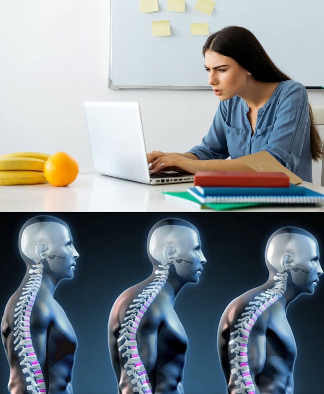
When posture collapses (slouching, rounded shoulders, forward head posture), several things can happen:
The chest may cave inward
The upper back may round more
The ribs may not expand as well
The diaphragm may not move as freely
The body may rely more on neck and shoulder muscles to breathe
UCLA Health explains that poor posture can cause the chest to cave in, affecting breathing mechanics (UCLA Health, 2024). Harvard also lists breathing difficulties among the less obvious problems linked to poor posture (Harvard Health Publishing, 2023).
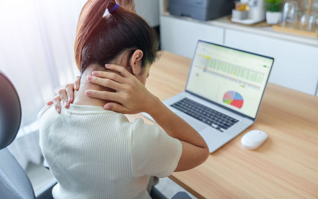
A research article on head-neck posture and respiratory function also found that posture changes can alter normal breathing mechanics, including diaphragm function. This matters because many people spend hours sitting at a desk, driving, or looking down at phones, which can reinforce forward head posture and rounded shoulders (Zafar et al., 2018).
Common signs that posture may be affecting your breathing
You may not always say, “I can’t breathe.” Instead, people often describe it like this:
“I can’t take a full deep breath”
“My chest feels tight when I sit”
“My neck and shoulders always feel tense”
“I sigh a lot”
“I feel winded faster than I should”
Sources on physical therapy and posture education also note a connection between poor posture and reduced diaphragm mobility, poor chest expansion, and shallow breathing (Capital Area PT, 2025; Total Health Chiropractic, 2022).
How Poor Posture Can Affect Digestion
Most people think digestion is only about food choices, enzymes, or stomach acid. Those are important, but body position matters too.
When you slouch, your abdomen compresses. That pressure can affect the stomach and intestines. UCLA Health notes that poor posture can slow digestion and increase abdominal pressure, which may trigger heartburn and acid reflux (UCLA Health, 2024).
BreatheWorks and other posture-focused digestive resources describe similar patterns: slouched alignment can increase abdominal pressure, affect swallowing and breathing coordination, and make reflux or bloating worse for some people (BreatheWorks, 2023a, 2023b).
Digestive symptoms that may be worse with slouching
Some common examples include:
Heartburn after meals
Acid reflux (GERD) symptoms when sitting or bending
Bloating or pressure in the upper abdomen
Feeling overly full
Constipation (especially with long periods of sitting)
Chiropractic and posture education sources (including Nolensville Chiropractic and BreatheWorks) often describe poor posture as a “compression” problem that can interfere with comfortable digestion and gut motility (Nolensville Chiropractic, 2025; BreatheWorks, 2023a).
The Breathing–Digestion Connection
Breathing and digestion are closely linked, and posture affects both simultaneously.
Here’s why:
The diaphragm supports both breathing and abdominal pressure control
The diaphragm is not just a breathing muscle. It also helps regulate pressure in the trunk. If it cannot move well, breathing becomes less efficient, and pressure control in the abdomen may change.
Poor posture can encourage shallow chest breathing
When breathing shifts more into the upper chest and neck, the body often feels more tense. In many people, this goes along with stress and “fight-or-flight” patterns, which can make digestion feel worse.
Slouching compresses the digestive area
A flexed, collapsed posture can reduce the space available to the stomach and intestines. That can be especially noticeable after eating.
BreatheWorks specifically describes how breathing coordination, alignment, and digestive comfort are connected, especially in people with reflux and bloating symptoms (BreatheWorks, 2023a, 2023b). El Paso Back Clinic and Dr. Jimenez’s educational content also emphasize this whole-body view, especially in patients with both musculoskeletal complaints and gut-related symptoms (Jimenez, n.d.; El Paso Back Clinic, n.d.).
Posture Patterns That Commonly Cause Problems
At El Paso Back Clinic, many patients dealing with neck, upper back, or shoulder pain also show posture patterns that can affect breathing and digestion. Dr. Jimenez’s educational content often highlights the same patterns in functional assessments (Jimenez, n.d.).
Forward head posture
This happens when the head moves in front of the shoulders. It increases neck strain and often leads to upper-chest breathing.
Rounded shoulders
Rounded shoulders can limit chest expansion and change rib cage motion.
Excessive upper-back rounding (kyphotic posture)
This can reduce thoracic mobility (mid-back motion), which is important for full breathing.
Slumped sitting posture
A tucked pelvis, a collapsed lower back, and a caved chest can increase abdominal pressure, making both breathing and digestion less efficient.
Why Integrative Chiropractic Care Can Help
A strong posture plan usually needs more than a quick reminder to “sit up straight.” Many people need a combination of mobility work, spinal/rib movement restoration, soft-tissue care, breathing retraining, and strength work to build lasting change.
That is why the El Paso Back Clinic approach is helpful for many people. The clinic’s posture and rehabilitation content describes a broader plan that can include:
Spinal adjustments
Mobility and stretching
Movement retraining
Soft-tissue care
Posture-focused exercises
Health coaching (El Paso Back Clinic, n.d.)
How this may improve breathing
When spinal and rib mobility improve, the chest can move more naturally during breathing. That can support deeper, more efficient breaths and reduce overuse of neck muscles.
How this may improve digestion
When posture improves, abdominal compression may decrease. Better alignment can also make it easier to breathe diaphragmatically, which may support calmer, more comfortable digestion in some patients.
Dr. Jimenez’s educational pages also describe the importance of posture, breathing mechanics, rib mobility, and functional movement in patients with reflux, bloating, and related complaints (Jimenez, n.d.).
Practical Steps to Improve Posture, Breathing, and Digestion
The good news is that small daily changes can make a real difference.
Reset your sitting posture
Try this simple “stacking” setup:
Feet flat on the floor
Hips level (not rolled backward)
The rib cage is stacked over the pelvis
Shoulders relaxed (not rounded forward)
Chin level (not poking forward)
Even a few posture resets per day can help reduce the long stretches of slouching that many people fall into while working or driving (UCLA Health, 2024).
Use posture breaks every 30–60 minutes
Long sitting is a major factor in the worsening of posture over time. A short break helps.
Quick break routine (2 minutes)
Stand up
Roll your shoulders back gently
Take 5 slow breaths
Walk for 1 minute
Reset your sitting position
This kind of movement break can reduce stiffness and help restore better breathing mechanics. General health and posture guidance consistently supports frequent movement to reduce the effects of prolonged sitting (Harvard Health Publishing, 2023; UCLA Health, 2024).
Practice diaphragmatic breathing
Diaphragmatic breathing can help train the body away from shallow chest breathing.
Simple drill (1–2 minutes)
Sit upright or lie on your back
Place one hand on your chest and one on your belly/ribs
Breathe in through your nose
Try to expand the lower ribs and belly gently
Exhale slowly and fully
Keep shoulders relaxed
Posture-focused breathing resources often recommend this type of drill to improve breathing efficiency and reduce tension (Capital Area PT, 2025; Total Health Chiropractic, 2022).
Improve meal posture
How you sit while eating matters, especially if you have reflux.
Better meal posture tips
Sit upright when eating
Avoid eating while slouched on a couch
Chew slowly
Stay upright after meals
Take a light walk after eating if possible
BreatheWorks and UCLA Health both discuss how posture can affect reflux and digestive comfort, especially in people who slouch during or after meals (BreatheWorks, 2023b; UCLA Health, 2024).
When to Get Medical Care Right Away
Posture can affect breathing and digestion, but some symptoms require medical evaluation and should not be blamed solely on posture.
Seek prompt medical care if you have:
Chest pain
Severe shortness of breath
Trouble swallowing
Vomiting blood
Black/tarry stools
Severe abdominal pain
Unexplained weight loss
Ongoing reflux that is not improving
These can be signs of a more serious condition and need a full medical workup (UCLA Health, 2024; Harvard Health Publishing, 2023).
Clinical Perspective from Dr. Alexander Jimenez, DC, APRN, FNP-BC
For the El Paso Back Clinic audience, the key message is simple: posture problems are often functional problems. In Dr. Jimenez’s educational content, posture is not treated as an isolated issue. It is part of a bigger clinical picture that includes spinal mechanics, rib motion, breathing patterns, stress load, and daily movement habits (Jimenez, n.d.).
That is why many patients feel better when care is more comprehensive. Instead of only focusing on pain, an integrative plan may help by:
Improving spinal and rib mobility
Restoring more natural breathing mechanics
Reducing neck and shoulder overuse
Addressing posture during work and meals
Supporting better movement and daily function
The El Paso Back Clinic posture and rehabilitation pages also describe a personalized approach using adjustments, exercise, stretching, and movement retraining, which fits well with this type of whole-body care model (El Paso Back Clinic, n.d.).
Final Takeaway
Poor posture can affect much more than the spine. Slouching and forward head posture can limit diaphragm movement, reduce chest expansion, and lead to shallow breathing. At the same time, abdominal compression can make digestion less comfortable and may worsen reflux, bloating, and constipation in some people.
The good news is that posture can improve. With the right plan—especially one that includes posture correction, breathing retraining, and integrative chiropractic care—many people can breathe better, move better, and feel more comfortable after meals.
For readers of El Paso Back Clinic, this is an important reminder: posture is not just about standing tall. It is about giving your body the space and mechanics it needs to function well.
IFM's Find A Practitioner tool is the largest referral network in Functional Medicine, created to help patients locate Functional Medicine practitioners anywhere in the world. IFM Certified Practitioners are listed first in the search results, given their extensive education in Functional Medicine