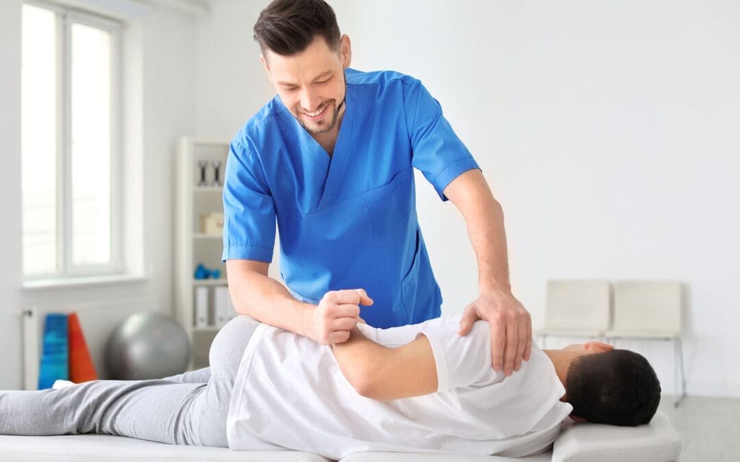
Sciatica Self-Massage at Home (The El Paso Back Clinic Approach to Safer Relief)
Sciatica is a nerve irritation pattern, not just a tight muscle. It often feels like burning, aching, tingling, or “electric” pain that can start in the low back or buttock and travel into the thigh, calf, and foot. Many people in El Paso experience sciatica after long hours of sitting, driving, or heavy lifting, or after an old injury that never fully healed. At El Paso Back Clinic, sciatica care is commonly described as integrative—meaning hands-on chiropractic care plus soft-tissue work, rehab, and (when appropriate) decompression strategies to reduce nerve pressure and help the body heal instead of just “chasing symptoms.”
Self-massage can be an effective home tool when done correctly. The goal is to relax the tissues around the irritated nerve pathway—especially the glutes, piriformis, low back muscles, hamstrings, and sometimes the calf—without smashing the nerve itself.
The safety rule that matters most: don’t “dig into” the sciatic nerve
If you press directly on the most “zappy” spot, you can flare symptoms. Instead, aim for gentle, targeted pressure that feels like a controlled release.
Use the “hurts good” rule:
Keep pressure 0–3 out of 10 (mild to moderate discomfort)
Avoid 4–10 out of 10 (too aggressive)
If symptoms worsen, stop right away and reduce pressure next time
Tools that work well at home
You do not need expensive equipment. These basic tools are enough for most people:
Tennis ball (beginner-friendly pressure)
Foam roller (great for slow myofascial release)
Two tennis balls taped together or in a sock (to work beside the spine more safely)
Heat pack (before or after)
Many sciatica massage guides recommend simple tools like tennis balls and foam rollers because they help you reach deep glute and hip muscles without overworking your hands.
Step-by-step: a simple self-massage routine for sciatica relief
Start with heat (optional, but helpful)
Apply heat to the lower back or glutes for 10–15 minutes. Heat can help muscles relax, so you do not need to apply as much pressure during a massage.
Tip: Heat should feel soothing, not scorching.
Trigger point release for the glutes and piriformis (tennis ball)
This is one of the most helpful self-massage steps because the piriformis and nearby glute muscles can tighten and irritate the sciatic nerve pathway.
How to do it:
Sit on the floor (or a firm bed) and place a tennis ball under one buttock.
Lean your weight into the ball until you find a tender “knot.”
Hold steady pressure for 20–45 seconds while breathing slowly.
Move the ball 1–2 inches and repeat on 2–4 spots.
Keep it safe:
If pain becomes sharp, numbness increases, or symptoms travel farther down the leg, stop immediately.
Low back muscle release (two tennis balls—NOT on the spine)
At El Paso Back Clinic, massage and soft-tissue work are considered a key part of sciatica treatment because relaxing tight tissues can reduce pressure on irritated structures. A safe home approach is to use two tennis balls so that pressure is applied beside the spine.
How to do it:
Tape two tennis balls together (or place them in a sock).
Lie on your back with knees bent.
Place the balls on either side of the spine, not on the bone.
Make tiny shifts and pauses—no fast rolling.
Work for 1–2 minutes, then rest.
Myofascial release for hamstrings (foam roller)
If your hamstrings are tight, they can “pull” on the pelvis and keep the low back and hip region tense. Slow foam rolling is often described as a form of self-myofascial release that warms and loosens tissue over time.
How to do it:
Sit with the roller under the back of your thigh.
Roll slowly and pause on tight spots for 20–30 seconds.
Don’t chase pain—stay in the 0–3/10 range.
Calf massage for referred pain (hands or roller)
Some sciatica patterns show up strongly in the calf or foot. Gentle calf work may help reduce guarding and improve comfort.
How to do it:
Use your hands to squeeze and glide from ankle toward knee.
Pause on a tender spot and breathe.
Keep pressure light to moderate.
What to avoid (so you don’t flare symptoms)
Heavy pressure on the “electric” pain spot
Fast rolling over the lower back or buttocks
Long sessions that leave you sore for 1–2 days
Pressing on the bone (spine, sacrum ridge, hip bone)
If you feel worse after self-massage, your body is telling you the dose was too high. Reduce pressure and shorten the next session.
Why chiropractic + massage often works better than either alone
Self-massage can help relieve muscle tension, but some cases of sciatica also involve spinal joint restriction, disc irritation, or nerve root pressure. That is why integrative chiropractic care is often paired with soft-tissue work.
On El Paso Back Clinic, sciatica care is described as focusing on addressing sources of pain (not only masking it), and the clinic also highlights combining chiropractic adjustments with therapeutic massage and non-surgical decompression options.
Common integrative components include:
Targeted chiropractic adjustments to improve motion and reduce irritation
Myofascial release/therapeutic massage to reduce spasms and improve circulation
Non-surgical spinal decompression (when appropriate) to reduce pressure on discs/nerve roots
Clinical observations from Dr. Alexander Jimenez
Across sciatica-focused education on the clinic’s site, the recurring theme is that lasting relief often improves when care addresses both sides of the problem:
tissue tension (glutes/piriformis/low back tightness), and
spinal mechanics (how joints/discs and nerve pathways are loading under stress).
When to stop home care and get evaluated quickly
Get urgent medical evaluation if you have:
New or worsening leg weakness
Loss of bowel or bladder control
Numbness in the saddle area
Severe pain with fever, unexplained weight loss, or major trauma
These may indicate a condition requiring immediate care beyond self-massage.
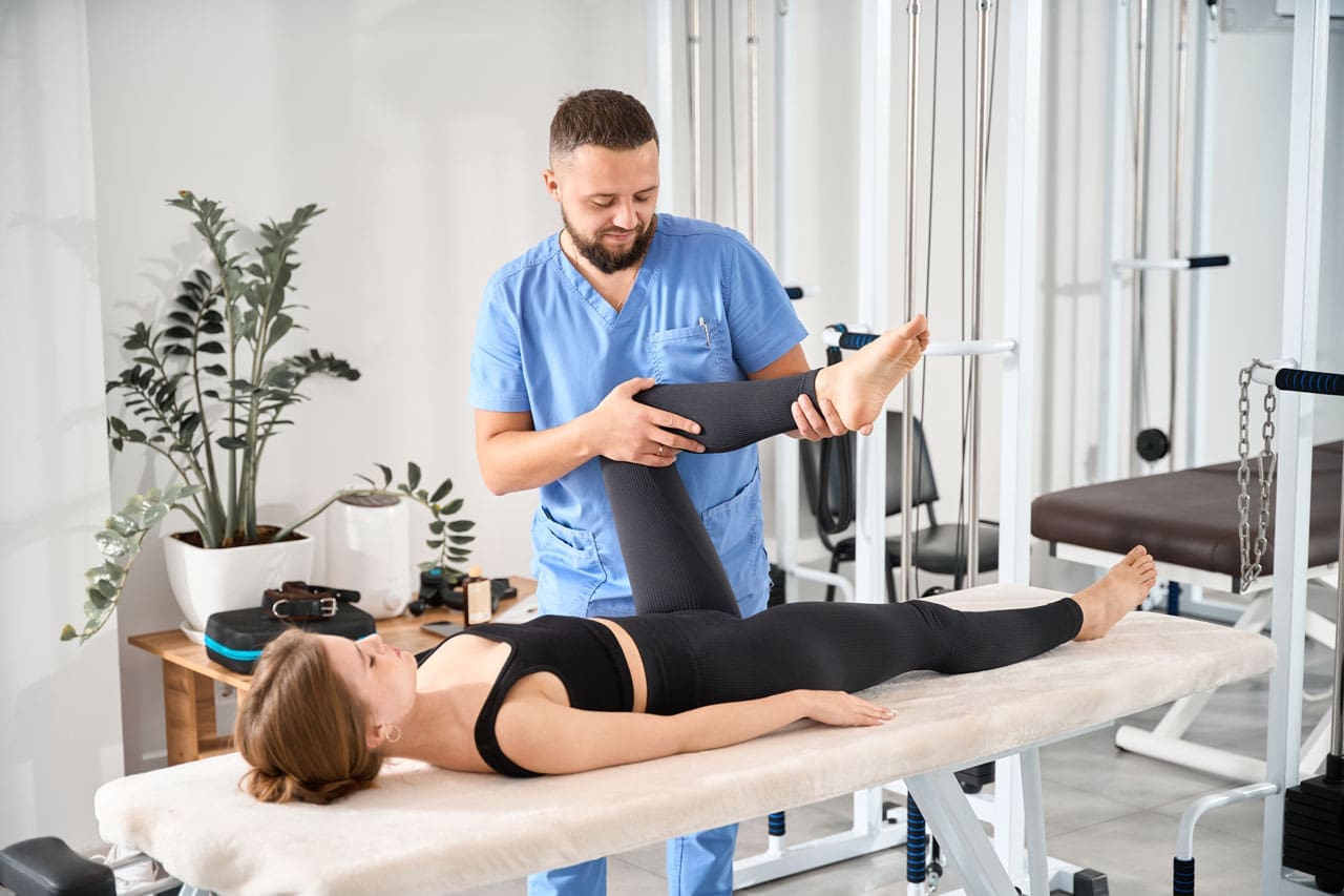
Fitness Optimization in El Paso, TX: How to Organize a Weekly Workout Plan With Warm-Ups, Cool-Downs, and Integrative Chiropractic Support
A woman doing her weekly workout
A weekly workout plan should do two things at the same time:
Help you get stronger, fitter, and more mobile
Help you stay consistent without getting hurt or burned out
That balance matters even more in El Paso, Texas, where heat, dry air, and busy schedules can make training feel harder than expected. A smart plan incorporates strength training, cardio, mobility, and recovery—and includes warm-ups and cool-downs in every session.
This guide is written for real life. It is geared to the El Paso Back Clinic approach: improving movement quality, addressing posture and joint mechanics, and supporting safer training through an integrative model that blends chiropractic and clinical assessment. ()
Why most people struggle with weekly workout planning
Many people start with motivation, then hit one of these problems:
They do too much too fast (and flare up pain)
They skip warm-ups and feel stiff or strained
They train hard but don’t recover well
They repeat the same muscle groups without enough rest
They don’t have a simple weekly structure that they can repeat
A better plan is not “perfect.” It is repeatable.
A common starting target for beginners and intermediate exercisers is 3–5 workout days per week, depending on schedule, recovery, and current fitness level. (Mayo Clinic, 2023; EōS Fitness, 2024) ()
What a balanced weekly workout plan includes
A strong weekly plan usually includes these building blocks:
Strength training (2–3 days/week)
Cardio (2–3 days/week)
Mobility (most days, even 5–10 minutes helps)
Recovery (at least 1 full rest day, plus lighter days)
Many gyms and fitness instructors recommend alternating training styles throughout the week—such as upper body, lower body, and cardio—to give muscles time to recover while you stay active. (Grinder Gym, 2025; ISSA, 2022)
El Paso-specific training: heat, hydration, and timing
El Paso’s climate can change how workouts feel, especially if you train outdoors. Dry air can increase fluid loss, and heat can accelerate fatigue.
Simple El Paso-friendly adjustments:
Train early morning or later evening outdoors when possible
Build hydration into your plan, not as an afterthought
Hydration tip: If you sweat heavily or train longer, you may need electrolytes—especially during hot weather—based on your personal needs and health status. (American College of Sports Medicine, 2007)
Warm-ups and cool-downs: the 5–10 minute habit that protects progress
If you only change one thing in your training week, make it this:
Warm up for 5–10 minutes (dynamic movement)
Cool down for 5–10 minutes (gradual slowdown + stretching/breathing)
Why warm-ups matter
Warm-ups help your body transition from rest to work. Mayo Clinic explains that warm-ups prepare the cardiovascular system, raise temperature, increase blood flow to muscles, and may lower injury risk. (Mayo Clinic, 2023) ()
Why cool-downs matter
Cooling down helps your body transition back toward rest. Mayo Clinic Press emphasizes that cooldown supports recovery and helps the body transition out of high-intensity exercise more smoothly. (Mayo Clinic Press, 2025) ()
A simple warm-up you can reuse for almost any workout (5–10 minutes)
Keep it easy. The goal is to feel warmer, looser, and more “ready,” not exhausted.
Warm-up (choose this as your default):
2 minutes of easy movement
brisk walk, light bike, easy row
2 minutes dynamic mobility (pick 3–4)
arm circles
hip circles
ankle rocks
thoracic (upper back) rotations
2–4 minutes workout-specific prep
strength day: 1–2 lighter sets of your first lift
cardio day: start slower and gradually build pace
Mayo Clinic Press notes that warm-up duration depends on intensity, but 5–10 minutes is a solid baseline for many people, with longer warm-ups for higher-intensity work. (Mayo Clinic Press, 2025) ()
A simple cool-down you can reuse (5–10 minutes)
Cool-downs work best when they are consistent.
Cool-down template:
3–5 minutes gradual slowdown
walk slowly, easy cycling, gentle movement
2–5 minutes stretching + breathing
hamstrings
hip flexors
calves
chest/shoulders
gentle low back rotation (if comfortable)
Mayo Clinic explains that warm-ups and cool-downs are often the same activity, performed at a lower intensity before and after the workout. (Mayo Clinic, 2023) ()
The best weekly workout schedules for beginners and intermediates (3–5 days/week)
Below are three schedules you can choose from. Pick the one you can follow most weeks.
Option A: 3-day plan (simple and sustainable)
This is perfect if you are starting again, staying consistent, or managing pain flare-ups.
Day 1 (Mon): Full-body strength + short walk
Day 2 (Wed): Cardio + mobility
Day 3 (Fri): Full-body strength + core
Weekend: 1 light activity day + 1 full rest day
Many weekly workout guides recommend 2–3 strength sessions and at least one rest day for recovery. (Health, n.d.) ()
Option B: 4-day plan (upper/lower split + cardio)
This is a popular plan for steady progress.
Mon: Lower body strength
Tue: Upper body strength
Thu: Lower body strength + core
Sat: Cardio + mobility (or a class)
Splitting upper/lower body helps prevent repeating the same muscle groups on back-to-back days and makes recovery easier to manage. (ISSA, 2022; Grinder Gym, 2025) ()
Option C: 5-day plan (shorter sessions, more frequency)
This works well if you like shorter workouts and a daily structure.
Mon: Strength (full body)
Tue: Cardio
Wed: Strength (upper)
Thu: Mobility + easy cardio
Fri: Strength (lower)
Sat: Optional class or easy walk
Sun: Rest
EōS Fitness emphasizes building a weekly plan based on your goals and starting level, often incorporating strength, cardio, and recovery. (EōS Fitness, 2024) ()
What to do inside each strength workout (so it’s organized)
A clean structure keeps you from wandering around the gym and doing random exercises.
Strength session structure (45–60 minutes):
Warm-up: 5–10 minutes
Main lift: 10–15 minutes
Assistance work: 15–25 minutes
Core: 5–10 minutes
Cool-down: 5–10 minutes
Main lift examples:
squat pattern (leg press or squat)
hinge pattern (deadlift variation or hip hinge)
press (dumbbell press)
pull (row or pulldown)
Assistance work examples:
glute bridges or hip thrusts
split squats or step-ups
face pulls or band work for shoulders
hamstring curls
carries (farmer carry)
This aligns with structuring training days around major patterns (push/pull/lower) to build balanced strength and avoid overuse. (Grinder Gym, 2025; ISSA, 2022) ()
Cardio planning: simple is better than perfect
Cardio should support your life, not crush you.
Great El Paso-friendly cardio options:
incline treadmill walking (easy on joints)
stationary bike
rowing machine
brisk outdoor walking (timing matters in heat)
Easy weekly cardio goals:
2 days of steady cardio (20–40 minutes)
1 optional interval day (shorter, only if you tolerate it)
Health.com outlines weekly schedules that combine strength and cardio while protecting recovery. (Health, n.d.) ()
Mobility and recovery: the glue that holds the week together
Recovery is not “doing nothing.” It is training your body to stay ready for the next workout.
Recovery habits that work:
sleep consistency
hydration plan
protein and balanced meals
walking on rest days
mobility work for hips, ankles, upper back, and shoulders
Simple mobility “micro-dose” (5 minutes):
1 minute hip flexor stretch (each side)
1 minute calf stretch (each side)
1 minute thoracic rotations
1 minute shoulder mobility
This kind of daily movement keeps joints from stiffening, especially if you sit a lot.
How integrative chiropractic supports routine optimization
Many people don’t need more willpower. They need:
better joint motion
better movement patterns
better recovery
fewer flare-ups
The El Paso Back Clinic approach: integrative care and movement-focused support
The El Paso Back Clinic describes an integrated model led by Dr. Alex Jimenez, DC, APRN, FNP-BC, combining chiropractic care and clinical assessment within a multidisciplinary setting. (El Paso Back Clinic, n.d.)
From a routine-optimization standpoint, that integrative approach can help people who struggle with:
recurring neck or low back tightness during training
posture-related strain (desk work, long driving, “tech neck”)
limited hip or shoulder mobility
compensation patterns (one side always “takes over”)
The clinic also discusses advanced collaboration and diagnostics, including imaging relationships when needed for complex cases—especially when symptoms do not match what someone expects from “normal soreness.” (El Paso Back Clinic, n.d.) ()
Clinical observations from Dr. Jimenez (fitness-focused takeaways)
Across the clinic’s educational content, Dr. Jimenez emphasizes:
improving posture and movement quality to reduce repeated strain patterns (El Paso Back Clinic, n.d.) ()
using mobility and functional training to build resilience and prevent re-injury (El Paso Back Clinic, n.d.) ()
integrating training structure with recovery so people can stay consistent long-term (El Paso Back Clinic, n.d.) ()
In simple terms: train with a plan, move better, recover better.
A weekly “checklist” you can follow
Use this to keep your week on track:
✅ 3–5 workouts completed (based on your plan)
✅ Warm-up done every workout (5–10 minutes) (Mayo Clinic, 2023)
pain that worsens with training, even after deloading
trouble figuring out what movements are safe for your body
If you want clinic support, El Paso Back Clinic provides contact and appointment options, including online scheduling information listed on their site. (El Paso Back Clinic, n.d.) ()
Navigating Car Accident Claims in El Paso, Texas: Pre-Existing Conditions, the Eggshell Skull Rule, and Care at El Paso Back Clinic
The doctor explains an X-ray to the patient and points at the computer screen. The patient wears a cervical collar
Car accidents are common in El Paso, Texas. They can cause new injuries or worsen existing ones. People often wonder if a past health issue, like back pain or arthritis, will block them from getting help after a crash. Texas law offers protection. You can still claim money for injuries even with prior conditions. This article covers the rules, what you can get paid for, and the steps to follow. It highlights the “eggshell skull rule” and why quick medical care is key. In El Paso, El Paso Back Clinic stands out for expert care for auto accidents and worsening conditions.
Understanding the Eggshell Skull Rule
The eggshell skull rule is an important legal concept. It means that if someone causes an accident, they must pay for all resulting damage. This applies even if the injured person had a weakness from a prior condition. It’s like breaking a fragile egg—you can’t blame the thin shell. The rule is also known as the “thin skull rule” or “take your victim as you find them” (Amtz Law, n.d.).
Simply put, the at-fault person takes full responsibility. They can’t use your old health problems to avoid paying. However, the accident must cause new damage or aggravate the existing issue. If your condition had worsened on its own, that might not be covered (Gutierrez Law Firm, n.d.a). For instance, if you had mild back arthritis and the crash resulted in severe pain that required therapy, the at-fault driver is responsible for covering that additional harm.
This rule originated in prior legal cases. It safeguards those who are more vulnerable. In Texas, it’s used in car accident lawsuits to ensure fair compensation (Reyes Law, n.d.).
Applying the Eggshell Skull Rule in Texas and El Paso
Texas fully supports the eggshell skull rule. In El Paso, if a car accident aggravates your pre-existing condition, you can pursue a claim. The law holds the at-fault party liable for all injuries resulting from the crash, including those amplified by prior issues (GDL Firm, n.d.).
El Paso has busy highways, such as I-10, which leads to frequent accidents. Local laws follow Texas standards. For example, if you had an old neck injury and a collision causes whiplash on top of it, the rule helps you recover costs. Insurance companies may argue that your pain stems solely from the prior condition to reduce payments (BHW Law Firm, n.d.). Strong evidence can counter this.
You have two years from the accident date to file in El Paso under the statute of limitations (No Bull Law, n.d.). Act fast to avoid missing out.
Typical Pre-Existing Conditions Impacted: Chronic back pain, sciatica, herniated discs, fibromyalgia, or degenerative disc disease.
Signs of Aggravation: Increased pain, new movement limitations, or the need for additional medical treatment.
El Paso-Specific Risks: Border traffic and dust storms increase crash chances, often affecting backs and necks.
Compensation Options for Aggravated Conditions
When an accident worsens your condition, Texas allows claims for various damages. The eggshell skull rule ensures coverage for the full extent of harm (Siegfried & Jensen, n.d.). This includes bills, lost income, and emotional distress.
Possible compensations include:
Medical Expenses: Costs for new therapies, adjustments, or surgeries due to the aggravation, plus future care.
Wage Loss: Earnings missed from work because of heightened symptoms.
Pain and Suffering: Payment for added physical discomfort and mental strain, such as stress from chronic pain.
Reduced Quality of Life: If daily activities or hobbies become harder.
Long-Term Disability: For permanent worsening, like ongoing sciatica.
Amounts depend on severity. Minor aggravations may yield smaller settlements, while persistent issues, such as the need for regular chiropractic care, may increase them (Reyes Law, n.d.). Age factors in—younger victims may experience greater impacts over time.
In El Paso, solid documentation boosts settlements (Abraham Watkins, n.d.).
Proving Your Case for Compensation
To win, show that the accident directly worsened your condition. Use medical records from before and after to illustrate changes (St. Louis Injury Law, n.d.). This “before-and-after” approach is crucial.
Key steps:
Seek Immediate Care: Visit a doctor soon after. Discuss your history and new symptoms.
Maintain Documentation: Collect bills, notes, and X-ray scans.
Log Daily Effects: Journal pain levels and activity changes.
Expert Testimony: Have a physician explain the connection.
Accident Evidence: Include reports, photos, and statements.
Honesty about your past is vital—concealing it can weaken your claim (Gage Mathers, n.d.). Courts assess if the aggravation ties to the crash or is a natural progression.
Handling Insurance Challenges
Insurers aim to minimize payouts. They may blame your pre-existing condition entirely. They could demand full records to deny claims (Romanow Law Group, n.d.). Avoid broad agreements without advice.
Strategies:
Use Legal Support: Have an attorney negotiate on your behalf.
Reject Low Offers: Initial proposals are often insufficient.
Challenge Rejections: Present evidence linking to the accident.
Recognize Strategies: Beware of their experts minimizing damage.
El Paso attorneys familiar with local rules can help (Ellis & Thomas, n.d.).
Benefits of Specialized Auto Accident Clinics in El Paso
Post-accident, choose a clinic expert in auto injuries. This ensures proper documentation and healing. El Paso Back Clinic excels in this, offering chiropractic care for whiplash, back pain, and aggravated conditions (El Paso Back Clinic, n.d.).
Their approach includes:
Chiropractic Adjustments: To align the spine and ease nerve pressure.
Physical Therapy: To rebuild strength and mobility.
Spinal Decompression: For herniated discs and sciatica.
Functional Medicine: Addressing root causes with nutrition and lifestyle.
Seeing them early helps record aggravations, aiding claims (Your Back in Line Now, n.d.). They coordinate with attorneys for seamless support.
Insights from Dr. Alexander Jimenez at El Paso Back Clinic
Dr. Alexander Jimenez, DC, APRN, FNP-BC, leads El Paso Back Clinic. With dual expertise in chiropractic and nursing, he treats complex cases like auto-aggravated back issues (El Paso Back Clinic, n.d.).
He observes that crashes often intensify conditions like degenerative discs or fibromyalgia. Treatments blend adjustments, acupuncture, and rehab. He emphasizes non-invasive methods, using diagnostic tools such as X-rays to establish links.
Patients praise quick relief. For example, Gale Grijalva recovered from accident-related back pain, resuming activities. Dr. Jimenez’s team offers personalized plans to prevent surgery.
The clinic’s 30,000+ sq ft facility includes gyms and meal prep, supporting full recovery.
Wrapping Up
Dealing with car accidents in El Paso is challenging, especially with pre-existing conditions. Texas’s eggshell skull rule allows compensation for aggravations with proper proof. Seek prompt medical attention, document everything, and consult legal counsel. El Paso Back Clinic, led by Dr. Jimenez, provides top chiropractic and rehab for healing and claims. Contact them at +1-915-850-0900 or visit https://elpasobackclinic.com/ for help.
References
Abraham Watkins. (n.d.). Do Pre-Existing Conditions Disqualify Me From Damages in a Personal Injury Case?Abraham Watkins.
Amtz Law. (n.d.). How Pre-Existing Conditions Affect Your Personal Injury Claim.Amtz Law.
BHW Law Firm. (n.d.). Pre-Existing Injury and Accident in Texas.BHW Law Firm.
Ketogenic Diet in 2026: A Smarter, Safer Approach for Metabolic Health, Brain Support, and Better Movement
In early 2026, the ketogenic (“keto”) diet is still widely used—but the way people use it has matured. Keto is no longer just a “trend diet.” It is now better understood as a structured clinical nutrition strategy that can help certain people with epilepsy, type 2 diabetes, and weight loss, while also being actively studied for brain health and mental health. At the same time, leading medical sources continue to warn that keto can raise LDL (“bad”) cholesterol in some people, and long-term heart outcomes are still not fully clear. That is why the modern keto plan in 2026 is less about “more fat” and more about better fat, better fiber, and better monitoring. (Harvard Health Publishing, 2024a, 2024b)
For a clinic focused on spine, mobility, and whole-body function—such as El Paso Back Clinic—this is important because metabolic and musculoskeletal health are interconnected. Many people who want to lose weight or improve blood sugar also deal with back pain, neck pain, joint stiffness, poor sleep, and high stress, which can make lifestyle changes harder to stick with. In 2026, the best outcomes usually come from a team approach: nutrition guidance (often supported by a nurse practitioner) plus chiropractic support for movement, posture, and nervous system regulation—so the person can keep moving and recover well while changing how they eat. (Masood et al., 2023)
What the Keto Diet Is (Plain and Simple)
A ketogenic diet is typically:
Very low in carbohydrates
Moderate in protein
Higher in fat
The goal is to achieve nutritional ketosis, in which the body uses fat and ketones for much of its energy rather than relying primarily on glucose (blood sugar). UC Davis describes keto as a strict high-fat plan with very low carbohydrates, and they emphasize that it is not right for everyone. (UC Davis Health, 2025)
Important safety note: Nutritional ketosis is not the same as diabetic ketoacidosis (DKA). DKA is a dangerous medical emergency, most often linked to type 1 diabetes and uncontrolled blood sugar. Keto dieting is not meant to create that state, which is why medical screening and medication review matter—especially for people taking diabetes medications. (Masood et al., 2023)
Why Keto Is Still Clinically Relevant in 2026
Epilepsy: Keto’s Most Established Medical Use
Keto has a long history in epilepsy care and is still used in specialized settings for seizure control. This is one reason keto has remained part of mainstream medical conversation, not just social media. (Masood et al., 2023)
Key point: When keto is used therapeutically for epilepsy, it is often monitored closely, with specific nutrition targets and professional oversight. (Masood et al., 2023)
Type 2 Diabetes and Insulin Resistance: Helpful for Some, Needs Monitoring
Keto can sharply reduce carbohydrate intake, which often improves blood glucose control for some people. Many people also experience changes in appetite and reduced cravings after adaptation. But if a person is taking glucose-lowering medications, the plan must be coordinated with a clinician to reduce the risk of hypoglycemia and to adjust medications safely. (Masood et al., 2023)
UC Davis also notes that keto can lead to weight loss and metabolic changes, but it is restrictive and should be approached carefully with individual health factors in mind. (UC Davis Health, 2025)
Weight Loss: Often Fast Early Results, Long-Term Success Requires a Real Plan
Keto is known for quick early weight change, often due to:
Lower carbohydrate intake
Water shifts early on
More structured eating patterns (fewer refined foods for many people)
But long-term outcomes depend less on “perfect keto” and more on:
Sleep
Stress
Food quality
Consistency you can sustain Harvard also points out that keto may work short-term, but long-term heart effects and sustainability are big concerns. (Harvard Health Publishing, 2024a)
Keto and the Brain in 2026: Mental Health and “Metabolic Therapy”
One of the most significant shifts in 2026 is the growing interest in the effects of keto on brain metabolism. Stanford Medicine reported results from a small pilot study in severe mental illness, suggesting potential improvements in metabolic health and psychiatric measures while participants followed a ketogenic diet under clinical guidance. (Stanford Medicine, 2024; Sethi et al., 2024)
Stanford also discussed “keto therapy” as a topic of active interest in psychiatry, while still emphasizing that larger trials are needed and keto is not a replacement for standard care. (Stanford Medicine, 2025)
The responsible 2026 takeaway: Keto may be promising for certain brain-related conditions, but it should not be oversold as a cure. It should be included in a medically supervised, whole-person plan when appropriate. (Stanford Medicine, 2025; Sethi et al., 2024)
Keto and Athletic Performance: It Depends on the Sport and the Person
In 2026, keto is often discussed differently for endurance athletes vs. power athletes. Some people report steadier energy after adaptation, whereas others struggle with high-intensity training, where rapid carbohydrate fuel helps performance.
A practical way to view this:
Keto may fit some endurance goals
Keto may be tougher during high-intensity bursts
Training quality, sleep, hydration, and recovery matter either way (UC Davis Health, 2025)
The Heart Question in 2026: Why Fat Quality Matters So Much
Here is the reality: keto can improve some markers (like triglycerides and HDL) while raising LDL in some people—especially when saturated fat intake is high. (McGaugh et al., 2022; Harvard Health Publishing, 2024a, 2024b)
Harvard Health warns that keto is associated with increased LDL cholesterol and that long-term heart outcomes remain uncertain. They also emphasize limiting saturated fat due to its association with increased risk of heart disease. (Harvard Health Publishing, 2024a)
A scientific review in PMC reports that ketogenic diets often lower triglycerides and raise HDL, but lipid responses can vary, and LDL may rise depending on diet composition and the person. (McGaugh et al., 2022)
“Better Keto” fats (heart-smarter choices)
If you do keto in 2026, many clinicians push the plan toward unsaturated fats and fiber-rich foods:
Extra-virgin olive oil
Avocados and avocado oil
Nuts and seeds (portion-aware)
Fatty fish (like salmon and sardines)
Lots of low-carb vegetables for fiber (Harvard Health Publishing, 2024a)
Fats to limit if LDL rises
Heavy reliance on butter, cream, and fatty processed meats
Frequent fried foods
A pattern where saturated fat dominates most meals (Harvard Health Publishing, 2024a)
Long-Term Keto and Longevity: Newer Cautions in 2026
A UT Health San Antonio-led study (animal research) found that continuous long-term ketogenic dieting in mice increased cellular senescence (“aged cells”) in multiple organs, with particular attention to heart and kidney tissues. The same research line reported that an intermittent approach appeared to prevent those pro-inflammatory senescence effects in their model. This does not automatically prove the same outcome in humans, but it supports today’s more careful “longevity-minded” keto planning. (UT Health San Antonio, 2024)
Practical meaning for 2026: Some people may do better with:
Cycles or planned breaks
A “modified keto” approach with more fiber and better fats
Regular lab monitoring and symptom tracking (UT Health San Antonio, 2024)
Who Should Be Careful (Or Avoid Keto)
Clinical education sources emphasize screening for contraindications and identifying patients who need close monitoring or personalized adjustments before starting keto. (Masood et al., 2023)
Situations that often require extra caution:
Diabetes medications that can cause low blood sugar (needs clinician review)
History of kidney stones or significant kidney disease (case-by-case)
Pregnancy or breastfeeding (specialized guidance)
History of eating disorders (risk of triggering restrictive patterns)
Certain rare metabolic conditions (Masood et al., 2023)
Why El Paso Back Clinic’s “Whole-Person” Angle Fits 2026, Keto
A common reason people fail on diet plans is not lack of motivation—it is pain, poor sleep, stress overload, and limited movement capacity. Back pain and neck pain can reduce daily activity. Reduced activity can worsen insulin resistance. Poor sleep can increase hunger hormones and cravings. Stress can increase comfort eating. These factors stack together.
That is why keto in 2026 works best with an integrated plan that addresses:
Nutrition structure
Mobility and function
Sleep and stress regulation
Realistic habit systems (Masood et al., 2023)
The Nurse Practitioner role: safety, labs, and medication review
The NP-guided side of keto commonly includes:
Reviewing medications and contraindications
Tracking metabolic markers and side effects
Personalizing protein, fiber, hydration, and electrolytes
Adjusting the plan based on response (Masood et al., 2023)
This is also consistent with the increasing involvement of nurse practitioners in contemporary obesity and metabolic care, which combines nutrition strategies with patient-centered support and medical interventions when appropriate. (American Association of Nurse Practitioners, 2025)
The Chiropractic role: movement, posture, and the “pain barrier” problem
When pain limits activity, people often get stuck. Chiropractic care can support the musculoskeletal side of the plan by helping patients:
Improve mobility and joint motion
Reduce mechanical stress that flares pain
Support posture and movement patterns
Build a more consistent foundation for walking, training, and rehab work
El Paso Back Clinic has published educational materials on ketogenic diets and brain-fuel concepts (ketones vs. glucose), as well as on basic principles, reflecting an integrative wellness approach that links nutrition and function. (El Paso Back Clinic, n.d.-a, n.d.-b)
A Practical “Safer Keto” Framework for 2026
Step 1: Choose your goal (and set a time frame)
Common goals:
Weight loss jump-start
Blood sugar improvement
Reduced cravings
A monitored metabolic therapy trial for brain or mood support (UC Davis Health, 2025; Stanford Medicine, 2025)
Step 2: Build your plate the 2026 way (quality first)
Protein: eggs, poultry, fish, leaner cuts as needed
Fluids/electrolytes: plan ahead (many people feel “keto flu” symptoms when hydration and sodium are too low) (UC Davis Health, 2025)
Step 3: Track the markers that matter
Beyond the scale:
Waist measurement
Blood pressure
Energy and sleep quality
Lipid panel (LDL, HDL, triglycerides)
A1C (for blood sugar trends) (Harvard Health Publishing, 2024a; McGaugh et al., 2022)
Step 4: Re-check at 6–12 weeks and personalize
If LDL climbs or symptoms worsen, a clinician may adjust:
Fat quality (shift from saturated → unsaturated)
Fiber intake
Carb target (sometimes “less strict” works better long-term)
Overall approach (including cycling or modified keto) (Harvard Health Publishing, 2024a; UT Health San Antonio, 2024)
Bottom Line: Keto Is Still Useful in 2026—But It Must Be Done Well
Keto remains a valuable tool for:
Epilepsy therapy (the most established medical use)
Some cases of type 2 diabetes and insulin resistance (with monitoring)
Fast early weight loss (especially when it reduces refined foods)
But in 2026, the higher-standard approach is clear:
Keto is not a cure-all
Heart markers matter
Fat quality matters
Long-term planning matters
A team approach helps people succeed safely (Harvard Health Publishing, 2024a; Masood et al., 2023)
For individuals pursuing weight loss and metabolic health while managing chronic back or neck problems, pairing clinical nutrition guidance with function-focused chiropractic care can remove key obstacles, making the plan not only possible but also sustainable.
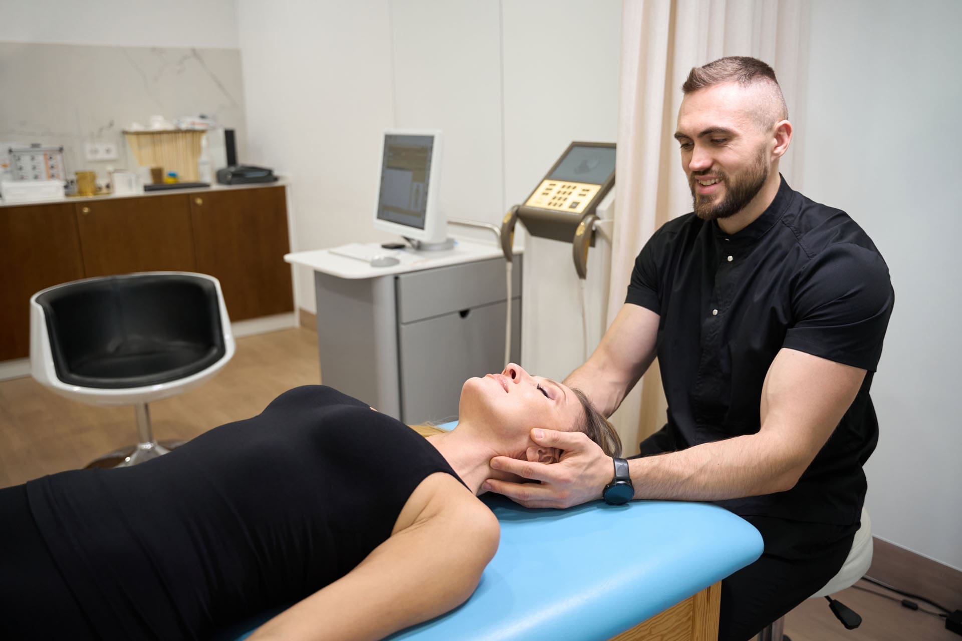
Optimal Joint Movement: Enhancing Mobility and Stability at El Paso Back Clinic
A chiropractor or Nurse Practitioner works with a patient in a rehabilitation center to improve joint mobility.
Optimal joint movement is essential for an active, pain-free life. At El Paso Back Clinic in El Paso, TX, we specialize in helping people achieve this through personalized chiropractic care. This article explains what optimal joint movement means, why it’s important, and how our clinic’s integrative approaches can restore it. Whether you’re dealing with back pain, sports injuries, or daily stiffness, our team, led by Dr. Alex Jimenez, DC, APRN, FNP-BC, uses spinal adjustments, rehabilitation, and functional medicine to get you moving better. Discover how we support joint health to improve function in everyday tasks and athletic pursuits.
Understanding Optimal Joint Movement
Optimal joint movement is the ability to move your joints through their full natural range of motion (ROM) smoothly, without pain, and with good control. It’s often referred to as high-quality mobility, blending flexibility with strength for daily activities and sports (University of Colorado Anschutz Medical Campus, n.d.).
At El Paso Back Clinic, we define it as moving joints efficiently while maintaining balance between mobility (active movement) and stability (joint control). This ensures muscles, ligaments, and tendons work together properly (National Academy of Sports Medicine, n.d.; Mainstay Medical, n.d.). For instance, a healthy shoulder should lift overhead to 180 degrees without strain, allowing you to reach shelves or throw a ball (Verywell Health, 2023a).
When injury or prolonged sitting disrupts this, mobility declines, leading to awkward movements elsewhere in the body (University of Colorado Anschutz Medical Campus, n.d.). Our clinic addresses this through holistic care, combining adjustments, soft-tissue therapy, and exercises to reduce inflammation and improve coordination.
Key Elements of Optimal Movement:
Full ROM: Joints reach their natural limits, like knee flexion to 140 degrees for squatting (The GO KNEE, n.d.).
Smooth Control: No jerking or pain, thanks to strong muscles and clear nerve signals.
Balance: Mobility for range, stability to prevent wobbles or injuries (ACE Fitness, n.d.a).
The Importance of Mobility and Stability Balance
At El Paso Back Clinic, we emphasize the balance between mobility and stability for peak performance. Mobility allows free movement, while stability keeps joints secure during activities (ACE Fitness, n.d.b). This synergy is key in our treatments.
Think of the body as a chain: Ankles and hips need mobility for steps, while knees and lower back provide stability (Motus Physiotherapy, n.d.; NASM, n.d.). If an ankle stiffens due to injury, the knee compensates, increasing the risk of pain (Physical Therapy at MJC, n.d.). Our chiropractic adjustments and rehab programs restore this chain, enhancing joint function.
Integrative care at our clinic—including spinal decompression and strength training—supports this balance, reducing the risk of injury and improving mobility (Peninsula Wellness Partners, n.d.).
Common Disruptions to Joint Mobility
Life factors can hinder optimal joint movement. Injuries cause swelling and tightness, limiting ROM (Frozen Shoulder Clinic, n.d.; Musculoskeletal Key, n.d.). A sedentary lifestyle, common in desk jobs, tightens muscles and stiffens joints (Dr. Ong Kee Leong, n.d.).
At El Paso Back Clinic, we see this in patients with back pain or sciatica, where poor posture leads to compensation and strain in other areas (OMassageT, n.d.). Aging, arthritis, or repetitive motions worsen it (Arthritis Foundation, n.d.; Chesapeake Regional, n.d.).
Typical Causes:
Trauma: Sprains create hard end-feels, stopping movement early (Physiopedia, n.d.c).
Inactivity: Shortens tissues, reducing flexibility (Dr Ong Kee Leong, n.d.).
Health Conditions: Arthritis limits ROM, causing bony sensations (Physiopedia, n.d.c).
Habits: Bad ergonomics unbalance the kinetic chain (OMassageT, n.d.).
Without correction, this increases fall risk and reduces quality of life. Our clinic’s diagnostic tools, such as digital X-rays, identify issues early.
Why Prioritize Optimal Joint Movement?
Good joint movement enhances everything from walking to sports. It prevents pain and boosts efficiency (OneStep, n.d.). At El Paso Back Clinic, we help athletes improve power and reduce injuries through better ROM (Activ Therapy, n.d.).
For daily life, it means easier tasks without fatigue (Baliston, n.d.). In walking, ankle flexion aids balance; poor ROM shortens strides (Baliston, n.d.). Our programs keep joints lubricated and muscles strong (Arthritis Foundation, n.d.).
At El Paso Back Clinic, maintenance starts with assessment. We measure ROM against norms using tools like goniometers (Physical Therapy at MJC, n.d.; Trainerize, n.d.). Then, we recommend exercises.
Regular activity, such as stretching, helps keep joints flexible (Arthritis Foundation, n.d.; Royal City Physiotherapy, n.d.). Our mobility drills focus on control for real-world use (Royal City Physiotherapy, n.d.).
Practical Tips:
Warm-Ups: Shoulder circles or ankle rolls (Chesapeake Regional, n.d.).
Stretching: Hold for 30 seconds on tight spots (Verywell Health, 2023a).
Strength Work: Squats for knee stability (ACE Fitness, n.d.b).
Activity: Low-impact, like swimming (Arthritis Foundation, n.d.).
Tools: Foam rollers for self-care (Muscle and Motion, n.d.).
Visit our East Side location for personalized plans.
Integrative Chiropractic Care at El Paso Back Clinic
Our clinic offers holistic chiropractic care to restore joint movement. Led by Dr. Alex Jimenez, we combine adjustments, therapy, and guidance (Peninsula Wellness Partners, n.d.; Evolved Health Chiropractic, n.d.).
Adjustments realign joints, easing inflammation and nerves (Rodgers Stein Chiropractic, n.d.a; Rodgers Stein Chiropractic, n.d.b). Soft tissue work and rehab build muscle support (Evolved Health Chiropractic, n.d.).
This approach enhances mobility, strengthens areas, and reduces risks (Core Integrative Health, n.d.; Duca Chiropractic, n.d.). Joint mobilization gently increases ROM (Smart Sports Medicine, n.d.).
Our Services:
Spinal Adjustments: Restore alignment for better ROM (Chiropractic Omaha, n.d.).
Functional Medicine: Addresses root causes, such as nutrition (TXMAC, n.d.).
Rehab: Exercises for long-term health (Duca Chiropractic, n.d.).
Clinical Insights from Dr. Alex Jimenez at El Paso Back Clinic
Dr. Alex Jimenez, DC, APRN, FNP-BC, heads El Paso Back Clinic, with over 30 years of experience in integrative care. At our facilities, he blends chiropractic, functional medicine, and rehab for joint issues (Jimenez, n.d.a; Jimenez, n.d.b).
His observations: Adjustments alleviate nerve-related issues, restoring ROM in cases of back pain or sciatica (Jimenez, n.d.a). Patients from accidents or sports regain mobility through tailored plans (Jimenez, n.d.a).
Dr. Jimenez focuses on root causes with nutrition and exercises, preventing surgery (Jimenez, n.d.b). For hips or knees, agility programs balance mobility and stability (Jimenez, n.d.a). Our holistic model empowers patients and aligns with evidence supporting better function (Jimenez, n.d.b).
At El Paso Back Clinic, optimal joint movement is achievable with our expert care. Balance mobility and stability to overcome disruptions. Visit elpasobackclinic.com or our El Paso locations for help from Dr. Jimenez’s team.
When to See a Primary Care Doctor vs. a Gastroenterologist for Stomach Problems
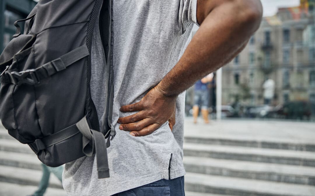
A senior man working in the office and having stomach pain
Stomach issues can range from minor annoyances to serious health concerns that affect daily life. Many people wonder whether to see their primary care doctor or a specialist when experiencing digestive discomfort. A primary care physician (PCP), also known as a general practitioner or family doctor, manages overall health and addresses common problems. In contrast, a gastroenterologist (GI doctor) has additional training to diagnose and treat complex digestive system conditions, including the esophagus, stomach, intestines, liver, and more. Understanding when to choose one over the other can help you get the right care faster and avoid unnecessary worry.
This article explores the key differences, the symptoms that guide your decision, and alternative options such as integrative care from nurse practitioners and chiropractors. We’ll also draw on clinical insights from experts, including Dr. Alexander Jimenez, to provide a well-rounded view.
Starting with Mild or New Digestive Issues: See Your Primary Care Doctor
For many stomach problems, your first stop should be a PCP. These doctors are trained to manage a wide array of health issues, including basic digestive complaints. They can perform initial exams, order simple tests, and prescribe treatments for everyday problems. If the issue proves more complex, they can refer you to a specialist.
Primary care doctors often treat short-lived or mild symptoms effectively. For example, if you have a brief episode of stomach flu, they can recommend hydration and rest. They also address common conditions such as occasional heartburn or mild constipation with over-the-counter remedies or lifestyle changes. This approach saves time and money, as PCP visits are usually easier to schedule and less specialized.
Here are some common scenarios where a PCP is the best choice:
Short-term stomach flu: If you have sudden nausea, vomiting, or diarrhea that lasts a few days, a PCP can check for dehydration and suggest fluids or anti-nausea meds.
Mild or occasional heartburn: Burning in your chest after meals, especially if it occurs rarely, can often be managed with dietary adjustments, such as avoiding spicy foods.
Light constipation: If you’re having trouble with bowel movements but it’s not chronic, a PCP might recommend more fiber or exercise.
Simple stomach aches: General discomfort from gas, indigestion, or overeating usually resolves with basic care from your regular doctor.
According to health experts, primary care providers can evaluate or begin treatment for mild or acute symptoms, such as occasional digestive upsets. They focus on your overall health, considering how stomach issues may be linked to other factors such as stress or medications. If symptoms don’t improve, they guide you to the next step.
PCPs play a key role in improving gut health through preventive measures. They can discuss diet, screen for basic issues, and monitor ongoing mild problems. In some cases, if symptoms persist, they may order tests such as blood work before referring you. This holistic oversight ensures nothing is overlooked early on.
When Symptoms Are Serious or Ongoing: Time for a Gastroenterologist
If your digestive problems are persistent, severe, or accompanied by warning signs, it’s best to see a gastroenterologist. These specialists complete additional years of training beyond medical school, specializing in the digestive tract. They use advanced tools, such as endoscopies and colonoscopies, to identify and treat conditions that a PCP may not address on their own.
Gastroenterologists are experts in conditions affecting the esophagus, stomach, small and large intestines, liver, pancreas, and gallbladder. They can manage chronic diseases and perform procedures to remove polyps or biopsy tissues. If you’re over 45, they often recommend routine screenings to catch problems early.
Key signs that point to needing a GI doctor include:
Trouble swallowing: If food feels stuck or causes pain, this may indicate esophageal issues such as GERD or strictures.
Constant abdominal pain: Ongoing discomfort that doesn’t respond to basic treatments may indicate ulcers, gallstones, or inflammation.
Blood in stool or rectal bleeding: Red or black stools can be a red flag for hemorrhoids, polyps, or even cancer.
Unexplained weight loss: Losing pounds without trying, especially with appetite changes, needs specialist evaluation.
Chronic diarrhea: Loose stools lasting more than four weeks may indicate IBS, IBD, or infection.
Recurrent heartburn: If it occurs frequently and over-the-counter medications don’t help, it may be GERD requiring advanced care.
Age 45 or older for screening: Even without symptoms, a colonoscopy is advised to prevent colorectal cancer.
Experts note that symptoms like rectal bleeding, frequent heartburn, or changes in bowel habits warrant a visit to a gastroenterologist for specialized care. For instance, ongoing diarrhea or constipation might stem from disorders like irritable bowel syndrome (IBS) or small intestinal bacterial overgrowth (SIBO), which GIs can diagnose with targeted tests.
Gastroenterologists also handle liver-related problems, such as fatty liver disease or hepatitis, and pancreatic issues like pancreatitis. Their training enables them to identify subtle signs that could lead to serious conditions if left unaddressed. If you have a family history of digestive diseases, seeing a GI early can be crucial for prevention.
Not Sure Where to Start? Begin with Your PCP for Guidance
If you’re unsure about your symptoms, it’s always safe to start with a primary care doctor. They act as your health coordinator, assessing the issue and deciding if a referral is needed. This step prevents jumping straight to a specialist when a simple fix might suffice.
PCPs can run initial tests, like stool samples or X-rays, to rule out common causes. If results show something unusual, they’ll refer you to a GI doctor. This system ensures efficient care and avoids overwhelming specialists with minor cases.
For example, mild heartburn might be managed by a PCP with lifestyle advice, but if it’s chronic, they’ll send you for further evaluation. Starting here also builds a complete health record, helping any specialist understand your full picture.
Exploring Integrative Options: Nurse Practitioners and Chiropractors for Holistic Care
Beyond traditional doctors, integrative approaches offer another path for managing stomach problems. Nurse practitioners (NPs), especially in functional or integrative medicine, provide patient-centered care with more time for in-depth discussions. They focus on root causes such as diet, stress, sleep, and nutrient deficiencies, often ordering advanced tests such as microbiome mapping or food sensitivity panels.
Functional medicine differs from conventional medicine in that it places greater emphasis on history and uses lab tests to address imbalances in the gut microbiome or leaky gut. NPs create personalized plans emphasizing whole foods, reduced sugar, and lifestyle changes to reduce inflammation and support digestion.
Integrative chiropractors take a whole-body view, linking spinal health to digestion through the gut-brain connection. Misalignments, or subluxations, can disrupt nerves that control the digestive system, leading to symptoms such as bloating or constipation. Adjustments restore nerve function, improve posture, and enhance blood flow to organs.
Key ways chiropractors help:
Gut-brain connection: Aligning the spine supports the autonomic nervous system, balancing stress responses that affect digestion.
Manual therapies: Techniques such as visceral manipulation gently realign organs to ease pain and improve movement.
Lifestyle guidance: Advice on anti-inflammatory diets, supplements, and exercises to boost gut health.
Studies show that chiropractic care can alleviate symptoms such as indigestion and abdominal pain by improving gastrointestinal function. At centers like Highland Wellness, precise adjustments promote nutrient absorption and reduce digestive disorders holistically.
Insights from Dr. Alexander Jimenez on Integrative Digestive Care
Dr. Alexander Jimenez, DC, APRN, FNP-BC, IFMCP, CFMP, brings over 30 years of experience in integrative chiropractic and functional medicine. He emphasizes addressing the root causes of digestive issues through detailed assessments of genetics, lifestyle, and environmental factors. His approach combines chiropractic adjustments with nutrition and detox protocols to treat chronic conditions like inflammation and autoimmunity, which often affect the gut.
Dr. Jimenez highlights the gut-brain connection, noting that spinal misalignments can affect digestion through nerve signals to the immune and endocrine systems. He uses non-invasive methods, such as spinal decompression and exercises, to restore balance and reduce symptoms. For instance, patients with back pain and digestive complaints benefit from movement-based recovery that links spine and gut health.
In his functional medicine practice, Dr. Jimenez promotes personalized nutrition to prevent chronic diseases and support gut microbiota. He integrates therapies such as acupuncture and stress management, referring patients to specialists as needed for collaborative care. His work underscores that holistic methods can complement traditional care, focusing on long-term wellness rather than just symptoms.
Balancing Traditional and Integrative Approaches for Better Outcomes
Combining PCPs, GIs, and integrative providers offers the best results for many. A PCP might start with basics, a GI handles diagnostics, and an NP or chiropractor adds lifestyle support. This team approach addresses both immediate symptoms and underlying causes.
For chronic issues like IBS, functional medicine’s focus on diet and stress can reduce flare-ups alongside GI treatments. Chiropractic care may alleviate pain associated with nerve issues, improving overall comfort.
Preventive care is key: regular check-ups with a PCP, GI screenings, and holistic habits help prevent escalation. Listen to your body—if symptoms change, seek help promptly.
In summary, for mild or new stomach problems, see a PCP. For chronic or severe ones, consult a gastroenterologist. Integrative options provide added support. Always prioritize your health by starting with professional advice.
Beginner Gym Workout Routine: Build Strength, Flexibility, and Avoid Injuries
Young hispanic man does a beginner gym workout with weights.
Starting a workout at a sports training gym can feel exciting but also a bit scary if you are new to it. A good beginner routine helps build strength in all parts of your body. It uses big movements that work many muscles at once. These are called compound exercises. Things like squats, lunges, push-ups, rows, and planks are key. Do this routine three times a week. Each exercise should have three sets of eight to twelve reps. This builds a strong base without too much strain (Planet Fitness, n.d.a).
The goal is to mix full-body strength training with some easy cardio. Low-impact cardio means activities that do not jar your joints too much, such as walking on a treadmill or using an elliptical. This helps you get fit without overdoing it. Adding chiropractic care can make it even better. It helps with movement, cuts injury risk, and speeds up recovery. Let’s break this down step by step.
Why Start with a Balanced Routine?
A good starting plan focuses on functional strength. This means exercises that help with everyday activities, like picking things up or climbing stairs. For beginners, full-body workouts are best. They work all major muscle groups without splitting days for arms or legs only. This way, you recover faster and see progress soon (Mikolo, 2024).
Experts say beginners should aim for consistency over intensity. Start slow to learn proper form. Bad form can lead to hurts. A routine with strength and cardio boosts heart health, muscle tone, and energy. It also helps with weight control and mood. But without good recovery, you might get sore or injured. That’s where things like stretching and chiropractic come in.
Key Exercises for Beginners
Here are some top exercises for a sports training gym. They build strength, flexibility, and stability. Most use bodyweight or simple machines. Do them in order for a full workout.
Squats: Stand with feet shoulder-width apart. Bend your knees and lower yourself as if you were sitting in a chair. Keep your chest up and knees over toes. Push back up. This works legs, glutes, and core (Refinery29, 2020).
Lunges: Step forward with one foot. Lower until both knees are bent at 90 degrees. Push back to start. Alternate legs. This exercise is beneficial for enhancing balance and building leg strength (Kong, 2024).
Push-ups: Start on your hands and toes or on your knees. Lower your chest to the ground, then push up. This hits the chest, arms, and shoulders. Modify by using a wall if needed (Magnus Method, 2023).
Rows: Use a machine or dumbbells. Pull weights toward your body, squeezing your shoulder blades. This exercise enhances back strength and improves posture (Planet Fitness, n.d.b).
Planks: Hold a push-up position on forearms. Keep your body straight. Hold for 20-30 seconds. Strengthens core for stability (Quora, n.d.).
Do three sets of 8-12 reps for each, except planks, which are timed. Rest 60 seconds between sets. Warm up with 5 minutes of light walking first.
Sample Weekly Routine
A three-day plan works well for beginners. Space days out, like Monday, Wednesday, and Friday. This gives time to rest. Each session lasts 30-45 minutes.
Day 1: Full Body Strength Focus
Warm-up: 5 min treadmill walk.
Squats: 3 sets of 10 reps.
Push-ups: 3 sets of 8 reps.
Rows: 3 sets of 10 reps.
Planks: 3 holds of 30 seconds.
Cool-down: Stretch legs and arms.
Day 2: Rest or Light Cardio
Walk or bike for 20 minutes at an easy pace.
Day 3: Lower Body Emphasis
Warm-up: 5 min elliptical.
Lunges: 3 sets of 10 per leg.
Glute bridges: 3 sets of 12.
Calf raises: 3 sets of 15.
Planks: 3 holds of 30 seconds.
This builds on basics. As you get better, add weights (Under Armour, n.d.). Track your progress in a notebook.
Adding Cardio for Endurance
Cardio is key for heart health and stamina. For beginners, start low-impact. Use machines like a treadmill or a rower. Aim for 15–20 minutes after strength training. Walk at a 5-8% incline on a treadmill to build legs without running (Kong, 2024). This burns calories and boosts recovery.
Mix it in: Do cardio on off days or at the end of your workout. Things like jumping jacks or brisk walking work too. Cardio helps with overall fitness, but do not overdo it. Too much can tire you out.
The Role of Integrative Chiropractic Care
Integrative chiropractic care is more than just spinal cracks. It looks at the whole body. Dr. Alexander Jimenez, a chiropractor with over 30 years of experience, notes it helps with injury prevention and better movement (Jimenez, n.d.a). He combines adjustments with exercises and nutrition.
For beginners, it identifies hidden issues such as muscle imbalances. These can lead to injuries if ignored. Adjustments fix joint problems, improving the range of motion. This lets you do exercises with better form (Pushasrx, n.d.).
Dr. Jimenez observes that chiropractic boosts nerve function. This helps muscles adapt faster and cuts pain. In his clinic, he uses functional assessments to identify weaknesses early (Jimenez, n.d.b). For sports training, it keeps you going without breaks.
Benefits of Chiropractic for Gym Beginners
Chiropractic makes starting safer. Here are key perks:
Injury Prevention: Spots imbalances before they hurt. Fixes tight muscles or stiff joints (Atlas Total Health, 2022).
Better Mobility: Improves joint range. It helps with squats or lunges without causing strain (Elevate to Life, 2023).
Faster Recovery: Uses soft-tissue work and exercises to help you heal more quickly. It also helps reduce soreness after workouts (Team Elite Chiropractic, 2022).
Dr. Jimenez stresses holistic care. He integrates chiropractic care with fitness, such as HIIT, to build strength. This prevents chronic issues and boosts performance (Jimenez, n.d.a).
When to Get Chiropractic Adjustments
Timing matters. Get adjusted before workouts to optimize nerve and muscle function. This prevents strain. After workouts, it aids recovery by reducing inflammation (Atlas Total Health, 2022). Dr. Jimenez recommends regular visits for long-term health.
Do at-home exercises too. Things like glute bridges or cat-cow stretches support treatment (Elevate to Life, 2023). These speed healing and keep balance.
Recovery Tips to Stay Injury-Free
Recovery is as important as working out. Add these to your routine:
Stretching: Do dynamic stretches before and static stretches after. This practice enhances your flexibility, according to 10 Fitness (n.d.).
Rest Days: Allow muscles to grow. Walk lightly if active rest.
Corrective Exercises: Fix imbalances. Hip openers or spine mobilizations prevent injury (Asheville Medical Massage, 2025).
Nutrition and Sleep: Eat protein-rich foods. Sleep 7-9 hours for repair (Squatwolf, n.d.).
If injured, stay fit with low-impact activities like swimming. Balance activity to heal (RP3 Rowing, n.d.). Chiropractic helps here, too, per Dr. Jimenez.
Putting It All Together
A good beginner workout at a sports training gym mixes strength, cardio, and care. Start with compounds three times a week. Add chiropractic for safety. Dr. Jimenez’s work shows this approach builds a strong, injury-free base (Jimenez, n.d.b). Stay consistent, listen to your body, and progress slowly. This makes fitness fun and lasting.
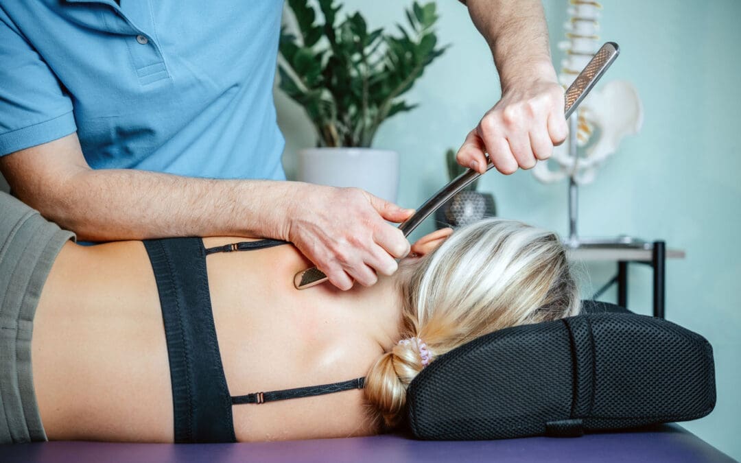
“Cleanse” Yourself of Stress? A Real-World Stress Detox Guide for El Paso Back Clinic Patients
A chiropractor/Nurse practitioner massages a patient’s neck, who is suffering from headaches, neck and shoulder pain, and stress.
Many people type things like “stress cleanse,” “mental detox,” or “cortisol detox” into Google because they feel worn down and want a reset that actually works. The good news is: yes, it’s possible to reduce stress load and feel calmer. The key is to define “cleanse” correctly.
A true “stress cleanse” is not about flushing cortisol out of your body like it’s poison. Cortisol is a normal hormone your body needs. What you can do is reduce constant triggers that keep cortisol and adrenaline elevated, improve sleep, relax tight muscles, and help your nervous system shift into recovery mode. That’s the practical meaning of “detox from stress,” and it’s the version that’s most helpful.
This article is geared for people in El Paso who want a clear, step-by-step plan—especially if stress is showing up as neck tension, headaches, shallow breathing, low back pain, poor sleep, or anxiety-like symptoms.
What “Stress Detox” Really Means (In Plain Language)
A “stress detox” is best thought of as a nervous system reset:
Less time stuck in fight-or-flight (sympathetic mode)
More time in rest-and-digest (parasympathetic mode)
Better sleep and recovery
Less muscle tension and pain flare-ups
More stable mood and focus
The Centers for Disease Control and Prevention emphasizes core stress-management basics that are simple yet powerful: move more, get enough sleep, and use healthy coping tools.
Why Stress Feels “Physical” (And Why Your Back Can Hurt More)
Stress is not “all in your head.” Chronic stress changes how your body holds itself:
Shoulders creep up
Jaw tightens
Breathing becomes shallow
Hip flexors and low back muscles stay tense
Sleep gets lighter, so your tissues recover less
Over time, those patterns can feed into pain cycles—especially in the neck and low back.
El Paso Back Clinic has written about the connection between chronic stress and physical tension, including how spinal alignment and supportive care may help reduce stress-related strain.
Cortisol: Helpful Hormone, Not a Toxin
Cortisol gets a bad reputation online. Here’s the reality:
Cortisol is needed for normal life (energy, metabolism, immune regulation)
It naturally rises and falls during the day
“High cortisol” as a trendy diagnosis is often oversimplified
A recent report found that most people don’t need to obsess over “controlling cortisol” in response to viral trends. When cortisol-related conditions are present, they require medical diagnosis and management—not detox drinks or random supplements.
So if you want a “cortisol detox,” the safest, most effective approach is: build habits that lower chronic stress signals and improve recovery.
The Nervous System Switch You’re Trying to Flip
Think of your body as having two main settings:
Fight-or-flight (Sympathetic)
Muscles brace
Heart rate rises
Digestion slows
Sleep gets lighter
Pain sensitivity can increase
Rest-and-digest (Parasympathetic)
Breathing slows
Digestion improves
Muscles soften
Sleep deepens
Recovery improves
Your “stress cleanse” goal is to spend more time in the rest-and-digest phase.
The Pillars of a Real Stress Cleanse
Sleep: The Fastest Way to Reset Stress Chemistry
If sleep is poor, everything gets harder—pain, mood, cravings, focus.
The CDC recommends that adults get 7+ hours of sleep and maintain a consistent sleep schedule.
Simple sleep upgrades
Keep the same wake-up time most days
Dim lights 60 minutes before bed
Keep the room cool and dark
Put your phone away (or at least out of your hand)
AdventHealth also supports the idea of a “mental cleanse” by encouraging people to step away from electronic devices and use calming routines to feel more whole.
Movement: Your Body Was Designed to Burn Off Stress
Exercise doesn’t have to be intense to help.
The CDC suggests building toward 2.5 hours per week (150 minutes), and even 20–30 minutes a day helps.
Good stress-friendly movement options
Brisk walking
Light strength training
Mobility work
Yoga or stretching
Mayo Clinic lists being active, eating well, laughing, connecting with others, and mindfulness as practical stress relievers.
Breathing: A “Remote Control” for Your Nervous System
When stress rises, breathing often becomes fast and shallow. Changing your breath can send a safety signal to your brain.
Try this for 2 minutes
Inhale through your nose for 4 seconds
Exhale slowly for 6–8 seconds
Repeat 10 rounds
This is simple, free, and surprisingly powerful when done daily.
Nutrition + Hydration: Don’t Let Blood Sugar Create More Stress
When you’re stressed, it’s easy to skip meals, snack on sugar, or live on caffeine. That rollercoaster can worsen feelings of anxiety.
Healthline outlines practical ways to support healthier cortisol patterns: sleep, regular exercise, balanced eating, and stress-management habits.
Supportive “stress reset” foods
Protein with breakfast (eggs, Greek yogurt, beans, tofu)
Fiber daily (berries, oats, vegetables, lentils)
Healthy fats (olive oil, avocado, nuts, salmon)
Plenty of water
Boundaries: The Most Underrated Detox Tool
A big source of chronic stress is constant availability—work messages, social media, and nonstop news.
Healthy boundary examples
No work email after 7 pm
A 30-minute phone-free morning
One tech-free block each day
A weekly “low stimulation” afternoon
This is the kind of change that reduces stress at the source rather than trying to “out-supplement” it.
Support: Your Nervous System Regulates Better With Safe People
Humans calm down faster when they feel supported.
Even one meaningful conversation per day can help lower your baseline stress.
Where Integrative Chiropractic Care Fits at El Paso Back Clinic
Let’s keep it honest and grounded: chiropractic care doesn’t “detox your liver.” Your liver and kidneys already do detoxification.
But chiropractic and integrative care can be very helpful when stress shows up as tension, posture strain, restricted movement, or pain that keeps your nervous system on high alert.
Henry Ford Health describes chiropractic care as a way to address physical tension patterns linked with stress, including muscle tightness and posture-related strain.
El Paso Back Clinic similarly discusses chiropractic care as part of a plan to manage chronic stress and support the body’s relaxation.
What this can look like in real life
A practical integrative plan often includes:
Chiropractic adjustments (when appropriate)
Soft tissue work/myofascial release
Mobility + rehab exercises
Posture and ergonomic coaching
Lifestyle support (sleep, movement, nutrition)
Some clinics also describe “detox pathways” in more holistic terms; the safest way to interpret this is to improve movement, circulation, breathing mechanics, and recovery habits.
A 24-Hour “Stress Cleanse” You Can Do This Week
This is for those moments when you feel overloaded and need a reset.
Morning
Wake up at a normal time
Drink water
Eat protein + fiber
10–20 minutes outside
10-minute walk
Midday
Lunch with protein + vegetables
5 minutes breathing
30–60 minutes phone-free block
Afternoon
20–40 minutes of movement
Short social connection (text/call someone safe)
Evening
Light dinner
No doom scrolling
Warm shower + dim lights
Go to bed at a consistent time
Mayo Clinic supports many of these basics (movement, healthy diet, breathing, time in nature, and reducing screen time).
A 7-Day Stress Detox Plan (Simple and Repeatable)
Daily anchors (do these every day)
20–30 minutes of movement
7+ hours sleep goal
5 minutes of slow breathing
Whole-food meals most of the time
Add one upgrade per day
Day 1: No phone for the first 30 minutes
Day 2: Caffeine cutoff (try noon)
Day 3: 20 minutes nature
Day 4: Mobility + stretching session
Day 5: Strength training (light/moderate)
Day 6: Support day (social connection)
Day 7: Plan your next week’s boundaries
Some lifestyle blogs promote “stress detox” challenges; use the helpful parts (movement, mindfulness, routine) and skip anything extreme or guilt-based.
When Stress Symptoms Need Medical Evaluation
Please don’t “detox” your way past red flags. Seek medical help if you have:
Chest pain, fainting, severe palpitations
Severe insomnia lasting weeks
Panic attacks that feel unmanageable
Depression symptoms, hopelessness, or thoughts of self-harm
Unexplained weight loss or major hormone concerns
If you’re worried about a true cortisol disorder, don’t self-diagnose with social media trends—get evaluated clinically.
Local CTA for El Paso Readers
If stress is showing up as neck pain, headaches, shallow breathing, shoulder tension, or low back flare-ups, a combined approach may help—especially when care includes both physical treatment and lifestyle coaching.
To learn more about your options, explore the clinic’s stress-related resources and integrative care topics on the El Paso Back Clinic website.
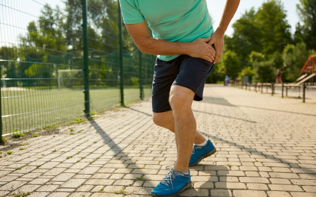
Anterior Hip and Leg Muscles: What They Are, What They Do, and Why They Hurt
A woman holds her aching anterior hip.
Pain in the front of the hip (often felt in the hip crease or groin area) and the front of the thigh is very common. It can show up when you stand up from a chair, climb stairs, run, kick, or even after sitting for a long time. The tricky part is this: front-hip pain is not always “just a tight hip flexor.” Sometimes it’s a muscle or tendon problem, but it can also be related to the hip joint, the pelvis, or the lower back.
This guide is written for everyday people in El Paso who want clear answers, plus a practical explanation of how an integrative chiropractic approach can help reduce pain and prevent flare-ups.
At El Paso Back Clinic, Dr. Alexander Jimenez and the team often observe a pattern: tight, overworked hip flexors, underactive glutes, and poor pelvic control—especially in people who sit a lot, train hard, or are recovering after an accident.
What “anterior hip and leg muscles” means
“Anterior” means the front side. The anterior hip and leg muscles are basically your “go-forward” and “stand-tall” muscles. They help you:
Lift your knee (hip flexion)
Step forward when walking or running
Stabilize your pelvis so your lower back doesn’t overwork
Straighten your knee (knee extension)
Control your leg when you climb stairs or squat
When these muscles get overloaded, they can feel tight, sore, weak, or sharp—depending on the cause.
The main anterior hip muscles (your hip flexors)
Hip flexors are not one muscle. They’re a group that works together.
Key hip flexor muscles
Iliopsoas (iliacus + psoas): the classic “deep hip flexor”
Rectus femoris: part of the quadriceps, crosses the hip and the knee
Sartorius: a long, strap-like muscle across the front of the thigh
Tensor fasciae latae (TFL): supports hip flexion and pelvic control
Pectineus (often grouped with hip flexors in clinical discussions)
Why iliopsoas matters so much
The iliopsoas helps:
Lift the thigh toward the trunk
Support the hip joint and pelvis
Add stability near the lumbar spine/pelvis connection
At El Paso Back Clinic, iliopsoas overuse is commonly discussed among athletes and active individuals who engage in sprinting, jumping, kicking, or repeated hip flexion.
The anterior thigh muscles (front of the thigh)
The main anterior thigh group is the quadriceps. They’re designed to extend the knee and help control motion during walking, stairs, squats, and landing.
Quadriceps muscles
Rectus femoris
Vastus medialis
Vastus lateralis
Vastus intermedius
The anterior thigh compartment is also supplied and controlled by key anatomical structures, such as the femoral nerve (often described as the L2–L4 roots) and the femoral artery system. That’s one reason pain patterns can sometimes feel confusing—muscles, nerves, and joints all influence the sensation you feel.
Why the anterior hip and leg muscles sometimes hurt sometimes
There are a few “big buckets” that explain most front-hip and front-thigh pain.
You’re asking the muscles to do too much, too often (overuse)
Overuse happens when the workload increases faster than your tissues can adapt. Common triggers include:
Sudden jump in running miles
More hills or speed work than usual
Lots of kicking (soccer, martial arts)
Heavy squats/lunges with poor control
Repetitive direction changes (basketball, football)
Overuse can irritate:
The muscle belly (soreness, tightness)
The tendon (tendinopathy-like pain)
The hip flexor attachment area near the front of the hip
Prolonged sitting keeps hip flexors in a “shortened” position
Sitting puts the hips into flexion. Over time, many people notice:
Hip tightness when standing up after sitting
A “pinchy” feeling in the front of the hip
Low back stiffness that shows up with hip tightness
Dr. Jimenez has emphasized in his recent writing that prolonged sitting can contribute to tight hip flexors and poor movement patterns, and that short movement breaks, along with targeted mobility work, can help many people feel better.
The hip flexors can be tight because other muscles are not doing their job
This is one of the most common “root causes” in stubborn cases:
Weak or underactive glutes
Weak deep core stabilizers
Limited hip mobility (the hip joint doesn’t move well)
Pelvic control issues (pelvis tips forward, rotates, or drops during gait)
El Paso Back Clinic explains that when the glutes weaken from inactivity and prolonged sitting, the hips and pelvis can become less stable and shift out of alignment, thereby increasing stress on surrounding tissues.
Sometimes the pain is not in the hip flexor at all
A major clinical point from family medicine guidelines is that hip pain often groups into:
Anterior (front)
Lateral (side)
Posterior (back)
…and the cause changes based on that pattern. Anterior hip pain may result from hip flexor injury, but it can also result from intra-articular hip joint problems (such as femoroacetabular impingement or labral pathology) or from referred pain.
A helpful “body map” concept is presented in educational videos that discuss what different hip pain locations can indicate, but a hands-on evaluation remains important when symptoms persist.
What the pain feels like: common patterns that guide the next step
These are not perfect rules, but they help you decide whether you’re dealing with a likely muscle/tendon issue or something deeper.
More likely muscle/tendon irritation (common hip flexor pattern)
Pain in the front hip crease
Worse with lifting the knee (stairs, marching)
Worse with running sprints, kicking, or hills
Tenderness in the front hip region
Feels tight after sitting
More likely hip joint involvement
Deep groin pain with hip rotation
Catching, clicking, locking, or “pinching”
Pain that persists despite basic stretching/rest
Range of motion feels blocked (especially flexion + rotation)
More likely low back/nerve referral
Front thigh pain plus low back symptoms
Numbness, tingling, and burning sensations
Symptoms that change with spine position
Why “stretching only” often fails
Stretching can feel good short-term, but it may not solve the real driver if the problem is:
Weak glutes and weak core control
A stiff hip joint or pelvic restriction
Poor movement strategy (how you squat, run, or stand)
A training load problem (too much too soon)
In other words, the hip flexors may be tight because they’re protecting you or compensating for something else.
How El Paso Back Clinic approaches anterior hip and leg pain
El Paso Back Clinic describes an integrative model that blends chiropractic care, rehabilitation concepts, and movement-based strategies, with a focus on mobility, flexibility, and the restoration of balanced function.
Here’s how that “integrative” approach commonly helps front-hip and front-thigh problems.
Identify the true driver (not just the sore spot)
A good evaluation typically includes:
History (training, sitting, injury, accident history)
Differentiation between hip joint vs. lumbar referral patterns
Dr. Jimenez has written about the importance of a structured hip evaluation to sort out the likely source of pain and match care to the pattern.
Restore joint motion and reduce protective “guarding”
When the pelvis/hip/lumbar spine isn’t moving well, the body often shifts load to the hip flexors and quads. Chiropractic-style care may focus on restoring smoother motion so the muscles stop overworking.
El Paso Back Clinic also discusses how muscle imbalance and chronic guarding can make it harder for muscles to “relax on their own,” especially after injuries.
Use soft tissue + targeted techniques to normalize muscle function
A common strategy is pairing hands-on care with neuromuscular techniques. El Paso Back Clinic specifically discusses assessing hip flexors with MET therapy (muscle energy technique) as part of reducing tightness and improving hip mobility.
Rebuild strength where it matters (glutes + core + hip control)
To prevent recurrence, the plan usually includes strengthening and control, especially:
Glute bridges and progressions
Hip abduction strength (side-lying or banded work)
Gradual reloading of hip flexors (instead of only stretching)
El Paso Back Clinic’s content repeatedly emphasizes that restoring balanced muscle function around the pelvis and hips supports daily movement and performance.
Practical tips you can start today (safe, simple, and realistic)
If your symptoms are mild and you’re not dealing with red flags, these are common first steps.
For desk workers and drivers (very common in El Paso)
Take 1–2 minute movement breaks every 30–60 minutes
Do a gentle hip flexor stretch (no sharp pinching)
Add a glute activation move (bridges or mini-band walks)
Keep your daily steps consistent (don’t go from 2,000 to 12,000 overnight)
For runners and athletes
Reduce aggravating volume for 1–2 weeks (not “stop forever,” just calm it down)
Avoid sprinting/kicking if it spikes sharp pain
Strengthen glutes and hip stabilizers 2–3x/week
Return to speed and hills gradually, not all at once
Quick self-check idea (mobility clue)
The Thomas Test is commonly used to screen for hip flexor tightness and may help distinguish whether the “tight feeling” is more iliopsoas- or quadriceps-based (rectus femoris). It’s not a diagnosis, but it can be a clue.
When you should get evaluated sooner rather than later
Don’t try to “stretch through it” if you have:
Severe pain after a fall or accident
Inability to bear weight
Fever or feeling unwell with hip pain
Worsening numbness/tingling or leg weakness
Persistent catching/locking and deep groin pain
A structured clinical examination is particularly important when hip pain may involve the hip joint or referral patterns.
The main takeaway
Your anterior hip and leg muscles—especially the hip flexors and quadriceps—are essential for walking, running, stairs, and posture. They often hurt because of:
Too much repeated load (overuse)
Too much sitting (hip flexors stay shortened)
Muscle imbalance (weak glutes/core causing hip flexors to overwork)
Hip joint or low back referral (pain “shows up” in the front)
An integrative chiropractic model—such as the one described in El Paso Back Clinic’s educational resources—focuses on identifying the underlying cause, restoring motion, improving muscle balance, and developing a plan to reduce the likelihood of recurrence.
Is It Safe to Wear a Backpack? Expert Tips on Spinal Health and Back Pain Prevention in the US and El Paso, TX
A woman walking, wearing a backpack with the recommended weight, and maintaining correct posture to prevent back pain and problems.
Back pain is a big issue for many people in the United States
Up to 80% of adults face low back pain at some point in their lives. This is one of the top reasons for doctor visits and missed workdays. The cost is huge too, with over $100 billion spent on spine problems each year. In El Paso, Texas, where people often have active jobs like industrial work or lots of driving, back pain questions focus on things like sciatica, herniated discs, and spinal stenosis. A common concern across the country, including in places like El Paso, is whether wearing a backpack is safe for the spine. The good news is that it can be safe if you follow some simple rules. This article focuses on backpack safety and then addresses other key questions about managing back pain, treatment options, and daily habits to keep your spine healthy.
Understanding Backpack Safety and Spinal Health
Wearing a backpack is common for carrying things, but if it’s too heavy or worn incorrectly, it can hurt your back. Heavy backpacks can strain muscles and joints in your back, neck, and shoulders. This might lead to pain or bad posture over time. However, backpacks do not cause scoliosis, a spinal curvature that affects about 2% to 3% of people. Scoliosis often starts in teens and is more common in girls, but it’s not linked to backpacks.
Is it safe? Yes, as long as you distribute the weight right and follow the tips to avoid strain. Improper use can cause muscle fatigue, poor posture (such as slouching), and even chronic pain if left unaddressed. In El Paso, where people might carry tools or bags for work, this is especially important to prevent issues such as sciatica, where pain radiates down the leg due to nerve pressure.
Here are some key tips for safe backpack use:
Choose the right backpack: Pick one with wide, padded straps and a padded back. It should fit your body size and have a waist strap for heavy loads. Lightweight materials help too.
Limit the weight: Keep the backpack under 10-15% of your body weight. For example, if you weigh 150 pounds, aim for no more than 15-22.5 pounds.
Distribute weight evenly: Put heavier items at the bottom and close to your back. Use compartments to balance things and stop shifting.
Wear it correctly: Always use both straps. Adjust them so the pack sits in the middle of your back, not sagging low. Bend your knees to lift it.
Make smart choices: Remove extra items often. Use lockers or storage if possible. For very heavy loads, try a rolling backpack or crossbody bag.
These steps help distribute the load across your strong back muscles and keep your spine aligned. If you feel pain, stop and adjust. In places like El Paso, with busy lifestyles, following these can help prevent accidents from becoming long-term back issues.
Common Causes of Back Pain in the US
Back pain affects millions. In the US, about 26% of adults have it at any time, and it’s more common after age 45. Among adults aged 50 and older, up to 45.6% experience it. Causes include muscle strains, ligament injuries, herniated discs (where the disc’s soft center protrudes), arthritis, and spinal stenosis (where the spinal canal narrows). Stress can make it worse by causing muscle spasms. Even factors such as obesity or infections can play a role.
Chronic back pain lasts more than 3 months and affects 8% of adults. It often comes from wear and tear on discs or joints. Poor sleep makes it worse because pain disrupts rest, and lack of sleep raises inflammation. In the US, this results in high costs, such as lost work and medical bills.
Symptoms vary. You might feel an ache in your lower back or sharp pain if it’s sciatica. Numbness, tingling, or weakness in the legs are red flags. Scoliosis, which affects 7 million Americans, can cause symptoms such as uneven shoulders or back pain; most cases are mild.
Muscle or ligament strain: From lifting incorrectly or sudden moves.
Disc problems: Bulges or herniations press on nerves.
Arthritis: Joint wear is common in older people.
Stenosis: Narrowing squeezes nerves, causing leg pain.
Stress and lifestyle: Tension builds up, leading to spasms.
Knowing these helps prevent pain. For example, strengthening your core muscles supports your spine and reduces strain from daily activities like wearing a backpack.
Managing Chronic Back Pain
Chronic back pain needs long-term plans. First, see if it’s new or ongoing. Most cases improve with rest and simple fixes, but if it lasts, get checked. Avoid bed rest; gentle movement helps recovery faster.
Daily habits matter. Exercise like walking or swimming builds strength. Maintain a healthy weight to reduce spinal load. Quit smoking, as it negatively affects spinal tissues and raises surgery risk by up to 50%. Good posture and ergonomic setups at work prevent strain.
In El Paso, with industrial jobs and driving, pain from accidents is common. Recovery focuses on building habits to avoid re-injury.
Stay active: Low-impact exercises like yoga or Pilates.
Watch your diet: Healthy foods reduce inflammation.
Manage stress: Deep breathing or mindfulness helps.
Sleep well: Use pillows to maintain spinal alignment.
Stretch daily: Loosen tight muscles, such as the hamstrings.
These steps reduce pain and improve quality of life.
Treatment Options: Surgery vs. Conservative Care
When pain doesn’t go away, choices include conservative care or surgery. Conservative means non-surgical options such as physical therapy, medications, injections, chiropractic care, or massage. These are tried first for 8-12 weeks. Surgery is indicated for severe cases, such as nerve damage or instability.
Ask your doctor: What causes my pain? What tests do I need? What are the risks and benefits? For surgery, ask about the surgeon’s experience, recovery time, and whether you’ll need help at home. Alternatives like spinal decompression stretch the spine to ease disc pressure.
Chiropractic vs. orthopedic: Chiropractors focus on spinal adjustments to realign the spine and relieve pain without medication. Orthopedists may recommend surgery for significant issues. Both can help, but chiropractic care is well-suited to conservative care.
In El Paso, many choose chiropractic for herniated discs or sciatica. It’s safe and effective for back pain, reducing symptoms by fixing alignment and boosting blood flow.
Spinal Health in El Paso, TX
El Paso has unique needs. Active lives, work injuries, and car accidents lead to questions about sciatica, where nerve pain goes down the leg, or spinal stenosis with leg weakness. Herniated discs are common from lifting or falls.
Lumbar stenosis FAQs: It causes leg pain or numbness when walking. Avoid high-impact exercises like running; try swimming instead. Treatments include therapy or decompression.
Local care often combines chiropractic and orthopedic care. Dr. Alexander Jimenez, a chiropractor in El Paso with over 30 years of experience, notes that integrative care is most effective. He uses adjustments, nutrition, and therapy for root causes. For example, a worker’s back pain improved by 50% within weeks with his plan. He stresses non-surgical options for sciatica and injuries, helping people stay active in El Paso’s environment.
Sciatica: From disc pressure; chiropractic eases it.
Chiropractic: Aligns the spine, safe for all ages.
Dr. Jimenez’s work shows personalized plans reduce pain without surgery.
Daily Habits to Prevent Spinal Injury
Preventing pain starts with habits. Lift by bending knees, not back. Stand every 15 minutes if sitting for long. For driving in El Paso, take breaks to stretch.
Core strength is key. Exercises like planks support your spine. Avoid smoking for better healing. Ergonomics: Screen at eye level, chair with back support.
For backpacks, combine with these: Even weight helps posture.
Lift right: Knees bent, close to body.
Posture: Stand tall, no slouch.
Exercise: Core and back focus.
Weight control: Less strain on the spine.
Breaks: Move often.
These reduce the risk of injury and tie into backpack safety.
Conclusion
Wearing a backpack is safe when done properly, with proper weight distribution and habits. This fits into broader questions about spinal health in the US and El Paso. Manage chronic pain with conservative care first, like chiropractic, and build daily routines to prevent issues. Experts like Dr. Jimenez show that integrative approaches work. Stay active, ask questions, and protect your spine for a better life.
IFM's Find A Practitioner tool is the largest referral network in Functional Medicine, created to help patients locate Functional Medicine practitioners anywhere in the world. IFM Certified Practitioners are listed first in the search results, given their extensive education in Functional Medicine
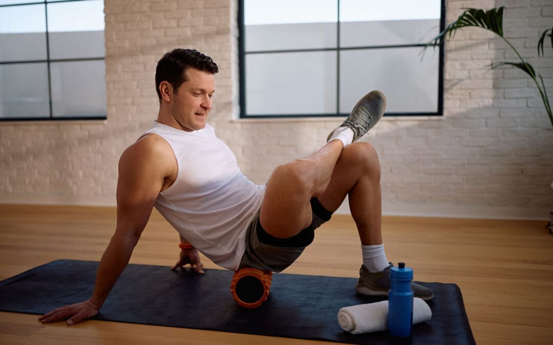
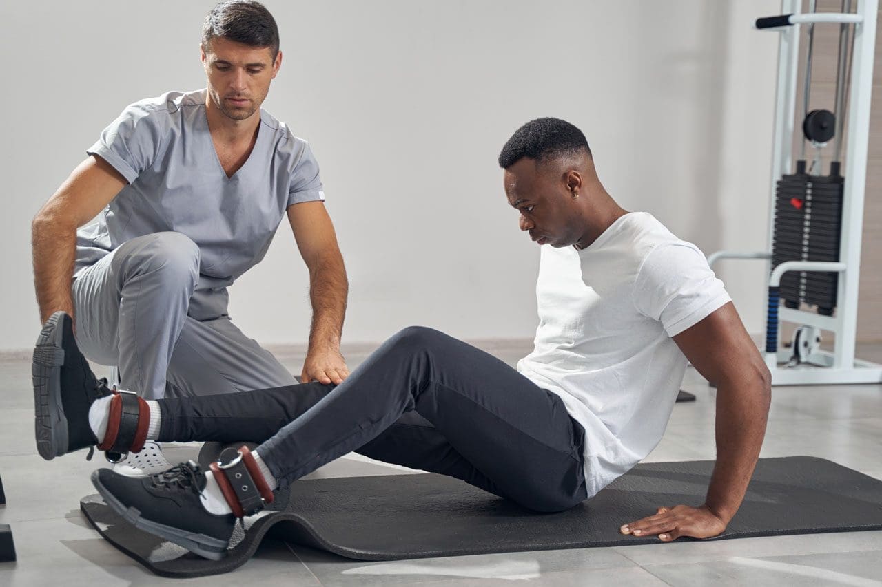 Sciatica is a nerve irritation pattern, not just a tight muscle. It often feels like burning, aching, tingling, or “electric” pain that can start in the low back or buttock and travel into the thigh, calf, and foot. Many people in El Paso experience sciatica after long hours of sitting, driving, or heavy lifting, or after an old injury that never fully healed. At El Paso Back Clinic, sciatica care is commonly described as integrative—meaning hands-on chiropractic care plus soft-tissue work, rehab, and (when appropriate) decompression strategies to reduce nerve pressure and help the body heal instead of just “chasing symptoms.”