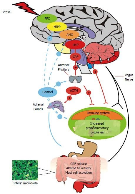
Ah� the bathroom, nature�s thinking room. The one place where we create many thoughts that turn into ideas, a place to read, and the place where we want peace and quiet. Known as many different names,� the bathroom is the one place where we flush out our systems after consuming quantities of food. However, did you know that the bile inside your body plays a huge important role in our system?
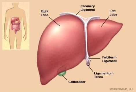
Bile is a vital body fluid that absorbs nutrients in our small intestines and flushes the toxins out of the liver. Bile continuously is being produced in the liver and then stored in the gallbladder. It also works as a signal molecule for both inside and outside the liver as well. When the liver and bile is disruptive, it can cause many problems for the body.
TCM (Traditional Chinese Medicine) stated that the liver is known as the �master organ� and it is the causative factor in many of the body�s ailments. When there are major imbalances on our bodies, it causes liver dysfunction. In TCM, the liver actually helps with detoxification; enzyme, hormone, and bile production, immune cell activation and stores vitamins and iron.
How Does the Liver Work?
The liver�s main job is to get rid of bile in our internal system. Not only that but the liver plays an important role by distributing nutrients by converting those nutrients into metabolizing energy. As well as storing vitamins and minerals for later use.
However, in today�s fast-paced society, the liver can�t perform right as we don�t give the liver it�s essential nutrients that it needs to function. Which causes blockage in the gallbladder, diseases, and digestive issues.
The Importance of Bile
As stated, bile has many functions that play an important part in the liver. Bile actually lubricates our small intestines and stool for easy disposal. But when there is less lubrication in our small intestines and stool, it can lead to constipation and too much lubrication can lead to diarrhea.
There is also a chance of blockage in bile where it can increase the accumulation of toxins in our body which creates oxidative stress and back up our waste matter. When we eat too much food and it�s still in our intestines, it will ferment and causes a leaky gut as toxic gasses penetrate the intestinal lining.
Another function is that the bile salts break down and processes fats that are essential for weight loss. Bile can also transport toxins out of our liver to feces while keeping everything free-flowing. When there isn�t enough bile being produced, cholesterol stones are forming.
Vitamins like, vitamin A, D, E and K can break down the excess fat in the body if there is an adequate amount of bile. But if patients had gallbladder surgery must be taken to account that they don�t have a bile storage system, and must manage their bile production.
Liver Functions for Hormonal Balance
Did you know that the liver and thyroid have a synergistic relationship? That�s because the T4 is converted to T3 for the liver. It turns out that the T3 is more potent in the thyroid hormone and is metabolized from iodine. When a patient has a thyroid problem, it could actually be the result of a bile or liver malfunction.
When our mood and mental state is imbalanced, it can be the results of a sick or fatty liver. Due to the fact that the liver influencing on our hormones. Because our mental state and our moods correlate to our hormones, it is important to keep the liver healthy but also finding a good place in our state of mind to make sure that we keep our liver healthy as well.
Signs of Liver Imbalance
There are many symptoms that can cause the liver to be imbalanced and can contribute to a low bile count:
Now that we understand what symptoms that causes our liver to be imbalanced, here are 5 ways to make sure that you are protecting your liver and producing a healthy amount of bile.
Removing Causative Factors
The first and foremost factor in protecting your liver is to get rid of the causative factors that are harming your liver. Things like an excessive amount of lactate in coffee, sugars, alcohol, and processed foods can disrupt your liver�s health. It�s ok to enjoy those things sparsely but, if you are trying to maintain a healthy liver, it�s best to ease up on the consumption of those lactate factors as much as possible.
Eating Liver Supportive Foods
There are many foods that actually help support the liver. Grapefruit, blueberries, cranberries, prickly pears, and bitter greens are just some of the foods that can actually help your liver and bile production. We here at Injury Medical Clinic, always inform our patients the importance of whole nutritious food to heal and repair what ailments in our patient�s bodies.
Practicing Deep Breathing Exercise
Practicing deep breathing exercises have been known to lower stress and diaphragmatic breathing exercises is no exception. Since the liver is positioned below the diaphragm, when we do any deep breathing exercises; these motions actually massages our liver and stimulate bile production. Hence why singers do these exercises when they are getting ready to perform. They are massaging their liver without even knowing it.
Intermittent Fasting
Since we now know that bile is continuously being produced in the liver and being stored into the gallbladder when we are not eating, intermittent fasting is a good way to increase the bile production in the gallbladder. Plus it has been known for weight loss and beneficial for the body to heal while getting rid of the junk in our cells.
Stress Less
Stress is one of the huge factors that can either cause our body to improve our immune system or causes the body to chronic illness. Emotional stress is known to cause a huge burden to our organs because it is tied to our hormones. When we get emotionally stressed, our liver cannot get rid of the excess toxins and hormones.� So we have to find ways to de-stressed ourselves so we won�t hurt our organs, especially the liver.
Conclusion
So when it comes to our liver, we must make sure that the bile that we are producing is turning into energy all the way to feces. So, when we have to go to the bathroom we can let out the toxins naturally without any complications. If we can lower the factors that are causing us to have an inadequate bowel movements, then we can improve our body�s ability to detoxify.
NCBI Resources
Western diets, high in sugar and fat, cause liver inflammation, especially in males, according to a new animal study in�The American Journal of Pathology. Inflammation was most pronounced in males that lacked farnesoid x receptor (FXR), a bile acid receptor.
One of the most recognized running injuries is runner�s knee. However, runner�s knee is not an injury but a result of different injuries. Running can hurt when improper mechanics, inadequate shoes, and cheap over the counter insoles are being used. It can lead to:
Plantar fasciitis
Achilles tendonitis
Medial tibial stress syndrome
Metatarsalgia
Runner�s knee
Runner�s knee is not an actual injury in of itself
It�s a broad description of knee pain caused by other knee injuries, which include:
Iliotibial band syndrome
Patellofemoral pain syndrome (PFPS)
Chondromalacia patella
This can keep runners sidelined, annually.
Runner�s knee is caused by usually one of two things: poor biomechanics and overuse.
Feet are the foundation of the body and if not taken care of, pain
Feet are 99% normal at birth.
Then life takes over and issues begin to arise.
8% develop foot conditions, then�by age 5 that number jumps to 41% and 80%� at twenty.
By forty pretty much everyone has a foot condition or some type of foot pain from work and activity.
These foot issues then begin to set up problems for the rest of the body, especially the generalized condition of back pain or leg/knee/hip problems.
Being able to foresee and realize a potential problem with the feet can PREVENT other injuries from beginning to present themselves and dodge any issues that can affect health and lifestyle.
Runners who are able to avoid injury are those that land the lightest, much like a cat and sustain the lowest level of impact.
Runners think about landing softly and adjust their stride so they land closer to the midfoot.
However, it’s easier said than done.
Most runners tend to be heel-strikers
Runners with excessive pronation that try to change to a forefoot strike pattern are more prone to inner foot and ankle injuries,
And runners with high arches that try to change to a forefoot strike pattern end up with sprained ankles and metatarsal fractures.
Runners try to treat the pain with stretches or exercises that target the area, but the source of the pain is actually elsewhere and they don’t realize it and are treating the wrong area.
A lot of the time the source of the imbalance comes from the feet.
An overlooked option for reducing knee injury and pain is the use of custom orthotics.
Custom orthotics align and support the foot/ankle in a more normal physiologic position for weight-bearing, which prevents dysfunction and improves the function of the body as a whole.
What they do is:
Make a symmetrical foundation that blocks pronation and supports supination
Give the heels shock absorption
Stops serial stress throughout the body
Enhances re-educating muscle-memory
Custom-made orthotics help reduce the impact of heel strikes when running or walking.
Shock absorption is a must when there is
Instability
Chronic degeneration
Inflammatory arthritis
Orthotics that are designed specifically to cushion the impact load from running can reduce pain triggers throughout the body.
Avoiding knee injuries
When orthotic care is indicated, custom-made functional orthotics can help reduce pain.
Look for orthotics that support the running gait and
Absorb shock at heel-strike
Support mid-stance
Provide propulsion at toe-off
Excessive Foot Pronation can Affect *FOOT POSTURE & MOBILITY* | El Paso, TX (2019)
The following video discusses how excessive foot pronation can ultimately have an effect on foot posture and mobility. Several things can impact foot posture and mobility, such as excessive foot pronation. Excessive foot pronation is most widespread among the overall populace, therefore, it’s regarded as one of the most frequent factors for abnormal foot posture and mobility, which can lead to a variety of health issues like overuse injuries. Excessive foot pronation and supination can ultimately impact general health and wellness.
What’s Afoot
Misalignment can be caused by many common runners� experiences including running on the same type of surface every day, running on a slanted surface such as a beach or replacing running shoes too infrequently. As a runner, you can work to vary your running surfaces and keep a better watch on your shoes, but your chiropractor will let you know if your body is in need of more balance.
NCBI Resources
If you are an active amateur or a competitive runner, using the services of a chiropractor can make a vast change in your overall health, reduce your pain from injuries and improve your alignment for a more effective run. Runners may not even realize that the tension they feel is the beginning of pain caused by being out of balance until it is adjusted. Chiropractic adjustments are often part and parcel of a runner�s training program to strengthen and improve performance. They can also help�recover from pregnancy�and postpartum bodily changes.
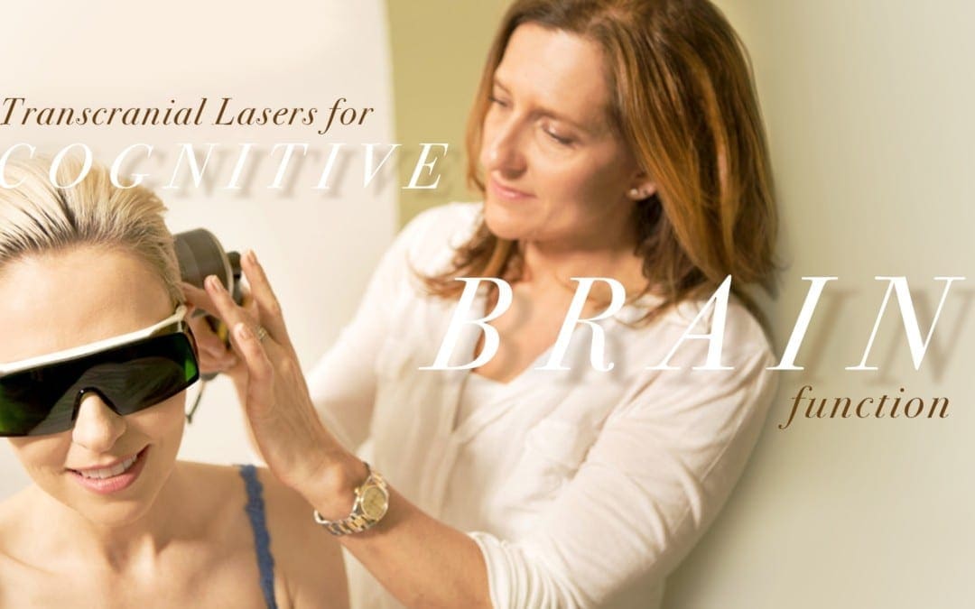
According to research studies, transcranial infrared laser stimulation, as well as other types of transcranial lasers, utilized on frontal cortex functions can improve sustained attention and working memory, among other brain functions. Transcranial laser stimulation with low-power density (mW/cm2) and high-energy density (J/cm2) monochromatic light in the near-infrared wavelengths regulates and maintains brain functions and may promote neurotherapeutic effects in a non-destructive and non-thermal manner. Researchers determined through the first controlled research study that transcranial laser stimulation improves human cognitive and emotional brain functions. �
In the field of low-level light/laser therapy or LLLT, developing a model to demonstrate how luminous energy from red-to-near-infrared wavelengths improves bioenergetics has been in development for the last 40 years. Previous LLLT research studies have demonstrated historical a variety of developments, principles and applications on the subject matter. The purpose of the following article is to demonstrate an update on LLLT’s neurochemical mechanisms supporting transcranial laser stimulation for cognitive-enhancing functions. We will describe the effect of LLLT on brain bioenergetics, briefly discussing its bioavailability and dose-response, and its effects on cognitive brain function. Although our focus is on prefrontal-related cognitive functions, LLLT should be able to improve other brain functions. By way of instance, stimulating different brain regions affect different functions associated with sensory and motor systems. �
Transcranial Lasers on Brain Bioenergetics
Near-infrared lasers and light-emitting diodes, or LEDs, affect brain function according to bioenergetics, a mechanism which is fundamentally different than other brain stimulation methods and techniques, such as electric and magnetic stimulation. LLLT has been demonstrated to regulate and maintain the function of neurons in cell cultures, and brain function in animals as well as cognitive and emotional functions in patients and health issues. Photoneuromodulation is associated with the absorption of photons by certain molecules in neurons which activate bioenergetic signaling pathways after being exposed to red-to-near-infrared light. The 600mm to 1150 nm wavelengths provide improved tissue penetration by photons because light is scattered at lower wavelengths and absorbed by water at higher wavelengths. Over 25 years ago, it was demonstrated that molecules which absorb LLLT wavelengths are part of the mitochondrial respiratory enzyme cytochrome oxidase in different oxidation states. Moreover, for red-to-near-infrared light, the main molecular photoacceptor of photon energy is cytochrome oxidase, also known as cytochrome c oxidase or cytochrome a-a3. �
Furthermore, photon energy absorption by cytochrome oxidase is the main neurochemical mechanism of action of LLLT in neurons. The more the enzymatic activity of cytochrome oxidase increases, the more metabolic energy which is developed through mitochondrial oxidative phosphorylation. LLLT provides the brain with metabolic energy in an analogous manner to the conversion of nutrients into metabolic energy with the utilization of light instead of nutrients developing the source for ATP-based metabolic energy. If an effective near-infrared light energy dose is provided, it stimulates brain ATP production and blood flow, ultimately fueling ATP-dependent membrane ion pumps, promoting greater membrane stability and resistance to depolarization, which has been demonstrated to transiently reduce neuronal excitability. Electromagnetic stimulation also directly affects the electrical excitability of neurons, as demonstrated in research studies. �
A long-lasting effect is provided by LLLT’s up-regulating the amount of cytochrome oxidase, which improved neuronal capacity for metabolic energy production which can be utilized to improve cognitive brain functions. In mice and rats, memory has been demonstrated to improve by LLLT and by methylene blue, a drug, which at low doses, provides electrons to cytochrome oxidase. Near-infrared light stimulates mitochondrial respiration by providing photons to cytochrome oxidase because cytochrome oxidase mainly accepts photons from red-to-near-infrared light in neurons. By stimulating cytochrome oxidase activity, transcranial LLLT promotes post-stimulation up-regulation of the amount of cytochrome oxidase in brain mitochondria. LLLT may also improve the conversion of luminous energy into metabolic energy during light exposure as well as the up-regulation of the mitochondrial enzymatic machinery to develop more energy after light exposure. �
Bioavailability and Hormetic Dose-Response by Transcranial Lasers
The most numerous metalloprotein found in nerve tissue is cytochrome oxidase and its absorption wavelengths are often associated with its enzymatic activity and ATP production. Increased LLLT bioavailability to the brain in vivo has been demonstrated in a variety of research studies by exposing brain cytochrome oxidase activity transcranially, resulting in improved extinction memory retention in healthy rats and improved visual discrimination in rats with impaired retinal mitochondrial function. Other LLLT research studies utilized a variety of wavelengths (633�1064 nm), daily doses (1�60 J/cm2), fractionation sessions (1�6), and power densities (2�250 mW/cm2) which ultimately characterized effective LLLT parameters for both rats and humans. �
By way of instance, researchers evaluated in rats the effects of different LLLT doses in vivo on brain cytochrome oxidase activity, at either 10.9, 21.6, 32.9 J/cm2, or no LLLT. Treatments were utilized for 20, 40, and 60 min through four 660-nm LED arrays with a power density of 9 mW/cm2. One day after the LLLT session, the brains of the rats were extracted, frozen, sectioned, and processed for cytochrome oxidase histochemistry. A 10.9 J/cm2 dose increased cytochrome oxidase activity by 13.6 percent. A 21.6 J/cm2 dose developed a 10.3 percent increase. A non-significant cytochrome oxidase increase of 3 percent was found after the highest 32.9 J/cm2 dose. Responses of brain cytochrome oxidase to LLLT in vivo were characterized by hormesis, with a low dose being stimulatory while higher doses were less effective. �
The first demonstration that LLLT increased oxygen consumption in the rat prefrontal cortex in vivo was demonstrated by another research study. Oxygen concentration in the cortex of rats was measured utilizing fluorescence-quenching during LLLT at 9 mW/cm2 and 660 nm. LLLT promoted a dose-dependent increase in oxygen consumption of 5 percent after 1 J/cm2 and 16 percent after 5 J/cm2. Because oxygen is utilized to develop water within mitochondria in a response developed by cytochrome oxidase, more cytochrome oxidase activity should promote more oxygen consumption. �
LLLT can also offer several benefits over other types of stimulation because LLLT non-invasively targets cytochrome oxidase, a fundamental enzyme utilized for energy production, with promoted expression associated with energy increase. LLLT is mechanistically specific and non-invasive while transcranial magnetic stimulation may be non-specific, prolonged forehead electrical stimulation may increase muscle spasms and deep brain or vagus nerve stimulations are invasive. �
Transcranial Lasers on Cognitive and Emotional Functions
LLLT through commercial low-power sources, such as FDA-cleared laser diodes and LEDs, is a highly promising, affordable, non-pharmacological alternative treatment option for improving cognitive brain function. LLLT offers safe doses of light energy which regulate and maintain neuronal functions, however, these are low enough to not damage the brain. In 2002, the FDA approved LLLT for pain relief in cases of head and neck pain, arthritis and carpal tunnel syndrome. LLLT has been utilized non-invasively in humans after ischemic stroke to improve neurological functions. It also improved recovery and ultimately reduced fatigue after exercise. One LLLT stimulation session to the forehead, as demonstrated in another research study, developed a considerable antidepressant effect in patients with depression. No adverse side effects were found either immediately nor at 2 or 4 weeks after LLLT. Therefore, LLLT treatments have been demonstrated to be safe and effective in humans. Although LLLT has been determined to be safe and it received FDA approval to be utilized for pain treatment, transcranial lasers for the augmentation of cognitive brain function should be limited for further research studies until further outcome measures support this application for clinical utilization. �
Transcranial laser stimulation to the forehead was utilized in a placebo-controlled, randomized research study, to demonstrate the effects of cognitive tasks associated with the prefrontal cortex, including a psychomotor vigilance task, or PVT, and a delayed match-to-sample, or DMS, memory task. The PVT evaluates sustained attention, with patients remaining vigilant during delay intervals, and pushing a button when visual stimulation appeared on a monitor. The laser stimulation targeted prefrontal regions which are believed to be utilized in the sustained attentional processes of the PVT. The DMS task supports the prefrontal cortex as part of a network of frontal and parietal brain regions. �
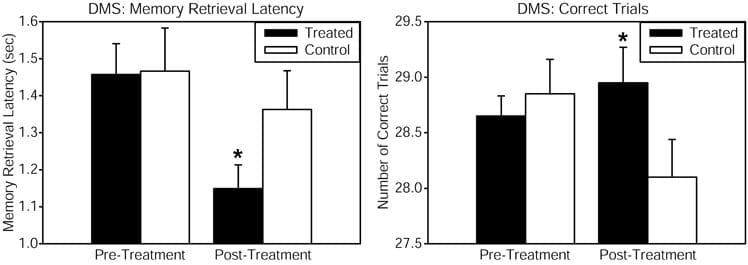
Healthy patients received consistent wave near-infrared light intersecting cytochrome oxidase’s absorption spectrum, targetted to the forehead utilizing a 1064 nm low-power laser diode, also known as �cold laser�, which increases tissue penetration due to its long wavelength and has been utilized in humans for other health issues. The power density or irradiance, 250 mW/cm2, and the cumulative energy density or fluence, 60 J/cm2, were identical which demonstrated the benefits of psychological effects in another research study. This laser exposure develops negligible heat and no physical damage at the low power level utilized. This laser apparatus is utilized safely in a clinical setting by the supplier of the laser. Reaction time in the PVT was improved by the laser treatment, as demonstrated by a considerable pre-post reaction time effect associated with the placebo group. The DMS memory task also demonstrated considerable improvements in measures of memory retrieval latency and number of correct trials, when comparing the LLLT-treated with the placebo group as demonstrated in Figure 1. Self-reported positive and negative affective or emotional states were also measured utilizing the PANAS-X questionnaire before and 2 weeks after laser treatment. As compared to the placebo, treated patients demonstrated considerably improved affective states. We suggest that this type of transcranial laser stimulation may serve as a non-invasive and efficacious method and technique to augment cognitive brain functions associated with attention, memory, and emotional functions. �
Figure 1. Cognitive performance in the delayed match-to-sample (DMS) memory task was improved after transcranial infrared stimulation to the right forehead. The DMS task involves presentation of a visual stimulus (grid pattern) on a screen. Then the stimulus disappears, and the participant must remember the stimulus through a delay. Then two choices appear, and the participant must decide which of these two is identical to the previous stimulus (the �match�). Treated subjects showed faster memory retrieval (left panel) and increased number of correct trials (right panel) out of 30 trials when attempting to choose the correct grid pattern. The function of frontal cortex regions, implicated in the attentional mode network utilized during this visuospatial memory task, was augmented by the laser treatment. Compared to baseline, this treatment also increased by 5% the oxyhemoglobin concentration of the prefrontal cortex as measured by near-infrared spectroscopy, both during the laser stimulation and during post-treatment DMS performance (in preparation). The data for the treated group consisted of n = 10 males and n = 10 females; the control group also consisted of n = 10 males and n = 10 females. *Significant treatment by pre-post score interaction, p < 0.05.
LLLT’s bioenergetics mechanisms associated with cognitive augmentation may also be associated in its neuroprotective effects. LLLT’s stimulation of mitochondrial respiration should improve cellular function due to increased metabolic energy and cellular survival after injury due to the antioxidant effects of increases in cytochrome oxidase and superoxide dismutase. �
Laser transmittance of the 1064-nm wavelength at the forehead LLLT site was estimated in a post-mortem human specimen, which demonstrated that approximately 2 percent of the light passed through the frontal bone. This yielded an absorption coefficient of a = 0.24, similar to the demonstrated a = 0.22 transmittance through cranial bone for this wavelength. Furthermore, it was estimated that about 1.2 J/cm2 of the 60 J/cm2 LLLT dose applied reached the surface of the prefrontal cortex. This value is similar to 1 J/cm2, the peak effective LLLT dose in neuron cultures for increasing cytochrome oxidase activity. �
Transcranial absorption of photon energy by cytochrome oxidase, the terminal enzyme in mitochondrial respiration, is associated as the bioenergetic mechanism of action of LLLT in the brain. Transcranial LLLT up-regulates cortical cytochrome oxidase and improves oxidative phosphorylation. LLLT improves prefrontal cortex-related cognitive functions, such as sustained attention, extinction memory, working memory, and affective state. Transcranial infrared stimulation may be utilized effectively to support neuronal mitochondrial respiration as a new non-invasive, cognition-improving intervention in animals and humans. This fascinating new treatment approach should also be able to affect other brain functions associated with the neuroanatomical site stimulated and the stimulation parameters utilized. �
Low-level laser therapy, or LLLT, and other types of transcranial lasers are non-invasive, low-powered lasers which are now being utilized in specific cortical regions of the brain to improve physiological responses and cognitive function. Many research studies have demonstrated that transcranial lasers can ultimately improve attention, memory, and reactions, where many other research studies have also demonstrated that these can also help improve depression and possibly even Alzheimer’s disease. Although further research studies are still required, the outcome measures are promising. – Dr. Alex Jimenez D.C., C.C.S.T. Insight
According to research studies, transcranial infrared laser stimulation, as well as other types of transcranial lasers, utilized on frontal cortex functions can improve sustained attention and working memory, among other brain functions. Transcranial laser stimulation regulates and maintains brain functions and may promote neurotherapeutic effects in a non-destructive and non-thermal manner. Researchers determined through the first controlled research study that transcranial laser stimulation improves human cognitive and emotional brain functions. The scope of our information is limited to chiropractic, musculoskeletal and nervous health issues as well as functional medicine articles, topics, and discussions. To further discuss the subject matter above, please feel free to ask Dr. Alex Jimenez or contact us at 915-850-0900 . �
Curated by Dr. Alex Jimenez �
Additional Topic Discussion: Chronic Pain
Sudden pain is a natural response of the nervous system which helps to demonstrate possible injury. By way of instance, pain signals travel from an injured region through the nerves and spinal cord to the brain. Pain is generally less severe as the injury heals, however, chronic pain is different than the average type of pain. With chronic pain, the human body will continue sending pain signals to the brain, regardless if the injury has healed. Chronic pain can last for several weeks to even several years. Chronic pain can tremendously affect a patient’s mobility and it can reduce flexibility, strength, and endurance.
Neural Zoomer Plus for Neurological Disease
�
Dr. Alex Jimenez utilizes a series of tests to help evaluate neurological diseases. The Neural ZoomerTM Plus is an array of neurological autoantibodies which offers specific antibody-to-antigen recognition. The Vibrant Neural ZoomerTM Plus is designed to assess an individual�s reactivity to 48 neurological antigens with connections to a variety of neurologically related diseases. The Vibrant Neural ZoomerTM Plus aims to reduce neurological conditions by empowering patients and physicians with a vital resource for early risk detection and an enhanced focus on personalized primary prevention. �
Formulas for Methylation Support
XYMOGEN�s Exclusive Professional Formulas are available through select licensed health care professionals. The internet sale and discounting of XYMOGEN formulas are strictly prohibited.
Proudly,�Dr. Alexander Jimenez makes XYMOGEN formulas available only to patients under our care.
Please call our office in order for us to assign a doctor consultation for immediate access.
If you are a patient of Injury Medical & Chiropractic�Clinic, you may inquire about XYMOGEN by calling 915-850-0900.
�
For your convenience and review of the XYMOGEN products please review the following link.*XYMOGEN-Catalog-Download �
* All of the above XYMOGEN policies remain strictly in force.
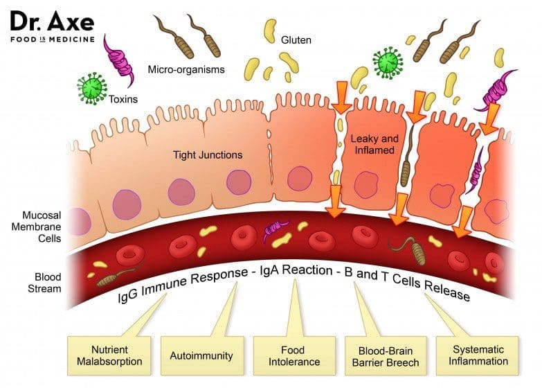
Ever wondered why you feel sluggish from a long day? Or feel sick to the stomach when you ate something bad or overindulged on your favorite food? Could it be that your gut is showing signs of stress and discomfort due to certain habits that you may encounter and didn�t even know about it?
In our previous article, we talked about the six types of food that our gut needs to be healthy. Since our gut contains trillions of microbiomes, both good and bad, these microbiomes play an important role in our overall health. A healthy microbiome improves our gut health, heart health, brain health, controls our weight and regulates our blood sugar. With the good bacteria in our gut, the bacteria benefit us with a good digestive system and destroys the harmful bacteria. But certain lifestyles and diet choices can actually increase the bad bacteria and lower the good bacteria and overall health.
Here are five surprisingly lifestyle choices that are hurting your gut:
Not Eating a Wide Range of Foods
Our gut plays an important role in our overall health. When we eat good whole foods, our gut is happier; we have more energy to complete any task that is thrown at us and we are getting nutrients for our gut flora. However, during the past couple of decades, we have been leaning more into processed foods due to the economic pressures of increased food productions. FOA stated that �75 percent of the world�s food is generated from only 12 plants and five animal species� and that is very bad to our gut flora.
Here at Injury Medical & Chiropractic Clinic, we inform our patients about the importance of eating nutritious, whole foods to promote not only a healthy gut but a healthy mind. When the body gets introduced to a wide variety of whole foods (with a high fiber content), our gut starts to repair the damage of processed food that we may have consumed internally.
However, when you disregard prebiotics to your diet, you are harming your digestive health. Without prebiotics, our digestive system slows down the development and diversity for our gut flora. So in order to have a healthy microbiome development, you need to incorporate foods filled with both digestible and indigestible fibers to your diet. Some foods included in this category are oats, nuts, onions, garlic, leeks, asparagus, bananas, pears, chickpeas, and beans.
Sticking to a high fiber diet maybe challenging however, there is the option of taking prebiotic supplements. If you have a food allergen or food sensitivity to any high enriched fiber foods, taking prebiotic supplements can actually help grow Bifidobacterium and Faecalibacterium in your gut and be beneficial to your health without the discomfort.
Excessive Alcohol Consumption
Every adult enjoys alcohol once in a while. Yes, it�s one of those beverages that help you relax a bit after a long day, however, too much of it can lead to alcohol abuse and addiction. So, did you know that consuming that much alcohol is bad for your heart, liver, and brain; thus hurting your gut health and giving you dysbiosis?
One study stated, that the alcoholics with dysbiosis had a lower median abundance of Bacteroidetes and a high abundance of Proteobacteria. The ones that weren�t alcoholics were not affected by the study.
However; there is some good news on limiting yourself to alcoholism and that it can be beneficial to your gut bacteria. If you moderately consumed red wine responsibly, the polyphenols in the wine can help benefit your gut flora. So, enjoy a glass of wine once in a while as a small treat that should not be taken for granted.
Inadequate Sleep
In one of the previous articles, we talked about how to achieve a good night sleep through herbs. When we get little to no sleep through our hectic lives, it affects us through various health problems, including heart disease and obesity. In a 2016 study, researchers discovered the effect of short-term sleep deprivation on the gut microbiota after two days.
When our body doesn�t receive the recommended 8 hours of sleep, our gut takes a huge toll as we feel sluggish and exhausted. So, to make sure that our gut microbiome will be taken care of, we recommended to turn off your electronical devices at least 30 minutes before you get ready to settle down for the night. Turn off all the lights, and don�t drink any liquids at least two hours before bed, close your eyes and take a deep breath in a meditative state, and relax as you drift off into slumber town.
Inadequate Exercise
Through our fast-paced lifestyle and stressful jobs, it�s hard to find time to exercise. But when we actually do find time to exercise, not only do our minds feel good; but our body and gut feel good as well. However, things always come up when we are in an exercise routine and we have to skip exercising altogether. It happens to all of us and it�s hard to pick up where we left off when we tried to exercise.
When we don�t exercise at least a couple of times out of the week, our bodies take a huge toll on us as we gained weight, our stress is way too high, and we have a higher chance of getting a chronic disease. When this happens our gut flora is a huge disadvantage. Here at the clinic, we strive to inform our patients about the importance of exercising and that it not only changes their lives but also changes their mood entirely.
However, don�t just go into a hard exercise routine where you will injure yourself. Start off with a low-intensity workout then build it up as you go because your gut flora will thank you for it.
As a final say, we here at Injury Medical want to keep you informed on nutrition and ways to help you improve your ailments with these 5 surprises. But to also educate you on what may be hurting your gut. With these surprises and slight changes to your daily life, your gut will be thanking you for the long haul.
NCBI Resources
According to evidence from a 2016 research study, the gut�s immune system is fundamental towards preventing a variety of diseases and it may often contribute to metabolic disorders. However, it might also help provide a treatment goal when observing systemic inflammation in insulin resistance. Moreover, modified gut immunity has been linked with changes to the gut microbiota, intestinal barrier function, gut-residing immune cells, and resistance to antigens which enter the gastrointestinal, or GI, system. Although this has been previously believed to raise the danger of esophageal ailments including, pathogenic infections and chronic inflammation, which may ultimately lead to chronic health issues.
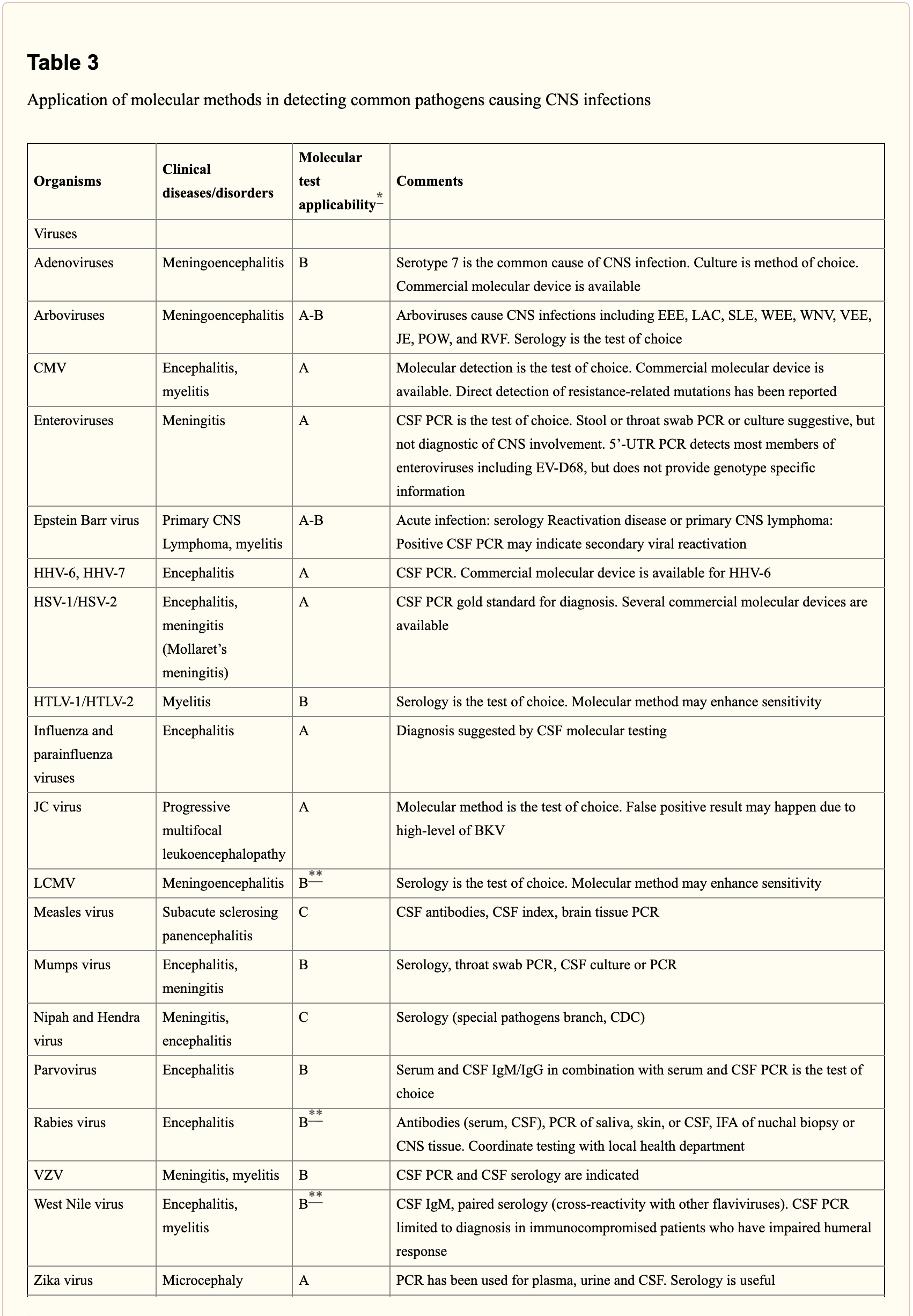
Central nervous system, or CNS, infections can be life-threatening if they are not diagnosed and treated early. Because CNS infections are non-specific, determining an accurate diagnosis can be challenging. The nucleic acid in vitro amplification-based molecular methods are starting to be utilized for routine microbial diagnosis. These molecular methods have improved beyond conventional diagnostic techniques with increased sensitivity and specificity. Moreover, molecular methods utilized on cerebrospinal fluid samples are considered the new standard for diagnosis of CNS infections caused by pathogens. �
Molecular methods for the diagnosis of CNS infections offers a variety of monoplex and multiplex PCR assays to diagnose several types of health issues. Pan-omic molecular platforms can also help diagnose CNS infections. Although molecular methods are utilized for the diagnosis of CNS infections, the outcome measures for these diagnostic techniques must be carefully identified by healthcare professionals. The following article discusses conventional diagnostic techniques and molecular methods utilized for the diagnosis of central nervous system infections, their application, and future approaches. �
Molecular Methods in the Diagnosis of CNS Infections
Because of increased sensitivity and specificity, nucleic acid in vitro amplification-based molecular methods has tremendously improved the ability to diagnose CNS infections in a reasonable and effective time frame. Several PCR-derived techniques have also ultimately increased the flexibility and rigor of currently available diagnostic techniques. �
Reverse transcriptase, or RT,-PCR was developed to increase RNA targets. Its utilization plays a fundamental role in the diagnosis of RNA-virus infections as well as managing their reaction to treatment. Timely access to enterovirus RT-PCR outcome measures has demonstrated shorter hospital stays, reduced unnecessary antibiotic utilization, and decreased ancillary laboratory evaluations and tests. Broad-range rRNA PCR techniques, which utilize a single pair of primers targeting conserved regions of genes, have been utilized to diagnose bacterial pathogens and herpes viruses in the CSF. Isothermal amplification-based techniques. including loop-mediated isothermal amplification or LAMP, have been developed to offer a diagnosis within several minutes to hours. Table 2 demonstrates commercial molecular in vitro diagnostic devices, or IVD, which have been cleared by the US Food and Drug Administration, or FDA, for diagnosis of microbial pathogens in CSF. �
Monoplex Assays
A conventional molecular method involves three phases: sample extraction, target nucleic acid amplification, and amplicon detection. One of the first molecular assays successfully utilized for the diagnosis of CNS infections was utilized for the diagnosis of HSV in cerebrospinal fluid or CSF. PCR became the test of choice when research studies demonstrated that CSF PCR was similar to culture of brain tissue for diagnosis of HSV encephalitis and meningitis. Many PCR based methods for the diagnosis of herpes and enteroviruses have become available with increased sensitivity compared to viral culture. �
Real-time PCR with nucleic acid amplification and amplicon detection further improved the transition to molecular methods in clinical laboratories. Unlike conventional PCR, the real-time system is a �closed� system and it overcomes the fundamental problem of carryover contamination. At the time of manuscript preparation, three molecular assays utilized to help diagnose HSV and enteroviruses in CSF have ultimately been approved by the FDA as demonstrated in Table 2 of the previous article. � Real-time PCR-based methods are the main diagnostic technique utilized to help diagnose the Zika virus, which was first reported in Uganda in 1947, and is now a worldwide concern after the virus spread widely in Brazil and Central America. Research studies developed a one-step RT-PCR assay utilized to diagnose the Zika virus in human serum with a limited detection of 7.7pfu/reaction. Along with plasma, the Zika virus RNA can be diagnosed through urine and plasma within the first 2 weeks after symptoms have manifested. In March 2016, the FDA approved a trioplex-PCR assay under emergency use authorization for the simultaneous diagnosis of Zika, Chikungunya, and Dengue viruses in serum, urine, CSF and amniotic fluid. The RT-PCR assay utilizes dual labeled hydrolysis probes with a LOD of 1.54�10 4 GCE/ ml of Zika virus in serum. �
Introduction of real-time PCR based diagnostic assays have affected early and effective diagnosis of several bacterial infections. Isothermal amplification-based molecular assays have excellent performance characteristics and they don’t require any specialized equipment. These assays are fundamental for the utilization of on or near point-of-care testing. LAMP-based methods have been utilized to diagnose Neisseria meningitis, Streptococcus pneumoniae, Haemophilus influenzae type b, M. tuberculosis, and JEV in the CSF. The Xpert MTB/RIF assay has tremendously improved regulation of tuberculosis by offering an integrated and automated system which allows quick clinical decision making in a POC or near-care context. Several research studies have utilized the Xpert MTB/RIF to evaluate the diagnosis of M. tuberculosis in CSF from TB meningitis. In a meta-analysis of thirteen research studies, the pooled sensitivity of the Xpert assay was 80.5 percent, or 95 percent CI 59.0 percent to 92.2 percent, against culture and 62.8 percent, or 95 percent CI 47.7 percent to 75.8 percent, against composite standard. Utilizing a large volume of sample, of at least 8�10 ml, is necessary for testing CSF and centrifugation can cause considerable improvements in yield. Despite the lack of standardization for sample processing, WHO has allowed testing CSF with the automated Xpert MTB/RIF assay as the first-line test over conventional microscopy. �
Multiplex Assays
Simplicity makes multiplex molecular assays fundamental for the diagnosis of a panel of microbial targets. Several multiplex PCR assays have been developed to diagnose bacterial pathogens in CSF targeting the most common causes of meningitis: S. pneumoniae, N. meningitis, H. influenzae, L. monocytogenes, S. agalactiae, S. aureus, E. coli, and M. pneumoniae. A multiplex PCR followed by Luminex suspension array can simultaneously diagnose eight bacterial and viral pathogens in CSF, including N. meningitis, S. pueumoniae, E. coli, S. aureus, L. monocytogenes, S. agalactiae, HSV-1/2, and VZV, among others. �
Considering the variety of pathogens involved in CNS infection, application of comprehensive molecular panels with multiple bacterial and viral targets have improved the efficiency of diagnosis. The BioFire FilmArray Meningitis/Encephalitis panel is currently the only FDA cleared multiplex assay utilized for the diagnosis of six bacterial, such as Escherichia coli K1, Haemophilus influenzae, Listeria monocytogenes, Neisseria meningitides, Streptococcus agalactiae and Streptococcus pneumoniae, seven viral, such as cytomegalovirus, enterovirus, HSV-1, HSV-2, human herpesvirus 6 or HHV-6, human parechovirus and VZV, as well as a single fungal, such as Cryptococcus neoformans/gattii, target in CSF as demonstrated in Table 2. The integrated FilmArray system takes about an hour, with only 2 minutes of hands-on time. During the preparation of the manuscript, two research studies demonstrated the performance of this assay. Utilizing 48 samples from gram stain negative CSF samples from suspected cases of meningitis, research studies demonstrated that this system diagnosed more viral pathogens, such as EBV. Four cases of WNV and a single case of Histoplasma were not diagnosed by this assay. Among HIV infected patients in Uganda, the test performance demonstrated increased sensitivity and specificity for the diagnosis of Cryptococcus. Although the FilmArray Meningitis/Encephalitis panel offers a quick diagnosis of CNS infections, further research studies are needed to determine its performance for a variety of targets and other high-risk populations. �
Co-infections are frequently found among immunocompromised patients and can ultimately be challenging to diagnose for clinicians. The multiplex design allows simultaneous diagnosis of multiple targets on the same sample. One research study utilized a panel of monoplex and multiplex molecular assays to conduct a prospective cohort research study in Uganda to comprehensively evaluate the etiology of meningitis among HIV-infected adults. Among the 314 HIV-infected patients with meningitis, EBV co-infection was diagnosed with Cryptococcus, M. tuberculosis, or other viral pathogens. EBV in CSF in these settings is not completely understood although a single research study associated increased EBV viral load as a marker of poor outcome measures in patients with bacterial meningitis and EBV co-infection/ reactivation, among others. �
Pan-Omic Molecular Assays
Technological improvements in metagenomic deep sequencing have increased its utilization for clinical diagnosis of CNS infections. Several research studies have demonstrated its ability to solve diagnostic technique problems which challenge the limits of traditional laboratory testing. Due to sterile status and protection by BBB, CSF and brain biopsies are fundamental to further explore the utilization of this technology for pathogen diagnosis. Metagenomics was successfully utilized to establish a diagnosis of neuroleptospirosis in a 14-year-old boy with severe combined immunodeficiency who also suffered from recurrent bouts of fever, headache, and coma. Similarly, high-throughput RNA sequencing performed on brain biopsy from an 18-month-old boy with encephalopathy diagnosed a new Astrovirus as the cause. Despite the utilization of metagenomics for the diagnosis of infectious disease, there are many technological and practical concerns which need to be addressed before this form of diagnostic testing can become mainstream and part of the clinical standard of care. �
Other promising advances have occurred in transcriptomics, proteomics and metabolomics. Host and microbial microRNA or miRNA, profiles have been utilized for a variety of inflammatory and infectious diseases. Two miRNAs, miR-155 and miRNA-29b, were reported as potential biomarkers for JEV infection and treatment targets for anti-JEV therapy. Host neural epidermal growth factor, including 2 and apolipoprotein B in CSF, was able to diagnose tuberculous meningitis with 83.3 percent to 89.3 percent sensitivity and 75 percent to 92 percent specificity. CSF metabolite profiling has been reported to be useful in the classification, diagnosis, epidemiology, and treatment assessment of CNS infections in HIV patients. CSF metabolic profile analysis demonstrated bioenergetic adaptation in regulating shifts of HIV-infected patients. �
Outcome Measures Associated with Diseases
Diagnosis of an etiologic agent in patients with CNS infections needs consideration of the most common causative organisms, the available diagnostic techniques and molecular methods for these agents, and the highest-yield clinical specimens for evaluation and testing. Knowledge of the epidemiology and clinical presentation of specific agents is fundamental in selecting which diagnostic methods are appropriate for patients. Animal or vector exposures, geographic location, recent travel history, season of the year, exposure of ill contacts, and occupational exposures should be considered. �
When selecting appropriate pathogen-specific molecular diagnostic methods, the following factors should be considered. CSF is the optimal specimen for PCR testing for patients with meningitis or meningoencephalitis. While indirect evidence can be determined by testing other specimen types, attempts should be made to obtain CSF samples early before treatment can compromise yield. Time of testing from the manifestation of symptoms is fundamental to understand and rule out false-negative results and recommend retesting within a certain time frame. By way of instance, HSV PCR can commonly render false-negative results if CSF sample is obtained very early or late in the process of HSE infection. Host health is also known to affect test performance characteristics. Immunocompromised patients are at risk for infection by a variety of opportunistic pathogens, by way of instance HHV-6, JC virus, Toxoplasma encephalitis in bone marrow transplant recipients and patients with HIV. Often, infection can be more severe, such as WNV, and challenging to diagnose in this population. Table 3 below demonstrates practical recommendations on application and pitfalls of molecular test for the diagnosis of CNS infections. �
Furthermore, a positive nucleic acid amplification testing results are considered to be complicated by the fact that some viruses survive latently in macrophages or neurologic tissues even if they’re incidentally diagnosed by sensitive molecular techniques without an actual pathogenic role which can potentially lead to overtreatment. Utilization of adjunctive biomarkers which help determine active replication is being explored to overcome this drawback in research studies. �
Historically, the diagnosis of microbiologic agents in patients with CNS infections has been hindered by the low yield of CSF culture for viral and fastidious bacterial organisms, delays in CNS production of organism-specific antibodies, and challenges in determining optimum samples for testing. The nucleic acid in vitro amplification-based molecular diagnostic methods and techniques have a wider and better application in clinical microbiology practice. The monoplex assay will likely be the main platform utilized for urgent, random-access, low throughput assays. Multiplex assays have the additional benefit of diagnosing multiple targets and mixed infections. As the volume of CSF sample retrieved is often small, multiplex assays enable comprehensive diagnostic analysis with a low amount of sample, obviating the need for repeated lumbar punctures. The clinical relevance and cost-effectiveness of simultaneous multi-pathogen diagnosis strategies need further research studies. Application of pan-omic techniques in challenging to diagnose CNS infections is the new exciting frontier, the technology is promising but routine implementation is expected to be slow due to various challenges, such as lack of applicable regulatory guidelines and adaptation in the clinical setting, although the outcome measures are promising. �
As previously mentioned, central nervous system, or CNS, infections can be life-threatening health issues if they are not accurately diagnosed and properly treated. However, determining a diagnosis of CNS infections can be challenging for many clinicians. Fortunately, a variety of diagnostic techniques and molecular methods can ultimately help determine the source of CNS infections and other health issues. These diagnostic techniques and molecular methods have tremendously improved over the years, as previously mentioned, and more of these evaluations are being utilized in clinical settings to accurately diagnose CNS infections for proper treatment. – Dr. Alex Jimenez D.C., C.C.S.T. Insight
In part 2 of our “Diagnosis of Central Nervous System Infections” article, we discussed the molecular methods and the pan-omic molecular assays which are utilized in the diagnosis of CNS infections as well as how specific testing outcome measures have ultimately been associated with clinical diseases and health issues. The scope of our information is limited to chiropractic, musculoskeletal and nervous health issues as well as functional medicine articles, topics, and discussions. To further discuss the subject matter above, please feel free to ask Dr. Alex Jimenez or contact us at 915-850-0900 . �
Curated by Dr. Alex Jimenez �
Additional Topic Discussion: Chronic Pain
Sudden pain is a natural response of the nervous system which helps to demonstrate possible injury. By way of instance, pain signals travel from an injured region through the nerves and spinal cord to the brain. Pain is generally less severe as the injury heals, however, chronic pain is different than the average type of pain. With chronic pain, the human body will continue sending pain signals to the brain, regardless if the injury has healed. Chronic pain can last for several weeks to even several years. Chronic pain can tremendously affect a patient’s mobility and it can reduce flexibility, strength, and endurance.
Neural Zoomer Plus for Neurological Disease
Dr. Alex Jimenez utilizes a series of tests to help evaluate neurological diseases. The Neural ZoomerTM Plus is an array of neurological autoantibodies which offers specific antibody-to-antigen recognition. The Vibrant Neural ZoomerTM Plus is designed to assess an individual�s reactivity to 48 neurological antigens with connections to a variety of neurologically related diseases. The Vibrant Neural ZoomerTM Plus aims to reduce neurological conditions by empowering patients and physicians with a vital resource for early risk detection and an enhanced focus on personalized primary prevention. �
Formulas for Methylation Support
XYMOGEN�s Exclusive Professional Formulas are available through select licensed health care professionals. The internet sale and discounting of XYMOGEN formulas are strictly prohibited.
Proudly,�Dr. Alexander Jimenez makes XYMOGEN formulas available only to patients under our care.
Please call our office in order for us to assign a doctor consultation for immediate access.
If you are a patient of Injury Medical & Chiropractic�Clinic, you may inquire about XYMOGEN by calling 915-850-0900.
�
For your convenience and review of the XYMOGEN products please review the following link.*XYMOGEN-Catalog-Download �
* All of the above XYMOGEN policies remain strictly in force.
Do you have symptoms of back pain, headaches, or stress? Pain medications work for only so long and don’t fix the problem. Visiting a chiropractor may help some or all of your symptoms. A chiropractic adjustment means that a chiropractor physically adjusts the vertebrae in the spine. This procedure creates positive effects without the stress or invasiveness of surgery.
A study in the Human Journal of Hypertension showed that chiropractic could give people with high/low blood pressure the same effect as taking blood pressure medication.
The effects of the adjustment would last for 6 months after the adjustment.
Medications can have negative side effects which include:
Fatigue
Nausea
Dizziness
Anxiety
Weight loss
Imagine that chiropractic can have the same effect as these medications, An adjustment might be something to consider.
Neck and Lower Back Pain
80% of all Americans experience some sort of lower back pain in their lifetime.
Medications and surgery is an option, but as we know from the opioid epidemic, they can be dangerous, expensive, and ineffective. Chiropractic treatment can definitely reduce back and neck pain and is also cheaper than other back pain management therapies saving you from mild and chronic pain while reducing medical costs.
Sciatica
Sciatica is pain that radiates from the lower back, down your legs that usually stems from a damaged, herniated, bulging disc or sciatic nerve pressure.
Because of the intensity of the pain, over-medicating can become a problem.
A chiropractic adjustment can relieve pressure on the sciatic nerve.
Studies show that patients receiving chiropractic care reduced the number of pain days.
They also experienced a reduction in the intensity of their pain.
Inflammation is Reduced
Inflammation is one of the main causes of pain, joint issues, and tension.
Chronic inflammation has been linked to diseases like:
Heart disease
Chronic pain
Cancer
Chiropractic adjustments have been shown to reduce inflammation that leads to positive benefits:
Chronic lower back pain relief
Reduced muscle tension
Relief of joint pain
Headaches
Other than back pain, headaches are the top complaint that chiropractors treat. Both tension and migraine headaches can be caused by neck and spine issues. Back/Spinal misalignment can cause muscle tension and pain.
Neurological Conditions
Chiropractic adjustments help increase blood flow to the brain, as well as, cerebral spinal fluid. This helps people suffering from neurological conditions like multiple sclerosis and epilepsy.
Child Benefits
Chiropractic care can significantly improve conditions that affect children. The big three that can be helped are
Colic
Acid reflux
Ear infections
Colic
Colic causes crying and fussiness in children and babies from abdominal discomfort and gas. Adjustments have been shown to help improve colic symptoms.
Acid Reflux
Nerves in the gut can be particularly sensitive along with certain food sensitivity. Chiropractic helps keep the natural flow of the intestines moving.
This also boosts the immune system, which helps prevent infection/s.
Athletic Performance
Athletes who depend on their body for their sport definitely benefit from chiropractic. With a reduction of inflammation, pain, and other conditions chiropractic helps get the body in top shape and keep it.
Sports teams and professional athletes have chiropractors as part of their team. Alignments reduce inflammation and boost the immune system to help athletic performance.
Vertigo
Dizziness and vertigo can make it impossible to do everyday tasks without feeling disoriented, nauseous and fatigued. This is a common condition after going through a head or neck injury.
A chiropractic adjustment specifically targets joints and vertebrae that are not moving correctly. Adjustments also bring the body into balance, thus reducing vertigo episodes.
Conclusion
A chiropractic adjustment can be a great way to improve multiple areas of the body, along with improving overall health with non-invasive treatment.
With a chiropractic adjustment, you can safely and easily get yourself into top health.
Difference Foot Orthotics Make to *REDUCE FOOT PAIN* & Correct Posture | El Paso, TX (2019)
Custom made foot orthotics can help control foot motion and posture. Healthcare professionals prescribe custom foot orthotics to help patients focus on their foot posture and mobility control. Research studies have ascertained that using custom foot orthotics for posture and mobility control can help fix excessive foot pronation and supination to prevent a variety of foot health problems. The subsequent video describes how custom foot orthotics will help control foot posture and mobility to improve health and wellness.
NCBI Resources
A chiropractor is a healthcare professional who focuses on the diagnosis, treatment, and prevention of neuromuscular and musculoskeletal disorders through the use of adjustments and manipulations of the spine. Chiropractors seek to reduce pain and enhance the performance of patients as well as to instruct them on how they can account for their health via ergonomics, exercise and other therapies to deal with their pain. Chiropractic is usually categorized as alternative medicine or complementary medicine.
The most important thing about summer is the food. Hotdogs and burgers on the grill and the seasonal fruits and vegetables that are ripe for the picking.� As much as we love the summer sun, it is still dangerous and can be harmful to our skin. We still put on sun cream, wear hats, and wear sun-protective clothing, but, did you know that certain foods can help heal your skin from sun damage and when possible can be eaten raw.
In the previous article, we talked about the 9 nutrients your skin needs to be protected from the harmful sun�s rays. Here is the top 9 food that will protect you from the sun and perfect for the summer.
Guava:
When we think of vitamin C, our minds think of any citrus fruit like oranges, lemon, limes, and grapefruit. But did you know that guava contains vitamin C as well? In fact, guava contains about 5 times more of vitamin C as much as any citrus fruit.
Guava contains about 228.3 mg of vitamin C and has antioxidants that attack free radicals and helps boost your immune system. Vitamin C has been known to battle scurvy. Plus guava can help improve your skin. By eating the fruit or using the guava leaves, your skin will be toned and the antioxidants from the fruit can keep your skin glowing, fight wrinkles and reduce signs of premature aging.
Sweet Potato:
Who doesn�t love potatoes? We eat them as fries, baked, saut�ed, mashed and use them as filling for pies. The sweet potato is no exception. There are many variations of sweet potatoes as they come in orange, white, and purple, depending on where you get them from and which region.
The sweet potatoes we are familiar with have an orange hue due to the carotenoids; which gives us that lovely orange color and has antioxidants to protect our skin from sun damage. Not only that but; sweet potatoes are very high in vitamin A, which is very good when they are cooked. Some people say that potatoes are known to be very starchy and can be used to soothe a sunburn by drawing out the heat from the skin.
Strawberries and Blueberries:
Both of these berries are great on their own but together, they are the dynamic duo to help our bodies combat the sun. Blueberries are richly filled with antioxidants as they combat the free radicals in our systems and can reduce the chances of cancer showing up.
Strawberries are really great as they are called �nature�s natural sunblock.� They contained about 108% of vitamin C as well as ellagic acid, which cleans up the free radicals and reduce sun-damaged pigmentation.� The Journal of Agricultural Food Chemistry stated that strawberries have anthocyanins, which gives the fruit its lovely red color to protect our cells.
Green Tea:
Who doesn�t love green tea? Not only it contains L-theanine, but it has many astounding health benefits that are wonderful and protects our body. Green tea can be consumed or used as a topical cream to soothe and hydrate your skin from the harsh sun rays. Green tea is jammed packed with vitamins B2 and E, as well as large amounts of polyphenol including, EGCG (Epigallocatechin Gallate).
These polyphenols help our inflammatory system repair our DNA from anything harsh in our bodies. Plus green tea has been known to lower the risk of various types of cancers.
Oatmeal:
Oatmeal is one of those foods that we all eat for breakfast. However, did you know that oatmeal can be used to soothe sunburns and exfoliate sun-damaged skin? Not only that but when oatmeal is finely grounded it is known as �colloidal oatmeal.�
You may have seen this type of oatmeal in the health/medical section in your local stores and it may be called, �Aveeno.� �Colloidal oatmeal has been approved by the FDA since 2003 and has been used as a topical ointment for anyone with eczema. Anyone with eczema experiences an abundance of itchiness when they are overly exposed by the sun�s rays or due to the heat of the summer knows this all too well.
With colloidal oatmeal, it helps relieve the symptoms of eczema by being applied with water and gently patting the topical on the source of eczema to lower the inflamed skin, thus calming it down.
Cucumber:
Cucumbers are used for anything that we can think of. In the spa, in our salads, or as a wonderful snack. This green vegetable is packed with vitamins C and K as well as, caffeic acid and potassium. Not only that but cucumbers are made up of 96% of water, which is very refreshing and great for the skin. Since our bodies lose water when we sweat and cucumbers actually replenishes our water intake and�helps cool off our bodies when we are sunburned.
Tomatoes:
Just like strawberries, tomatoes contain lycopene, which gives tomatoes that gorgeous red color and has vitamins C. K1, and B9 and potassium. Tomatoes can be eaten raw and are rich with antioxidants that help balance our bodies pH balance. As well as, protecting our skin from the sun.
Watermelon:
Oh, watermelon� not only you are the most consumed fruit for the 4th of July but you are one of the best summer fruits to be consumed. Watermelons contain not only vitamins A, B6 and C; but they also contained lycopene like tomatoes. Which helps our skin from photoaging from the sun but it�s in the top 30 most hydrating foods, next to cucumbers with 92% of water for excellent hydration properties for our skin.
Carrots:
Carrots are not only good for our eyes but did you know that carrots are jammed pack with beta-carotene, which turns to vitamin A when we eat it. Plus the sun exposure gives carrots vitamin C to help us protect our skin. Carrots have a wonderful source of carotenoids to produce photoprotection for our skin health.
Here at the clinic, we strive to inform our patients about the nutrients that food provides to our bodies. As well as, making our patients feel good with whole, nutritious options. Whether it is by adjustments or leading them to different food options for a healthy life, these top 9 foods not only help protect your skin from the sun but they also taste really good. So enjoy the summer months but remember to eat your photoprotective food.
NCBI Resources
A healthy diet is the cornerstone of good health.�You should maintain a diet�that includes lean meats, fresh fruits and vegetables, and whole grains. The key is choosing fresh, seasonal foods that are local to your area. Foods grown in their season have certain vitamins and minerals that the body needs for the time of year in which they are ripe and ready.
Anti-inflammatory activities of colloidal oatmeal (Avena sativa) contribute to the effectiveness of oats in the treatment of itch associated with dry, irritated skin: https://www.ncbi.nlm.nih.gov/pubmed/25607907
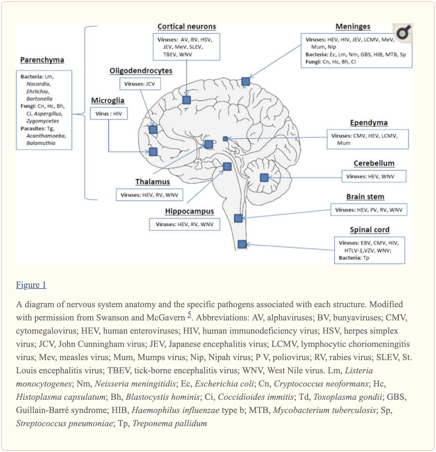
The central nervous system, or CNS, plays a fundamental role in the pathogenesis of infection. The CNS is regulated by the blood-brain barrier or BBB, however, it can still be exposed to a microbial invasion from a contiguous focus, hematogenous dissemination, or intraneural passage of organisms. A variety of environmental or commensal bacteria, viruses, fungi, protozoa, or parasites can enter the CNS and cause a variety of infections and health issues. Central nervous system infections can ultimately cause headache, stiff neck, vomiting, fever, photophobia, and focal neurological symptoms. �
What are Central Nervous System Infections?
CNS infections are characterized according to their affected region. Infection of the brain, spinal cord, and meninges results in meningitis, encephalitis, brain abscess, and myelitis. Infections can affect single or multiple regions of the brain, such as meningoencephalitis and encephalomyelitis. Moreover, CNS infections are characterized as acute, sub-acute, chronic, or recurrent based on their duration. Meningitis can cause headache, neck stiffness, fever, and photophobia over a period of hours to days. Encephalitis can cause brain parenchymal inflammation which can ultimately cause lethargy to coma. Last but not least, Myelitis can cause inflammation of the spinal cord including headache, fever, and paraparesis or paralysis. �
One of the most fatal CNS infections, acute bacterial meningitis, with three to five cases for every 100,000 people in the United States, has a mortality rate of 6 percent to 26 percent. Approximately 4,000 cases of acute bacterial meningitis occur in the U.S. every year with about 500 deaths. The frequent cause of acute bacterial meningitis includes Streptococcus pneumoniae, group B Streptococcus, Neisseria meningitides, Haemophilus influenzae, and Listeria monocytogenes. �
CNS infections caused by viruses are more common and are mostly mild and self-limited. However, these can manifest as meningitis and/or encephalitis. CNS infections caused by viruses can vary due to region and season. Non-polio enteroviruses are responsible for the majority of meningitis and/or encephalitis cases from late spring to fall. CNS infections due to herpes simplex viruses, or HSV, are associated with sporadic encephalitis and meningitis with severe sequelae if left untreated. �
Diagnosis of CNS Infections
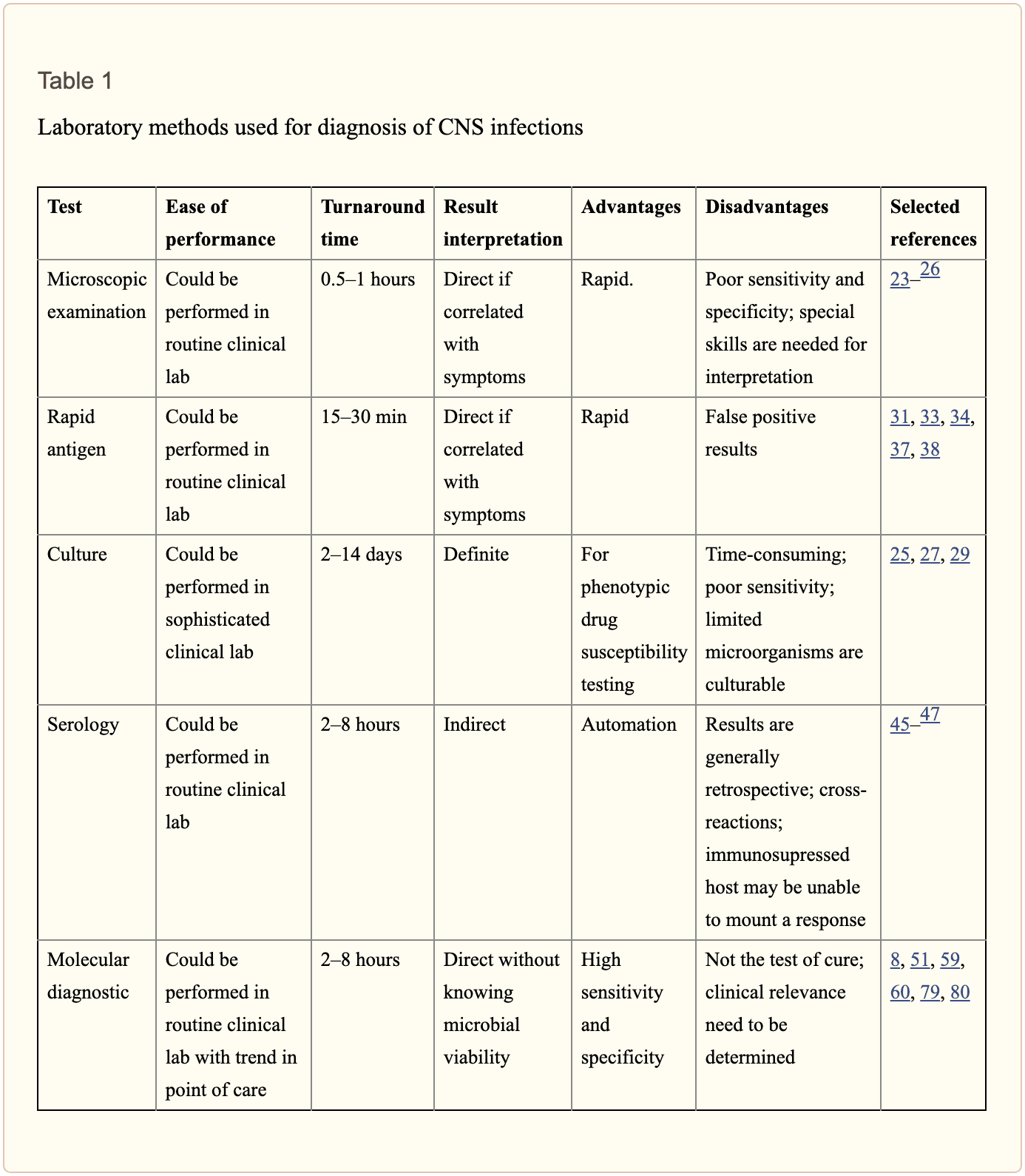
Diagnosis of microbial pathogens is fundamental for treatment. Methods and techniques utilized in clinical microbiology laboratories include direct microscopic examination, and culture techniques as well as antigen and antibody detection assays. However, each method and technique has several essential limitations. By way of instance, direct microscopic examination of CSF restricted sensitivity and specificity. The sensitivity of culture for enteroviruses is between 65 percent to 75 percent with average retrieval time of 3.7 to 8.2 days. Moreover, several serotypes of enteroviruses, especially Coxsackievirus A strains, are well-known to be non-cultivable and frequently grow poorly. Because enteroviruses are missing a common antigen found throughout a variety of serotypes, universal antigen and/or antibody diagnosis is impossible. Diagnosis of CNS HSV infections through methods and techniques utilized to determine culture sensitivity from CSF is tremendously poor. The presence of HSV IgG antibodies in CSF can ultimately be utilized to determine a diagnosis, however, the production is delayed until day 10 or day 12 after infection and it is not considered ideal for early diagnosis.
Diagnostic techniques, especially PCR based amplification, have developed a variety of mainstay tools to help determine the diagnosis of microbial pathogens in CSF. Molecular methods have demonstrated greater diagnosis rates than other diagnostic techniques. One research study demonstrated that 16S rRNA PCR-based assays were able to diagnose the causative organism in 65 percent of banked CSF samples compared to 35 percent when utilizing culture and microscopy. In another research study, diagnosis based on diagnostic techniques like molecular methods were utilized to optimize antibiotic treatment of patients with infectious meningitis when conventional methods and techniques demonstrated a negative outcome measure. Molecular methods and diagnostic techniques utilized on CSF samples are a fundamental standard when compared to the culture standard in the diagnosis of CNS infections caused by viruses which are challenging to diagnose. �
The diagnosis of CNS infections has tremendously changed over the last several years. PCR-based molecular methods have become a fundamental element in the clinical microbiology laboratory, providing tools for an accurate diagnosis. As demonstrated in Table 2, a variety of commercial molecular assays have been cleared by the Food and Drug Administration, or FDA, for the diagnosis of microbial pathogens. The approved assays for pathogen detection in the CNS are shown below. �
However, there are still several challenges in molecular diagnostic techniques and methods. Utilizing a combination of conventional diagnostic techniques and molecular methods, research studies demonstrated that in approximately 62 percent of patients with encephalitis, an etiologic organism could not be identified. Researchers have started to focus on developing advanced techniques and methods. In the following series of articles, we will demonstrate an update on the current conventional and molecular platforms utilized for the diagnosis of CNS infections. We will also demonstrate a preview on the potential clinical application of future technologies, including pan-omic assays. The emphasis of the following series of articles is to demonstrate optimal test selection in the clinical scenario for the diagnosis of CNS infection. �
Conventional Microbiology Methods and Techniques
Microscopic Examination
A positive CSF Gram stain confirms the diagnosis of bacterial meningitis. The sensitivity of the Gram stain for the diagnosis of bacterial meningitis is approximately 60 percent to 80 percent in patients not on antimicrobial treatment and approximately 40 percent to 60 percent in patients on antibacterial treatment. In one research study, Gram stain diagnosed as much as 90 percent Streptococcus pneumoniae and 50 percent Listeria monocytogenes in CSF collected from patients with bacterial meningitis confirmed by PCR 26 techniques and methods. Two organisms which are frequently diagnosed by microscopy include Mycobacterium tuberculosis by acid-fast bacillus, or AFB, staining and Cryptococcus neoformans by India ink or Gram stain. However,� the sensitivities of these techniques and methods are poor and culture is generally utilized instead. �
Culture
Culture of brain tissue can demonstrate a positive diagnosis of CNS infections, however, getting biopsies are tremendously invasive and frequently avoided unless a clinician determines that they are absolutely necessary. CSF sampling is most commonly performed to diagnose CNS infection. CSF viral, bacterial, including mycobacterial, and fungal cultures are fundamental in the diagnosis of infectious meningitis. However, CSF cultures in these cases are extremely low. Another disadvantage of CSF bacterial culture is that it generally requires up to 72 hours to determine a final diagnosis. A recent research study demonstrated that CSF mycobacterial culture had a sensitivity of 22 percent and a specificity of 100 percent in the diagnosis of tuberculosis meningitis. For viruses, utilizing monoclonal antibodies through culture increased the speed and specificity. However, due to time and sensitivity, CSF viral culture is frequently unable to determine a diagnosis. �
Rapid Antigen Detection
Cryptococcal antigen is the most commonly utilized antigen assay for CNS infections. The test utilizes Cryptococcus capsular polysaccharide antigens in CSF through enzyme immunoassay to determine a diagnosis. In a single research study which evaluated patients less than 35 years of age with CNS cryptococcosis, overall sensitivity and specificity of 93 percent to 100 percent and 93 percent to 98 percent were shown. Cryptococcus is a neurotropic fungus. Polysaccharide serum antigen titers with host immune status are frequently utilized to determine the need for a lumbar puncture to evaluate the patient for CNS health issues. The baseline peak titer of polysaccharide antigen in serum or CSF has demonstrated fundamental prognostic significance with an increased titer, or peak titer less than 1:1024, associated with antifungal therapy failure. �
The diagnosis of galactomannan, or GM, antigen and 1,3 ?-D-glucan, or BDG, in CSF, can help in the diagnosis of CNS aspergillosis or other invasive fungal infection such as fusariosis. Increased BDG in serum and CSF is associated with fungal infections. Measuring the levels of BDG is a beneficial biomarker in the evaluation of fungal CNS infection. It was recently demonstrated that patients receiving effective antifungal therapy demonstrated a decrease in CSF BDG concentrations with less than 31pg/ml and for this reason, BDG titers in CSF are a beneficial biomarker when monitoring response to treatment. �
For acute bacterial meningitis, a rapid antigen assay can help diagnose for a pneumococcal capsular antigen. Several research studies have demonstrated the utilization of M. tuberculosis-specific antigens in CSF for the diagnosis of tuberculosis meningitis. M. tuberculosis Early Secreted Antigenic Target 6, or ESAT-6, has been utilized for tuberculosis meningitis. �
Serology
Serological diagnosis of CNS infections is determined by identifying IgM antibodies or by demonstrating an increase in neutralizing antibody titers between acute- and convalescent-phase CSF. Due to a delay in antibody response when symptoms have manifested, a negative antibody test cannot be utilized to rule out infections and retesting may be required. Moreover, in specific populations, such as immunocompromised patients, the tests may not offer optimum sensitivity. In most instances, nucleic acid amplification tests have surpassed antibody-based detection as the test of choice. For several CNS infections, these assays play a fundamental role. CSF IgM is the most commonly utilized test for West Nile virus, or WNV, infections. Antibodies may manifest in as soon as 3 days and may continue for up to 3 months. However, its accuracy is challenged by high cross-reactivity with other flaviviruses and associated vaccines. Antibodies in recombinant WNV E proteins can determine where cross-reacting viruses co-circulate or determine which patients have been immunized. �
Fundamental serological assays for CNS infections are utilized for the diagnosis of neurosyphilis. Neurosyphilis is determined by a positive CSF venereal disease research laboratory, or VDRL, test. Diagnosis of varicella-zoster virus, or VZV, IgG in CSF is the most common technique and/or method for the diagnosis of VZV associated with CNS infection. �
Central nervous system, or CNS, infections can ultimately be life-threatening health issues if they are not diagnosed and treated early. Determining an accurate diagnosis of CNS infections can be challenging. Fortunately, a variety of diagnostic techniques and molecular methods can help determine the source of CNS infections. These diagnostic techniques and molecular methods have tremendously improved over the years and more and more of these evaluations are being utilized in clinical settings to accurately diagnose CNS infections for early treatment. – Dr. Alex Jimenez D.C., C.C.S.T. Insight
In part 2 of our “Diagnosis of Central Nervous System Infections” article, we will ultimately discuss the molecular methods and the pan-omic molecular assays which are utilized in the diagnosis of CNS infections as well as discuss how specific testing outcome measures are associated with clinical diseases and health issues. The scope of our information is limited to chiropractic, musculoskeletal and nervous health issues as well as functional medicine articles, topics, and discussions. To further discuss the subject matter above, please feel free to ask Dr. Alex Jimenez or contact us at 915-850-0900 . �
Curated by Dr. Alex Jimenez �
Additional Topic Discussion: Chronic Pain
Sudden pain is a natural response of the nervous system which helps to demonstrate possible injury. By way of instance, pain signals travel from an injured region through the nerves and spinal cord to the brain. Pain is generally less severe as the injury heals, however, chronic pain is different than the average type of pain. With chronic pain, the human body will continue sending pain signals to the brain, regardless if the injury has healed. Chronic pain can last for several weeks to even several years. Chronic pain can tremendously affect a patient’s mobility and it can reduce flexibility, strength, and endurance.
Neural Zoomer Plus for Neurological Disease
� �
Dr. Alex Jimenez utilizes a series of tests to help evaluate neurological diseases. The Neural ZoomerTM Plus is an array of neurological autoantibodies which offers specific antibody-to-antigen recognition. The Vibrant Neural ZoomerTM Plus is designed to assess an individual�s reactivity to 48 neurological antigens with connections to a variety of neurologically related diseases. The Vibrant Neural ZoomerTM Plus aims to reduce neurological conditions by empowering patients and physicians with a vital resource for early risk detection and an enhanced focus on personalized primary prevention. �
Formulas for Methylation Support
XYMOGEN�s Exclusive Professional Formulas are available through select licensed health care professionals. The internet sale and discounting of XYMOGEN formulas are strictly prohibited.
Proudly,�Dr. Alexander Jimenez makes XYMOGEN formulas available only to patients under our care.
Please call our office in order for us to assign a doctor consultation for immediate access.
If you are a patient of Injury Medical & Chiropractic�Clinic, you may inquire about XYMOGEN by calling 915-850-0900.
�
For your convenience and review of the XYMOGEN products please review the following link.*XYMOGEN-Catalog-Download �
* All of the above XYMOGEN policies remain strictly in force.
Everyone in the world wants healthy skin. We see it advertised on television with lotions and vitamin supplements. When we exercise and change our eating habits, we see our skin getting firmer with the foods we consume. However, whenever we are stressed, anxious, consuming junk food, or staying out in the sun too long; our skin takes a huge toll on our body. Our skin is the largest organ that covers our entire skeleton structure. When we expose our skin to harsh environments or have skin ailments that we contracted during our birth, our skin is depleted with the certain nutrients that our skin needs.
Glutathione:
Glutathione is known as the �wonder drug� for skin lightening. For some darker toned individuals, it will lighten up their natural melanin. This stigma has been popularized by media influences so people can have �porcelain skin.� However, glutathione actually made up of three amino acids:
Glutamine
Glycine
Cysteine
Melanin
This powerful antioxidant fights off free radicals in our immune system and is compatible with Vitamin E and C. For a natural way to make sure that your body keeps the glutathione nutrients when you get older with age, here are some vegetables that are enriched with glutathione:
Garlic
Onions
Avocado
Cabbage
Okra
Spinach
Kale
Cauliflower
Omega-3:
Omega-3s is one of the most common supplements that is known for healthy skin. This supplement keeps the body healthy as well as preventing inflammation. Omega-3s are mostly in:
Fish
Legumes
Walnuts
Avocados
Eggs
Spinach
But, there are certain limitations on taking Omega-3 supplements if you have a seafood allergy or an egg allergy. People with these types of food allergens can talk with their physician about taking the omega-3 supplements in a pill form in low dosages or eat omega-3 enriched food.
Other patients with omega-3 deficiency have been known to have psoriasis, thus using a topical lotion infused with omega-3s have been known to calm down the inflammation.
Biotin is the three-for-one supplements that target your nails, hair, and skin. This supplement can be found in vitamin pills at your local stores and is highly recommended by dermatologists. However, some people have biotin and zinc deficiency that can be linked to skin abnormalities, thus, biotin plays an important role in our skin health.
You can either take the vitamin pill or incorporate certain food groups like eggs, nuts, whole grains, some dairy products, and certain vegetables in your diet to get the beneficiary nutrients to keep your skin healthy.
Niacin:
Also known as vitamin B3, has been known to support skin health. This nutrient has many beneficial effects to promote skin wellness. It is one of the most essential nutrients we consume since our bodies can�t produce it on their own. Some of the food groups are in the meat department and vegetarian department:
Mushrooms
Potatoes
Legumes
Whole grains
Meat
Fish
Eggs
Milk
Vitamin A:
Vitamin A is filled with nutrients as it contains beta-carotene, thus it is mostly fruits and vegetables that contain this supplement. This supplement plays an important role as it helps repair any skin deficiencies and eye health. Some of the foods that boost up vitamin A are:
Carrots
Broccoli
Cantaloupe
Squash
Vitamin C:
Vitamin C is one of the most top tiers of improving skin health and has many beneficial factors in our immune system. Some patients develop scurvy when they don�t have enough vitamin C in their system. It is mostly found in citrus fruit, which is one of the best ways to consume the vitamin into your system.
But, there is a catch when you are taking vitamin C. Vitamin C when exposed to light, can oxidize and become unstable. So if you are taking the supplement, it should be stored in a dark place and the PH should be at 3.5.
Zinc:
Zinc is one of the supplements that support healthy skin. This micronutrient can protect our skin from the sun and supports our inflammatory system. Some of the food that actually can help us prevent sun damage and give us a zinc supplement boost include seeds, meat, shellfish, dairy and dark chocolate.
When our skin needs these 9 nutrients, they are thanking us for taking the time to get the necessary supplements to make sure our bodies are still functional and that we live a long healthy life. Granted that the media has televised about many ways to promote skin health, but it actually starts with eating the right foods that our body craves. When we eat processed food and ingest artificial sugars into our bodies, we feel sluggish, our skin takes a toll on the lack of nutrients we are not giving and so many health problems that we will face.
Yes, we can take topical creams and lotions to nourish our skin and combat the dryness that our skin faces. But that can only go for so long unless we change our eating styles. Some people may freak out because they hear the word, �diet� and are limited to what they can eat. However, when it�s a health issue and our physicians tell us that we need to eat healthier, we give it a go. Therefore, eating right is a lifestyle choice and it starts with these 9 nutrients to make sure our largest organ is taken care of as well as the rest of our body system. When we cut back on the bad food and focusing on good food, our bodies feel so much better.
NCBI Resources
Living a healthy lifestyle and eating your basic food groups; whether it be plant-based or omnivorous, as well as, exercising a couple of times out of the year. A bad healthy lifestyle is eating processed food and not exercising, which leads to obesity and cardiac arrest. Depending on the person and the efforts that they are willing to maintain a healthy lifestyle, they can achieve longevity by taking care of their gut first and foremost.
Vibrant America is at the forefront of modern medicine and is a leader in autoimmune diagnostics. Vibrant America has the ability and technology to get high-quality, accurate test results down to the peptide level.
One of the many tests we utilize from Vibrant America at Injury Medical & Chiropractic Clinic is Food Sensitivity. With this test, we are able to analyze 96 of the most common foods that are consumed. This is important because food sensitivities can occur hours or days after the food is consumed. Many individuals who suffer from digestive orders, migraines, weight gain, and inflammation might not realize it is from food sensitivities.
Why test for Food Sensitivity?
Not only do food sensitivities leave you with uncomfortable side effects days after ingestion, but they are damaging your gut. In a healthy gut, the intestinal lining will provide immunity to food antigens. The food we eat is not supposed to cross the intestinal barrier. However, in an unhealthy gut, the intestinal lining becomes damaged due to inflammation and an under abundance of healthy flora. This allows food particles to pass through into the bloodstream which then causes an IgG antibody response. This is commonly referred to as a “leaky gut”. This can result in further inflammation and can contribute to some diseases.
At Injury Medical, we want all of our patients to feel their best. On top of chiropractic care, we also offer functional medicine. When a patient mentions to us that they are having symptoms correlated to “leaky gut” such as abdominal pain, bloating, gas, skin itchiness, rashes like eczema, nausea, vomiting, joint pain, muscle stiffness, or feeling weak, we recommend running a food sensitivity panel.
How it works:
Once we sit down and have a detailed conversation, we send you to the labs with a “kit”. This “kit” provides the phlebotomist with everything they need in order to draw blood for the tests we order.
From here, the blood gets sent to Vibrant America. The blood is then examined all the way down to the peptide level. This allows a team of clinicians to discover the IgG and IgA antibody numbers in reaction to 96 foods (including dairy, meat, seafood, fruits, vegetables, grains, nuts, etc.).
Next, the results are sent back to us at Injury Medical Clinic. Dr. Jimenez and I ( Kenna Vaughn, Senior Health Coach), study these results to determine a proper treatment protocol. In addition to this, we have a team of clinicians that review each case with us, in order to fully understand what each result looks like and how to best approach it with the patient’s specific lifestyle.
Below is an example of how a patients test results would look:
Why this works
As mentioned above, food sensitivities have a delayed response. This testing works because it allows us to see what foods are responsible for the symptoms/reactions without the patient having to do an elimination diet.� With these results, the patient can start to make adjustments to their diet with our help in order to start feeling better and reducing their symptoms. The key to a healthy life is a healthy gut.
No one should have to live with uncomfortable symptoms or pain. If you or someone you know is experiencing these symptoms, come into Injury Medical Clinic where we can assess your needs and help you get on track to feeling better! – Kenna Vaughn, Senior Health Coach
Disclaimer: It is important to remember that food sensitivity testing and food allergy testing are not the same. Food allergies involve an IgE antibody and are an immediate reaction and can occur within minutes.
IFM's Find A Practitioner tool is the largest referral network in Functional Medicine, created to help patients locate Functional Medicine practitioners anywhere in the world. IFM Certified Practitioners are listed first in the search results, given their extensive education in Functional Medicine